me234—hw3 project
TRANSCRIPT
Final Project ME234, Introduction to Neuromechanics, Fall 2016 K. C. Gregoriou
NEURODEGENERATIVE PRION-LIKE PROPAGATION FOLLOWING RMTBI: CONSIDERATION FOR MMA K. C. Gregoriou
Department of Mechanical Engineering, Stanford University Stanford, California
Abstract. Traumatic Brain Injury (TBI) is the leading cause of death and long-term disability among adults younger than 45. Neurodegenerative disorders play a major role in the alarmingly high morbidity and mortality rates of TBI, including both single and repeated mild TBI (rmTBI). Despite the known and similar severities of these two TBI sub-types, rmTBI receives much less research effort, and thus fewer advancements in treatment and prevention [6]. The present study considers rmTBI with special respect to mixed martial arts (MMA) athletes. MMA requires concentrated study due to highly variable impact scenarios and time-dependency. Currently, the initial state—the forces sustained upon impact—as well as the final state—neurodegenerative pathology—of rmTBI due to MMA is known. However, there currently exists no universally validated mechanism for propagation of their neurodegenerative pathology. Therefore, we examine the most well-researched, proposed mechanism—prion-like propagation [3]. We analyze both the biological and kinetic models of prion-like spread, with respect to time-dependency. Finally, we extrapolate variables from the kinetic model that affect the transition of this pathology from pre-clinical to clinical, and hypothesize development of treatment and prevention methods relevant to rmTBI in MMA.
Final Project ME234, Mechanics of the Brain, Fall 2016 K. C. Gregoriou
Introduction. As both the leading cause of long-term disability and the leading cause of death among individuals younger than 45, Traumatic Brain Injury (TBI) presents an alarming global health crisis. By 2020, TBI will be the third leading cause of death worldwide. While current research indicates progress made toward understanding the biological and mechanical pathways of severe and moderate TBI, both mild (mTBI) and repeated mild (rmTBI) TBI are vastly underrepresented in current research efforts [6].
In the current review, we focus on rmTBI in the context of MMA—repeated blows to the head in exceptionally short spans of time. Due to the nature of MMA, these repeated blows can result from hundreds of different modes of cerebral impact. Thus, forces inflicted on the brain may consist of a vast number of combinations of linear, horizontal, and rotational forces, all of varying magnitude. Furthermore, the athlete may be in varying physical positions when he or she sustains the impact. Thus, modeling the initial conditions at the time of the impact is tedious and unlikely to produce meaningful results that apply to more than a handful of circumstances.
Therefore, we look next at the final conditions—the state of the individual weeks, months, and years after the impacts. Research into rmTBI indicates that this ‘final state’ shares common features among individuals with rmTBI, regardless of conditions of impact, or the ‘initial state’ [1]. Accumulation of misfolded proteins, signs of neurodegeneration, white matter propagation, and cognitive impairments are all long-term hallmarks of rmTBI [1, 3].
Thus, given the homogeneity of this ‘final state’ with various input factors at the ‘initial state,’ we now look to the mechanism that connects these two states. At the present, the propagation mechanism of neurodegeneration in rmTBI remains to be determined. In this paper, we consider the most well-studied mechanism—prion-like propagation—and its time-dependency in order to attempt to draw connections between the time between impacts in MMA rmTBI as well as potential treatment and prevention methods.
Finally, based on this prion-like model, we consider how additional knowledge of time-dependent mechanisms can open doors for improved treatment at the time of injury, or shortly thereafter. Additionally, we consider potential changes in MMA regulations for recovery time during fights, as well as parameters for neurological evaluation to return to contact practice.
Current Knowledge: Neurodegeneration in moderate and severe TBI. The buildup of two misfolded proteins—amyloid-β (Aβ) plaques and hyperphosphorylated tau(p-tau)—characterize neurodegeneration following mild and severe TBI. The presence of Aβ and p-tau buildup are indicators of Alzheimer’s Disease (AD) and Chronic Traumatic Encephalopathy (CTE), respectively [1,9]. Current research suggests a prion-like mechanism propagates these misfolded proteins via synapses. Thus, the extent of damage throughout the brain following a TBI may be defined by individual white matter structure. This simple model may elucidate the link among propagation of many different neurodegenerative diseases [10].
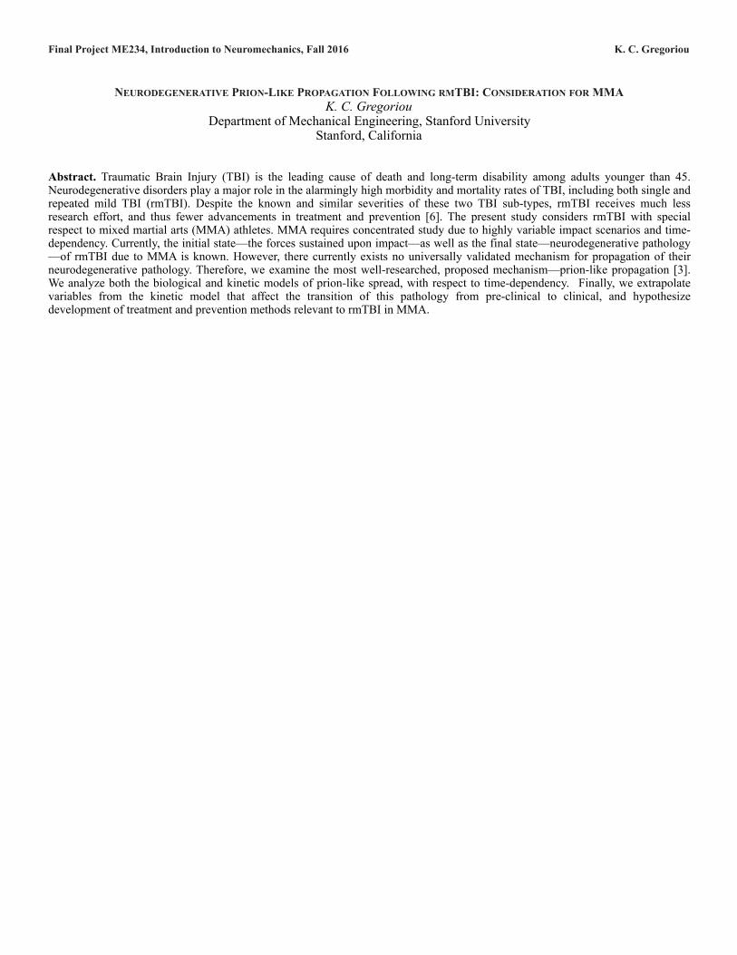
Current Knowledge: Neurodegeneration in rmTBI. Similar to moderate and severe TBI, rmTBI consists of a significant accumulation of Αβ and p-tau, also leading to AD and CTE pathology. Additionally, neurodegeneration is thought to propagate with the same prion-like mechanism. Research also shows significant radial diffusivity in white matter, suggesting this prion-like mechanism in rmTBI acts via the synapses [2].
Furthermore, in acute TBI, Αβ accumulates within just hours of the injury, indicating almost immediate neurodegeneration initiation [3]. A separate study by Briggs et al. confirms similar pathology. They induced rmTBI in 40 mice, and subsequently examined Aβ and p-tau accumulation, as well as neurological and cognitive impairments. One finding from this study—righting reflex recovery time—however, is particularly alarming and requires closer consideration to the criteria used to evaluate MMA athletes post-injury [1].
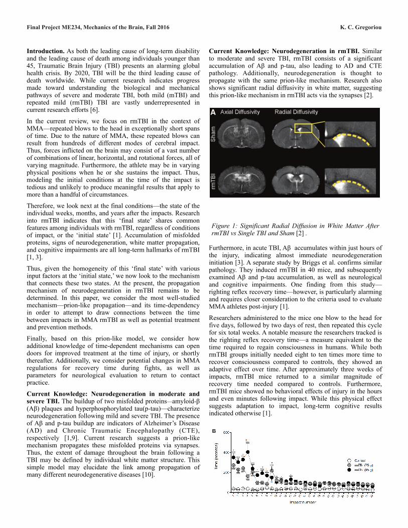
Researchers administered to the mice one blow to the head for five days, followed by two days of rest, then repeated this cycle for six total weeks. A notable measure the researchers tracked is the righting reflex recovery time—a measure equivalent to the time required to regain consciousness in humans. While both rmTBI groups initially needed eight to ten times more time to recover consciousness compared to controls, they showed an adaptive effect over time. After approximately three weeks of impacts, rmTBI mice returned to a similar magnitude of recovery time needed compared to controls. Furthermore, rmTBI mice showed no behavioral effects of injury in the hours and even minutes following impact. While this physical effect suggests adaptation to impact, long-term cognitive results indicated otherwise [1].
Figure 1: Significant Radial Diffusion in White Matter After rmTBI vs Single TBI and Sham [2] .
Final Project ME234, Mechanics of the Brain, Fall 2016 K. C. Gregoriou
Researchers examined the subjects’ brains two months after the final impact to reveal the following significant changes: increased depression-like behavior, further cognitive dysfunction, neurological pathology present in white matter far from the point of impact, and further increased Aβ and p-tau accumulation. Although researchers observed changes in behavior and coordination mid-study, these changes persisted contrary to the time needed to recover consciousness [1].
With the knowledge that neurodegeneration begins close to the time of impact, despite absence of symptoms until potentially months later, we must consider the most effective way to treat, reverse, and even prevent the spread of pathology. Thus, the time-dependent mechanism—prion-like propagation—by which neurodegenerative pathology in rmTBI spreads is critical to understand, especially in the context of MMA. Moreover, due to the highly variable nature of rmTBI impacts, elucidating the common prion-like mechanism behind every impact that leads to neurodegenerative pathology will unite treatment, recovery, and prevention efforts.
Biology of Prion Propagation. In order to more appropriately evaluate the kinetic model of prion propagation, we must first look to the biological model for foundational knowledge of the mechanism. Given the assumption we make that Aβ and p-tau proteins follow prion-like propagation, we adapt the following model for their biological propagation, for the current analysis.
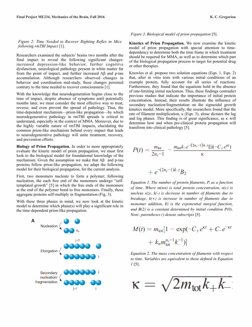
First, two monomers nucleate to form a polymer; following nucleation, the each free end of the monomers undergo “self-templated growth” [5] in which the free ends of the monomers at the end of the polymer bond to free monomers. Finally, these aggregate proteins self-multiply in fragmentation (Fig. 3).
With these three phases in mind, we now look at the kinetic model to determine which phase(s) will play a significant role in the time-dependent prion-like propagation.
Kinetics of Prion Propagation. We now examine the kinetic model of prion propagation with special attention to time-dependency to determine both the time frame in which treatment should be required for MMA, as well as to determine which part of the biological propagation process to target for potential drug or other therapies.
Knowles et al. propose two solution equations (Eqn. 1, Eqn. 2) that, after in vitro tests with various initial conditions of an example protein, fully account for all series of reactions. Furthermore, they found that the equations hold in the absence of rate-limiting initial nucleation. Thus, these findings contradict previous studies that indicate the importance of initial protein concentration. Instead, their results illustrate the influence of secondary nucleation/fragmentation on the sigmoidal growth kinetics model. More specifically, the researchers found that the rate of filament multiplication, κ (Eqn. 3), alone dictates the lag and log phases. This finding is of great significance, as κ will determine how and when pre-clinical protein propagation will transform into clinical pathology [5].
Figure 2: Time Needed to Recover Righting Reflex in Mice following rmTBI Impact [1].
$
Figure 3. Biological model of prion propagation [5].
Equation 1. The number of protein filaments, P, as a function of time. Where m(tot) is total protein concentration, n(c) is nucleus size, k(-) is decrease in number of filaments due to breakage, k(+) is increase in number of filaments due to monomer addition, Ei is the exponential integral function, and B(2) is a constant determined by initial condition P(0). Note: parentheses () denote subscripts [5].
$
Equation 2. The mass concentration of filaments with respect to time. Variables are equivalent to those defined in Equation 1 [5].
$
$
Final Project ME234, Mechanics of the Brain, Fall 2016 K. C. Gregoriou
Because κ depends only on the total protein concentration, the increase in the number of filaments due to monomer addition, and the decrease in the number of filaments due to breakage, we must pay special attention to these three variables with respect to time [5].
Injury and Propagation Timeline. As shown in Figure 4, prion-like diseases become pathogenic at the plateau phase within the sigmoidal growth kinetics model. Thus, there exists a clinical window of opportunity most notably at the lag phase. Here, prions are degraded by biological elements such as macrophages; thus, in this phase it is still possible to prevent and reverse propagation of misfolded proteins [12].
However, degradation does reach saturation, in this case before net prion accumulation reaches saturation. As a result, future study must investigate mechanisms to elevate the saturable degradation level—for instance, creating a biological pathway to input and sustain additional macrophages. Furthermore, supplementary research must investigate how to slow the lag phase, moderated by κ, taking advantage of its driving properties: decreasing total protein concentration, slowing secondary nucleation, and slowing secondary fragmentation resulting in additional nucleation [5].
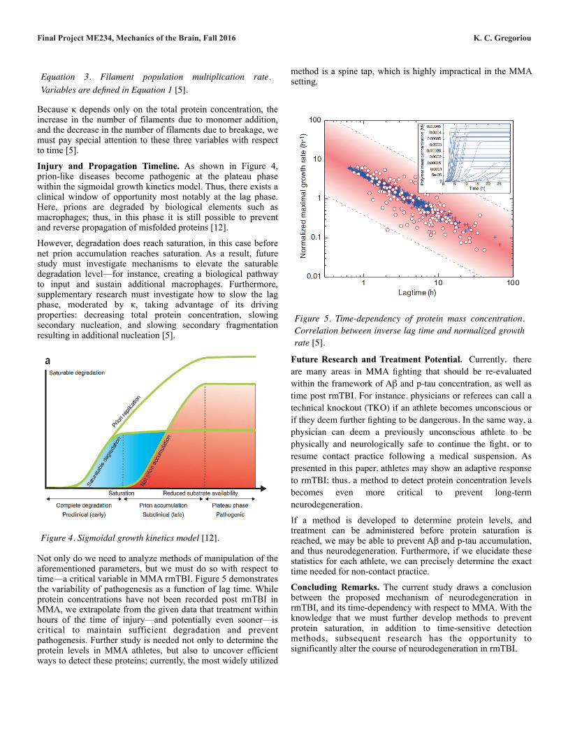
Not only do we need to analyze methods of manipulation of the aforementioned parameters, but we must do so with respect to time—a critical variable in MMA rmTBI. Figure 5 demonstrates the variability of pathogenesis as a function of lag time. While protein concentrations have not been recorded post rmTBI in MMA, we extrapolate from the given data that treatment within hours of the time of injury—and potentially even sooner—is critical to maintain sufficient degradation and prevent pathogenesis. Further study is needed not only to determine the protein levels in MMA athletes, but also to uncover efficient ways to detect these proteins; currently, the most widely utilized
method is a spine tap, which is highly impractical in the MMA setting.
Future Research and Treatment Potential. Currently, there are many areas in MMA fighting that should be re-evaluated within the framework of Aβ and p-tau concentration, as well as time post rmTBI. For instance, physicians or referees can call a technical knockout (TKO) if an athlete becomes unconscious or if they deem further fighting to be dangerous. In the same way, a physician can deem a previously unconscious athlete to be physically and neurologically safe to continue the fight, or to resume contact practice following a medical suspension. As presented in this paper, athletes may show an adaptive response to rmTBI; thus, a method to detect protein concentration levels becomes even more critical to prevent long-term neurodegeneration.
If a method is developed to determine protein levels, and treatment can be administered before protein saturation is reached, we may be able to prevent Aβ and p-tau accumulation, and thus neurodegeneration. Furthermore, if we elucidate these statistics for each athlete, we can precisely determine the exact time needed for non-contact practice.
Concluding Remarks. The current study draws a conclusion between the proposed mechanism of neurodegeneration in rmTBI, and its time-dependency with respect to MMA. With the knowledge that we must further develop methods to prevent protein saturation, in addition to time-sensitive detection methods, subsequent research has the opportunity to significantly alter the course of neurodegeneration in rmTBI.
Equation 3. Filament population multiplication rate. Variables are defined in Equation 1 [5].
Figure 4. Sigmoidal growth kinetics model [12].
$
Figure 5. Time-dependency of protein mass concentration. Correlation between inverse lag time and normalized growth rate [5].
$
Final Project ME234, Mechanics of the Brain, Fall 2016 K. C. Gregoriou
References.
[1] Briggs, D.I., Angoa-Pérez, M., Kuhn, D.M. “Prolonged Repetitive Head Trauma Induces a Singular Chronic Traumatic Encephalopathy—Like Pathology in White Matter Despite Transient Behavioral Abnormalities.” American Journal of Pathology 186.11 (2016): 2869-2886.
[2] Donovan et al. “Repeated traumatic brain injury results in long-term white-matter disruption.” Journal of Cerebral Blood Flow and Metabolism 34 (2014).
[3] G. Edwards III et al. “Amyloid-beta and tau pathology following repetitive mild traumatic brain injury.” Biochemical and Biophysical Research Communications (2016): 1-6.
[4] Iliff et al. “Impairment of Glymphatic Pathway Function Promotes Tau Pathology After Traumatic Brain Injury.” The Journal of Neuroscience 34 (2014): 16180-16193.
[5] Knowles et al. “An Analytical Solution to the Kinetics of Breakable Filament Assembly.” Science (2009): 1533-1537.
[6] Meaney, D. F., Morrison, B., Bass, C.D. “The Mechanics of Traumatic Brain Injury: A Review of What We Know and What We Need to Know for Reducing Its Societal Burden.” Journal of Biomechanical Engineering 136 (2014): 1-14.
[7] Pöschel, Thorsten, Nikolai V. Brilliantov, and Cornelius Frömmel. “Kinetics of Prion Growth.” Biophysical Journal 85.6 (2003): 3460–3474.
[8] “Prion Diseases.” Center for Disease Control and Prevention. 5 Feb. 2014. https://www.cdc.gov/prions/.
[9] Rubenstein et al. “A Novel, Ultrasensitive Assay for Tau: Potential for Assessing Traumatic Brain Injury in Tissues and Biofluids.” Journal of Neurotrauma 32 (2015): 342-352.
[10] Sharp et al. “Network dysfunction after traumatic brain injury.” Nature Reviews Neurology 10 (2014): 156-166.
[11] Shultz et al. “The potential for animal models to provide insight into mild traumatic brain injury: Translational challenges and strategies.” Neuroscience & Biobehavioral Reviews (2016): 1-19.
[12] Aguzzi, A. and Falsig, J. “Prion propagation, toxicity, and degradation.” Nature Neuroscience (2012): 936-939.