measuring physiological similarity of closely related
TRANSCRIPT

1
The following supplement accompanies the article
Measuring physiological similarity of closely related littorinid species: a proteomic insight
Arina L. Maltseva*, Marina A. Varfolomeeva, Arseniy A. Lobov, Natalia A. Mikhailova, Paul E. Renaud, Alexey V. Grishankov, Kseniia Y. Volovik, Andrei I. Granovitch
*Corresponding author: [email protected]
Marine Ecology Progress Series 552: 177–193 (2016)
Supplement 1.
Fig. S1. The division of intertidal area for sampling. Three zones were demarcated based on the position of the fucoid belt (as described in ‘Materials and methods’ in the main article): lower zone — the gravel below the fucoid belt; middle zone — macrophyte sand from gravel under macrophytes within fucoid belt; upper zone — the rocky zone with occasional fucoids above the fucoid belt.
2
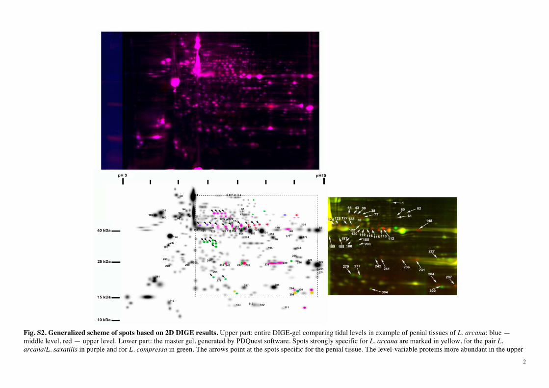
Fig. S2. Generalized scheme of spots based on 2D DIGE results. Upper part: entire DIGE-gel comparing tidal levels in example of penial tissues of L. arcana: blue —middle level, red — upper level. Lower part: the master gel, generated by PDQuest software. Spots strongly specific for L. arcana are marked in yellow, for the pair L. arcana/L. saxatilis in purple and for L. compressa in green. The arrows point at the spots specific for the penial tissue. The level-variable proteins more abundant in the upper

3
level are marked by red “plus” symbols, in the middle/lower level by blue “plus”. Right part: a fragment of the example 2D DIGE gel (shown on the right) of penial tissue samples of L. saxatilis (red coloration) and L. compressa (green coloration) from the lower tidal level. Species-differentiating spots are marked by arrows. Numbers indicate spot IDs. The corresponding area at the master gel is outlined by dashed lines.
Fig S3. The similarity trees among all the samples. Clustering was based on Jaccard dissimilarities for binary data (left tree) or Euclidean distance for quantitative data (right tree). The first part of the abbreviated label is species (arc - L. arcana; comp - L. compressa; sax - L. saxatilis); the second part is tidal level (L - lower; M - middle; U - upper); the third part is sex (f - female; m - male); the forth one is body part (fo - foot; he - head; pe - penis). Low support values for the first two divisions are shown in italic font.