mech vent monitoring
TRANSCRIPT
Mechanical Ventilation: Monitoring
IM 2013 (AVM)

Ventilator management algorithmInitial intubation• FiO2 = 50%• PEEP = 5
• RR = 12 – 15• VT = 8 – 10 ml/kg
SaO2 < 90% SaO2 > 90%
SaO2 > 90%• Adjust RR to maintain PaCO2
= 40• Reduce FiO2 < 50% as
tolerated• Reduce PEEP < 8 as tolerated• Assess criteria for SBT daily
SaO2 < 90%• Increase FiO2 (keep
SaO2>90%)• Increase PEEP to max 20• Identify possible acute lung
injury• Identify respiratory failure
causes
Acute lung injury
No injury
Fail SBT
Acute lung injury• Low TV (lung-protective)
settings• Reduce TV to 6 ml/kg• Increase RR up to 35 to
keep pH > 7.2, PaCO2 < 50• Adjust PEEP to keep FiO2 <
60%SaO2 < 90% SaO2 > 90%
SaO2 < 90%• Dx/Tx associated conditions
(PTX, hemothorax, hydrothorax)
• Consider adjunct measures (prone positioning, HFOV, IRV)
SaO2 > 90%• Continue lung-
protective ventilation until:
•PaO2/FiO2 > 300•Criteria met for
SBT
Persistently fail SBT• Consider tracheostomy• Resume daily SBTs with CPAP
or tracheostomy collar
Pass SBT
Airway stableExtubate
Intubated > 2 wks
• Consider PSV wean (gradual reduction of pressure support)
• Consider gradual increases in SBT duration until endurance improves
Prolonged ventilator dependence
Pass SBT
Pass SBT
Airway stable
Modified from Sena et al, ACS Surgery: Principles and Practice (2005).

Trouble Shooting
Post Initial Settings
• Obtain an ABG (arterial blood gas) about 30 minutes after you set your patient up on the ventilator.• information about any changes that may need to be
made to keep the patient’s oxygenation and ventilation status within a physiological range.
• First 30 – 60 minutes: evaluate • vital signs, • breath sounds, • ventilator parameters, • lung compliance and resistance, • the artificial airway, • patient response to therapy

Improving Ventilation / Oxygenation
1. Correcting PaCO2 Abnormalities2. Oxygenation Using FiO2 and PEEP
Correcting PaCO2 Abnormalities
• A change in VE will often be needed when a patient is first placed on mechanical ventilation to correct for respiratory alkalosis or acidosis; this is facilitated by making a change in VT or rate (f)

Correcting PaCO2 Abnormalities
• Methods of Changing Ventilation Based on PaCO2 and pH
• If it is appropriate to keep rate (f) constant and change VT, the equations is as follows:
Desired VT = Known PaCO2 x Known VT
Desired PaCO2
• If it is appropriate to keep VT the same and change rate (f), then the equations is as follows:
Desired f = Known PaCO2 x Known f Desired PaCO2
Correcting PaCO2 Abnormalities
• (Respiratory Acidosis)– Volume and Pressure Ventilation Changes
• When PaCO2 is elevated (>45 mm Hg) and pH is decreased (<7.35), respiratory acidosis is present and VA is not adequate
– Causes• PE, Pneumonia• Airway disease (e.g., severe asthma attack)• Pleural abnormalities (e.g., effusions)• Chest wall abnormalities • Neuromuscular disease• CNS problems
– Treatment:• VT to 8 – 12 mL/kg ideal body weight (based on patient’s pulmonary
problem)• Maintain plateau pressure <30 cm H2O• If VT is already high and/or Pplateau are already high, then f should be
increased
.

Correcting PaCO2 Abnormalities
• (Respiratory Alkalosis)– Volume and Pressure Ventilation Changes
• When PaCO2 is decreased (<35 mm Hg) and pH increases (>7.35), then respiratory alkalosis is present and alveolar ventilation is excessive
– Causes• Hypoxia with compensatory hyperventilation• Parenchymal lung disease• Medications • Mechanical ventilation• CNS disorders• Anxiety• Metabolic disorders
– Treatment:• Volume ventilation: f, and if necessary, VT
• Pressure ventilation: f, and if necessary, pressure

Correcting PaCO2 Abnormalities
• Metabolic Acidosis and Alkalosis– Treatment of metabolic acidosis and alkalosis
should focus on identifying those metabolic factors that can cause these acid-base disturbances
– These patients are often struggling to lower their PaCO2 to compensate for the metabolic acidemia. As a consequence, these patients are at risk for developing respiratory muscle fatigue
– If the patient is losing the struggle to maintain high with spontaneous breathing, assisted ventilation may be necessary to avoid respiratory failure. It is then appropriate to keep the pH (7.35 – 7.45)
Correcting PaCO2 Abnormalities
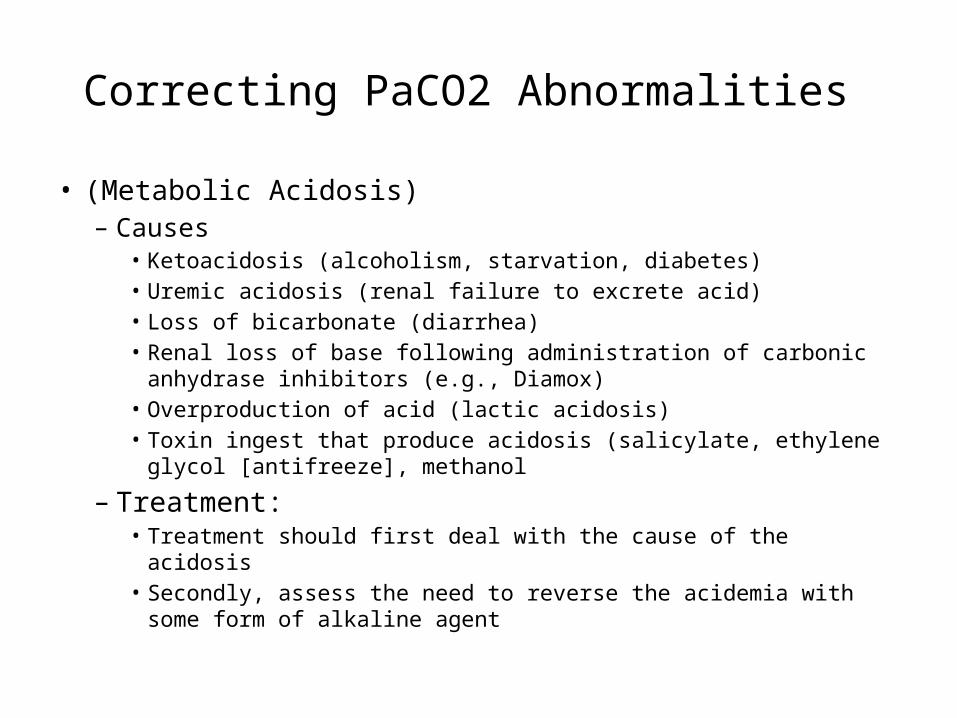
• (Metabolic Acidosis)– Causes
• Ketoacidosis (alcoholism, starvation, diabetes)• Uremic acidosis (renal failure to excrete acid)• Loss of bicarbonate (diarrhea)• Renal loss of base following administration of carbonic
anhydrase inhibitors (e.g., Diamox)• Overproduction of acid (lactic acidosis)• Toxin ingest that produce acidosis (salicylate, ethylene
glycol [antifreeze], methanol
– Treatment:• Treatment should first deal with the cause of the acidosis • Secondly, assess the need to reverse the acidemia with
some form of alkaline agent

Correcting PaCO2 Abnormalities
• (Metabolic Alkalosis)– Causes
• Loss of gastric fluid and stomach acids (vomiting, nasogastric suctioning)
• Acid loss in the urine (diuretic administration)• Acid shift into the cells (potassium deficiency)• Lactate, acetate, citrate administration• Excessive bicarbonate loads (bicarbonate administration)
– Treatment:• Treatment should first deal with the cause of the acidosis • Secondly, assess the need to reverse the acidemia with some form of
alkaline agent• Treatment involves correcting the underlying cause and reversing those
factors leading to the alkalosis. In severe cases, carbonic anhydrate inhibitors, acid infusion, and low bicarbonate dialysis my be required
– Only in rare circumstances does partial respiratory compensation of metabolic alkalosis occur – PaCO2 will usually not rise higher than 55 mm Hg (Remember that as the CO2 rises, the PaO2 falls)

Correcting PaCO2 Abnormalities
• Increased Physiological Dead Space– If pure respiratory acidosis persists even
after alveolar ventilation has been increased, the patient may have a problem with increased dead space
– Causes• Pulmonary emboli• Low cardiac output low pulmonary perfusion• High alveolar pressure (PEEP) pulmonary blood
flow• Air trapping pulmonary perfusion

Correcting PaCO2 Abnormalities
• Increased Metabolism and Increased CO2 Production– Metabolic rate and VCO2 are increased in the following
patients:• Fever • Sepsis• Burns• Multiple trauma and multiple surgical procedures• Hyperthyroidism• Seizures
– In these patients VE is increased and WOB is elevated
– Treatment:• Increase machine rate to WOB: may cause auto-peep• Add pressure support for spontaneous breaths to WOB through ET and
circuit• Switch to PC-CMV, use sedation to WOB
.

Correcting PaCO2 Abnormalities
• Intentional Iatrogenic Hyperventilation– Definition
• Deliberate hyperventilation in patients with acute head injury and increased intracranial pressure (ICP)
– Hyperventilation reduces PaCO2 which causes vasoconstriction of cerebral blood vessels and decreases blood flow to the brain and is believed to lower increased intracranial pressure ICP
– Current therapy guideline for head injuries with increased ICP do not recommend prophylactic hyperventilation (PaCO2 <25 mm Hg) during the first 24 hours - may cause cerebral ischemia and cerebral hypoxemia
– Treatment:• Hyperventilation may be needed for brief periods when acute
neurological deterioration is present and ICP elevated• Mild hyperventilation (PaCO2 30 – 35 mm Hg) may be used for
longer periods in a situation in which increased ICP is refractory to standard treatment

Correcting PaCO2 Abnormalities
• Permissive Hypercapnia (PHY)
– Definition• Deliberate limitation of ventilatory support to avoid lung
overdistention and injury of lung– ARDS or Status asthmaticus
• PaCO2 values are allowed to rise above normal– ≥50 – 150 mm Hg
• pH values are allowed to fall below normal– ≥7.10 – 7.30– Most researchers agree pH ≥7.25 is acceptable
– PaCO2 accompanied PaO2• O2 administration must be provided and monitored closely
– PaCO2 stimulates the drive to breath• Appropriate to provide sedation to patients in whom PHY is
being employed
Correcting PaCO2 Abnormalities
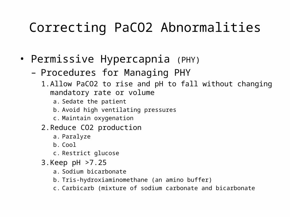
• Permissive Hypercapnia (PHY)
– Procedures for Managing PHY1. Allow PaCO2 to rise and pH to fall without changing
mandatory rate or volumea. Sedate the patient b. Avoid high ventilating pressuresc. Maintain oxygenation
2. Reduce CO2 productiona. Paralyzeb. Coolc. Restrict glucose
3. Keep pH >7.25a. Sodium bicarbonateb. Tris-hydroxiaminomethane (an amino buffer)c. Carbicarb (mixture of sodium carbonate and bicarbonate

Correcting PaCO2 Abnormalities
• Permissive Hypercapnia (PHY)
– Contraindications and Effects of PHY• Head trauma• Intracranial disease• Intracranial lesions
– Relatively contraindicated in the following• Cardiac ischemia• Left ventricular compromise• Pulmonary hypertension• Right heart failure
– The use of PHY is restricted to situations in which the target airway pressure is at its maximum and the highest possible rates are being used
– The risks of hypercapnia are considered by some to be preferable to the high Pplat required to achieve normal CO2 levels

Oxygenation Using FiO2 and PEEP
• Adjusting FiO2– Every attempt should be made to maintain the FiO2 <0.40
to 0.50 to prevent the complications of O2 toxicity while keeping the PaO2 between 60 and 90 mm Hg• This goal is not always possible and sometimes a higher FiO2 is required
– The SpO2 can be used to titrate FiO2, with the goal of maintaining the SpO2 >90%• The SaO2 on an ABG is used to establish the relationship with the
current SpO2
– ABGs are obtained after mechanical ventilation is initiated and compared with FiO2 being delivered and the SpO2 to establish their relationships
– A linear relationship exists between PaO2 and FiO2 as long as VE, CO, Shunt, VD/VT remain fairly constant (cardiopulmonary status)
.

Oxygenation Using FiO2 and PEEP
• Adjusting FiO2– Because of the linear correlation between PaO2
and FiO2 the following equation can be used to select the desired FiO2 to achieve a desired PaO2:
– Desired FiO2 = PaO2 (desired) x FiO2 (known)PaO2 (known)
– Exercise:After being supported on a ventilator for 30 minutes, a patient’s PaO2 is 40 mm Hg on an FiO2 of 0.50. Acid-base status is normal and all other ventilator parameters are within the acceptable range. What FiO2 is required to achieve a desired PaO2 of 60 mm Hg?

Oxygenation Using FiO2 and PEEP
Desired FiO2 = PaO2 (desired) x FiO2 (known) PaO2 (known)
Desired FiO2 = (60 mm Hg) (0.50 FiO2) 40 mm Hg
Desired FiO2 = 0.75
Oxygenation Using FiO2 and PEEP
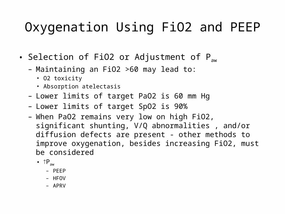
• Selection of FiO2 or Adjustment of Paw
– Maintaining an FiO2 >60 may lead to:• O2 toxicity• Absorption atelectasis
– Lower limits of target PaO2 is 60 mm Hg– Lower limits of target SpO2 is 90%– When PaO2 remains very low on high FiO2, significant
shunting, V/Q abnormalities , and/or diffusion defects are present - other methods to improve oxygenation, besides increasing FiO2, must be considered• Paw
– PEEP– HFOV– APRV
Oxygenation Using FiO2 and PEEP
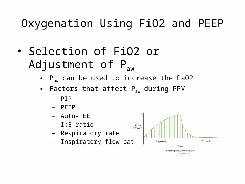
• Selection of FiO2 or Adjustment of Paw
• Paw can be used to increase the PaO2
• Factors that affect Paw during PPV– PIP– PEEP– Auto-PEEP– I:E ratio– Respiratory rate– Inspiratory flow patterns
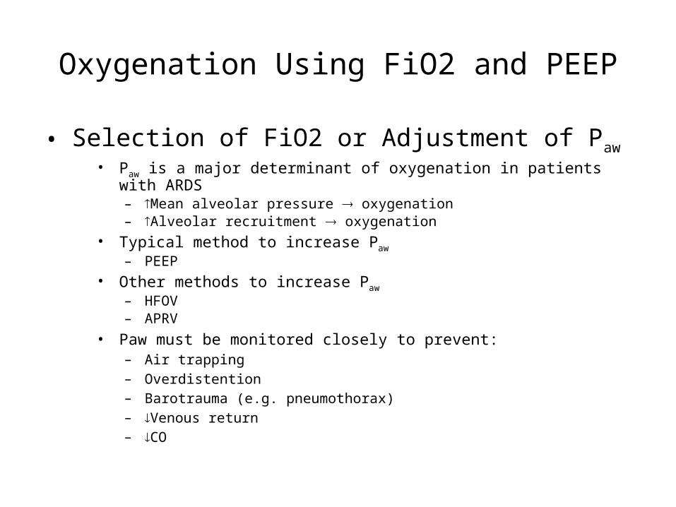
Oxygenation Using FiO2 and PEEP
• Selection of FiO2 or Adjustment of Paw• Paw is a major determinant of oxygenation in patients with
ARDS– Mean alveolar pressure oxygenation– Alveolar recruitment oxygenation
• Typical method to increase Paw
– PEEP• Other methods to increase Paw
– HFOV– APRV
• Paw must be monitored closely to prevent:– Air trapping– Overdistention– Barotrauma (e.g. pneumothorax)– Venous return– CO
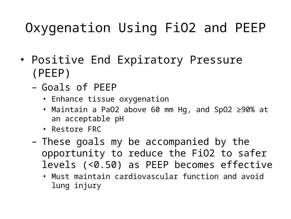
Oxygenation Using FiO2 and PEEP
• Positive End Expiratory Pressure (PEEP)– Goals of PEEP
• Enhance tissue oxygenation• Maintain a PaO2 above 60 mm Hg, and SpO2 ≥90%
at an acceptable pH• Restore FRC
– These goals my be accompanied by the opportunity to reduce the FiO2 to safer levels (<0.50) as PEEP becomes effective• Must maintain cardiovascular function and avoid
lung injury
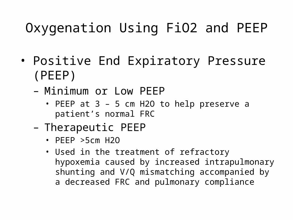
Oxygenation Using FiO2 and PEEP
• Positive End Expiratory Pressure (PEEP)– Minimum or Low PEEP
• PEEP at 3 – 5 cm H2O to help preserve a patient’s normal FRC
– Therapeutic PEEP• PEEP >5cm H2O• Used in the treatment of refractory hypoxemia
caused by increased intrapulmonary shunting and V/Q mismatching accompanied by a decreased FRC and pulmonary compliance
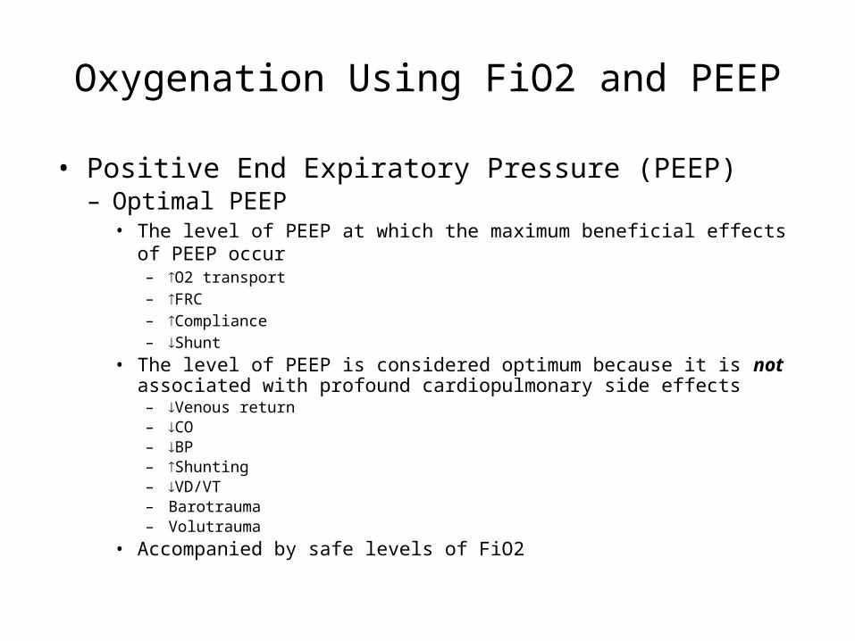
Oxygenation Using FiO2 and PEEP
• Positive End Expiratory Pressure (PEEP)– Optimal PEEP
• The level of PEEP at which the maximum beneficial effects of PEEP occur
– O2 transport– FRC– Compliance– Shunt
• The level of PEEP is considered optimum because it is not associated with profound cardiopulmonary side effects
– Venous return– CO– BP– Shunting– VD/VT– Barotrauma– Volutrauma
• Accompanied by safe levels of FiO2

Oxygenation Using FiO2 and PEEP
• Positive End Expiratory Pressure (PEEP)– Indications for PEEP Therapy
• Bilateral infiltrates on chest radiograph• Recurrent atelectasis• Reduced CL
• PaO2 <60 mm Hg on high FiO2 of >0.5• PaO2/FiO2 ratio <200 for ARDS and <300 for
ALI• Refractory hypoxemia: PaO2 increases <10 with
FiO2 increase of 0.2
Oxygenation Using FiO2 and PEEP
• Positive End Expiratory Pressure (PEEP)– Specific clinical disorders that may
benefit from PEEP• ALI• ARDS• Cardiogenic PE• Bilateral, diffuse pneumonia
Oxygenation Using FiO2 and PEEP
• Positive End Expiratory Pressure (PEEP)– Application of PEEP
• Increased in increments of 3 – 5 cm H2O in adults, 2 – 3 cm H2O in infants• Target acceptable PaO2/FiO2 ratio at a safe FiO2
– >300 (e.g., PaO2 = 100, with FiO2 = 0.33(optimal, but not always realistic)
• Patient Appearance– Color, level of consciousness, anxiety – a sudden deterioration may indicate
cardiovascular collapse or pneumothorax
• Blood Pressure– BP of 20 mm Hg systolic drop is significant
• Breath Sounds– Barotrauma, e.g., pneumothorax
• Ventilator Parameters– VT, Flow, PIP, plateau pressure, VE
• Static Compliance (CS)– As PEEP progressively restores FRC, compliance should increase– Too Much PEEP Overdistention CS

Volume
Pressure
Zone ofOverdistention
“Safe”Window
Zone ofDerecruitmentand Atelectasis
Injury
Injury
Optimized Lung Volume “Safe Window”
• Overdistension – Edema fluid accumulation– Surfactant degradation– High oxygen exposure– Mechanical disruption
• Derecruitment, Atelectasis– Repeated closure / re-expansion– Stimulation inflammatory response– Inhibition surfactant– Local hypoxemia– Compensatory overexpansion

Oxygenation Using FiO2 and PEEP
• Positive End Expiratory Pressure (PEEP)– Application of PEEP
• Arterial PO2, FiO2, and PaO2/FiO2– The usual approach to the management of FiO2 and PEEP is to start with
high FiO2 and incrementally decrease it as PEEP improves oxygenation
• Arterial to End-Tidal Carbon Dioxide Tension Gradient– Normal P(a-et)CO2 gradient is 4.5 ± 2.5 (Pilbeam)– Is lowest when gas exchange units are maximally recruited without being
overdistended– If P(a-et)CO2 gradient increases minimal acceptable values, it signifies
that too much PEEP has been added and is producing a drop in cardiac output and in increase in VD/VT
• Arterial-to-Venous Oxygen Difference (C(a-v)O2) reflects O2 utilization by the tissues
– Normal value is 5 vol%– Increases in C(a-v)O2 with increases in PEEP may indicate hypovolemia,
cardiac malfunction, decreased venous return to the heart, and decreased cardiac output from PEEP

Oxygenation Using FiO2 and PEEP
• Positive End Expiratory Pressure (PEEP)– Application of PEEP
• Mixed Venous O2 Tension or Saturation– Normal PvO2 = 35–40 mm Hg (minimal acceptable is 28 mm Hg)– Normal SvO2 = 75%
(minimal acceptable is 50%)– PEEP usually improves PvO2 and SvO2– When PvO2 and/or SvO2 decrease, with a increase C(a-v)O2 increase,
this indicates a decrease in cardiac output – TOO MUCH PEEP
• Cardiac Output– Cardiac output provide key information about the body’s response to
PEEP– PEEP improves V/Q Oxygenation CO– Too much PEEP Overdistention Venous return CO
Oxygenation Using FiO2 and PEEP
• Positive End Expiratory Pressure (PEEP)– Application of PEEP
• Pulmonary Vascular Pressure Monitoring– When using PEEP >15 cm H2O, it is important to
closely evaluate the patient’s hemodyamic status, which may require the placement of a pulmonary artery catheter
– If pulmonary artery occluding pressure (PAOP), also known as “wedge pressure,” rises markedly as PEEP is increased, the lungs may be overinflated
– On the other hand, when PEEP rises, PAOP may be markedly decreased because of pulmonary blood flow is reduced as a result of decreased venous return to the right side of the heart
Oxygenation Using FiO2 and PEEP
• Positive End Expiratory Pressure (PEEP)– Weaning From PEEP
• Patient should demonstrate an acceptable PaO2 on an FiO2 of <0.40• Must be hemodynamically stable and nonseptic• Lung conditions should have improved
– CS, PaO2/FiO2 ratio
• Reduce PEEP in 5 cm H2O increments• Evaluate SpO2 within 3 minutes to determine effect – if it falls <20%
from previous PEEP level, the patient is ready to tolerate lower PEEP level. If SpO2 drops >20% place PEEP at previous level
• Wait between reductions in PEEP and reevaluate the initial criteria. If the patient is stable, reduce PEEP by another 5 cm H2O. This may take 1 hour or may require as long as 6 hours or more
• When the patient is at 5 cm H2O, an additional evaluation is necessary. If reducing the PEEP to zero result is a worsening of the patient, then it may be appropriate to leave the patient at 5 cm H2O until extubation

Indications for extubation
• Clinical parameters• Resolution/Stabilization of
disease process• Hemodynamically stable• Intact cough/gag reflex• Spontaneous respirations• Acceptable vent settings
• FiO2< 50%, PEEP < 8, PaO2 > 75, pH > 7.25
• General approaches• SIMV Weaning• Pressure Support Ventilation
(PSV) Weaning• Spontaneous breathing trials
• Demonstrated to be superior
No weaning parameter completely accurate when used alone
Numerical Parameters
Normal Range
Weaning Threshold
P/F > 400 > 200
Tidal volume 5 - 7 ml/kg 5 ml/kg
Respiratory rate 14 - 18 breaths/min
< 40 breaths/min
Vital capacity 65 - 75 ml/kg 10 ml/kg
Minute volume 5 - 7 L/min < 10 L/min
Greater Predictive Value
Normal Range
Weaning Threshold
NIF (Negative Inspiratory Force)
> - 90 cm H2O > - 25 cm H2O
RSBI (Rapid Shallow Breathing Index) (RR/TV)
< 50 < 100
Marino P, The ICU Book (2/e). 1998.

WEANING FROM THE VENTILATOR • Weaning strategy:
– no single proven approach– ? easier from PSV (decrease 2–4 cm H2O q12h) or using
intermittent T-piece trials than from SIMV (decrease RR 2–4 breaths/min q12h + backup 5 cm H2O PSV)
• Method of weaning is not as relevant as knowing appropriate time to wean.– Identify patients who can breathe spontaneously

WEANING FROM THE VENTILATOR
• Consider these factors: WEANS NOW mnemonic– Wakefulness or sedation reversed– Electrolytes– Acid–base status– Neurologic state– Secretions minimal, cough adequate– Nutrition– Oxygenation:
• PAO2 >60 on FiO2, 0.4; • PaO2/FiO2 > 200, • PEEP </= 5 cm H2O
– Work of breathing: • volume support stable, • VE < 12 L/min, • vital capacity > 10 ml/kg

WEANING FROM THE VENTILATOR
• No weaning parameters are perfect. – Rapid Shallow Breathing Index (RSBI) – = (f/VT) = respiratory rate / tidal volume (in
liters)• If the RSBI is <105 after 1 hr of CPAP with
PSV 5 & PEEP 5, patient is often weanable.• If RSBI > 105 predicts failure
– Spontaneous breathing trial (e.g., CPAP × 1–2 hrs)
• Failure if: deteriorating ABGs, high RR, high or low HR, high or low BP, diaphoresis, anxiety
• If none of these events occurs consider extubation

Spontaneous Breathing Trials
• Settings• PEEP = 5, PS = 0 – 5, FiO2 < 40%• Breathe independently for 30 –
120 min• ABG obtained at end of SBT
• Failed SBT Criteria• RR > 35 for >5 min• SaO2 <90% for >30 sec• HR > 140, sustained 20% increase• Systolic BP > 180 or < 90mm Hg• Sustained increased work of
breathing• Cardiac dysrhythmia• Anxiety, Diaphoresis• pH < 7.32
SBTs do not guarantee that airway is stable or pt can self-clear secretions
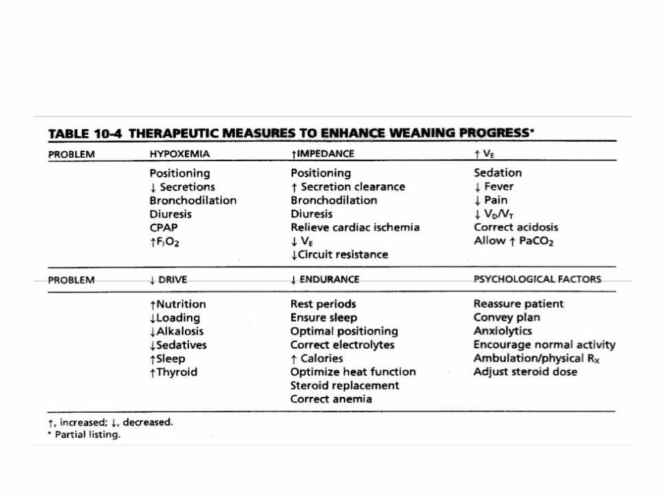
Causes of Failed SBTs
Treatments
Anxiety/Agitation Benzodiazepines or haldol
Infection Diagnosis and tx
Electrolyte abnormalities (K+, PO4-)
Correction
Pulmonary edema, cardiac ischemia
Diuretics and nitrates
Deconditioning, malnutrition
Aggressive nutrition
Neuromuscular disease Bronchopulmonary hygiene, early consideration of trach
Increased intra-abdominal pressure
Semirecumbent positioning, NGT
Hypothyroidism Thyroid replacement
Excessive auto-PEEP (COPD, asthma)
Bronchodilator therapySena et al, ACS Surgery: Principles and Practice (2005).
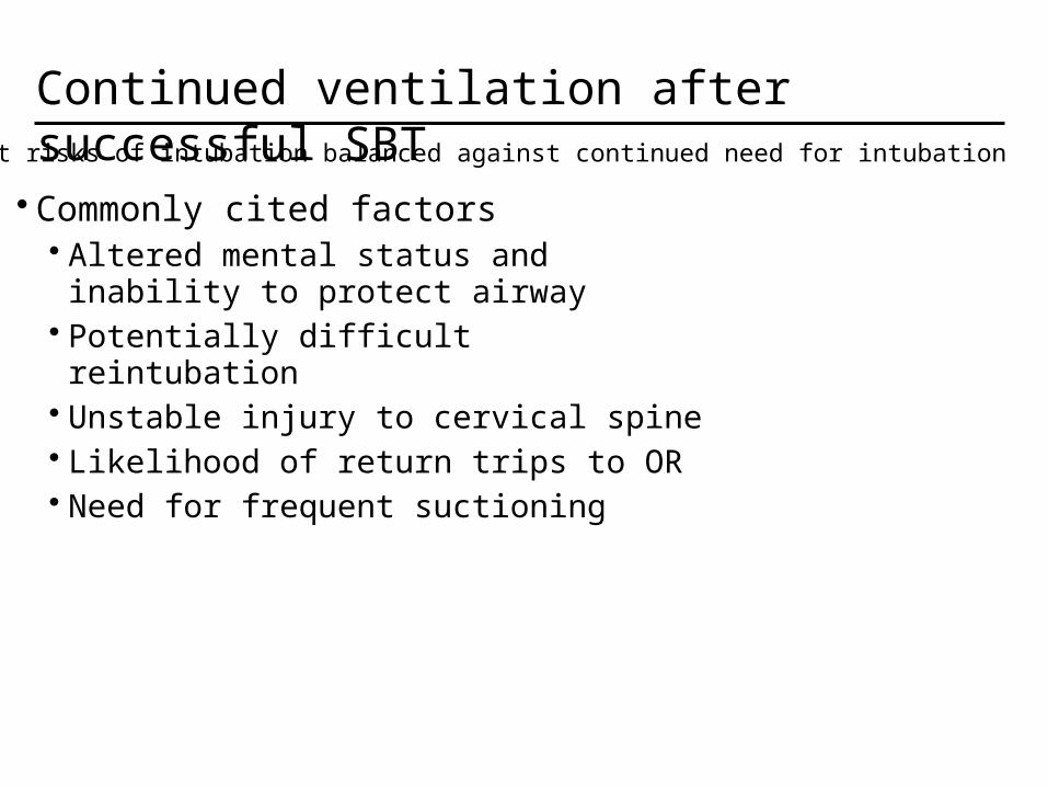
Continued ventilation after successful SBT
• Commonly cited factors• Altered mental status and inability to protect airway
• Potentially difficult reintubation• Unstable injury to cervical spine• Likelihood of return trips to OR• Need for frequent suctioning
Inherent risks of intubation balanced against continued need for intubation

Need for tracheostomy
• Advantages• Issue of airway stability can be
separated from issue of readiness for extubation• May quicken decision to
extubate• Decreased work of breathing• Avoid continued vocal cord injury• Improved bronchopulmonary
hygiene• Improved pt communication
• Disadvantages• Long term risk of tracheal stenosis• Procedure-related complication
rate (4% - 36%)
Prolonged intubation may injure airway and cause airway edema
1 - Vocal cords. 2 - Thyroid cartilage. 3 - Cricoid cartilage. 4 - Tracheal cartilage. 5 - Balloon cuff.

References
• Mechanical Ventilation for Nursing by M Dearing and C Shelley (ppt)
• Mechanical Ventilation Handout by DM Lieberman and AS Ho (ppt)
• Mechanical Ventilation by Marc Charles Parent (ppt)• Principles of Mechanical Ventilation RET 2284 by
Stultz (ppt)• Sena, MJ et al. Mechanical Ventilation. ACS Surgery:
Principles and Practice 2005; pg. 1-16.• Marino, PL. The ICU Book. 2nd edition. 1998.• Byrd, RP. Mechanical ventilation. Emedicine, 6/6/06.