mechanical characterization and stimulation solutions for ... · cartilage (knee) injuries through...
TRANSCRIPT

Mechanical Characterization and Stimulation
Solutions for Biomaterials

BioDynamic Instruments

Biomaterials and Tissue Characterization
Application Examples

Clinical Need: Understand how age impacts bone fragility
Research Need: Testing of small bone specimens to examine age-related effects of collagen on the mechanical properties of bone
ElectroForce Application: A 3200 with a standard system load cell and displacement sensor used to apply specific load levels to micro-machined human cortical bone and measure the corresponding displacement changes.
Bone Bending Creep Test
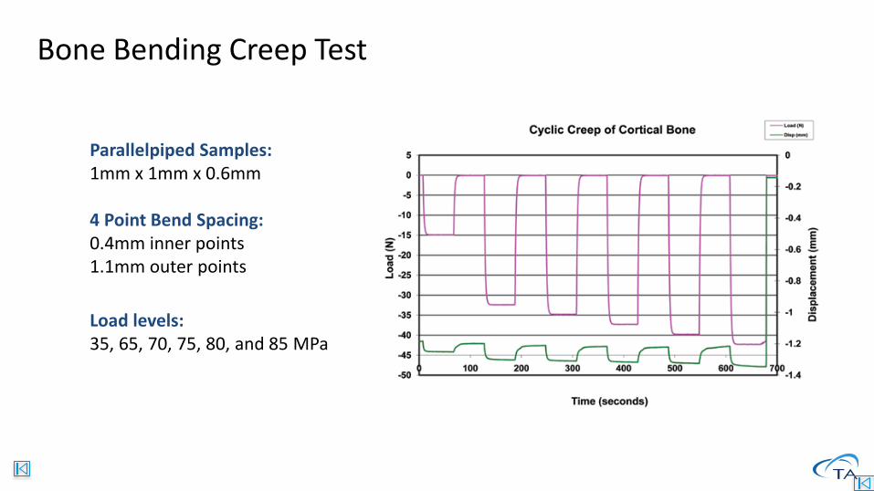
Parallelpiped Samples: 1mm x 1mm x 0.6mm
4 Point Bend Spacing: 0.4mm inner points1.1mm outer points
Load levels:35, 65, 70, 75, 80, and 85 MPa
Bone Bending Creep Test

Clinical Need: Understand the impact of changes in dentin in an aging population
Research Need:
Characterize fatigue properties of dentin using two methods:
1) bending
2) crack growth
ElectroForce application:
Using the ElectroForce 3200 to perform small amplitude fatigue tests on micro-samples of human dentin samples
Dental Biomaterials tests
Crack Growth
4-Point Bending
D. Arola et.al., “Effects of aging on the mechanical behavior of human dentin”, Biomaterials 2004DOI: 10.1016/j.biomaterials.2004.10.029

Results: Stress-Strain plots show stronger and tougher results in young specimens
Clear trends in Max Stress and Energy
Dental Biomaterials tests
OldYoung
D. Arola et.al., “Effects of aging on the mechanical behavior of human dentin”, Biomaterials 2004DOI: 10.1016/j.biomaterials.2004.10.029

Clinical Need: Better understanding of biochemical and biomechanical response of bone to mechanical loading
Research Need: Develop and apply a model for in vivo bone loading that is quantifiable and reliable
ElectroForce Application (model calibration): Compression loading of small animal ulna which creates quantifiable bending strains within bone
Calibrations were performed using strain gaged bones during axial loading with ElectroForce 3200 & 3300 instruments
In Vivo Bone Loading
A. Baumann et al., “Development of an in vivo rabbit ulnar loading model” Bone 2015. http://doi.org/10.1016/j.bone.2015.01.022

ElectroForce Application (In Vivo Loading): Cyclic 2Hz uniaxial compression for 360cycles/day with ElectroForce 3200Multiple levels of peak compression strains from 3000 to 5250 microstrain (~60 to 125N applied loads)
InVivo Loading Results: Periosteal bone formation was measured in response to different strain levels:
In Vivo Bone Loading
Control & <3000µε No measurable bone formation
3500µε Detectable but weak formation
4000 & 4500µε New lamellar bone
5250µε Significant woven bone formationA. Baumann et al., “Development of an in vivo rabbit ulnar loading model” Bone 2015. http://doi.org/10.1016/j.bone.2015.01.022

Clinical Need: Determine mechanical properties of cartilage to better understand what will be needed for replacement material.
Research Need: Understand the impact of indentation tip geometry and size on the results of indentation tests.
ElectroForce Application: Determine Young’s modulus utilizing an ElectroForce3100 or 3200, indenters, and saline bath.
Bovine articular cartilage was compared to elastic foam and urethane rubber searching for suitable alternative for future studies.
Cartilage Indentation
Ref: Application Brief: “1Characterizing Mechanical Properties of Cartilage in Situ App Brief”, TA instruments

Testing Solution: ElectroForce system used to conduct indentation test on urethane, foam and cartilage specimens:
• Preconditioned with cyclic indentation of -0.25/-
0.125mm at 5 Hz for 20 cycles
• 40 minute recovery
• Indenter surface contact and then indent sample
0.15mm at 1.5 mm/sec rate
• 1200 sec displacement hold
Conclusion
• Data on urethane and foam compared favorably to cartilage and should prove useful in simulations
Cartilage Indentation
Ref: Application Brief: “1Characterizing Mechanical Properties of Cartilage in Situ App Brief”, TA instruments
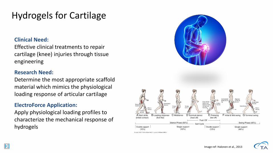
Clinical Need: Effective clinical treatments to repair cartilage (knee) injuries through tissue engineering
Research Need: Determine the most appropriate scaffold material which mimics the physiological loading response of articular cartilage
ElectroForce Application: Apply physiological loading profiles to characterize the mechanical response of hydrogels
Image ref: Halonen et al., 2013
Hydrogels for Cartilage

• Samples punched from polyethylene glycol hydrogel sheets (Medline Ind.)
• Sample preloaded to 0.1 N
• Walking gait compression waveform was imported to WinTest
Hydrogels for Cartilage
Ref: TA internal study

Waveform of strain vs. gait cycle based on simulation of human walking
Walking speed of 5 km/hGait cycle of 1.1 sec
Hydrogels for Cartilage
Ref: TA internal study

ElectroForce 5500 Test Instrument with a multi-specimen fixture used to apply walking waveform compression on hydrogels• Fixture used in combination
with 24-well plate
Samples punched out of PEG hydrogel sheets• 12 mm diameter, 1.2 mm height
Samples placed in saline-filled wells
Hydrogels for Cartilage
Ref: TA internal study

Samples subjected to two loading waveforms:• Sinusoidal • Walking Gait (Custom)
Experimental Conditions
1 Specimen/LoadedSinusoidal
&Custom Gait
24 Specimens/LoadedSinusoidal
&Custom Gait
12 Specimens/Loaded&
12 Specimens/Unloaded
Custom Gait
*3 samples each from loaded and unloaded groups tested to
failure with single pair of platens
Hydrogels for Cartilage
Ref: TA internal study

Hydrogels for Cartilage
Ref: TA internal study

Two sets of 24 samples were tested, to compare unloaded vs cyclically loaded specimen strength
Unloaded samples had a higher fracture load than loaded samples
Hydrogels for Cartilage
Ref: TA internal study

5110 and 5210 Mechanical Simulation BioreactorsProduct Details
Combining sterile biologic environment with mechanical stimulation and measurements
Cell-culture incubator compatible
One or four-chamber versions (5170 or 5270)
Mechanical Forces up to 200 N
3 Fixture packages: Tubular, Strips and Disc
Flexible, sterilizable chambers and flow-loops
Peristaltic pump included: 0.1-280 mL/min

5170 and 5270 Mechanical Simulation BioreactorsProduct Details
Combining sterile biologic environment with mechanical stimulation and measurements
Cell-culture incubator compatible
One or four-chamber versions (5170 or 5270)
Mechanical Forces up to 200 N
3 Fixture packages: Tubular, Strips and Disc
Flexible, sterilizable chambers and flow-loops
Dynamic Pulsatile Pump assembly: Up to 8.8 mL/pulse plus 1760 mL/min mean flow

BioDynamic InstrumentsChamber Details

Clinical Need: Determine the most appropriate biocompatible polymers which have different properties for different applications (tissue support or drug delivery)
Research Need: Currently, biodegradation of scaffolds is assessed under static conditions, but the materials are subjected to a dynamic physiological environment once implanted
ElectroForce Application: Investigate the degradation of a common biomaterial when subjected to long-term, dynamic loading
Ref: Application Brief: “Degradation of PLGA Scaffolds Under Dynamic Loading”, TA instruments
Degradation of PLGA

Test Groups:
• Dynamic: sinusoidal compression (5 to 30 grams at 1 Hz) and static perfusion
• + Perfusion: static perfusion only
• - Perfusion: stagnant saline (no perfusion)
• Control: completely dry
Degradation of PLGA
Ref: Application Brief: “Degradation of PLGA Scaffolds Under Dynamic Loading”, TA instruments

Degradation of PLGA
Ref: Application Brief: “Degradation of PLGA Scaffolds Under Dynamic Loading”, TA instruments

Clinical Need: Alternatives to chemical/drug treatment to stimulate bone formation
Research Need:Stimulation differentiation and mineralized matrix production of hMSCs via compressive loading
ElectroForce Application: hMSC-seeded scaffolds (polyurethane) were periodically compressed (dynamic) using the 3200 Test Instrument with a BioDynamic chamber
Dr. Gwen Reilly’s GroupUniversity of Sheffield
Bone Tissue Engineering
K. Mallick et.al, “Three-dimensional porous bioscaffolds for bone tissue regeneration”, Journal of Biomedical Materials Research 2012;
DOI: 10.1002/jbm.a.34238

Bone Tissue Engineering
K. Mallick et.al, “Three-dimensional porous bioscaffolds for bone tissue regeneration”, Journal of Biomedical Materials Research 2012;
DOI: 10.1002/jbm.a.34238

Clinical Need: Develop alternative treatments and therapies for tendon repair
Research Need: Drive tenogenic differentiation of hMSCs cultured on scaffold made of braided electrospunpoly(l-lactic acid) nanofibers.
ElectroForce Application: Use the 5210 BioDynamic Test System to (i) characterize and refine mechanical properties of scaffolds and (ii) direct stem cell differentiation with mechanical cues
Dr. Wan-Ju Li’s LaboratoryUniversity of Wisconsin – Madison
1Stem Cell Differentiation App Brief
Tendon Tissue Engineering
J. Barber, et.al., “Braided Nanofibrous Scaffold for Tendon and Ligament Tissue Engineering”, Tissue Engineering: Part A; 2011DOI: 10.1089/ten.tea.2010.0538

Tendon Tissue Engineering
J. Barber, et.al., “Braided Nanofibrous Scaffold for Tendon and Ligament Tissue Engineering”, Tissue Engineering: Part A; 2011DOI: 10.1089/ten.tea.2010.0538

Tendon Tissue Engineering
J. Barber, et.al., “Braided Nanofibrous Scaffold for Tendon and Ligament Tissue Engineering”, Tissue Engineering: Part A; 2011DOI: 10.1089/ten.tea.2010.0538

Dr. Showan Nazhat’s LaboratoryMcGill University
Clinical Need: Treatment for tracheal trauma or disease
Research Need: Understand the effect of shear stress and circumferential strain on airway SMCs
ElectroForce Application: Tissue engineered airway construct cultured under different mechanical conditions (Perfusion BioDynamic System). Circumferential strength measured after culture (3200 Test System)
Airway Tissue Engineering
C. Gheezi, et.al., “An airway smooth muscle cell niche under physiological pulsatile flow…”, Biomaterials 2013DOI: http://dx.doi.org/10.1016/j.biomaterials.2012.11.025

Airway Tissue Engineering
C. Gheezi, et.al., “An airway smooth muscle cell niche under physiological pulsatile flow…”, Biomaterials 2013DOI: http://dx.doi.org/10.1016/j.biomaterials.2012.11.025
