mechanical force-triggered drug delivery · mechanical force-triggered drug delivery yuqi...
TRANSCRIPT
Mechanical Force-Triggered Drug DeliveryYuqi Zhang,†,‡,§,⊥ Jicheng Yu,†,‡,⊥ Hunter N. Bomba,† Yong Zhu,*,†,§ and Zhen Gu*,†,‡,∥
†Joint Department of Biomedical Engineering, University of North Carolina at Chapel Hill and North Carolina State University,Raleigh, North Carolina 27695, United States‡Center for Nanotechnology in Drug Delivery and Division of Molecular Pharmaceutics, UNC Eshelman School of Pharmacy,University of North Carolina at Chapel Hill, Chapel Hill, North Carolina 27599, United States§Department of Mechanical and Aerospace Engineering, North Carolina State University, Raleigh, North Carolina 27695, UnitedStates∥Department of Medicine, University of North Carolina at Chapel Hill, Chapel Hill, North Carolina 27599, United States
ABSTRACT: Advanced drug delivery systems (DDS) enhance treatment efficacy ofdifferent therapeutics in a dosage, spatial, and/or temporal controlled manner. To date,numerous chemical- or physical-based stimuli-responsive formulations or devices forcontrolled drug release have been developed. Among them, the emerging mechanicalforce-based stimulus offers a convenient and robust controlled drug release platform andhas attracted increasing attention. The relevant DDS can be activated to promote drugrelease by different types of mechanical stimuli, including compressive force, tensile force,and shear force as well as indirect formats, remotely triggered by ultrasound and magneticfield. In this review, we provide an overview of recent advances in mechanically activatedDDS. The opportunities and challenges regarding clinical translations are also discussed.
CONTENTS
1. Introduction 125362. Compressive/Tensile Force-Triggered Drug Deliv-
ery 125382.1. Deformation 125382.2. Mechanochemical Change 12541
3. Shear Force-Induced Drug Delivery 125413.1. Disaggregation 125423.2. Deformation 12542
4. Ultrasound-Activated Drug Delivery 125444.1. Microbubbles 125454.2. Liposomes 125474.3. Nanoparticles 125494.4. Micro/nanomotors 125514.5. Microcapsules 125514.6. Injectable Depots 12551
5. Magnetic Force-Triggered Drug Delivery 125536. Conclusions and Future Perspectives 12555Author Information 12555
Corresponding Authors 12555Author Contributions 12555Notes 12555Biographies 12556
Acknowledgments 12556References 12556
1. INTRODUCTION
Drug delivery systems (DDS) are of paramount importance toenhance the treatment efficacy of medications.1−4 The last twodecades have witnessed great progress in sustained drugdelivery.5−11 Nevertheless, there are many clinical situationsrequiring more than a zero-ordered, continuous drug releaseprofile, but an on-demand control of therapeutics. Dosage-,spatial-, and temporal-controllable drug release, which can targetdiseased tissue, enhance uptake efficiency, prolong action time,and improve bioavailability of agents, has been heavily pursued inrecent years.12−22 Such DDS can respond to either endogenousenvironmental conditions, including pH,23−28 redox,29−32
hypoxia,33−35 ATP,36−39 glucose,40−43 and enzymes,44−50 orexogenous signals such as temperature,51−53 light,54−57 magneticfield,58−61 and electric current.62−64
Mechanical force, with a subclassification of compressive,tensile, and shear forces (Figure 1a),65,66 is ubiquitously achievedin the body or easily applied externally.67 Force sources rangefrom intrinsic compression/stretching via joint movements tointernal shear force in vascular systems, as well as exterioracoustic and magnetic force remotely applied through the skin(Figure 1b). Easy access of mechanical stimuli facilitatestreatment in a variety of conditions with convenient commands.Especially in occasions of emergence, such as heart attack andhypoglycemia, the mechanical force-mediated trigger could
Received: June 13, 2016Published: September 29, 2016
Review
pubs.acs.org/CR
© 2016 American Chemical Society 12536 DOI: 10.1021/acs.chemrev.6b00369Chem. Rev. 2016, 116, 12536−12563
enable prompt delivery of therapeutics by patients in a self-administered manner. Moreover, compared to chemical orbiological triggers, application of mechanical force provides arelatively predictable control in direction and an adjustablemagnitude management toward precision release of therapeutics.In addition, patient-controlled drug delivery may potentiallyincrease patient satisfaction compared with intermittentadministration, since the patients themselves can convenientlyregulate the release dosage and timing of the drug.
Collectively, mechanical force is a promising candidate torealize controlled drug release.67,68 To be specific, thecompressive and tensile forces can be readily obtained by usinghands or from simple daily motions, such as tension in muscles,tendons, and bone joints, as well as compression in cartilage andbones.69 Therefore, DDS integrated with mechanoresponsivemodality offers self-administration therapy without requiringadditional instruments, while the shear force generated by bloodflow in vascular systems70 holds promise for noninvasive
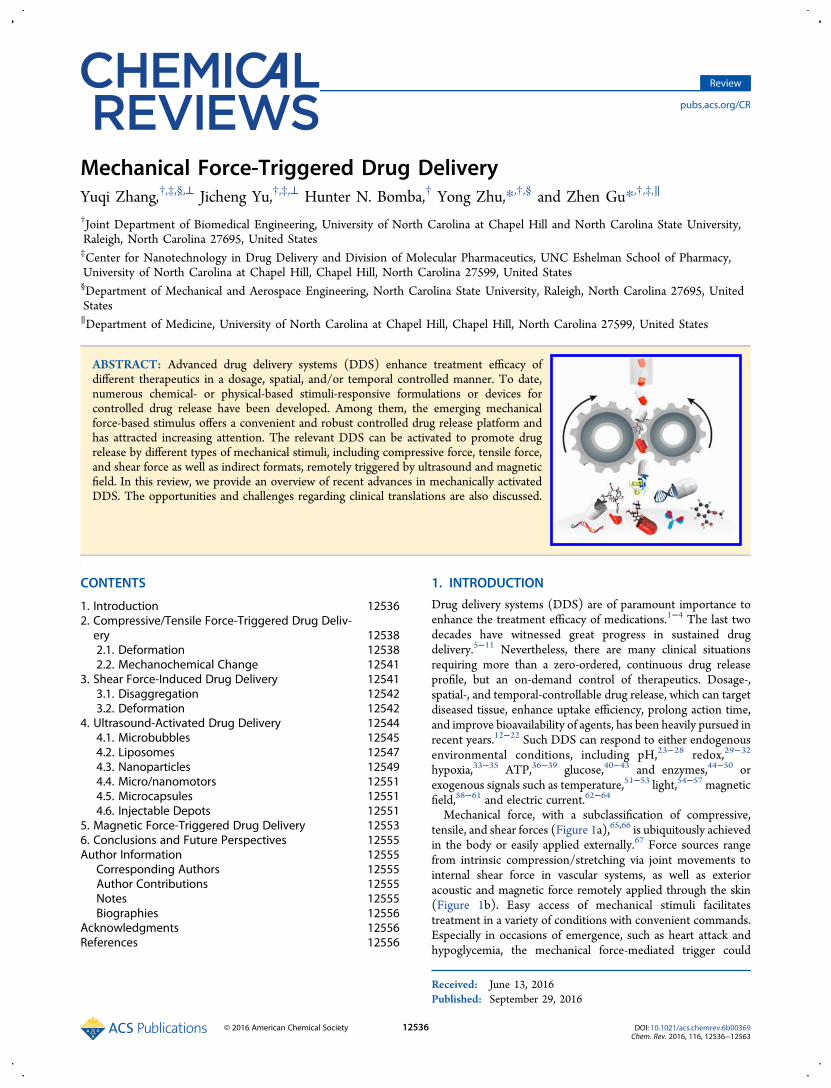
Figure 1. Different forms of mechanical force. (a) Three main types of mechanical force. (b) Mechanical force can be typically obtained in the humanbody or induced externally by ultrasound or magnetic field.
Figure 2. Schematic illustration of diverse mechanical force-triggered drug delivery systems. (a) Compressive or tensile force can induce drug release. (i)Physically loaded drugs can be released by deformation of carriers under compression. (ii) Tensile force can deform mechanoresponsive particlesembedded in the matrices and release the encapsulated drugs. (iii) Materials can be degraded upon exposure to related enzymes under strain. (iv)Compressive force can break the chemical bond. (b) Drug release can be triggered by shear force caused by (i) dissociation of aggregates or (ii)deformation of particles. (c) Ultrasound can facilitate drug release by cavitation or radiation. (d) External magnetic field can trigger drug release bydeforming ferrogels.
Chemical Reviews Review
DOI: 10.1021/acs.chemrev.6b00369Chem. Rev. 2016, 116, 12536−12563
12537

cardiovascular drug delivery.71,72 The design features offormulations or devices based on the two directly interactingtriggers generally involve physical deformation or mechano-chemical changes of drug carriers. On the other hand, ultrasound,as a highly efficient and noninvasive trigger, has been widelyexplored in the drug delivery field. The longitudinal forceproduced by ultrasound can induce hyperthermia, cavitation, andradiation, resulting in destruction of drug carriers and drugrelease by either thermal or nonthermal functions.73−75 Addi-tionally, magnetic force induced by a magnetic field can activatethe magnetoresponsive components. Integrated with thermo-sensitive materials or flexible matrices, the activated magneto-responsive particles can achieve a controlled release of cargoes bydisassociation or morphology variation.76
This review surveys the mechanical force-triggered DDS,mainly classified as direct interaction forces, includingcompressive, tensile, and shear forces as well as induced forcesgenerated by other stimuli like ultrasound and magnetic field(Figure 2). We will describe representative examples of eachstimulus signal, focused on the latest examples. Theopportunities and challenges associated with clinical translationswill also be discussed.
2. COMPRESSIVE/TENSILE FORCE-TRIGGERED DRUGDELIVERY
Compressive or tensile force-responsive DDS are mostly basedon stretchable materials, such as hydrogels and elastomers.68,77,78
Many formulations and devices, including nanoparticles andmicrogel depots, have been exploited to be hybridized into aflexible substrate to achieve on-demand release in response tomechanical stimuli. Typical mechanisms for releasing entrappedagents from systems are based on either physical deformation ofcarriers or chemical changes of materials, such as breakage ofchemical bonds.
2.1. Deformation
Thermosensitive hydrogels have been extensively utilized forspatiotemporal-controlled drug delivery.15 However, an externalheat source is usually required to activate the release. Pioletti andco-workers79 attempted to employ the dissipative properties ofhydrogel as an internal heat source (Figure 3a). The hydrogelmatrix, which was formed with thermoresponsive nanoparticles,produced self-heat after a cyclic mechanical loading for 5 min,causing a temperature increase from 36 to 37 °C (Figure 3b).The temperature increase led to shrinkage of the nanoparticles,followed by contents release after 5−8 min (Figure 3c). Thishybrid hydrogel with high dissipation holds promise in thedelivery of growth factors by mechanical activation. Mooney and
Figure 3. Mechanically triggered drug release from thermoresponsive self-heating hydrogel. (a) Illustration of drug release from thermoresponsivenanoparticle-loaded hydrogel with dissipation as an internal heat source. Thermoresponsive nanoparticles (blue dots) and drug (red dots) are loadedinside the hydrogel (gray). (b) Temperature changes after cyclic compressive load. (Top) Temperature increases to 37 °C after 5 min loading frominitial temperature (36 °C). (Bottom) Temperature increases from initial temperature (34 °C) to 34.7 °C after 5 min loading. (c) (Top) Temporal-controlled release of xylene cyanol FF triggered by dissipation properties of the hydrogel. (Bottom) Effect of nanoparticles on xylene cyanol FF releaseduring mechanical loading. Reprinted with permission from ref 79. Copyright 2014 Elsevier.
Chemical Reviews Review
DOI: 10.1021/acs.chemrev.6b00369Chem. Rev. 2016, 116, 12536−12563
12538

co-workers80 developed a controlled delivery system based onsynthetic extracellular matrices (ECMs). The natural ECMs oftissues, as depots of growth factors, are responsive to manyphysiological signals and can correspondingly release factors tosurrounding cells. To mimic the behavior of natural ECMs, theydesigned a hydrogel with reversible binding to drugs. Thereversible chemical binding was aimed at overcoming the limitedsensing period, since physically loaded hydrogel usually cannotrespond to compression after several release cycles due to rapiddepletion of drugs from the hydrogel.81 In this situation,unbound vascular endothelial growth factor (VEGF) wasreleased upon each compression. However, with re-equilibrationof bound VEGF turning into unbound VEGF, the hydrogel couldcontinuously control the release of VEGF via compressive force.Chemically cross-linked hydrogels integrated with mechano-
sensitive block copolymer micelles (BCMs) were reported tohave the capability to modulate drug release by mechanical force.Jia and co-workers82 applied drug-loaded BCMs with reactivehandles as the microscopic cross-linkers instead of traditionalmolecular cross-linkers for poly(acrylamide) (PAAm)-basednetworks (Figure 4a). The restricted BCMs provided themechanoresponsive elements for BCM-PAAm gels, and theforce-induced deformation of BCMs led to drug release uponstrain application. At a strain of 200%, the BCMs presented amore elongated appearance with an average dimension ratio of1.53 ± 0.23 (Figure 4b). Furthermore, the BCMs returned totheir original morphology after removal of the external force dueto reversible structural deformation. Strain-dependent shapealternation could effectively control the drug release rate fromthe BCM-cross-linked gel (Figure 4c). The same group alsoprepared hyaluronic acid (HA) gels covalently embedded with
dexamethasone (DEX)-loaded BCMs to control inflammationwithin mechanically stressed tissues.83 The HA gels markedlyreduced the initial burst release and allowed sustained release in astress-dependent manner, which offers an alternative strategy totake advantage of mechanical stress to promote tissue repairduring the wound-healing process.Jeong and co-workers84 developed a strain-controlled system
that released molecules from an array of stretchable micro-capsules based on a poly(dimethylsiloxane) (PDMS) elastomersubstrate using a hydrogel pattern fabrication technique (Figure5a). The mechanical strain applied on the substrate led todeformation of the polystyrene (PS) microcapsules (Figure 5b),followed by pumped-out preload molecules. A strain of 2.5% onthe substrate reduced the microcapsule volume by 17%, while ahigher strain of 7.5% decreased the volume by 98%. The dosageof released molecules was highly dependent on the degree ofstrain (Figure 5c). The deformed PS capsules could return totheir initial shape and volume upon release from stretching,which favored repeatable drug diffusion from the patch (Figure5d).In another example, Di et al.85 recently exploited a
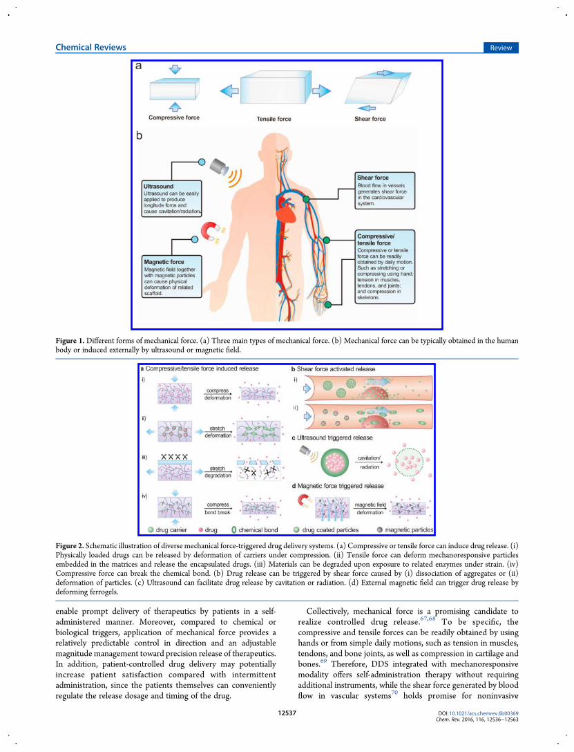
multipurpose wearable elastomer that could mechanicallypromote release of therapeutics to achieve “elastic drug delivery”(Figure 6). The stretchable elastomer was integrated withmicrogel depots that contained drug-loaded nanoparticles. Thedrugs could be temporarily stored in the microdepots. Whenstretching was applied, the drug could continuously diffuse outdue to deformation of the microgels, which was attributed to theenlarged surface area for diffusion and Poisson’s ratio-inducedcompression toward the microdepots. Therefore, an on-demanddrug delivery can be realized by convenient daily body motion.
Figure 4. Mechanoresponsive hydrogels based on block copolymer micelles (BCMs). (a) Synthesis of BCM-PAAm hydrogels. (b) Cross-sectionaltransmission electron microscopie (TEM) images and schematics of unstretched, stretched, and recovered BCM-PAAm gels. (c) Effects of (i) periodicstretching forces on (ii) release rate and (iii) cumulative release of pyrene from BMC-PAAm gels. Reprinted with permission from ref 82. Copyright2012 Royal Society of Chemistry.
Chemical Reviews Review
DOI: 10.1021/acs.chemrev.6b00369Chem. Rev. 2016, 116, 12536−12563
12539

On the other hand, pulsating release can be achieved throughintentional administration, which allows patients to control therelease timing and dose of drug on their own. Furthermore, whencombined with microneedle arrays, this device could be utilizedto regulate blood glucose levels (BGLs) by on-demandtranscutaneous insulin delivery for type 1 diabetic mice. Afterseveral cycles of stretching and releasing, the BGLs of micequickly reduced to a normoglycemic level within half an hour.Meanwhile, pulsating regulation of BGLs was observed whenstrain was applied at 4 h intervals. With this technology, diabeticpatients can easily maintain BGLs via simple joint movementinstead of traditional, painful insulin injection. Kim et al.86 alsodesigned a stretchable reservoir-based patch-type system forsmart control of drug delivery. The liquid drop-based reservoirloaded in a PDMS elastomer underwent a decrease in volumeand subsequently released the drug when the substrate wasdeformed by external strains. Furthermore, the empty rubberreservoir was readily refilled with a microsyringe to achieve long-term release.Besides volume change, crack propagation generated by an
applied strain can also be utilized to trigger drug release. On thebasis of this concept, Grinstaff and co-workers87 reported asuperhydrophobic polymer composite for a stretch-triggeredDDS (Figure 7). In their device, the drug-loaded hydrophilicmesh core was encased by two superhydrophobic coatings withdifferent mechanical properties. This mismatch led tomechanicalfailure of the coatings under applied tension, and the resultingcrack propagation allowed the cargo to diffuse from the meshcore. The localized ex vivo study of this stretch-responsive device
incorporated with a metal esophageal stent indicated that themodel drug could be released to the esophageal epithelial mucosalayer from the expanded system.Lavalle and co-workers88 also developed a multilayer film with
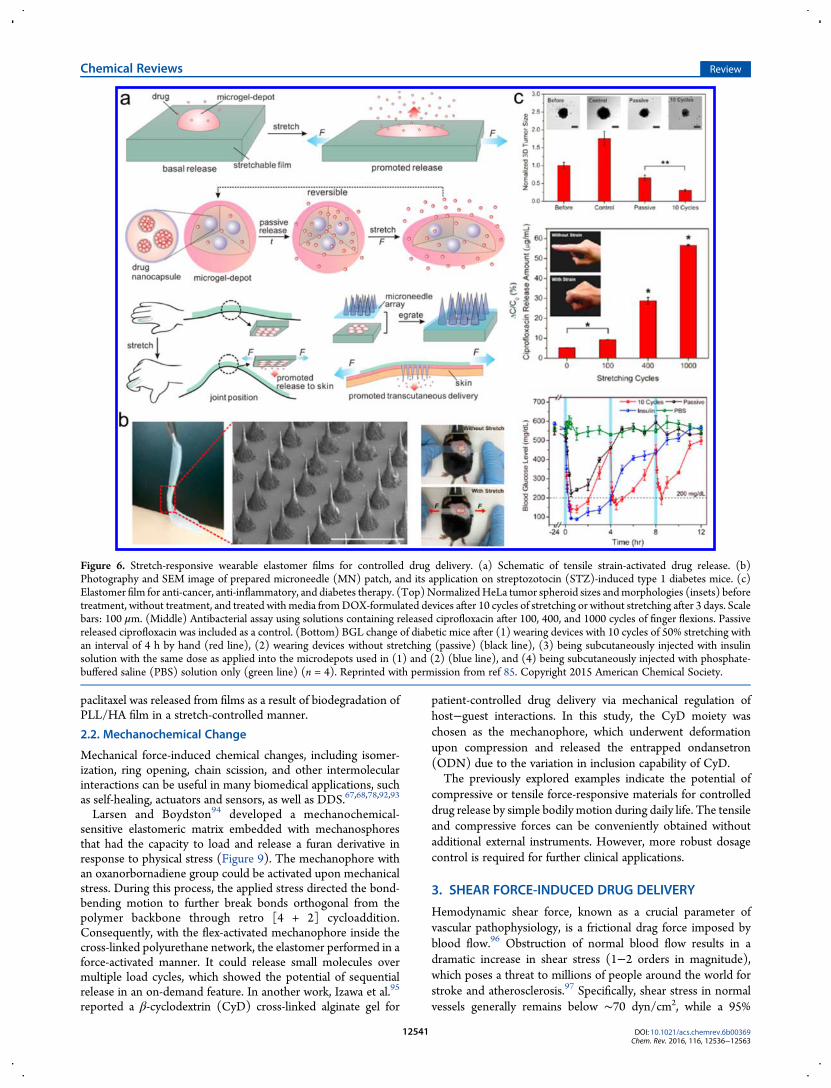
a mechanotransductive surface for reversible biocatalysisactivation. The multilayer film, made by a layer-by-layer (LbL)method, consisted of a poly(L-lysine)/hyaluronic acid (PLL/HA) film as a reservoir, which was loaded with alkalinephosphatase (ALP) enzymes and capped with a poly-(diallyldimethylammonium)/poly(styrenesulfonate) (PDAD-MA/PSS) film as a barrier (Figure 8a). ALP, a hydrolaseenzyme, can dephosphorylate fluorescein diphosphate (FDP).Upon stretching, the loaded ALP was exposed through thebarrier; then the biocatalysis was switched on and the progresswas shown in confocal fluorescent images (Figure 8b). Besidestriggering of biocatalysis via stretching, biocatalytic activity canalso be controlled by the thickness of the barrier film. Withrelatively thicker barrier layers, fewer enzymes could be broughtto the substrates, resulting in a lower jump of fluorescence(Figure 8c). Reversible stretch-triggered biocatalytic activationwas achieved in this LbL system. The same group further appliedsimilar biodegradable polyelectrolyte multilayers as a mechanor-esponsive drug delivery platform.89−91 Paclitaxel was embeddedin the PLL/HA reservoir, capped with a poly(allylaminehydrochloride)/poly(styrenesulfonate) (PAH/PSS) film as thebarrier. When immerged in trypsin solution, PLL inside thereservoir was cleaved by trypsin once the PAH/PSS barrier wasopened by mechanical stretch. Therefore, the entrapped
Figure 5. (a) Schematic of preparation of arrayed microcapsules. (b) Changes in the dimensions of microcapsules observed by AFM images and cross-section analysis: (i) before and (ii) after stretching. (c) Release behavior of (1) FITC-labeled dextran and (2) rhodamine B under different strains. (d)Amount of rhodamine B released upon stretching after consecutive stretching and release events. Repinted with permission from ref 84. Copyright 2011Wiley−VCH.
Chemical Reviews Review
DOI: 10.1021/acs.chemrev.6b00369Chem. Rev. 2016, 116, 12536−12563
12540
paclitaxel was released from films as a result of biodegradation ofPLL/HA film in a stretch-controlled manner.
2.2. Mechanochemical Change
Mechanical force-induced chemical changes, including isomer-ization, ring opening, chain scission, and other intermolecularinteractions can be useful in many biomedical applications, suchas self-healing, actuators and sensors, as well as DDS.67,68,78,92,93
Larsen and Boydston94 developed a mechanochemical-sensitive elastomeric matrix embedded with mechanosphoresthat had the capacity to load and release a furan derivative inresponse to physical stress (Figure 9). The mechanophore withan oxanorbornadiene group could be activated upon mechanicalstress. During this process, the applied stress directed the bond-bending motion to further break bonds orthogonal from thepolymer backbone through retro [4 + 2] cycloaddition.Consequently, with the flex-activated mechanophore inside thecross-linked polyurethane network, the elastomer performed in aforce-activated manner. It could release small molecules overmultiple load cycles, which showed the potential of sequentialrelease in an on-demand feature. In another work, Izawa et al.95
reported a β-cyclodextrin (CyD) cross-linked alginate gel for
patient-controlled drug delivery via mechanical regulation ofhost−guest interactions. In this study, the CyD moiety waschosen as the mechanophore, which underwent deformationupon compression and released the entrapped ondansetron(ODN) due to the variation in inclusion capability of CyD.The previously explored examples indicate the potential of
compressive or tensile force-responsive materials for controlleddrug release by simple bodily motion during daily life. The tensileand compressive forces can be conveniently obtained withoutadditional external instruments. However, more robust dosagecontrol is required for further clinical applications.
3. SHEAR FORCE-INDUCED DRUG DELIVERY
Hemodynamic shear force, known as a crucial parameter ofvascular pathophysiology, is a frictional drag force imposed byblood flow.96 Obstruction of normal blood flow results in adramatic increase in shear stress (1−2 orders in magnitude),which poses a threat to millions of people around the world forstroke and atherosclerosis.97 Specifically, shear stress in normalvessels generally remains below ∼70 dyn/cm2, while a 95%
Figure 6. Stretch-responsive wearable elastomer films for controlled drug delivery. (a) Schematic of tensile strain-activated drug release. (b)Photography and SEM image of prepared microneedle (MN) patch, and its application on streptozotocin (STZ)-induced type 1 diabetes mice. (c)Elastomer film for anti-cancer, anti-inflammatory, and diabetes therapy. (Top)NormalizedHeLa tumor spheroid sizes andmorphologies (insets) beforetreatment, without treatment, and treated with media fromDOX-formulated devices after 10 cycles of stretching or without stretching after 3 days. Scalebars: 100 μm. (Middle) Antibacterial assay using solutions containing released ciprofloxacin after 100, 400, and 1000 cycles of finger flexions. Passivereleased ciprofloxacin was included as a control. (Bottom) BGL change of diabetic mice after (1) wearing devices with 10 cycles of 50% stretching withan interval of 4 h by hand (red line), (2) wearing devices without stretching (passive) (black line), (3) being subcutaneously injected with insulinsolution with the same dose as applied into the microdepots used in (1) and (2) (blue line), and (4) being subcutaneously injected with phosphate-buffered saline (PBS) solution only (green line) (n = 4). Reprinted with permission from ref 85. Copyright 2015 American Chemical Society.
Chemical Reviews Review
DOI: 10.1021/acs.chemrev.6b00369Chem. Rev. 2016, 116, 12536−12563
12541

constricted stenotic or thrombosed artery possesses stress above1000 dyn/cm2.98−100
Although there are a multitude of stimuli-responsive DDS,covering redox, temperature, and external signals,101,102 as well asmany formulations such as liposomes and micelles,103−106 it stillremains challenging to construct on-demand drug delivery forcardiovascular diseases.107,108 Given the distinguishable shearforce in vascular fluid, shear-sensitive DDS may be an emergingoutlet for noninvasive therapy of vascular-related diseases.109−111
In this section, shear force-promoted DDS will be reviewed thatare based on either disaggregation or deformation of drugcarriers.3.1. Disaggregation
Platelets, components in blood with a long circulation time,aggregate and coagulate to cease bleeding during blood vesselinjuries.112 Once shear force increases, platelets sense this changeand respond by activating and adhering to the vascular wall atthese narrowed sites.99 Inspired by the physiological response ofplatelets, Ingber and co-workers72 designed microscale aggre-gates of nanoparticles to mimic platelets, which could remainintact in normal physiological flow. However, they broke up intoindividual nanocomponents once activated by high shear forceand then adhered and accumulated at stenotic regions. Thedisaggregated nanoparticles were exposed to much lower dragforce compared to the microscale aggregates, allowing forlocalization and accumulation on endothelial cells (Figure 10).On the other hand, an increase in total exposed surface area of theparticles further facilitated release of the payload that wasimmobilized on the nanoparticle surface. In vivo experimentsimplied that the microaggregates coated with tissue plasminogenactivator (tPA) rapidly dissolved the clot in 5 min in a mesentericinjury model (Figure 10c) and remarkably enhanced the survivalrate up to 80% in another fatal mouse pulmonary embolismmodel.
Most recently, Chen et al.113 took advantage of shear force as aweapon for antithrombus therapy (Figure 11a). Heparin, ananticoagulant drug, was formed into nanoparticles via hybrid-ization with polypeptide PLL. These positively charged heparinnanoparticles (cNPs) were then absorbed to negatively chargedred blood cells (RBCs) via electrostatic attraction for long-termcirculation. The large size of RBCs prevented the cNPs fromextravascular diffusion and benefited the drug’s intravascularrelease. On thrombus sites, the shear stress significantly increaseddue to narrowing of the blood vessels, which led to site-specificrelease of cNPs from RBCs. Scanning electron microscopic(SEM) images indicated that, with higher shear stress (10 Pa vs 1Pa), more desorption of cNPs from RBCs was observed after 24and 48 h (Figure 11b). With the help of RBCs as the drugdelivery vehicle, the drug could be delivered to targetdestinations with a prolonged circulation time.3.2. Deformation
Another strategy is to utilize shear force-induced deformation ofcarriers to release therapeutics. Budtova and co-workers114 firstpresented a shear-induced controlled release from hydrogels andmicrocapsules. They observed the solvent release from eitherhydrogels or microcapsules upon exposure to silicon oil flow, andover 30% deformation was generated by the flow shear stress.Their results showed that there was a threshold for shear stress toproduce deformation. Once above the critical point, the particleswould break up and release the cargo inside, which indicates thatshear force-induced release is feasible.115 Furthermore, theydesigned a microgel that could release contained fullerene by thefunction of shear and swelling.116 Holme et al.71 demonstratedthat lenticular-shaped vesicles could sense elevated shear stress(Figure 12a). The lenticular liposome was prepared from theartificial phospholipid Pad-PC-Pad through an extrusion method(Figure 12b). The two amide bonds of lipid can increase thestability of the polar region on the membrane via hydrogen
Figure 7. Stretch-triggered drug delivery based on superhydrophobic polymer composites. (a) Schematic of preparation and mechanism of tension-responsive system. The electrosprayed barrier coating (yellow) prevents unwanted drug release from the core (green). (b) SEM image of device cross-section and advancing water contact angles for electrosprayed hydrophobic (top, PCL) and superhydrophobic (bottom, PCL:PGC-C18 1:1) coatings.(c) Contrast-enhanced microcomputed tomography (mCT) images observed before and after tensile strain application and sequential images duringstretching along the x-axis. (d) Release profiles of model hydrophilic dye from the tension-responsive system via crack propagation. Reprinted withpermission from ref 87. Copyright 2016 Wiley−VCH.
Chemical Reviews Review
DOI: 10.1021/acs.chemrev.6b00369Chem. Rev. 2016, 116, 12536−12563
12542
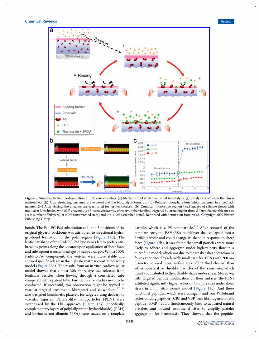
bonds. The Pad-PC-Pad substitution at 1- and 3-positions of theoriginal glycerol backbone was attributed to directional hydro-gen-bond formation in the polar region (Figure 12d). Thelenticular shape of the Pad-PC-Pad liposomes led to preferentialbreaking points along the equator upon application of shear forceand subsequent transient leakage of trapped cargos. With a 100%Pad-PC-Pad component, the vesicles were more stable andshowed specific release in the high-shear-stress constricted-arterymodel (Figure 12c). The results from an in vitro cardiovascularmodel showed that almost 30% more dye was released fromlenticular vesicles when flowing through a constricted tubecompared with a patent tube. Further in vivo studies need to beconducted. If successful, this observation might be applied invascular-targeted treatment. Mitragotri and co-workers117,118
also designed biomimetic platelets for targeted drug delivery tovascular injuries. Platelet-like nanoparticles (PLN) weresynthesized by the LbL approach (Figure 13a). Specifically,complementary layers of poly(allylamine hydrochloride) (PAH)and bovine serum albumin (BSA) were coated on a template
particle, which is a PS nanoparticle.118 After removal of thetemplate core, the PAH/BSA multlilayer shell collapsed into aflexible particle and could change its shape in response to shearforce (Figure 13b). It was found that small particles were morelikely to adhere and aggregate under high-velocity flow in amicrofluidmodel, which was due to the weaker shear detachmentforce experienced by relatively small particles. PLNs with 200 nmdiameter covered more surface area of the fluid channel thaneither spherical or disc-like particles of the same size, whichmainly contributed to their flexible shape under shear. Moreover,with targeted peptide modification on their surfaces, the PLNsexhibited significantly higher adhesion to injury sites under shearstress in an in vitro wound model (Figure 13c). And thesefunctional peptides, which were collagen- and von Willebrandfactor-binding peptides (CBP and VBP) and fibrinogen-mimeticpeptide (FMP), could simultaneously bind to activated naturalplatelets and injured endothelial sites to amplify plateletaggregation for hemostasis. They showed that the peptide-
Figure 8. Stretch-activated biodegradation of LbL reservoir films. (a) Mechanism of stretch-activated biocatalysis: (i) Catalysis is off when the film isunstretched. (ii) After stretching, enzymes are exposed and the biocatalysis turns on. (iii) Released phosphate ions inhibit enzymes in a feedbackmanner. (iv) After rinsing, the enzymes are reactivated for further catalysis. (b) Confocal microscope section (x,z) images of silicone sheets withmultilayer films loaded with ALP enzymes. (c) Biocatalytic activity of reservoir/barrier films triggered by stretching for three different barrier thicknesses(m = number of bilayers). α = 0% (unstretched state) and α = 110% (stretched state). Reprinted with permission from ref 88. Copyright 2009 NaturePublishing Group.
Chemical Reviews Review
DOI: 10.1021/acs.chemrev.6b00369Chem. Rev. 2016, 116, 12536−12563
12543

modified PLNs could reduce the tail bleeding time of mice by∼65% in a tail amputation test (Figure 13d).Besides the shear-induced deformation of drug carriers, shear
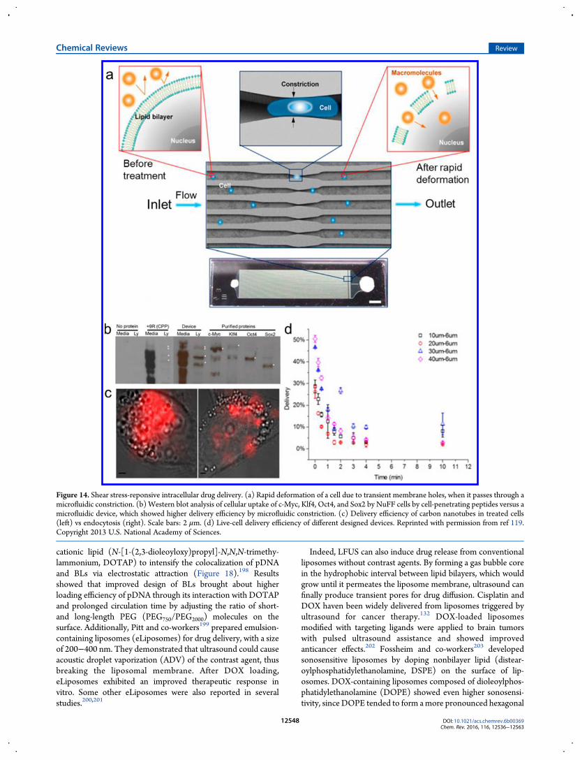
force can also be employed to temporarily disrupt cellmembranes for intracellular drug delivery.119 The Langer andJensen laboratories119 designed a microfluidic device that candeliver a range of materials, including macromolecules, into cells(Figure 14a). A relatively narrow channel (30−80% smaller thancell diameter) was fabricated in the device to producecompression and shear force. When cells, such as embryonicstem cells and immune cells, passed through the constrictedchannel, transient holes on the cell membrane were generated,which further enabled the diffusion of surrounding cargos acrossthe membrane. Moreover, it demonstrated 10-fold enhanceddelivery efficiency of transcription factors compared to cell-penetrating peptides (Figure 14b). Besides application in cellreprogramming, this technique also enabled direct delivery ofquantum dots, carbon nanotubes, and proteins to cell cytosol forprobe-based sensing (Figure 14c). The diffusion deliverymechanism was proved and characterized by monitoring thedelivery kinetics, which indicated 70−90% cargo delivery withinthe first minute (Figure 14d). The membrane disruption-basedmechanism offers flexibility in delivery, since it does not rely onexogenous materials or endocytotic pathways. Furthermore,unlike current existing methods, the microfluidic device showedno damage to either payloads or cells, and hence improvedbiological activities. Such capabilities of the technique expand itsprospects in therapeutic applications, and a company (SQZBiotech) was established on the basis of this concept, aiming toimprove the CellSqueeze platform for efficient cell therapy.
Thereafter, relevant research on interactions between micro-fluid and platelet mimetics or vesicles was conducted to promoteshear-responsive DDS.120−124 PEG [poly(ethylene glycol)]coating, multilamellar shell, and surface charge all affect thestability and uptake efficacy of carriers. The influence of plateletendothelial cell adhesion molecules (PECAMs) on shear stressdifferently regulated endothelial uptake of nanocarriers was alsoevaluated.125,126 These targeting agents, including PECAMs andglycoproteins, were adhered to nanocarriers for targetedendothelial drug delivery.127,128
The research discussed above strengthens the feasibility ofshear force-induced drug delivery for cardiovascular diseasetreatment. Shear force-sensitive drug carriers are able to achievespatial- and temporal-controlled release in a noninvasive manner.However, their clinical application is still limited due to severalchallenges such as short circulation time and safety issues.
4. ULTRASOUND-ACTIVATED DRUG DELIVERY
Ultrasound is a sound wave with frequencies above 20 kHz.129 Itcan generate longitudinal force/pressure that induces mechanicalforce or/and local heating in a noninvasive manner.130
Ultrasound has been widely used for therapeutic purposessince the beginning of the 20th century, including tissue ablation,kidney stone shattering, imaging, liposuction, and transdermaldrug delivery.131−135
Ultrasonic waves can map the location and promote drugrelease from carriers by causing localized hyperthermia, acousticcavitation, or/and acoustic radiation forces as designed.130 Theacoustic force can change the permeability or absorption oftissues and “push” the drug into the cells or across the tissue.Furthermore, it can also change the chemical properties of
Figure 9. (a) Schematics of flex activation in an oxanorbornadiene-based mechanophore. The oxanorbornadiene group is released upon mechanicalstress as a result of breaking bonds orthogonal from the polymer backbone through retro[4 + 2] cycloaddition. (b) Synthesis of polyurethane networks.Reprinted with permission from ref 94. Copyright 2014 American Chemical Society.
Chemical Reviews Review
DOI: 10.1021/acs.chemrev.6b00369Chem. Rev. 2016, 116, 12536−12563
12544

materials. In 1989, Kost et al.136 reported that ultrasound couldfacilitate the degradation process of polymer materials andsubsequently enhance the release of incorporated substances.Afterward, drug-loaded microbubbles were illustrated to beintentionally ruptured by ultrasound, which presented effectivespatiotemporal control of drug release with a nondestructivefeature.137 To date, a wide range of drug carriers, such asliposomes, micelles, nanoparticles, and micromotors, have beenexplored for ultrasound-triggered drug delivery.75,138−140
Ultrasound with frequency between 20 and 100 kHz is definedas low-frequency ultrasound (LFUS), which is usually used toinduce sonophoresis and affect drug carriers such as lip-osomes.132,141,142 LFUS was also applied to temporarily enhancepermeability of skin for transdermal drug delivery.143,144
Mitragotri et al.145,146 and others147 reported that large molecularproteins, such as insulin (∼6 kDa) and erythropoietin (∼48kDa), as well as other drugs can be transported across the skindue to the disorganization of tissue induced by LFUS. Recently, itwas reported that LFUS could locally promote drug delivery ingastrointestinal systems.148 In both mouse and pig models, itshowed treatment efficacy without significant safety issues.High-frequency ultrasound (HFUS), which has a frequency
greater than 1 MHz, is used to trigger oscillations inmicrobubbles.149 Additionally, it has been shown that the
acoustic pressure associated with cavitation derived fromultrasound waves can induce carrier destruction, drug release,and temporary increase in membrane permeability, thusfacilitating the cellular uptake of therapeutic agents.150−152
The following sections will mainly focus on nonthermallyactivated drug delivery. Since microbubbles have been widelydiscussed in a number of reviews,74,153−155 here we brieflyintroduce the most recent progress.
4.1. Microbubbles
Microbubbles, which are gas-filled spheres dispersed in aqueousmedium with an average size between 1 and 8 μm, have beenwidely employed in ultrasound imaging as ultrasound contrastagents.74,156 The bubbles experience oscillations under a HFUSfield when driven by a frequency near resonance and even canend up with a rupture.137,157 By decreasing the threshold forcavitation, the collapse of drug-encapsulating microbubbles canalso be used in ultrasound-activated drug delivery (Figure 15).158
Furthermore, Dayton and co-workers159 discovered that micro-bubbles serve as cavitation nuclei and can enhance ultrasoundenergy deposition in cells and tissues, thus facilitating intra-cellular drug transport by disruption of their membranes.160,161
Recently, numerous studies have been reported on theapplication of microbubbles for drug or gene delivery,particularly in brain tumor in situ/targeted treatment and
Figure 10. Shear force-triggered system for targeted delivery to obstructed blood vessels. (a) Preparation of platelets mimicking microscale aggregates.(b) Schematic of hemodynamic shear targeting to stenotic vessels by use of platelet mimetics. (c) Change of clot size after injection of tPA-coated shear-activated nanotherapeutics (SA-NTs) (left) compared to PBS treatment (right) in a partially occluded mesenteric artery. Scale bar, 100 mm. Reprintedwith permission from ref 72. Copyright 2012 American Association for the Advancement of Science.
Chemical Reviews Review
DOI: 10.1021/acs.chemrev.6b00369Chem. Rev. 2016, 116, 12536−12563
12545

cardiovascular therapy.73,155,159,162 Effective drug delivery tobrain is limited due to the blood−brain barrier (BBB) and theblood−tumor barrier (BTB).163−165 Focused ultrasound hasshown to be capable of transiently opening the BBB in thepresence of microbubbles.166−168 To date, a large number oftherapeutic agents have overcome the barrier and have beendelivered into the brain or a brain tumor.169,170 Yeh and co-workers171 designed superparamagnetic iron oxide nanoparticlesconjugating with doxorubicin-loaded microbubbles (DOX-SPIO-MBs) for imaging-guided drug delivery to brain tumors(Figure 16). It was found that the superparamagnetic/acousticproperties of DOX-SPIO-MBs were suitable for imaging.Ultrasound concurrently induced BBB opening and promoteddrug delivery, which enhanced the brain tumor treatment effect.They further modified targeting ligands on microbubbles tofacilitate directing chemotherapeutic agents to regions of tumorvasculature.172 Kovacs et al.173 also demonstrated that micro-bubbles might serve as an effective method for glioblastomamultiforme (GBM) treatment after investigating ultrasonicmicrobubble therapy in two GBM mouse models.Researchers have also attempted to apply ultrasound-triggered
microbubbles in ovarian cancer and prostate cancer treatment, aswell as in lymph nodes and in other tumor therapy.174−176
Attributed to the spatial and temporal control of drug deliverythat is feasible by ultrasound, microbubbles can aid in a variety ofdiseases therapy besides cancer. For example, drugs could bedelivered to the inner ear through the round window membrane,resulting in 3.5−38 times higher delivery by ultrasound than thatby spontaneous diffusion.177 In addition, genetic drugs could bespecifically delivered to the skeletal muscles of mice.178
Besides drugs encapsulated in microbubbles, some researchersdeveloped a new formulation with drug-loaded nanocarrierscoated on microbubbles. As reported in the literature, nano-particles have been linked to microbubbles through avidin−biotin interaction; however, the washing steps may influence thestability of the microbubbles.179−181 On the other hand, self-
Figure 11. Shear force-triggered system for antithrombus therapy. (a)Schematic of preparation of cNPs adsorbed to red blood cells (RBC-cNPs) and mechanism of shear-induced release of cNPs. (b) SEMimages showing shear force-triggered desorption of heparin/polypep-tide hybrid nanoparticles cross-linked with disulfide bonds (cNPs) fromred blood cells (RBCs) as a function of time. (A) RBC-adsorbed cNPs(RBC-cNPs) under static conditions. (B, C) RBC-cNPs under 10 Pashear-stress treatment for (B) 24 and (C) 48 h. (D, E) RBC-cNPs under1 Pa shear-stress treatment for (D) 24 and (E) 48 h. Scale bar = 4 μm.Reprinted with permission from ref 113. Copyright 2016 Wiley−VCH.
Figure 12. Shear force-responsive lenticular vesicles for targeted drug delivery. (a) Mechanism of shear force-activated drug delivery. (b) Cryo-TEM ofPad-PC-Pad vesicles. (c) Release behavior of L-α-phosphatidylcholine (eggPC) vesicles with different Pad-PC-Pad at 37 °C before (untreated) and afterone pass only through either healthy or constricted arterial models. (d) Chemical structure of phospholipid Pad-PC-Pad. Reprinted with permissionfrom ref 71. Copyright 2012 Nature Publishing Group.
Chemical Reviews Review
DOI: 10.1021/acs.chemrev.6b00369Chem. Rev. 2016, 116, 12536−12563
12546

assembled microbubbles loaded with liposome have recentlyshown their potential as a promising drug delivery carrier tocancer tissues, which could eliminate cancer cells with a very lowdose of therapeutics (0.5 μg/mL DOX) as demonstrated by DeSmedt and co-workers.182 Drug-loaded liposomes, formed withfunctionalized (SPDP-)PEG-lipids, were conjugated to themicrobubbles containing the hydrophobic perfluorobutane(C4F10) gas through thiol−maleimide linkages. Approximately600−1300 liposomes were bound per single microbubble, andthe size of the microbubbles increased from 3.6 to 4.0 μm afterconjugation with liposomes (diameter around 200 nm),indicating the formation of a single liposome layer coated onthe surface of the microbubbles. The same group183 designed N-cadherin-targeted liposome-loaded microbubbles for circulatingtumor cell (CTC)-specific drug delivery (Figure 17). Theirresults confirmed that the targeted antibodies specificallyadhered to the target cells and that focused ultrasound helpedtransport small molecules (propidium iodide) into the cells.Sanders and co-workers184 further proved that liposome-decorated microbubbles could enhance ultrasound-mediateddrug delivery in vivo. They chose indocyanine green (ICG) as amodel drug for intravenous injection inmice andmonitored localICG deposition post-ultrasonic therapy via fluorescence imaging.
4.2. Liposomes
The relatively large size of microbubbles results in a shorterretention time compared to nanocarriers. The inevitableretention in the lungs also restricts the use of microbubbles in
cardiovascular targets and tumor endothelia. Hence, acousticallyactivated liposomes, sometimes called nanobubbles, havebecome more prominent in ultrasonic drug delivery.132,185,186
This kind of ultrasound-responsive liposomes (echogenicliposomes), which are typically formed with a gas core coveredwith a lipid monolayer, were initially studied for ultrasoundimaging as contrast agents.129,187,188 Lin and Thomas189 foundthat LFUS could control the release of the payload from drug-encapsulating echogenic liposomes with nonthermal effects.Under the appropriate ultrasound frequency, echogenic lip-osomes rapidly grew, oscillated, collapsed, and spewed payloadby cavitation. Several factors like wave frequency, membraneconstituents, encapsulated gas or drug, presented media, and sizeand shell thickness of liposomes can affect the stability ofliposomes and liposomal drug release.190−193
Liposomes containing monolayer lipid-covered bubbles areknown as bubble liposomes (BLs).194−196 Aramaki and co-workers197 employed BLs along with ultrasound exposure todirectly deliver plasmid DNA (pDNA) to skeletal muscles bylocal injection for peripheral artery disease treatment. PEG-modified BLs with perfluoropropane gas were easily fabricatedthrough a reverse-phase evaporation method. It was found thathigh gene transfection efficiency was achieved in the muscleadministrated with BLs and ultrasound in a hind-limb ischemiamice model, as well as an increase in capillary density and bloodflow. Furthermore, in order to deliver genes to deeper tissues,novel BLs were explored for systemic injection, by use of a
Figure 13. (a) Synthesis of platelet-like nanoparticles (PLNs). (b) SEM images of (i) sacrificial 200 nm spherical polystyrene (PS) templates, (ii) PAH/BSA-coated PS templates, and (iii) final PLNs. Scale bars = 200 nm. (c) In vitro binding of PLNs with and without functional peptides to targetingsurface and nontargeting BSA surface (control) under shear stress. (d) Bleeding time was reduced after treatment of PLNs in a tail amputation model inmice. Reprinted with permission from ref 118. Copyright 2014 American Chemical Society.
Chemical Reviews Review
DOI: 10.1021/acs.chemrev.6b00369Chem. Rev. 2016, 116, 12536−12563
12547
cationic lipid (N-[1-(2,3-dioleoyloxy)propyl]-N,N,N-trimethy-lammonium, DOTAP) to intensify the colocalization of pDNAand BLs via electrostatic attraction (Figure 18).198 Resultsshowed that improved design of BLs brought about higherloading efficiency of pDNA through its interaction with DOTAPand prolonged circulation time by adjusting the ratio of short-and long-length PEG (PEG750/PEG2000) molecules on thesurface. Additionally, Pitt and co-workers199 prepared emulsion-containing liposomes (eLiposomes) for drug delivery, with a sizeof 200−400 nm. They demonstrated that ultrasound could causeacoustic droplet vaporization (ADV) of the contrast agent, thusbreaking the liposomal membrane. After DOX loading,eLiposomes exhibited an improved therapeutic response invitro. Some other eLiposomes were also reported in severalstudies.200,201
Indeed, LFUS can also induce drug release from conventionalliposomes without contrast agents. By forming a gas bubble corein the hydrophobic interval between lipid bilayers, which wouldgrow until it permeates the liposome membrane, ultrasound canfinally produce transient pores for drug diffusion. Cisplatin andDOX haven been widely delivered from liposomes triggered byultrasound for cancer therapy.132 DOX-loaded liposomesmodified with targeting ligands were applied to brain tumorswith pulsed ultrasound assistance and showed improvedanticancer effects.202 Fossheim and co-workers203 developedsonosensitive liposomes by doping nonbilayer lipid (distear-oylphosphatidylethanolamine, DSPE) on the surface of lip-osomes. DOX-containing liposomes composed of dioleoylphos-phatidylethanolamine (DOPE) showed even higher sonosensi-tivity, since DOPE tended to form amore pronounced hexagonal
Figure 14. Shear stress-reponsive intracellular drug delivery. (a) Rapid deformation of a cell due to transient membrane holes, when it passes through amicrofluidic constriction. (b) Western blot analysis of cellular uptake of c-Myc, Klf4, Oct4, and Sox2 by NuFF cells by cell-penetrating peptides versus amicrofluidic device, which showed higher delivery efficiency by microfluidic constriction. (c) Delivery efficiency of carbon nanotubes in treated cells(left) vs endocytosis (right). Scale bars: 2 μm. (d) Live-cell delivery efficiency of different designed devices. Reprinted with permission from ref 119.Copyright 2013 U.S. National Academy of Sciences.
Chemical Reviews Review
DOI: 10.1021/acs.chemrev.6b00369Chem. Rev. 2016, 116, 12536−12563
12548

structure due to its unsaturated acyl chains.204 Thus, uponexposure to ultrasound, the phase transition of DOPE inducedmembrane perturbations, followed by drug release fromliposomes.
4.3. Nanoparticles
Polymeric micelles typically assembled by amphiphilic blockpolymers have been ubiquitously used in drug delivery fordecades.13 Micelles made from thermosensitive polymers can bedisrupted by ultrasound; therefore, ultrasound-assisted micellardrug delivery has been recently studied.205 Furthermore,mechanical action caused by pulsed ultrasound can also triggerdrug release from micelles. Many micelle-based DDS incorpo-rated with local sonication were reported as efficient tumor-selective treatments.206−208 Xia and co-workers209 studiedultrasound-responsive behavior of PLA-b-PEG [poly(lacticacid)-b-poly(ethylene glycol)] copolymer micelles under high-intensity focused ultrasound (HIFU) (Figure 19). Resultsrevealed that generated transient cavitation led to thedegradation of PLA-b-PEG under HIFU at the ultrasound focalspot. They also invented a dual-responsive micelle with a
disulfide bond that could be broken under reductive con-ditions.210 Thus, this micelle can be remotely controlled byHIFU and simultaneously stimulated by an intracellularbioreductive environment.Several publications have also reported that ultrasound can
promote gene delivery from nanoparticles. Gene transfection wasenhanced when complexes of genes and cationic lipids orpolymers were treated by ultrasound.211−213 Wang et al.214
investigated various acoustic parameters to facilitate the deliveryof microRNA from PLGA−PEG [poly(lactic-co-glycolic acid)−poly(ethylene glycol)] nanoparticles to the tumor site incooperation with microbubbles. Evidence showed a 7.9-foldincrease in delivery efficiency compared to treatment withoutultrasound. Concurrent application of ultrasound and sodiumlauryl sulfate could also enhance the passive transdermal deliveryefficacy of nanoparticles.215 In addition, mesoporous silicananoparticles (MSNs), as ultrasound contrast agents, were alsoreported to be useful for ultrasonic drug delivery.216 Resultsdemonstrated that the target antibody (herceptin) functionalizedMSNs could potentially be used for ultrasound activated imagingand tumor-specific treatment for breast cancer.
Figure 15. Illustration of application of drug-loaded microbubbles in drug delivery systems. Microbubbles can prevent drugs from degradation in thebloodstream and avoid uptake in unwanted areas. At the target site, the drug can be released and penetrate cell membranes by application of ultrasound.Reprinted with permission from ref 158. Copyright 2012 Elsevier.
Figure 16. Schematic of controlled release of DOX-SPIO-MBs into brain tissues triggered by ultrasound and enhanced SPIO deposition by externalmagnet targeting. Reprinted with permission from ref 171. Copyright 2013 Elsevier.
Chemical Reviews Review
DOI: 10.1021/acs.chemrev.6b00369Chem. Rev. 2016, 116, 12536−12563
12549

It has also been shown that nanodroplets (NDs) formed withlipid-stabilized low-boiling perfluorocarbon could release theircargo via interaction with ADV caused by ultrasound. Acousticdroplets were reported for ultrasound-mediated targeted drugdelivery.217−219 Yeh and co-workers220 prepared folate-deco-rated NDs (FA-CPT-NDs) entrapping camptothecin (CTP) asan acoustic drug carrier for targeted tumor theranostics in amouse xenograft model. The FA-CTP-NDs underwent a
transition shift from liquid to gas upon ADV, followed by releaseof CTP and apoptosis of the targeted cancer cells. Thisformulation exhibited enhanced chemical and mechanicalantitumor effects. Superparamagnetic iron oxide could also beembedded into nanodroplets to assist inmagnetic control of drugdeposition.221
Emulsion is known as a two-phase mixture with a dispersedphase surrounded by a continuous phase. Hydrocarbon orfluorocarbon liquids are two common dispersed phases that cancarry hydrophobic agents.162,222 Perfluorocarbon nanoemulsionsstabilized by copolymers could deliver lipophilic therapeutics tosolid tumors.223 Ultrasound would trigger ADV and reversiblytransport droplets to gas in PFCE (perfluoro-15-crown-5-ether)nanoemulsions. Desired therapeutic effects were observed inboth breast and pancreatic cancer animal models with ultrasonicdelivery of paclitaxel loaded in PFCE nanoemulsions. Thebiodistribution of nanoemulsions could be simultaneouslymonitored via ultrasonography. Stone and co-workers224
developed an ultrasound-sensitive gas-in-oil-in-water-in-oil tripleemulsion through a microfluidic method. The triple emulsionswere stable in the atmosphere; thus it was possible to reinforcethe emulsions through polymerization by UV light outside thedevice and turn it into gas-in-liquid-in-solid compound particles.When ultrasound was applied, the gas core induced breakage ofemulsions and released the encapsulated contents.
Figure 17. Schematic depictions of (a) a targeted liposome-loadedmicrobubble and (b) intercellular uptake into circulating tumor cells.Adapted with permission from ref 183. Copyright 2013 Wiley−VCH.
Figure 18. Schematic presentation of systemic gene delivery systems using bubble liposomes (BLs) entrapping ultrasound contrast gas. Thiscombination of BLs and ultrasound exposure provides an effective way for the delivery of plasmid DNA (pDNA) into skeletal muscles. Reprinted withpermission from ref 198. Copyright 2012 American Chemical Society.
Figure 19. Scheme of HIFU and redox dual-responsive process of PEG-S-S-PLA micelle. Upon HIFU irradiation or glutathione treatment,degradation of the PEG-S-S-PLA chain and disruption of micelles cantrigger drug release. Reprinted with permission from ref 210. Copyright2010 Royal Society of Chemistry.
Chemical Reviews Review
DOI: 10.1021/acs.chemrev.6b00369Chem. Rev. 2016, 116, 12536−12563
12550

4.4. Micro/nanomotors
Micro/nanomotors are a class of miniaturized syntheticmachines, which are capable of converting chemical or externalenergy to mechanical motion.225 Over the past decade,numerous artificial micro/nanomotors based on diversepropulsion mechanisms have been developed in a wide varietyof medical applications ranging from bioanalytics to drugdelivery.226−229 However, previously reported synthetic motorswere mostly propelled by chemically catalyzed gradients or byejecting bubbles that usually contained toxic ingredients likeH2O2.
230,231
Mallouk and co-workers232 utilized continuous or pulsedultrasound for the propulsion of metallic micromotors. Asultrasound-powered micromotors showed considerable promisein penetrating into deep tissues, Mallouk and co-workers233
further employed rod-shaped nanomotors into HeLa cells. Theydemonstrated that the desired intercellular uptake of goldnanorods was achieved with the assistance of ultrasound andwithout any chemical fuel, and insignificant toxicity was detectedtoward cells. Intrigued by their work, Wang and co-workers234
developed ultrasound-poweredmicrobullets that could penetrateand deform cellular tissues and deliver drugs to diseaseddestinations (Figure 20). These tubular microbullets loaded
with ultrasound contrast agent (perfluorocarbons, PFCs) insidethe conical-shaped microtubes could produce sufficient force tocontribute to the ADV of PFCs. Consequently, a “bulletlike”velocity of over 6 ms−1, approximately 100 times faster thancommon micromotors, was assessed, offering PFC-loadedmicromotors as a possible platform to deliver drugs throughthe tissue barrier. The same group developed a multifunctionalnanowire motor consisting of three metallic segments (Au−Ni−
Au) that was propelled by ultrasound and guided by magnet-ism.235 The pH-sensitive eletropolymerized polypyrrole−poly-(styrenesulfonate) (PPy−PSS) was linked to the Au−Ni−Aunanowire, serving as a drug carrier for a model drug, brilliantgreen (BG). Similarly, they used ultrasound to drive nanoporousAu nanowires with the drug inside toward the cancer tissue andtriggered the release by near-infrared light (NIR).236 Wu et al.237
fabricated polymer-based multilayer tubular nanorockets via LbLassembly assisted with a nanoporous template, which wereverified to efficiently transport drugs into cancer cells. Poly-(diallyldimethylammonium chloride)-stabilized platinum nano-paticles were incorporated into the inner layer (as framework ofthe nanorockets), while DOX was loaded into the outer layer (ascontainer of the nanorockets). The self-propelling nanorocketswere shown to partially penetrate into the HeLa cells and releaseDOX through ultrasound-mediated breaks of the outer layers.Recently, Wang and co-workers238 further developed a kind ofmicromotor based on natural particulates by inserting iron oxidenanoparticles inside red blood cells to prolong circulation timeand improve biocompatibility.
4.5. Microcapsules
Van Hest and co-workers239 fabricated a series of hollowbiodegradable polymeric microcapsules and studied themechanical effects of PFO−PLLA [perfluorooctanol−poly(lacticacid)] capsules with varied ratios of shell thickness to diameterunder different acoustic pressures. Thus, drug release fromPFO−PLLA capsules could be triggered independently in astepwise or selective fashion by control of mechanical index. Inanother work, Wang et al.240 utilized alginate microcapsules asdrug carriers incorporated with pulsating ultrasound to deliverdiclofenac for anti-inflammatory purposes, obtaining a 30%higher release rate in vivo.Mohwald and co-workers241 fabricatedpolyelectrolyte microcapsules with ZnO nanoparticles embed-ded in the shell, which resulted in considerably higher ultrasoundsensitivity. It was demonstrated that the shell thickness androughness is dependent on the number of ZnO nanoparticlelayers. Their different mechanical properties affected sensitivityto ultrasonic treatment. This ZnO nanocomposite showed itspotential as a drug carrier with the possibility of opening with theapplication of ultrasound.Moreover, lasers can also generate focused ultrasound for
spatiotemporal control of drug delivery. Gu and co-workers242
demonstrated a laser-generated focused ultrasound (LGFU)-triggered drug delivery device. Compared to the conventionalHIFU, LGFU can focus on a tight spot with a higher resolutionand reduce the heating effect on tissue. Different types of drugs,such as DOX for anticancer treatment and ciprofloxacin forantibacterial treatment, were encapsulated in PLGA nano-particles. Then the PLGA nanoparticles were further loadedinto alginate microgels. Upon applying LGFU onto microgels, asignificant promoted drug release was observed at the focal pointdue to the cavitation effect. They demonstrated thatspatiotemporally remote control of drug release could beachieved in in vitro antitumor and antibacterial studies viaapplication of LGFU.
4.6. Injectable Depots
Recently, several injectable and ultrasound-responsive depot-based DDS have also been developed for long-term admin-istration. For example, Gu and co-workers243 reported a smartnanonetwork that was able to regulate BGLs by ultrasound-triggered insulin release (Figure 21a). The gel-like 3D nano-scaffold was formed with alginate- and chitosan-coated PLGA
Figure 20. (a) Preparation of PFC-loaded microbullets. (b) Computer-aided graphics and coresponding experimental images of PFH-loadedmicrobullets (i) penetrating, (ii) cleaving, and (iii) expanding a tissuefollowing a pulsed ultrasound signal. Reprinted with permission from ref234. Copyright 2012 Wiley−VCH.
Chemical Reviews Review
DOI: 10.1021/acs.chemrev.6b00369Chem. Rev. 2016, 116, 12536−12563
12551

nanoparticles via electrostatic attraction. Insulin was loaded inthe nanoparticles, and the formed cohesive nanonetwork couldbe subcutaneously injected through a universal syringe due toreduced cohesive forces under high shear rate. The nanonetworkcould remain stably underneath the skin with tight adhesion ofoppositely charged nanoparticles. Once focused ultrasound wasapplied, the nanonetwork was partially dissociated, and storedinsulin diffused out from the microchannels in the porousscaffold (Figure 21b). In particular, they demonstrated that therelease rate could be controlled by tuning the parameters offocused ultrasound such as input voltage, pulse duration, andultrasound administration time (Figure 21d). In vivo experi-ments showed that the insulin-loaded nanonetwork helpedregulate BGLs of type 1 diabetic mice at normal glycemia over aweek after treatment with focused ultrasound (Figure 21c).Kohane and co-workers244 developed an in situ cross-linking
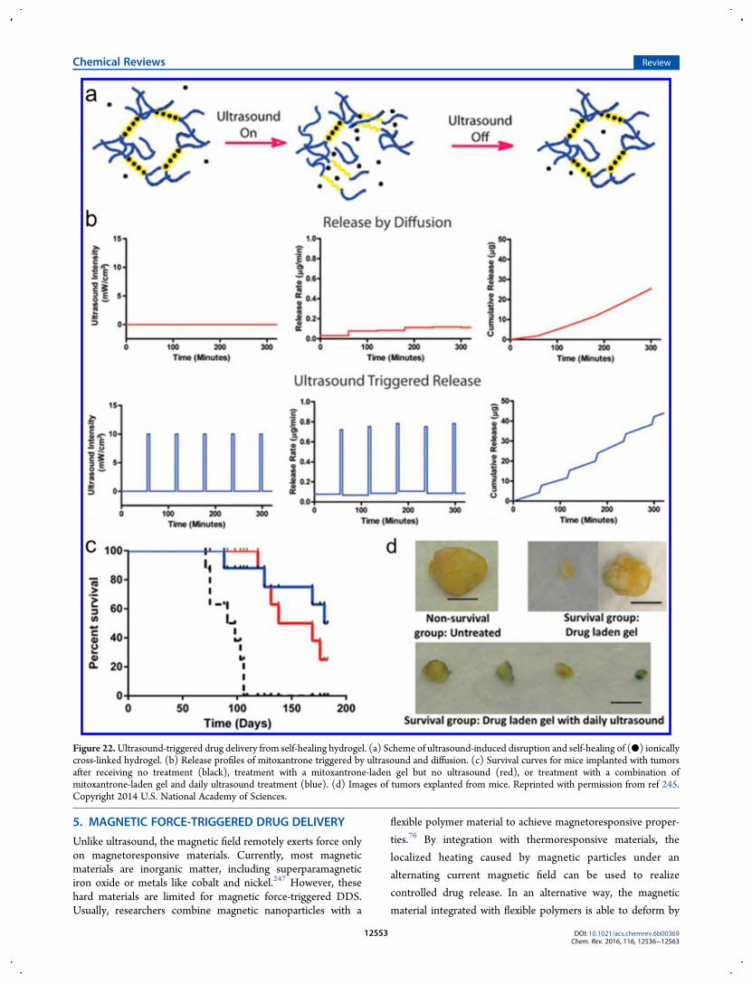
hydrogel for repeated on-demand ultrasound-triggered drugdelivery. This injectable hydrogel composed of drug-containingliposomes and gas-filled microbubbles presented a low baselinerate of drug release and prolonged the ultrasound triggerabilityfor over 14 days. Another ultrasound-responsive hydrogelinvented by Mooney and co-workers245 was reported as apromising DDS for sustained controlled release (Figure 22a).With the disruption induced by ultrasound and self-healing of
ionically cross-linked hydrogels, the switch of drug releasecorrespondingly turned on and off (Figure 22b,c). Resultsindicated a remarkable decrease of tumor size after dailyultrasound treatment compared to the control group (Figure22d). Similar polymers that can be ionically cross-linked arepossible candidates for ultrasonic drug delivery using thisapproach. Grinstaff and co-workers246 prepared superhydropho-bic meshes with poly(ε-caprolactone) (PCL) and poly(glycerolmonostearate-co-caprolactone) (PGC-C18) for ultrasonic drugdelivery. The air entrapped both at the material surface andwithin the structure of the 3D meshes could be removed upontreatment with HIFU, allowing cargo release from meshes.During recent decades, numerous ultrasound-sensitive drug
carriers have been developed for various medical applications.Compared to compressive/tensile and shear force-triggered drugdelivery systems, the ultrasound-activated method can realizeprecise and repeatable dosage control. Meanwhile, the highlyselective region for treatment can further avoid side effects.Despite this, the requirement for an external ultrasound sourcestill hardly satisfies the need for a convenient and practicaltreatment.
Figure 21. (a) Schematic illustration of ultrasound-mediated insulin delivery system using a nanonetwork. (b) Schematic of regulating BGLs in anoninvasive manner via ultrasonic therapy. (c) BGLs of STZ mice administrated with three cycles of ultrasound treatment. Solid columns indicate theadministration window, including 5 min anesthesia and 0.5 min FUS treatment. (d) Tunable ultrasound parameters that can affect insulin release rate.Reprinted with permission from ref 243. Copyright 2014 Wiley−VCH.
Chemical Reviews Review
DOI: 10.1021/acs.chemrev.6b00369Chem. Rev. 2016, 116, 12536−12563
12552
5. MAGNETIC FORCE-TRIGGERED DRUG DELIVERY
Unlike ultrasound, the magnetic field remotely exerts force onlyon magnetoresponsive materials. Currently, most magneticmaterials are inorganic matter, including superparamagneticiron oxide or metals like cobalt and nickel.247 However, thesehard materials are limited for magnetic force-triggered DDS.Usually, researchers combine magnetic nanoparticles with a
flexible polymer material to achieve magnetoresponsive proper-
ties.76 By integration with thermoresponsive materials, the
localized heating caused by magnetic particles under an
alternating current magnetic field can be used to realize
controlled drug release. In an alternative way, the magnetic
material integrated with flexible polymers is able to deform by
Figure 22.Ultrasound-triggered drug delivery from self-healing hydrogel. (a) Scheme of ultrasound-induced disruption and self-healing of (●) ionicallycross-linked hydrogel. (b) Release profiles of mitoxantrone triggered by ultrasound and diffusion. (c) Survival curves for mice implanted with tumorsafter receiving no treatment (black), treatment with a mitoxantrone-laden gel but no ultrasound (red), or treatment with a combination ofmitoxantrone-laden gel and daily ultrasound treatment (blue). (d) Images of tumors explanted from mice. Reprinted with permission from ref 245.Copyright 2014 U.S. National Academy of Sciences.
Chemical Reviews Review
DOI: 10.1021/acs.chemrev.6b00369Chem. Rev. 2016, 116, 12536−12563
12553

stretching, compressing, or bending to release the drug ondemand upon exposure to a magnetic field.248
Ferrogel, obtained by association of a ferrofluid and a hydrogel,was first reported as a magnetic force-responsive system byZrinyi et al. in 1996.248 They distributed monodomain magneticparticles with a diameter of 10 nm in a polymeric flexible networkby adhesive forces. The resulting gel, namely ferrogel, had theability to generate strain under a nonuniform magnetic field,leading to a controllable change in shape. Since then, a great dealof work on magnetic field-controlled drug delivery has beenreported by taking advantage of the deformation of ferrogelsunder a magnetic field.137,249−251 For instance, a poly(vinylalcohol) (PVA) hydrogel with Fe3O4 magnetic particles wascapable of accumulating drug when an external magnetic fieldwas applied.249,252 In contrast, the accumulated drug rapidlydiffused out of the ferrogel once themagnetic field was turned off.Due to high water content in the ferrogel, effective
incorporation of hydrophobic drugs is limited.253 In order toaddress this issue, Muhammed and co-workers254 appliedPluronic copolymer [poly(ethylene oxide)−poly(propyleneoxide)−poly(ethylene oxide)] as a gelling material toencapsulate hydrophobic drugs. Pluronic is a triblock copolymerconsisting of one poly(propylene oxide) (PPO) block and twopoly(ethylene oxide) (PEO) blocks. After incorporation ofindomethacin, a model hydrophobic drug, PPO segments canform a hydrophobic core to entrap it. In vitro release experiments
showed that the release rate significantly increased upon applyinga magnetic field. Besides small molecular drugs, Mooney and co-workers255 further applied the ferrogel as an active depot ofvarious cells to achieve on-demand cell delivery. Homogeneouspores with suitable size for cell adhesion were prepared byfreezing ferrogels at a specific temperature (Figure 23a). Unlikenanoporous ferrogel, the resulting macroporous gel showedreversible deformation at a compressive strain of 80% (Figure23b). A larger deformation was also achieved in macroporousferrogel under the same magnetic field (Figure 23c,d). Cell-binding peptides were further covalently conjugated onto thepolymer to support cell adhesion and viability. The results of invitro and in vivo tests indicated human dermal fibroblasts ormouse mesenchymal stem cells could be released under externalmagnetic stimulation. In particular, multiple stimulationparameters, including the strength of external magnetic field,frequency of stimulation, and number of cycles, are tunable tocontrol the cell release in this delivery system.In addition, an oscillating or alternating magnetic field (AMF)
can cause vibration of magnetic particles. By taking advantage ofthis phenomenon, Hu et al.256 designed a drug reservoir with amagnetism-sensitive shell. A fluorescent dye was encapsulated inthe silica core as a model drug, with a thin layer ofpoly(vinylpyrrolidone) (PVP)/Fe3O4 coating on the surface(Figure 24). The core/shell nanosphere was stable in aqueoussolution with no significant dye release after 24 h. Once exposed
Figure 23. (a) Hierarchical structure of macroporous ferrogels. Ferrogels with different pore sizes were prepared by freezing gels at differenttemperatures. (b) Stress vs strain curves for nanoporous and macroporous ferrogels subjected to compression tests. (c) Shape change in (top)nanoporous ferrogel and (bottom) macroporous ferrogel when subjected to a vertical magnetic field. (d) SEM images of a freeze-dried macroporousferrogel in the undeformed and deformed states. (Scale bar 500 μm) Reprinted with permission from ref 255. Copyright 2011 U.S. National Academy ofSciences.
Chemical Reviews Review
DOI: 10.1021/acs.chemrev.6b00369Chem. Rev. 2016, 116, 12536−12563
12554
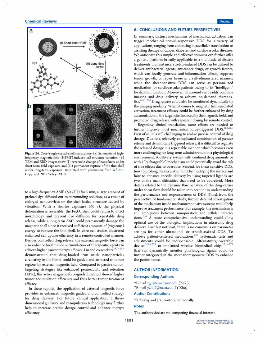
to a high-frequency AMF (50 kHz) for 5 min, a large amount ofpreload dye diffused out to surrounding solution, as a result ofenlarged nanocrevices on the shell lattice structure caused byvibration. With a shorter exposure (60 s), the physicaldeformation is reversible; the Fe3O4 shell could return to intactmorphology and prevent dye diffusion for repeatable drugrelease, while a long-term AMF could permanently damage themagnetic shell since it received sufficient amounts of (vigorous)energy to rupture the thin shell. In vitro cell studies illustratedenhanced cell uptake efficiency in a remote-controlled manner.Besides controlled drug release, the external magnetic force canalso enhance local tumor accumulation of therapeutic agents toachieve higher cancer therapy efficacy. Liu and co-workers257−259
demonstrated that drug-loaded iron oxide nanoparticlescirculating in the blood could be guided and attracted to tumorregions by external magnetic field. Compared to passive tumor-targeting strategies like enhanced permeability and retention(EPR), this active magnetic force-guided method showed highertumor accumulation efficiency and thus better tumor treatmentefficacy.In these reports, the application of external magnetic force
provides an enhanced magnetic guided and controlled strategyfor drug delivery. For future clinical application, a three-dimensional guidance and manipulation technology may furtherhelp to increase precise dosage control and enhance therapyefficiency.
6. CONCLUSIONS AND FUTURE PERSPECTIVES
In summary, distinct mechanisms of mechanical actuation cantrigger mechanical stimuli-responsive DDS for a variety ofapplications, ranging from enhancing intracellular transfection toassisting therapy of cancer, diabetes, and cardiovascular diseases.We anticipate this simple and effective stimulus can further offera generic platform broadly applicable to a multitude of diseasetreatments. For instance, stretch-induced DDS can be utilized todeliver antibacterial agents, anticancer drugs, or growth factors,which can locally generate anti-inflammation effects, suppresstumor growth, or repair tissue in a self-administered manner;while the shear-sensitive DDS can serve as personalizedmedication for cardiovascular patients owing to its “intelligent”localization function. Moreover, ultrasound can readily combineimaging and drug delivery to achieve on-demand theranos-tics.260−263 Drug release could also be monitored dynamically bythe imaging modality. When it comes to magnetic field-mediatedactivation, treatment efficacy could be further enhanced by drugaccumulation in the target site, induced by themagnetic field, andpromoted drug release with repeated dosing by remote control.Regarding clinical translation, more efforts are needed to
further improve most mechanical force-triggered DDS.264,265
First of all, it is still challenging to realize precise control of drugdosage. Due to a relatively complicated combination of passiverelease and dynamically triggered release, it is difficult to regulatethe released dosage in a repeatable manner, which becomes evenmore challenging for long-term administration in a physiologicalenvironment. A delivery system with confined drug amounts orwith a “rechargeable”mechanism could potentially avoid the riskof side effects due to overdose. Second, for shear-sensitive DDS,how to prolong the circulation time by modifying the surface andhow to enhance specific delivery by using targeted ligands aretwo of the main difficulties that need to be addressed. Moredetails related to the dynamic flow behavior of the drug carrierunder shear flow should be taken into account in understandingthe performance and responsiveness of DDS. Third, from theperspective of fundamental study, further detailed investigationof the mechanism inside mechanoresponsive systems would helpimprove treatment performance. For example, the mechanism isstill ambiguous between sonoporation and cellular interac-tions.266 A more comprehensive understanding could allowoptimal use of the biological implications in ultrasonic drugdelivery. Last but not least, there is no consensus on parametersettings for either ultrasound- or stretch-assisted DDS. Toachieve patient-centered medications,267 systematic tests andadjustments could be indispensable. Alternatively, wearabledevices268−270 or implanted wireless biomedical chips271−273
that can dynamically monitor physiological signals could befurther integrated in the mechanoresponsive DDS to enhancethe performance.
AUTHOR INFORMATION
Corresponding Authors
*E-mail [email protected] (Z.G.).*E-mail [email protected] (Y.Zhu).
Author Contributions⊥Y.Zhang and J.Y. contributed equally.
Notes
The authors declare no competing financial interest.
Figure 24. Core/single-crystal-shell nanosphere. (a) Schematic of high-frequency magnetic field (HFMF)-induced cell structure variation. (b)TEM and XRD images show (I) reversible change of nanofaults undershort-term field exposure and (II) permanent rupture of the thin shellunder long-term exposure. Reprinted with permission from ref 256.Copyright 2008 Wiley−VCH.
Chemical Reviews Review
DOI: 10.1021/acs.chemrev.6b00369Chem. Rev. 2016, 116, 12536−12563
12555

Biographies
Yuqi Zhang obtained her B.S. in chemistry in 2015 from NanjingUniversity. She is currently a Ph.D. student, cosupervised by ProfessorZhen Gu in the Joint Department of Biomedical Engineering at theUniversity of North Carolina at Chapel Hill and North Carolina StateUniversity and Professor Yong Zhu in the Department of Mechanicaland Aerospace Engineering at North Carolina State University. Herresearch interests include stimuli-responsive biomaterials and controlleddrug delivery.
Jicheng Yu received his B.S. in chemistry in 2011, followed by a M.S. inpolymer chemistry and physics fromNanjing University. He is currentlya Ph.D. student in Professor Zhen Gu’s laboratory in the JointDepartment of Biomedical Engineering at the University of NorthCarolina at Chapel Hill andNorth Carolina State University. His currentresearch interests include nanomedicine and bioinspired materials fordiabetes and cancer therapy.
Hunter Bomba currently attendsNorth Carolina State Univeristy, whereshe is working toward a B.S. in biomedical engineering. Hunter joinedProfessor Zhen Gu’s laboratory in the Joint Department of BiomedicalEngineering at the University of North Carolina at Chapel Hill andNorth Carolina University in 2014. Her research interests includecontrolled drug delivery and immuno-onocology.
Yong Zhu received his M.S. and Ph.D. in mechanical engineering fromNorthwestern University. After a postdoctoral fellow position at theUniversity of Texas at Austin, he joined the Department of Mechanicaland Aerospace Engineering at North Carolina State University in 2007,where he is currently an associate professor. His group conductsresearch in MEMS/NEMS, nanomechanics, and nanomaterial-enabledstretchable/wearable electronics.
Zhen Gu obtained his Ph.D. at the University of California, Los Angeles,under the guidance of Professor Yi Tang in the Department of Chemicaland Biomolecular Engineering. He was a postdoctoral associate workingwith Professor Robert Langer at MIT and Harvard Medical School. Heis currently an associate professor in the Joint Department of BiomedicalEngineering at University of North Carolina at Chapel Hill and NorthCarolina State University. He also holds a joint position in the EshelmanSchool of Pharmacy and Department of Medicine at UNC. His groupstudies controlled drug delivery, bioinspired materials, and nano-biotechnology, especially for cancer and diabetes treatment.
ACKNOWLEDGMENTSThis work was supported by grants from the American DiabetesAssociation (ADA; 1-14-JF-29 and 1-15-ACE-21), JDRF (3-SRA-2015-117-Q-R), and NC TraCS, NIH’s Clinical andTranslational Science Awards (CTSA) at UNC-CH(1UL1TR001111) to Z.G. and by the National ScienceFoundation (NSF) through ASSIST Engineering ResearchCenter at NC State (EEC-1160483) and EFRI-1240438 to Y.Z.
REFERENCES(1) Mitragotri, S.; Lahann, J. Materials for Drug Delivery: InnovativeSolutions to Address Complex Biological Hurdles. Adv. Mater. 2012, 24(28), 3717−3723.(2) Chow, E. K.-H.; Ho, D. Cancer Nanomedicine: from DrugDelivery to Imaging. Sci. Transl. Med. 2013, 5 (216), 216rv4.(3) Wilhelm, S.; Tavares, A. J.; Dai, Q.; Ohta, S.; Audet, J.; Dvorak, H.F.; Chan, W. C. Analysis of Nanoparticle Delivery to Tumours.Nat. Rev.Mater. 2016, 1, 16014.(4) Tibbitt, M. W.; Dahlman, J. E.; Langer, R. Emerging Frontiers inDrug Delivery. J. Am. Chem. Soc. 2016, 138 (3), 704−717.(5) Allen, T. M.; Cullis, P. R. Drug Delivery Systems: Entering theMainstream. Science 2004, 303 (5665), 1818−1822.
(6) Rosen, H.; Abribat, T. The Rise and Rise of Drug Delivery. Nat.Rev. Drug Discovery 2005, 4 (5), 381−385.(7) Peer, D.; Karp, J. M.; Hong, S.; Farokhzad, O. C.; Margalit, R.;Langer, R. Nanocarriers as an Emerging Platform for Cancer Therapy.Nat. Nanotechnol. 2007, 2 (12), 751−760.(8) Farokhzad, O. C.; Langer, R. Impact of Nanotechnology on DrugDelivery. ACS Nano 2009, 3 (1), 16−20.(9) Hu, Q.; Sun, W.; Wang, C.; Gu, Z. Recent Advances of CocktailChemotherapy by Combination drug Delivery Systems. Adv. DrugDelivery Rev. 2016, 98, 19−34.(10) Anselmo, A. C.; Mitragotri, S. Impact of Particle Elasticity onParticle-Based Drug Delivery Systems. Adv. Drug Delivery Rev. 2016,DOI: 10.1016/j.addr.2016.01.007.(11) Nishiyama, N.; Kataoka, K. Current State, Achievements, andFuture Prospects of Polymeric Micelles as Nanocarriers for Drug andGene Delivery. Pharmacol. Ther. 2006, 112 (3), 630−648.(12) de las Heras Alarcon, C.; Pennadam, S.; Alexander, C. StimuliResponsive Polymers for Biomedical Applications. Chem. Soc. Rev. 2005,34 (3), 276−285.(13) Rapoport, N. Physical Stimuli-Responsive Polymeric Micelles forAnti-Cancer Drug Delivery. Prog. Polym. Sci. 2007, 32 (8), 962−990.(14) Fleige, E.; Quadir, M. A.; Haag, R. Stimuli-Responsive PolymericNanocarriers for the Controlled Transport of Active Compounds:Concepts and Applications. Adv. Drug Delivery Rev. 2012, 64 (9), 866−884.(15) Qiu, Y.; Park, K. Environment-Sensitive Hydrogels for DrugDelivery. Adv. Drug Delivery Rev. 2012, 64, 49−60.(16) Mura, S.; Nicolas, J.; Couvreur, P. Stimuli-Responsive Nano-carriers for Drug Delivery. Nat. Mater. 2013, 12 (11), 991−1003.(17) Lu, Y.; Sun, W.; Gu, Z. Stimuli-Responsive Nanomaterials forTherapeutic Protein Delivery. J. Controlled Release 2014, 194, 1−19.(18) Brudno, Y.; Mooney, D. J. On-Demand Drug Delivery from LocalDepots. J. Controlled Release 2015, 219, 8−17.(19) Torchilin, V. P. Multifunctional, Stimuli-Sensitive Nano-particulate Systems for Drug Delivery. Nat. Rev. Drug Discovery 2014,13 (11), 813−827.(20) Karimi, M.; Ghasemi, A.; Zangabad, P. S.; Rahighi, R.; Basri, S. M.M.; Mirshekari, H.; Amiri, M.; Pishabad, Z. S.; Aslani, A.; Bozorgomid,M.; et al. Smart Micro/Nanoparticles in Stimulus-Responsive Drug/Gene Delivery Systems. Chem. Soc. Rev. 2016, 45 (5), 1457−1501.(21) Caldorera-Moore, M.; Peppas, N. A. Micro- and Nano-technologies for Intelligent and Responsive Biomaterial-Based MedicalSystems. Adv. Drug Delivery Rev. 2009, 61 (15), 1391−1401.(22) Park, K. Controlled Drug Delivery Systems: Past Forward andFuture Back. J. Controlled Release 2014, 190, 3−8.(23) Felber, A. E.; Dufresne, M.-H.; Leroux, J.-C. pH-SensitiveVesicles, Polymeric Micelles, and Nanospheres Prepared withPolycarboxylates. Adv. Drug Delivery Rev. 2012, 64 (11), 979−992.(24) Gao, W.; Chan, J. M.; Farokhzad, O. C. pH-ResponsiveNanoparticles for Drug Delivery. Mol. Pharmaceutics 2010, 7 (6),1913−1920.(25) Liu, J.; Huang, Y.; Kumar, A.; Tan, A.; Jin, S.; Mozhi, A.; Liang, X.J. pH-Sensitive Nano-Systems for Drug Delivery in Cancer Therapy.Biotechnol. Adv. 2014, 32 (4), 693−710.(26) Pillai, P. P.; Huda, S.; Kowalczyk, B.; Grzybowski, B. A. ControlledpH Stability and Adjustable Cellular Uptake of Mixed-ChargeNanoparticles. J. Am. Chem. Soc. 2013, 135 (17), 6392−6395.(27) Zhang, S.; Bellinger, A. M.; Glettig, D. L.; Barman, R.; Lee, Y.-A.L.; Zhu, J.; Cleveland, C.; Montgomery, V. A.; Gu, L.; Nash, L. D.; et al.A pH-Responsive Supramolecular Polymer Gel as an Enteric Elastomerfor Use in Gastric Devices. Nat. Mater. 2015, 14 (10), 1065−1071.(28) Liu, J.; Huang, Y.; Kumar, A.; Tan, A.; Jin, S.; Mozhi, A.; Liang, X.-J. pH-Sensitive Nano-Systems for Drug Delivery in Cancer Therapy.Biotechnol. Adv. 2014, 32 (4), 693−710.(29) McCarley, R. L. Redox-Responsive Delivery Systems. Annu. Rev.Anal. Chem. 2012, 5, 391−411.(30) Phillips, D. J.; Gibson, M. I. Redox-Sensitive Materials for DrugDelivery: Targeting the Correct Intracellular Environment, Tuning
Chemical Reviews Review
DOI: 10.1021/acs.chemrev.6b00369Chem. Rev. 2016, 116, 12536−12563
12556

Release Rates, and Appropriate Predictive Systems. Antioxid. RedoxSignaling 2014, 21 (5), 786−803.(31) Wang, M.; Sun, S.; Neufeld, C. I.; Perez-Ramirez, B.; Xu, Q.Reactive Oxygen Species-Responsive Protein Modification and ItsIntracellular Delivery for Targeted Cancer Therapy. Angew. Chem., Int.Ed. 2014, 53 (49), 13444−13448.(32) Ong, W.; Yang, Y.; Cruciano, A. C.; McCarley, R. L. Redox-Triggered Contents Release from Liposomes. J. Am. Chem. Soc. 2008,130 (44), 14739−14744.(33) Yu, J.; Zhang, Y.; Hu, X.; Wright, G.; Gu, Z. Hypoxia-SensitiveMaterials for Biomedical Applications. Ann. Biomed. Eng. 2016, 44 (6),1931−1945.(34) Perche, F.; Biswas, S.; Wang, T.; Zhu, L.; Torchilin, V. P. Hypoxia-Targeted siRNA Delivery. Angew. Chem., Int. Ed. 2014, 53 (13), 3362−3366.(35) Yu, J.; Zhang, Y.; Ye, Y.; DiSanto, R.; Sun, W.; Ranson, D.; Ligler,F. S.; Buse, J. B.; Gu, Z. Microneedle-Array Patches Loaded withHypoxia-Sensitive Vesicles Provide Fast Glucose-Responsive InsulinDelivery. Proc. Natl. Acad. Sci. U. S. A. 2015, 112 (27), 8260−8265.(36) Sun, W.; Gu, Z. ATP-Responsive Drug Delivery Systems. ExpertOpin. Drug Delivery 2016, 13, 311−314.(37) Mo, R.; Jiang, T.; DiSanto, R.; Tai, W.; Gu, Z. ATP-TriggeredAnticancer Drug Delivery. Nat. Commun. 2014, 5, No. 3364.(38) Lai, J.; Shah, B. P.; Zhang, Y.; Yang, L.; Lee, K.-B. Real-TimeMonitoring of ATP-Responsive Drug Release UsingMesoporous-Silica-CoatedMulticolor Upconversion Nanoparticles. ACSNano 2015, 9 (5),5234−5245.(39) Liao, W.-C.; Lu, C.-H.; Hartmann, R.; Wang, F.; Sohn, Y. S.;Parak, W. J.; Willner, I. Adenosine Triphosphate-Triggered Release ofMacromolecular and Nanoparticle Loads from Aptamer/DNA-Cross-Linked Microcapsules. ACS Nano 2015, 9 (9), 9078−9086.(40) Mo, R.; Jiang, T.; Di, J.; Tai, W.; Gu, Z. Emerging micro- andNanotechnology Based Synthetic Approaches for Insulin Delivery.Chem. Soc. Rev. 2014, 43 (10), 3595−3629.(41) Veiseh, O.; Tang, B. C.; Whitehead, K. A.; Anderson, D. G.;Langer, R. Managing Diabetes with Nanomedicine: Challenges andOpportunities. Nat. Rev. Drug Discovery 2015, 14 (1), 45−57.(42) Wu, Q.; Wang, L.; Yu, H.; Wang, J.; Chen, Z. Organization ofGlucose-Responsive Systems and Their Properties. Chem. Rev. 2011,111 (12), 7855−7875.(43) Webber, M. J.; Anderson, D. G.; Langer, R. EngineeringSynthetically Modified Insulin for Glucose-Responsive DiabetesTherapy. Expert Rev. Endocrinol. Metab. 2015, 10 (5), 483−489.(44) Rooseboom, M.; Commandeur, J. N. M.; Vermeulen, N. P. E.Enzyme-Catalyzed Activation of Anticancer Prodrugs. Pharmacol. Rev.2004, 56 (1), 53−102.(45) Hu, J.; Zhang, G.; Liu, S. Enzyme-Responsive PolymericAssemblies, Nanoparticles and Hydrogels. Chem. Soc. Rev. 2012, 41(18), 5933−5949.(46) Hu, Q.; Katti, P. S.; Gu, Z. Enzyme-Responsive Nanomaterials forControlled Drug Delivery. Nanoscale 2014, 6 (21), 12273−12286.(47) Gu, Z.; Dang, T. T.; Ma, M.; Tang, B. C.; Cheng, H.; Jiang, S.;Dong, Y.; Zhang, Y.; Anderson, D. G. Glucose-Responsive MicrogelsIntegrated with Enzyme Nanocapsules for Closed-Loop InsulinDelivery. ACS Nano 2013, 7 (8), 6758−6766.(48) Bernardos, A.; Mondragon, L.; Aznar, E.; Marcos, M. D.;Martínez-Manez, R.; Sancenon, F.; Soto, J.; Barat, J. M.; Perez-Paya, E.;Guillem, C.; et al. Enzyme-Responsive Intracellular Controlled ReleaseUsing Nanometric Silica Mesoporous Supports Capped with “Saccha-rides. ACS Nano 2010, 4 (11), 6353−6368.(49) Fischel-Ghodsian, F.; Brown, L.; Mathiowitz, E.; Brandenburg,D.; Langer, R. Enzymatically Controlled Drug Delivery. Proc. Natl. Acad.Sci. U. S. A. 1988, 85 (7), 2403−2406.(50) Sun, W.; Jiang, T.; Lu, Y.; Reiff, M.; Mo, R.; Gu, Z. Cocoon-LikeSelf-Degradable DNA Nanoclew for Anticancer Drug Delivery. J. Am.Chem. Soc. 2014, 136 (42), 14722−14725.(51) Choi, S. W.; Zhang, Y.; Xia, Y. A Temperature-Sensitive DrugRelease System Based on Phase-Change Materials. Angew. Chem., Int.Ed. 2010, 49 (43), 7904−7908.
(52) Yoo, J.-W.; Mitragotri, S. Polymer Particles that Switch Shape inResponse to a Stimulus. Proc. Natl. Acad. Sci. U. S. A. 2010, 107 (25),11205−11210.(53) Jeong, B.; Kim, S. W.; Bae, Y. H. Thermosensitive Sol−GelReversible Hydrogels. Adv. Drug Delivery Rev. 2002, 54 (1), 37−51.(54) Timko, B. P.; Dvir, T.; Kohane, D. S. Remotely Triggerable DrugDelivery Systems. Adv. Mater. 2010, 22 (44), 4925−4943.(55) Rai, P.; Mallidi, S.; Zheng, X.; Rahmanzadeh, R.; Mir, Y.;Elrington, S.; Khurshid, A.; Hasan, T. Development and Applications ofPhoto-Triggered Theranostic Agents. Adv. Drug Delivery Rev. 2010, 62(11), 1094−1124.(56) Melancon, M. P.; Zhou, M.; Li, C. Cancer Theranostics withNear-Infrared Light-Activatable Multimodal Nanoparticles. Acc. Chem.Res. 2011, 44 (10), 947−956.(57) Zhang, Y.; Yin, Q.; Yin, L.; Ma, L.; Tang, L.; Cheng, J. Chain-Shattering Polymeric Therapeutics with On-Demand Drug-ReleaseCapability. Angew. Chem., Int. Ed. 2013, 52 (25), 6435−6439.(58) Dobson, J. Magnetic Micro-and Nano-Particle-Based Targetingfor Drug and Gene Delivery. Nanomedicine 2006, 1 (1), 31−37.(59) Polyak, B.; Friedman, G. Magnetic Targeting for Site-SpecificDrug Delivery: Applications and Clinical Potential. Expert Opin. DrugDelivery 2009, 6 (1), 53−70.(60) Cai, K.; Luo, Z.; Hu, Y.; Chen, X.; Liao, Y.; Yang, L.; Deng, L.Magnetically Triggered Reversible Controlled Drug Delivery fromMicrofabricated Polymeric Multireservoir Devices. Adv. Mater. 2009, 21(40), 4045−4049.(61) Stanley, S. A.; Gagner, J. E.; Damanpour, S.; Yoshida, M.; Dordick,J. S.; Friedman, J. M. Radio-Wave Heating of Iron Oxide NanoparticlesCan Regulate Plasma Glucose in Mice. Science 2012, 336 (6081), 604−608.(62) Lakshmanan, S.; Gupta, G. K.; Avci, P.; Chandran, R.; Sadasivam,M.; Jorge, A. E. S.; Hamblin, M. R. Physical Energy for Drug Delivery;Poration, Concentration and Activation. Adv. Drug Delivery Rev. 2014,71, 98−114.(63) Ge, J.; Neofytou, E.; Cahill, T. J., III; Beygui, R. E.; Zare, R. N.Drug Release from Electric-Field-Responsive Nanoparticles. ACS Nano2012, 6 (1), 227−233.(64) Murdan, S. Electro-Responsive Drug Delivery from Hydrogels. J.Controlled Release 2003, 92 (1), 1−17.(65) Callister, W. D.; Rethwisch, D. G. Materials Science andEngineering: An Introduction; Wiley: New York, 2007; pp 665−715.(66) Bird, J.; Ross, C. Mechanical Engineering Principles; Routledge:Abingdon, U.K., 2014; pp 45−167.(67) Ariga, K.; Mori, T.; Hill, J. P. Mechanical Control ofNanomaterials and Nanosystems. Adv. Mater. 2012, 24 (2), 158−176.(68) Wiggins, K. M.; Brantley, J. N.; Bielawski, C. W. Methods forActivating and Characterizing Mechanically Responsive Polymers.Chem. Soc. Rev. 2013, 42 (17), 7130−7147.(69) McGinnis, P. M. Biomechanics of Sport and Exercise; HumanKinetics: Champaign, IL, 2013; pp 239−262.(70) Davies, P. F. Flow-Mediated Endothelial Mechanotransduction.Physiol. Rev. 1995, 75 (3), 519−560.(71) Holme, M. N.; Fedotenko, I. A.; Abegg, D.; Althaus, J.; Babel, L.;Favarger, F.; Reiter, R.; Tanasescu, R.; Zaffalon, P.-L.; Ziegler, A.; et al.Shear-Stress Sensitive Lenticular Vesicles for Targeted Drug Delivery.Nat. Nanotechnol. 2012, 7 (8), 536−543.(72) Korin, N.; Kanapathipillai, M.; Matthews, B. D.; Crescente, M.;Brill, A.; Mammoto, T.; Ghosh, K.; Jurek, S.; Bencherif, S. A.; Bhatta, D.;et al. Shear-Activated Nanotherapeutics for Drug Targeting toObstructed Blood Vessels. Science 2012, 337 (6095), 738−742.(73) Frenkel, V. Ultrasound Mediated Delivery of Drugs and Genes toSolid Tumors. Adv. Drug Delivery Rev. 2008, 60 (10), 1193−1208.(74)Hernot, S.; Klibanov, A. L.Microbubbles in Ultrasound-TriggeredDrug and Gene Delivery. Adv. Drug Delivery Rev. 2008, 60 (10), 1153−1166.(75) Sirsi, S. R.; Borden, M. A. State-of-the-Art Materials forUltrasound-Triggered Drug Delivery. Adv. Drug Delivery Rev. 2014,72, 3−14.
Chemical Reviews Review
DOI: 10.1021/acs.chemrev.6b00369Chem. Rev. 2016, 116, 12536−12563
12557

(76) Thevenot, J.; Oliveira, H.; Sandre, O.; Lecommandoux, S.Magnetic Responsive Polymer Composite Materials. Chem. Soc. Rev.2013, 42 (17), 7099−7116.(77) Hoffman, A. S. Bioconjugates of Intelligent Polymers andRecognition Proteins for Use in Diagnostics and Affinity Separations.Clin. Chem. 2000, 46 (9), 1478−1486.(78) Caruso, M. M.; Davis, D. A.; Shen, Q.; Odom, S. A.; Sottos, N. R.;White, S. R.; Moore, J. S. Mechanically-Induced Chemical Changes inPolymeric Materials. Chem. Rev. 2009, 109 (11), 5755−5798.(79)Moghadam,M. N.; Kolesov, V.; Vogel, A.; Klok, H.-A.; Pioletti, D.P. Controlled Release from aMechanically-Stimulated ThermosensitiveSelf-Heating Composite Hydrogel. Biomaterials 2014, 35 (1), 450−455.(80) Lee, K. Y.; Peters, M. C.; Anderson, K. W.; Mooney, D. J.Controlled Growth Factor Release from Synthetic ExtracellularMatrices. Nature 2000, 408 (6815), 998−1000.(81) Lee, K. Y.; Peters, M.;Mooney, D. Controlled DrugDelivery fromPolymers by Mechanical Signals. Adv. Mater. 2001, 13 (11), 837−839.(82) Xiao, L.; Zhu, J.; Londono, J. D.; Pochan, D. J.; Jia, X. Mechano-Responsive Hydrogels Crosslinked by Block Copolymer Micelles. SoftMatter 2012, 8 (40), 10233−10237.(83) Xiao, L.; Tong, Z.; Chen, Y.; Pochan, D. J.; Sabanayagam, C. R.;Jia, X. Hyaluronic Acid-Based Hydrogels Containing CovalentlyIntegrated Drug Depots: Implication for Controlling Inflammation inMechanically Stressed Tissues. Biomacromolecules 2013, 14 (11), 3808−3819.(84) Hyun, D. C.; Moon, G. D.; Park, C. J.; Kim, B. S.; Xia, Y.; Jeong, U.Strain-Controlled Release of Molecules from Arrayed MicrocapsulesSupported on an Elastomer Substrate. Angew. Chem., Int. Ed. 2011, 50(3), 724−727.(85) Di, J.; Yao, S.; Ye, Y.; Cui, Z.; Yu, J.; Ghosh, T. K.; Zhu, Y.; Gu, Z.Stretch-Triggered Drug Delivery from Wearable Elastomer FilmsContaining Therapeutic Depots. ACS Nano 2015, 9 (9), 9407−9415.(86) Kim, B.; Yoo, S.; Kim, Y. J.; Park, J.; Kang, B.; Haam, S.; Kang, S.W.; Kang, K.; Jeong, U. A Strain-Regulated, Refillable Elastic Patch forControlled Release. Adv. Mater. Interfaces 2016, 3 (9), No. 1500803.(87) Wang, J.; Kaplan, J. A.; Colson, Y. L.; Grinstaff, M. W. Stretch-Induced Drug Delivery from Superhydrophobic Polymer Composites:Use of Crack Propagation Failure Modes for Controlling Release Rates.Angew. Chem. 2016, 128 (8), 2846−2850.(88)Mertz, D.; Vogt, C.; Hemmerle, J.; Mutterer, J.; Ball, V.; Voegel, J.-C.; Schaaf, P.; Lavalle, P. Mechanotransductive Surfaces for ReversibleBiocatalysis Activation. Nat. Mater. 2009, 8 (9), 731−735.(89) Mertz, D.; Hemmerle, J.; Mutterer, J.; Ollivier, S.; Voegel, J.-C.;Schaaf, P.; Lavalle, P. Mechanically Responding Nanovalves Based onPolyelectrolyte Multilayers. Nano Lett. 2007, 7 (3), 657−662.(90) Barthes, J.; Mertz, D.; Bach, C.; Metz-Boutigue, M.-H. l. n.;Senger, B.; Voegel, J.-C.; Schaaf, P.; Lavalle, P. Stretch-InducedBiodegradation of Polyelectrolyte Multilayer Films for Drug Release.Langmuir 2012, 28 (38), 13550−13554.(91) Vogt, C.; Mertz, D.; Benmlih, K.; Hemmerle, J.; Voegel, J.-C.;Schaaf, P.; Lavalle, P. Layer-by-Layer Enzymatic Platform for Stretched-Induced Reactive Release. ACS Macro Lett. 2012, 1 (7), 797−801.(92) Brantley, J. N.; Bailey, C. B.; Wiggins, K. M.; Keatinge-Clay, A. T.;Bielawski, C. W. Mechanobiochemistry: Harnessing Biomacromole-cules for Force-ResponsiveMaterials. Polym. Chem. 2013, 4 (14), 3916−3928.(93) Davis, D. A.; Hamilton, A.; Yang, J.; Cremar, L. D.; Van Gough,D.; Potisek, S. L.; Ong, M. T.; Braun, P. V.; Martínez, T. J.; White, S. R.;et al. Force-Induced Activation of Covalent Bonds in Mechanores-ponsive Polymeric Materials. Nature 2009, 459 (7243), 68−72.(94) Larsen, M. B.; Boydston, A. J. Successive MechanochemicalActivation and Small Molecule Release in an Elastomeric Material. J. Am.Chem. Soc. 2014, 136 (4), 1276−1279.(95) Izawa, H.; Kawakami, K.; Sumita, M.; Tateyama, Y.; Hill, J. P.;Ariga, K. β-Cyclodextrin-Crosslinked Alginate Gel for Patient-Controlled Drug Delivery Systems: Regulation of Host-GuestInteractions with Mechanical Stimuli. J. Mater. Chem. B 2013, 1 (16),2155−2161.
(96) Malek, A. M.; Alper, S. L.; Izumo, S. Hemodynamic Shear Stressand Its Role in Atherosclerosis. JAMA 1999, 282 (21), 2035−2042.(97) Nesbitt, W. S.; Westein, E.; Tovar-Lopez, F. J.; Tolouei, E.;Mitchell, A.; Fu, J.; Carberry, J.; Fouras, A.; Jackson, S. P. A ShearGradient-Dependent Platelet Aggregation Mechanism Drives Throm-bus Formation. Nat. Med. 2009, 15 (6), 665−673.(98) Strony, J.; Beaudoin, A.; Brands, D.; Adelman, B. Analysis of ShearStress and Hemodynamic Factors in a Model of Coronary ArteryStenosis and Thrombosis. Am. J. Physiol. Heart Circ. Physiol. 1993, 265(5), H1787−H1796.(99) Wootton, D. M.; Ku, D. N. Fluid Mechanics of Vascular Systems,Diseases, and Thrombosis. Annu. Rev. Biomed. Eng. 1999, 1 (1), 299−329.(100) Bark, D. L.; Ku, D. N. Wall Shear over High Degree StenosesPertinent to Atherothrombosis. J. Biomech. 2010, 43 (15), 2970−2977.(101) Madamanchi, N. R.; Runge, M. S. Redox Signaling inCardiovascular Health and Disease. Free Radical Biol. Med. 2013, 61,473−501.(102) Sutton, J. T.; Haworth, K. J.; Pyne-Geithman, G.; Holland, C. K.Ultrasound-Mediated Drug Delivery for Cardiovascular Disease. ExpertOpin. Drug Delivery 2013, 10 (5), 573−592.(103) Fitzgerald, K. T.; Holladay, C. A.; McCarthy, C.; Power, K. A.;Pandit, A.; Gallagher, W. M. Standardization of Models and MethodsUsed to Assess Nanoparticles in Cardiovascular Applications. Small2011, 7 (6), 705−717.(104) Galvin, P.; Thompson, D.; Ryan, K. B.; McCarthy, A.; Moore, A.C.; Burke, C. S.; Dyson, M.; MacCraith, B. D.; Gun’ko, Y. K.; Byrne, M.T.; et al. Nanoparticle-Based Drug Delivery: Case Studies for Cancerand Cardiovascular Applications. Cell. Mol. Life Sci. 2012, 69 (3), 389−404.(105) Bowey, K.; Tanguay, J.-F.; Tabrizian, M. Liposome Technologyfor Cardiovascular Disease Treatment and Diagnosis. Expert Opin. DrugDelivery 2012, 9 (2), 249−265.(106) Rao, S.; Tan, A.; Thomas, N.; Prestidge, C. A. Perspective andPotential of Oral Lipid-Based Delivery to Optimize PharmacologicalTherapies against Cardiovascular Diseases. J. Controlled Release 2014,193, 174−187.(107) Nieuwlaat, R.; Schwalm, J.-D.; Khatib, R.; Yusuf, S. Why Are WeFailing to Implement Effective Therapies in Cardiovascular Disease?Eur. Heart J. 2013, 34 (17), 1262−1269.(108) Cicha, I.; Singh, R.; Garlichs, C. D.; Alexiou, C. Nano-Biomaterials for Cardiovascular Applications: Clinical Perspective. J.Controlled Release 2016, 229, 23−36.(109) Wootton, D. M.; Alevriadou, B. R. The Shear Stress of BustingBlood Clots. N. Engl. J. Med. 2012, 367 (14), 1361−1363.(110) Lavik, E.; Ustin, J. Leveraging Shear Stress to Bust Clots withNanoparticles. Science 2012, 337 (6095), 658−659.(111) Barenholz, Y. Nanomedicine: Shake up the Drug Containers.Nat. Nanotechnol. 2012, 7 (8), 483−484.(112) Ruggeri, Z. M. Platelets in Atherothrombosis. Nat. Med. 2002, 8(11), 1227−1234.(113) Chen, C.; Li, S.; Liu, K.; Ma, G.; Yan, X. Co-Assembly of Heparinand Polypeptide Hybrid Nanoparticles for Biomimetic Delivery andAnti-Thrombus Therapy. Small 2016, 12 (34), 4719−4725.(114) Zanina, A.; Vilesov, A.; Budtova, T. Shear-Induced SolventRelease from Gel Particles: Application to Drug-Delivery Systems. Int. J.Pharm. 2002, 242 (1), 137−146.(115) Zeo, U.; Tarabukina, E.; Budtova, T. Kinetics of Shear-InducedGel Deswelling/Solvent Release. J. Controlled Release 2005, 108 (1),73−83.(116) Tarabukina, E.; Zoolshoev, Z.; Melenevskaya, E.; Budtova, T.Delivery of Fullerene-Containing Complexes via Microgel Swelling andShear-Induced Release. Int. J. Pharm. 2010, 384 (1), 9−14.(117) Doshi, N.; Orje, J. N.; Molins, B.; Smith, J. W.; Mitragotri, S.;Ruggeri, Z. M. Platelet Mimetic Particles for Targeting Thrombi inFlowing Blood. Adv. Mater. 2012, 24 (28), 3864−3869.(118) Anselmo, A. C.; Modery-Pawlowski, C. L.; Menegatti, S.; Kumar,S.; Vogus, D. R.; Tian, L. L.; Chen, M.; Squires, T. M.; Sen Gupta, A.;Mitragotri, S. Platelet-Like Nanoparticles: Mimicking Shape, Flexibility,
Chemical Reviews Review
DOI: 10.1021/acs.chemrev.6b00369Chem. Rev. 2016, 116, 12536−12563
12558
and Surface Biology of Platelets to Target Vascular Injuries. ACS Nano2014, 8 (11), 11243−11253.(119) Sharei, A.; Zoldan, J.; Adamo, A.; Sim, W. Y.; Cho, N.; Jackson,E.; Mao, S.; Schneider, S.; Han, M.-J.; Lytton-Jean, A.; et al. A Vector-Free Microfluidic Platform for Intracellular Delivery. Proc. Natl. Acad.Sci. U. S. A. 2013, 110 (6), 2082−2087.(120) Pommella, A.; Caserta, S.; Guido, S. Dynamic Flow Behaviour ofSurfactant Vesicles under Shear Flow: Role of a MultilamellarMicrostructure. Soft Matter 2013, 9 (31), 7545−7552.(121) Hosta-Rigau, L.; Stadler, B. Shear Stress and Its Effect on theInteraction of Myoblast Cells with Nanosized Drug Delivery Vehicles.Mol. Pharmaceutics 2013, 10 (7), 2707−2712.(122) Teo, B. M.; van der Westen, R.; Hosta-Rigau, L.; Stadler, B. CellResponse to PEGylated Poly (dopamine) Coated LiposomesConsidering Shear Stress. Biochim. Biophys. Acta, Gen. Subj. 2013,1830 (10), 4838−4847.(123) Bjornmalm, M.; Yan, Y.; Caruso, F. Engineering and EvaluatingDrug Delivery Particles in Microfluidic Devices. J. Controlled Release2014, 190, 139−149.(124) Godoy-Gallardo, M.; Ek, P.; Jansman, M.; Wohl, B.; Hosta-Rigau, L. Interaction between Drug Delivery Vehicles and Cells underthe Effect of Shear Stress. Biomicrofluidics 2015, 9 (5), 052605.(125) Han, J.; Zern, B. J.; Shuvaev, V. V.; Davies, P. F.; Muro, S.;Muzykantov, V. Acute and Chronic Shear Stress Differently RegulateEndothelial Internalization of Nanocarriers Targeted to Platelet-Endothelial Cell Adhesion Molecule-1. ACS Nano 2012, 6 (10),8824−8836.(126) Han, J.; Shuvaev, V. V.; Davies, P. F.; Eckmann, D. M.; Muro, S.;Muzykantov, V. R. Flow Shear Stress Differentially RegulatesEndothelial Uptake of Nanocarriers Targeted to Distinct Epitopes ofPECAM-1. J. Controlled Release 2015, 210, 39−47.(127) Lin, A.; Sabnis, A.; Kona, S.; Nattama, S.; Patel, H.; Dong, J. F.;Nguyen, K. T. Shear-Regulated Uptake of Nanoparticles by EndothelialCells and Development of Endothelial-Targeting Nanoparticles. J.Biomed. Mater. Res., Part A 2010, 93A (3), 833−842.(128) Kona, S.; Dong, J.-F.; Liu, Y.; Tan, J.; Nguyen, K. T.Biodegradable Nanoparticles Mimicking Platelet Binding as a Targetedand Controlled Drug Delivery System. Int. J. Pharm. 2012, 423 (2),516−524.(129) Lentacker, I.; De Smedt, S. C.; Sanders, N. N. Drug LoadedMicrobubble Design for Ultrasound Triggered Delivery. Soft Matter2009, 5 (11), 2161−2170.(130) Coussios, C. C.; Roy, R. A. Applications of Acoustics andAavitation to Noninvasive Therapy and Drug Delivery. Annu. Rev. FluidMech. 2008, 40, 395−420.(131) Hynynen, K. Ultrasound for Drug and Gene Delivery to theBrain. Adv. Drug Delivery Rev. 2008, 60 (10), 1209−1217.(132) Schroeder, A.; Kost, J.; Barenholz, Y. Ultrasound, Liposomes,and Drug Delivery: Principles for Using Ultrasound to Control theRelease of Drugs from Liposomes. Chem. Phys. Lipids 2009, 162 (1), 1−16.(133) Deelman, L. E.; Decleves, A.-E.; Rychak, J. J.; Sharma, K.Targeted Renal Therapies through Microbubbles and Ultrasound. Adv.Drug Delivery Rev. 2010, 62 (14), 1369−1377.(134) Azagury, A.; Khoury, L.; Enden, G.; Kost, J. UltrasoundMediated Transdermal Drug Delivery. Adv. Drug Delivery Rev. 2014, 72,127−143.(135) Mitragotri, S. Healing Sound: the Use of Ultrasound in DrugDelivery and Other Therapeutic Applications. Nat. Rev. Drug Discovery2005, 4 (3), 255−260.(136) Kost, J.; Leong, K.; Langer, R. Ultrasound-Enhanced PolymerDegradation and Release of Incorporated Substances. Proc. Natl. Acad.Sci. U. S. A. 1989, 86 (20), 7663−7666.(137) Liu, Y.; Miyoshi, H.; Nakamura, M. Encapsulated UltrasoundMicrobubbles: Therapeutic Application in Drug/Gene Delivery. J.Controlled Release 2006, 114 (1), 89−99.(138) Ferrara, K. W.; Borden, M. A.; Zhang, H. Lipid-Shelled Vehicles:Engineering for Ultrasound Molecular Imaging and Drug Delivery. Acc.Chem. Res. 2009, 42 (7), 881−892.
(139) Unga, J.; Hashida, M. Ultrasound Induced Cancer Immuno-therapy. Adv. Drug Delivery Rev. 2014, 72, 144−153.(140) Kwok, C. S.; Mourad, P. D.; Crum, L. A.; Ratner, B. D. Self-Assembled Molecular Structures as Ultrasonically-Responsive BarrierMembranes for Pulsatile Drug Delivery. J. Biomed. Mater. Res. 2001, 57(2), 151−164.(141) Mitragotri, S.; Kost, J. Low-Frequency Sonophoresis: aNoninvasive Method of Drug Delivery and Diagnostics. Biotechnol.Prog. 2000, 16 (3), 488−492.(142) Ogura, M.; Paliwal, S.; Mitragotri, S. Low-FrequencySonophoresis: Current Status and Future Prospects. Adv. Drug DeliveryRev. 2008, 60 (10), 1218−1223.(143) Mitragotri, S.; Edwards, D. A.; Blankschtein, D.; Langer, R. AMechanistic Study of Ultrasonically-Enhanced Transdermal DrugDelivery. J. Pharm. Sci. 1995, 84 (6), 697−706.(144) Prausnitz, M. R.; Mitragotri, S.; Langer, R. Current Status andFuture Potential of Transdermal Drug Delivery.Nat. Rev. Drug Discovery2004, 3 (2), 115−124.(145) Mitragotri, S.; Blankschtein, D.; Langer, R. Ultrasound-Mediated Transdermal Protein Delivery. Science 1995, 269 (5225),850−853.(146) Mitragotri, S.; Kost, J. Transdermal Delivery of Heparin andLow-Molecular Weight Heparin Using Low-Frequency Ultrasound.Pharm. Res. 2001, 18 (8), 1151−1156.(147) Polat, B. E.; Deen,W.M.; Langer, R.; Blankschtein, D. A PhysicalMechanism to Explain the Delivery of Chemical Penetration Enhancersinto Skin During Transdermal Sonophoresis-Insight into the ObservedSynergism. J. Controlled Release 2012, 158 (2), 250−260.(148) Schoellhammer, C. M.; Schroeder, A.; Maa, R.; Lauwers, G. Y.;Swiston, A.; Zervas, M.; Barman, R.; DiCiccio, A. M.; Brugge, W. R.;Anderson, D. G.; et al. Ultrasound-Mediated Gastrointestinal DrugDelivery. Sci. Transl. Med. 2015, 7 (310), No. 310ra168.(149) Sboros, V. Response of Contrast Agents to Ultrasound. Adv.Drug Delivery Rev. 2008, 60 (10), 1117−1136.(150) Paliwal, S.; Mitragotri, S. Ultrasound-Induced Cavitation:Applications in Drug and Gene Delivery. Expert Opin. Drug Delivery2006, 3 (6), 713−726.(151) Wu, J.; Nyborg, W. L. Ultrasound, Cavitation Bubbles and TheirInteraction with Cells. Adv. Drug Delivery Rev. 2008, 60 (10), 1103−1116.(152) Foley, J. L.; Vaezy, S.; Crum, L. A. Applications of High-IntensityFocused Ultrasound in Medicine: Spotlight on Neurological Applica-tions. Applied acoustics 2007, 68 (3), 245−259.(153) Blomley, M. J.; Cooke, J. C.; Unger, E. C.; Monaghan, M. J.;Cosgrove, D. O. Microbubble Contrast Agents: a New Era inUltrasound. Br. Med. J. 2001, 322 (7296), 1222.(154) Sirsi, S. R.; Borden, M. A. Advances in Ultrasound MediatedGene Therapy Using Microbubble Contrast Agents. Theranostics 2012,2 (12), 1208−1222.(155) Unger, E.; Porter, T.; Lindner, J.; Grayburn, P. CardiovascularDrug Delivery with Ultrasound and Microbubbles. Adv. Drug DeliveryRev. 2014, 72, 110−126.(156) Polat, B. E.; Hart, D.; Langer, R.; Blankschtein, D. Ultrasound-Mediated Transdermal Drug Delivery: Mechanisms, Scope, andEmerging Trends. J. Controlled Release 2011, 152 (3), 330−348.(157) Morgan, K. E.; Allen, J. S.; Dayton, P. A.; Chomas, J. E.; Klibaov,A.; Ferrara, K. W. Experimental and Theoretical Evaluation ofMicrobubble Behavior: Effect of Transmitted Phase and Bubble Size.IEEE Trans. Ultrason. Ferroelectr. Freq. Control 2000, 47 (6), 1494−1509.(158) Geers, B.; Dewitte, H.; De Smedt, S. C.; Lentacker, I. CrucialFactors and Emerging Concepts in Ultrasound-Triggered DrugDelivery. J. Controlled Release 2012, 164 (3), 248−255.(159) Lum, A. F.; Borden, M. A.; Dayton, P. A.; Kruse, D. E.; Simon, S.I.; Ferrara, K. W. Ultrasound Radiation Force Enables TargetedDeposition of Model Drug Carriers Loaded on Microbubbles. J.Controlled Release 2006, 111 (1), 128−134.
Chemical Reviews Review
DOI: 10.1021/acs.chemrev.6b00369Chem. Rev. 2016, 116, 12536−12563
12559

(160) Kooiman, K.; Vos, H. J.; Versluis, M.; de Jong, N. AcousticBehavior ofMicrobubbles and Implications for DrugDelivery.Adv. DrugDelivery Rev. 2014, 72, 28−48.(161) Rychak, J. J.; Klibanov, A. L. Nucleic Acid Delivery withMicrobubbles and Ultrasound. Adv. Drug Delivery Rev. 2014, 72, 82−93.(162) Rapoport, N. Y.; Kennedy, A. M.; Shea, J. E.; Scaife, C. L.; Nam,K.-H. Controlled and Targeted Tumor Chemotherapy by Ultrasound-Activated Nanoemulsions/Microbubbles. J. Controlled Release 2009, 138(3), 268−276.(163) Aryal, M.; Arvanitis, C. D.; Alexander, P. M.; McDannold, N.Ultrasound-Mediated Blood-Brain Barrier Disruption for TargetedDrug Delivery in the Central Nervous System. Adv. Drug Delivery Rev.2014, 72, 94−109.(164) Cloughesy, T. F.; Black, K. L. Pharmacological Blood-BrainBarrier Modification for Selective Drug Delivery. J. Neuro-Oncol. 1995,26 (2), 125−132.(165) Khil, M. S.; Kolozsvary, A.; Apple, M.; Kim, J. H. IncreasedTumor Cures Using Combined Radiosurgery and BCNU in theTreatment of 9LGlioma in the Rat Brain. Int. J. Radiat. Oncol., Biol., Phys.2000, 47 (2), 511−516.(166) Hynynen, K.; McDannold, N.; Vykhodtseva, N.; Jolesz, F. A.Noninvasive MR Imaging-guided Focal Opening of the Blood-BrainBarrier in Rabbits 1. Radiology 2001, 220 (3), 640−646.(167) Kang, J.; Wu, X.; Wang, Z.; Ran, H.; Xu, C.; Wu, J.; Wang, Z.;Zhang, Y. Antitumor Effect of Docetaxel-Loaded Lipid MicrobubblesCombined with Ultrasound-Targeted Microbubble Activation on VX2Rabbit Liver Tumors. J. Ultrasound Med. 2010, 29 (1), 61−70.(168) Mitragotri, S. Devices for Overcoming Biological Barriers: theUse of Physical Forces to Disrupt the Barriers. Adv. Drug Delivery Rev.2013, 65 (1), 100−103.(169) Ting, C.-Y.; Fan, C.-H.; Liu, H.-L.; Huang, C.-Y.; Hsieh, H.-Y.;Yen, T.-C.; Wei, K.-C.; Yeh, C.-K. Concurrent Blood-Brain BarrierOpening and Local Drug Delivery Using Drug-Carrying Microbubblesand Focused Ultrasound for Brain Glioma Treatment. Biomaterials2012, 33 (2), 704−712.(170) Li, P.; Zheng, Y.; Ran, H.; Tan, J.; Lin, Y.; Zhang, Q.; Ren, J.;Wang, Z. Ultrasound Triggered Drug Release from 10-Hydroxycamp-tothecin-Loaded Phospholipid Microbubbles for Targeted TumorTherapy in Mice. J. Controlled Release 2012, 162 (2), 349−354.(171) Fan, C.-H.; Ting, C.-Y.; Lin, H.-J.; Wang, C.-H.; Liu, H.-L.; Yen,T.-C.; Yeh, C.-K. SPIO-Conjugated, Doxorubicin-Loaded Micro-bubbles for Concurrent MRI and Focused-Ultrasound EnhancedBrain-Tumor Drug Delivery. Biomaterials 2013, 34 (14), 3706−3715.(172) Fan, C.-H.; Ting, C.-Y.; Liu, H.-L.; Huang, C.-Y.; Hsieh, H.-Y.;Yen, T.-C.; Wei, K.-C.; Yeh, C.-K. Antiangiogenic-Targeting Drug-Loaded Microbubbles Combined with Focused Ultrasound for GliomaTreatment. Biomaterials 2013, 34 (8), 2142−2155.(173) Kovacs, Z.; Werner, B.; Rassi, A.; Sass, J. O.; Martin-Fiori, E.;Bernasconi, M. Prolonged Survival upon Ultrasound-EnhancedDoxorubicin Delivery in Two Syngenic Glioblastoma Mouse Models.J. Controlled Release 2014, 187, 74−82.(174) Niu, C.; Wang, Z.; Lu, G.; Krupka, T. M.; Sun, Y.; You, Y.; Song,W.; Ran, H.; Li, P.; Zheng, Y. Doxorubicin Loaded SuperparamagneticPLGA-Iron Oxide Multifunctional Microbubbles for Dual-Mode US/MR Imaging and Therapy of Metastasis in Lymph Nodes. Biomaterials2013, 34 (9), 2307−2317.(175) Eggen, S.; Fagerland, S.-M.; Mørch, Y.; Hansen, R.; Søvik, K.;Berg, S.; Furu, H.; Bøhn, A. D.; Lilledahl, M. B.; Angelsen, A.; et al.Ultrasound-Enhanced Drug Delivery in Prostate Cancer Xenografts byNanoparticles Stabilizing Microbubbles. J. Controlled Release 2014, 187,39−49.(176) Liu, L.; Chang, S.; Sun, J.; Zhu, S.; Yin,M.; Zhu, Y.;Wang, Z.; Xu,R. X. Ultrasound-Mediated Destruction of Paclitaxel and OxygenLoaded Lipid Microbubbles for Combination Therapy in OvarianCancer Xenografts. Cancer Lett. 2015, 361 (1), 147−154.(177) Shih, C.-P.; Chen, H.-C.; Chen, H.-K.; Chiang,M.-C.; Sytwu, H.-K.; Lin, Y.-C.; Li, S.-L.; Shih, Y.-F.; Liao, A.-H.; Wang, C.-H. Ultrasound-Aided Microbubbles Facilitate the Delivery of Dugs to the Inner Ear via
the Round Window Membrane. J. Controlled Release 2013, 167 (2),167−174.(178) Sanches, P. G.; Muhlmeister, M.; Seip, R.; Kaijzel, E.; Lowik, C.;Bohmer, M.; Tiemann, K.; Grull, H. Ultrasound-Mediated GeneDelivery of Naked Plasmid DNA in Skeletal Muscles: a Case for BolusInjections. J. Controlled Release 2014, 195, 130−137.(179) Kheirolomoom, A.; Dayton, P. A.; Lum, A. F.; Little, E.; Paoli, E.E.; Zheng, H.; Ferrara, K. W. Acoustically-Active MicrobubblesConjugated to Liposomes: Characterization of a Proposed DrugDelivery Vehicle. J. Controlled Release 2007, 118 (3), 275−284.(180) Klibanov, A. L.; Shevchenko, T. I.; Raju, B. I.; Seip, R.; Chin, C.T. Ultrasound-Triggered Release of Materials Entrapped in Micro-bubble-Liposome Constructs: a Tool for Targeted Drug Delivery. J.Controlled Release 2010, 148 (1), 13−17.(181) De Temmerman, M.-L.; Dewitte, H.; Vandenbroucke, R. E.;Lucas, B.; Libert, C.; Demeester, J.; De Smedt, S. C.; Lentacker, I.;Rejman, J. mRNA-Lipoplex Loaded Microbubble Contrast Agents forUltrasound-Assisted Transfection of Dendritic Cells. Biomaterials 2011,32 (34), 9128−9135.(182) Geers, B.; Lentacker, I.; Sanders, N. N.; Demeester, J.; Meairs, S.;De Smedt, S. C. Self-Assembled Liposome-Loaded Microbubbles: TheMissing Link for Safe and Efficient Ultrasound TriggeredDrug-Delivery.J. Controlled Release 2011, 152 (2), 249−256.(183) Geers, B.; DeWever, O.; Demeester, J.; Bracke, M.; De Smedt, S.C.; Lentacker, I. Targeted Liposome-Loaded Microbubbles for Cell-Specific Ultrasound-Triggered Drug Delivery. Small 2013, 9 (23),4027−4035.(184) Cool, S. K.; Geers, B.; Roels, S.; Stremersch, S.; Vanderperren,K.; Saunders, J. H.; De Smedt, S. C.; Demeester, J.; Sanders, N. N.Coupling of Drug Containing Liposomes to Microbubbles ImprovesUltrasound Triggered Drug Delivery in Mice. J. Controlled Release 2013,172 (3), 885−893.(185) Huang, S.-L. Liposomes in Ultrasonic Drug and Gene Delivery.Adv. Drug Delivery Rev. 2008, 60 (10), 1167−1176.(186) Hitchcock, K. E.; Caudell, D. N.; Sutton, J. T.; Klegerman, M. E.;Vela, D.; Pyne-Geithman, G. J.; Abruzzo, T.; Cyr, P. E.; Geng, Y.-J.;McPherson, D. D. Ultrasound-Enhanced Delivery of TargetedEchogenic Liposomes in a Novel Ex Vivo Mouse Aorta Model. J.Controlled Release 2010, 144 (3), 288−295.(187) Unger, E.; Shen, D.-K.; Fritz, T.; Lund, P.; Wu, G.-L.; Kulik, B.;DeYoung, D.; Standen, J.; Ovitt, T.; Matsunaga, T. Gas-FilledLiposomes as Echocardiographic Contrast Agents in Rabbits withMyocardial Infarcts. Invest. Radiol. 1993, 28 (12), 1155−1159.(188) Alkan-Onyuksel, H.; Demos, S. M.; Lanza, G. M.; Vonesh, M. J.;Klegerman, M. E.; Kane, B. J.; Kuszak, J.; McPherson, D. D.Development of Inherently Echogenic Liposomes as an UltrasonicContrast Agent. J. Pharm. Sci. 1996, 85 (5), 486−490.(189) Lin, H.-Y.; Thomas, J. L. PEG-Lipids and Oligo (EthyleneGlycol) Surfactants Enhance the Ultrasonic Permeabilizability ofLiposomes. Langmuir 2003, 19 (4), 1098−1105.(190) Kabalnov, A.; Klein, D.; Pelura, T.; Schutt, E.; Weers, J.Dissolution of Multicomponent Microbubbles in the Bloodstream: 1.Theory. Ultrasound Med. Biol. 1998, 24 (5), 739−749.(191) Kabalnov, A.; Bradley, J.; Flaim, S.; Klein, D.; Pelura, T.; Peters,B.; Otto, S.; Reynolds, J.; Schutt, E.; Weers, J. Dissolution ofMulticomponent Microbubbles in the Bloodstream: 2. Experiment.Ultrasound Med. Biol. 1998, 24 (5), 751−760.(192) De Jong, N.; Emmer, M.; Chin, C. T.; Bouakaz, A.; Mastik, F.;Lohse, D.; Versluis, M. Compression-Only” Behavior of Phospholipid-Coated Contrast Bubbles.Ultrasound Med. Biol. 2007, 33 (4), 653−656.(193) Buchanan, K. D.; Huang, S.; Kim, H.; MacDonald, R. C.;McPherson, D. D. Echogenic Liposome Compositions for IncreasedRetention of Ultrasound Reflectivity at Physiologic Temperature. J.Pharm. Sci. 2008, 97 (6), 2242−2249.(194) Suzuki, R.; Takizawa, T.; Negishi, Y.; Hagisawa, K.; Tanaka, K.;Sawamura, K.; Utoguchi, N.; Nishioka, T.; Maruyama, K. Gene Deliveryby Combination of Novel Liposomal Bubbles with Perfluoropropaneand Ultrasound. J. Controlled Release 2007, 117 (1), 130−136.
Chemical Reviews Review
DOI: 10.1021/acs.chemrev.6b00369Chem. Rev. 2016, 116, 12536−12563
12560

(195) Wang, Y.; Li, X.; Zhou, Y.; Huang, P.; Xu, Y. Preparation ofNanobubbles for Ultrasound Imaging and Intracelluar Drug Delivery.Int. J. Pharm. 2010, 384 (1), 148−153.(196) Suzuki, R.; Namai, E.; Oda, Y.; Nishiie, N.; Otake, S.; Koshima,R.; Hirata, K.; Taira, Y.; Utoguchi, N.; Negishi, Y.; et al. Cancer GeneTherapy by IL-12 Gene Delivery Using Liposomal Bubbles andTumoral Ultrasound Exposure. J. Controlled Release 2010, 142 (2), 245−250.(197) Negishi, Y.; Matsuo, K.; Endo-Takahashi, Y.; Suzuki, K.;Matsuki, Y.; Takagi, N.; Suzuki, R.; Maruyama, K.; Aramaki, Y. Deliveryof an Angiogenic Gene into Ischemic Muscle by Novel BubbleLiposomes Followed by Ultrasound Exposure. Pharm. Res. 2011, 28 (4),712−719.(198) Negishi, Y.; Endo-Takahashi, Y.; Matsuki, Y.; Kato, Y.; Takagi,N.; Suzuki, R.; Maruyama, K.; Aramaki, Y. Systemic Delivery systems ofAngiogenic Gene by Novel Bubble Liposomes Containing CationicLipid and Ultrasound Exposure.Mol. Pharmaceutics 2012, 9 (6), 1834−1840.(199) Lin, C.-Y.; Javadi, M.; Belnap, D. M.; Barrow, J. R.; Pitt, W. G.Ultrasound Sensitive eLiposomes Containing Doxorubicin for DrugTargeting Therapy. Nanomedicine 2014, 10 (1), 67−76.(200) Javadi, M.; Pitt, W. G.; Belnap, D. M.; Tsosie, N. H.; Hartley, J.M. Encapsulating Nanoemulsions inside eLiposomes for UltrasonicDrug Delivery. Langmuir 2012, 28 (41), 14720−14729.(201) Javadi, M.; Pitt, W. G.; Tracy, C. M.; Barrow, J. R.; Willardson, B.M.; Hartley, J. M.; Tsosie, N. H. Ultrasonic Gene and Drug DeliveryUsing eLiposomes. J. Controlled Release 2013, 167 (1), 92−100.(202) Yang, F.-Y.; Wong, T.-T.; Teng, M.-C.; Liu, R.-S.; Lu, M.; Liang,H.-F.; Wei, M.-C. Focused Ultrasound and Interleukin-4 Receptor-Targeted Liposomal Doxorubicin for Enhanced Targeted Drug Deliveryand Antitumor Effect in Glioblastoma Multiforme. J. Controlled Release2012, 160 (3), 652−658.(203) Evjen, T. J.; Nilssen, E. A.; Rognvaldsson, S.; Brandl, M.;Fossheim, S. L. Distearoylphosphatidylethanolamine-Based Liposomesfor Ultrasound-Mediated Drug Delivery. Eur. J. Pharm. Biopharm. 2010,75 (3), 327−333.(204) Evjen, T. J.; Nilssen, E. A.; Barnert, S.; Schubert, R.; Brandl, M.;Fossheim, S. L. Ultrasound-Mediated Destabilization and Drug Releasefrom Liposomes Comprising Dioleoylphosphatidylethanolamine. Eur. J.Pharm. Sci. 2011, 42 (4), 380−386.(205) Husseini, G. A.; Pitt, W. G. Micelles and Nanoparticles forUltrasonic Drug and Gene Gelivery. Adv. Drug Delivery Rev. 2008, 60(10), 1137−1152.(206) Husseini, G. A.; Myrup, G. D.; Pitt, W. G.; Christensen, D. A.;Rapoport, N. Y. Factors Affecting Acoustically Triggered Release ofDrugs from Polymeric Micelles. J. Controlled Release 2000, 69 (1), 43−52.(207) Marin, A.; Sun, H.; Husseini, G. A.; Pitt, W. G.; Christensen, D.A.; Rapoport, N. Y. Drug Delivery in Pluronic Micelles: Effect of High-Frequency Ultrasound on Drug Release from Micelles and IntracellularUptake. J. Controlled Release 2002, 84 (1), 39−47.(208) Husseini, G. A.; Velluto, D.; Kherbeck, L.; Pitt, W. G.; Hubbell, J.A.; Christensen, D. A. Investigating the Acoustic Release of Doxorubicinfrom Targeted Micelles. Colloids Surf., B 2013, 101, 153−155.(209) Zhang, H.; Xia, H.; Wang, J.; Li, Y. High Intensity FocusedUltrasound-Responsive Release Behavior of PLA-b-PEG CopolymerMicelles. J. Controlled Release 2009, 139 (1), 31−39.(210) Li, Y.; Tong, R.; Xia, H.; Zhang, H.; Xuan, J. High IntensityFocused Ultrasound and Redox Dual Responsive Polymer Micelles.Chem. Commun. 2010, 46 (41), 7739−7741.(211) Hosseinkhani, H.; Tabata, Y. Ultrasound Enhances In VivoTumor Expression of Plasmid DNA by PEG-Introduced CationizedDextran. J. Controlled Release 2005, 108 (2), 540−556.(212) Zhou, Q.-H.; Miller, D. L.; Carlisle, R. C.; Seymour, L. W.;Oupicky, D. Ultrasound-Enhanced Transfection Activity of HPMA-Stabilized DNA Polyplexes with Prolonged Plasma Circulation. J.Controlled Release 2005, 106 (3), 416−427.(213) Chumakova, O. V.; Liopo, A. V.; Andreev, V. G.; Cicenaite, I.;Evers, B. M.; Chakrabarty, S.; Pappas, T. C.; Esenaliev, R. O.
Composition of PLGA and PEI/DNA Nanoparticles ImprovesUltrasound-Mediated Gene Delivery in Solid Tumors In Vivo. CancerLett. 2008, 261 (2), 215−225.(214) Wang, T.-Y.; Choe, J. W.; Pu, K.; Devulapally, R.; Bachawal, S.;Machtaler, S.; Chowdhury, S. M.; Luong, R.; Tian, L.; Khuri-Yakub, B.;et al. Ultrasound-Guided Delivery of microRNA loaded Nanoparticlesinto Cancer. J. Controlled Release 2015, 203, 99−108.(215) Lopez, R. F.; Seto, J. E.; Blankschtein, D.; Langer, R. Enhancingthe Transdermal Delivery of Rigid Nanoparticles Using the Simulta-neous Application of Ultrasound and Sodium Lauryl Sulfate.Biomaterials 2011, 32 (3), 933−941.(216)Milgroom, A.; Intrator, M.; Madhavan, K.; Mazzaro, L.; Shandas,R.; Liu, B.; Park, D. Mesoporous Silica Nanoparticles as a Breast-CancerTargeting Ultrasound Contrast Agent. Colloids Surf., B 2014, 116, 652−657.(217) Kripfgans, O. D.; Fowlkes, J. B.; Miller, D. L.; Eldevik, O. P.;Carson, P. L. Acoustic Droplet Vaporization for Therapeutic andDiagnostic Applications. Ultrasound Med. Biol. 2000, 26 (7), 1177−1189.(218) Chen, C. C.; Sheeran, P. S.; Wu, S.-Y.; Olumolade, O. O.;Dayton, P. A.; Konofagou, E. E. Targeted Drug Delivery with FocusedUltrasound-Induced Blood-Brain Barrier Opening Using Acoustically-Activated Nanodroplets. J. Controlled Release 2013, 172 (3), 795−804.(219) Wang, C.-H.; Kang, S.-T.; Lee, Y.-H.; Luo, Y.-L.; Huang, Y.-F.;Yeh, C.-K. Aptamer-Conjugated and Drug-Loaded Acoustic Dropletsfor Ultrasound Theranosis. Biomaterials 2012, 33 (6), 1939−1947.(220) Chen, W.-T.; Kang, S.-T.; Lin, J.-L.; Wang, C.-H.; Chen, R.-C.;Yeh, C.-K. Targeted Tumor Theranostics Using Folate-Conjugated andCamptothecin-Loaded Acoustic Nanodroplets in a Mouse XenograftModel. Biomaterials 2015, 53, 699−708.(221) Wang, C.-H.; Kang, S.-T.; Yeh, C.-K. Superparamagnetic IronOxide and Drug Complex-Embedded Acoustic Droplets for UltrasoundTargeted Theranosis. Biomaterials 2013, 34 (7), 1852−1861.(222) Sarker, D. K. Engineering of Nanoemulsions for Drug Delivery.Curr. Drug Delivery 2005, 2 (4), 297−310.(223) Rapoport, N.; Nam, K.-H.; Gupta, R.; Gao, Z.;Mohan, P.; Payne,A.; Todd, N.; Liu, X.; Kim, T.; Shea, J.; et al. Ultrasound-MediatedTumor Imaging and Nanotherapy Using Drug Loaded, BlockCopolymer Stabilized Perfluorocarbon Nanoemulsions. J. ControlledRelease 2011, 153 (1), 4−15.(224) Chen, H.; Li, J.; Wan, J.; Weitz, D. A.; Stone, H. A. Gas-CoreTriple Emulsions for Ultrasound Triggered Release. Soft Matter 2013, 9(1), 38−42.(225) Rao, K. J.; Li, F.; Meng, L.; Zheng, H.; Cai, F.; Wang, W. A Forceto Be Reckoned With: A Review of Synthetic Microswimmers Poweredby Ultrasound. Small 2015, 11 (24), 2836−2846.(226) Paxton, W. F.; Kistler, K. C.; Olmeda, C. C.; Sen, A.; St. Angelo,S. K.; Cao, Y.; Mallouk, T. E.; Lammert, P. E.; Crespi, V. H. CatalyticNanomotors: Autonomous Movement of Striped Nanorods. J. Am.Chem. Soc. 2004, 126 (41), 13424−13431.(227)Mallouk, T. E.; Sen, A. Powering Nanorobots. Sci. Am. 2009, 300(5), 72−77.(228) Sengupta, S.; Ibele, M. E.; Sen, A. Fantastic Voyage: DesigningSelf-Powered Nanorobots. Angew. Chem., Int. Ed. 2012, 51 (34), 8434−8445.(229) Guix, M.; Mayorga-Martinez, C. C.; Merkoci, A. Nano/Micromotors In (Bio) Chemical Science Applications. Chem. Rev.2014, 114 (12), 6285−6322.(230) Sundararajan, S.; Lammert, P. E.; Zudans, A. W.; Crespi, V. H.;Sen, A. Catalytic Motors for Transport of Colloidal Cargo. Nano Lett.2008, 8 (5), 1271−1276.(231) Baraban, L.; Makarov, D.; Streubel, R.; Monch, I.; Grimm, D.;Sanchez, S.; Schmidt, O. G. Catalytic Janus Motors on MicrofluidicChip: Deterministic Motion for Targeted Cargo Delivery. ACS Nano2012, 6 (4), 3383−3389.(232)Wang, W.; Castro, L. A.; Hoyos, M.; Mallouk, T. E. AutonomousMotion of Metallic Microrods Propelled by Ultrasound. ACS Nano2012, 6 (7), 6122−6132.
Chemical Reviews Review
DOI: 10.1021/acs.chemrev.6b00369Chem. Rev. 2016, 116, 12536−12563
12561

(233) Wang, W.; Li, S.; Mair, L.; Ahmed, S.; Huang, T. J.; Mallouk, T.E. Acoustic Propulsion of Nanorod Motors Inside Living Cells. Angew.Chem., Int. Ed. 2014, 53 (12), 3201−3204.(234) Kagan, D.; Benchimol, M. J.; Claussen, J. C.; Chuluun-Erdene,E.; Esener, S.; Wang, J. Acoustic Droplet Vaporization and Propulsion ofPerfluorocarbon-Loaded Microbullets for Targeted Tissue Penetrationand Deformation. Angew. Chem. 2012, 124 (30), 7637−7640.(235) Garcia-Gradilla, V.; Orozco, J.; Sattayasamitsathit, S.; Soto, F.;Kuralay, F.; Pourazary, A.; Katzenberg, A.; Gao, W.; Shen, Y.; Wang, J.Functionalized Ultrasound-Propelled Magnetically Guided Nano-motors: Toward Practical Biomedical Applications. ACS Nano 2013, 7(10), 9232−9240.(236) Garcia-Gradilla, V.; Sattayasamitsathit, S.; Soto, F.; Kuralay, F.;Yardımcı, C.; Wiitala, D.; Galarnyk, M.; Wang, J. Ultrasound-PropelledNanoporous Gold Wire for Efficient Drug Loading and Release. Small2014, 10 (20), 4154−4159.(237) Wu, Z.; Wu, Y.; He, W.; Lin, X.; Sun, J.; He, Q. Self-PropelledPolymer-Based Multilayer Nanorockets for Transportation and DrugRelease. Angew. Chem., Int. Ed. 2013, 52 (27), 7000−7003.(238) Wu, Z.; Li, T.; Li, J.; Gao, W.; Xu, T.; Christianson, C.; Gao, W.;Galarnyk, M.; He, Q.; Zhang, L.; et al. Turning Erythrocytes intoFunctional Micromotors. ACS Nano 2014, 8 (12), 12041−12048.(239) Lensen, D.; Gelderblom, E. C.; Vriezema, D. M.; Marmottant,P.; Verdonschot, N.; Versluis, M.; De Jong, N.; Van Hest, J. C.Biodegradable Polymeric Microcapsules for Selective Ultrasound-Triggered Drug Release. Soft Matter 2011, 7 (11), 5417−5422.(240) Wang, C.-Y.; Yang, C.-H.; Lin, Y.-S.; Chen, C.-H.; Huang, K.-S.Anti-Inflammatory Effect with High Intensity Focused Ultrasound-Mediated Pulsatile Delivery of Diclofenac. Biomaterials 2012, 33 (5),1547−1553.(241) Kolesnikova, T. A.; Gorin, D. A.; Fernandes, P.; Kessel, S.;Khomutov, G. B.; Fery, A.; Shchukin, D. G.; Mohwald, H. Nano-composite Microcontainers with High Ultrasound Sensitivity. Adv.Funct. Mater. 2010, 20 (7), 1189−1195.(242) Di, J.; Kim, J.; Hu, Q.; Jiang, X.; Gu, Z. Spatiotemporal DrugDelivery Using Laser-Generated-Focused Ultrasound System. J.Controlled Release 2015, 220, 592−599.(243) Di, J.; Price, J.; Gu, X.; Jiang, X.; Jing, Y.; Gu, Z. Ultrasound-Triggered Regulation of Blood Glucose Levels Using Injectable Nano-Network. Adv. Healthcare Mater. 2014, 3 (6), 811−816.(244) Epstein-Barash, H.; Orbey, G.; Polat, B. E.; Ewoldt, R. H.;Feshitan, J.; Langer, R.; Borden, M. A.; Kohane, D. S. AMicrocompositeHydrogel for Repeated On-Demand Ultrasound-Triggered DrugDelivery. Biomaterials 2010, 31 (19), 5208−5217.(245) Huebsch, N.; Kearney, C. J.; Zhao, X.; Kim, J.; Cezar, C. A.; Suo,Z.; Mooney, D. J. Ultrasound-Triggered Disruption and Self-Healing ofReversibly Cross-Linked Hydrogels for Drug Delivery and EnhancedChemotherapy. Proc. Natl. Acad. Sci. U. S. A. 2014, 111 (27), 9762−9767.(246) Yohe, S. T.; Kopechek, J. A.; Porter, T. M.; Colson, Y. L.;Grinstaff, M. W. Triggered Drug Release from SuperhydrophobicMeshes using High-Intensity Focused Ultrasound. Adv. HealthcareMater. 2013, 2 (9), 1204−1208.(247) Dai, Q.; Nelson, A. Magnetically-Responsive Self AssembledComposites. Chem. Soc. Rev. 2010, 39 (11), 4057−4066.(248) Zrínyi, M.; Barsi, L.; Buki, A. Deformation of Ferrogels Inducedby Nonuniform Magnetic Fields. J. Chem. Phys. 1996, 104 (21), 8750−8756.(249) Francois, N. J.; Allo, S.; Jacobo, S. E.; Daraio, M. E. Compositesof Polymeric Gels and Magnetic Nanoparticles: Preparation and DrugRelease Behavior. J. Appl. Polym. Sci. 2007, 105 (2), 647−655.(250) Liu, T.-Y.; Hu, S.-H.; Liu, K.-H.; Liu, D.-M.; Chen, S.-Y. Study onControlled Drug Permeation of Magnetic-Sensitive Ferrogels: Effect ofFe 3 O 4 and PVA. J. Controlled Release 2008, 126 (3), 228−236.(251) Muzzalupo, R.; Tavano, L.; Rossi, C. O.; Picci, N.; Ranieri, G. A.Novel pH Sensitive Ferrogels as New Approach in Cancer Treatment:Effect of the Magnetic Field on Swelling and Drug Delivery. ColloidsSurf., B 2015, 134, 273−278.
(252) Liu, T.-Y.; Hu, S.-H.; Liu, T.-Y.; Liu, D.-M.; Chen, S.-Y.Magnetic-Sensitive Behavior of Intelligent Ferrogels for ControlledRelease of Drug. Langmuir 2006, 22 (14), 5974−5978.(253) Hoare, T. R.; Kohane, D. S. Hydrogels in Drug Delivery:Progress and Challenges. Polymer 2008, 49 (8), 1993−2007.(254) Qin, J.; Asempah, I.; Laurent, S.; Fornara, A.; Muller, R. N.;Muhammed, M. Injectable Superparamagnetic Ferrogels for ControlledRelease of Hydrophobic Drugs. Adv. Mater. 2009, 21 (13), 1354−1357.(255) Zhao, X.; Kim, J.; Cezar, C. A.; Huebsch, N.; Lee, K.; Bouhadir,K.; Mooney, D. J. Active Scaffolds for On-Demand Drug and CellDelivery. Proc. Natl. Acad. Sci. U. S. A. 2011, 108 (1), 67−72.(256) Hu, S. H.; Chen, S. Y.; Liu, D. M.; Hsiao, C. S. Core/Single-Crystal-Shell Nanospheres for Controlled Drug Release via a Magneti-cally Triggered Rupturing Mechanism. Adv. Mater. 2008, 20 (14),2690−2695.(257) Li, Z.; Yin, S.; Cheng, L.; Yang, K.; Li, Y.; Liu, Z. MagneticTargeting Enhanced Theranostic Strategy Based on MultimodalImaging for Selective Ablation of Cancer. Adv. Funct. Mater. 2014, 24(16), 2312−2321.(258) Li, Z.; Wang, C.; Cheng, L.; Gong, H.; Yin, S.; Gong, Q.; Li, Y.;Liu, Z. PEG-Functionalized Iron Oxide Nanoclusters Loaded withChlorin e6 for Targeted, NIR Light Induced, Photodynamic Therapy.Biomaterials 2013, 34 (36), 9160−9170.(259) Wang, C.; Sun, X.; Cheng, L.; Yin, S.; Yang, G.; Li, Y.; Liu, Z.Multifunctional Theranostic Red Blood Cells For Magnetic-Field-Enhanced in vivo Combination Therapy of Cancer.Adv. Mater. 2014, 26(28), 4794−4802.(260) Jokerst, J. V.; Gambhir, S. S. Molecular Imaging with TheranosticNanoparticles. Acc. Chem. Res. 2011, 44 (10), 1050−1060.(261) Terreno, E.; Uggeri, F.; Aime, S. Image Guided Therapy: theAdvent of Theranostic Agents. J. Controlled Release 2012, 161 (2), 328−337.(262) Son, S.; Min, H. S.; You, D. G.; Kim, B. S.; Kwon, I. C. EchogenicNanoparticles for Ultrasound Technologies: Evolution from DiagnosticImaging Modality to Multimodal Theranostic Agent.Nano Today 2014,9 (4), 525−540.(263) Kiessling, F.; Fokong, S.; Bzyl, J.; Lederle, W.; Palmowski, M.;Lammers, T. Recent Advances in Molecular, Multimodal andTheranostic Ultrasound Imaging. Adv. Drug Delivery Rev. 2014, 72,15−27.(264) Barnett, S. B.; Ter Haar, G. R.; Ziskin, M. C.; Rott, H.-D.; Duck,F. A.; Maeda, K. International Recommendations and Guidelines for theSafe Use of Diagnostic Ultrasound in Medicine. Ultrasound Med. Biol.2000, 26 (3), 355−366.(265) Miller, D. L.; Smith, N. B.; Bailey, M. R.; Czarnota, G. J.;Hynynen, K.; Makin, I. R. S.; Medicine, B. C. o. t. A. I. o. U. i.. Overviewof Therapeutic Ultrasound Applications and Safety Considerations. J.Ultrasound Med. 2012, 31 (4), 623−634.(266) Lentacker, I.; De Cock, I.; Deckers, R.; De Smedt, S.; Moonen,C. Understanding Ultrasound Induced Sonoporation: Definitions andUnderlying Mechanisms. Adv. Drug Delivery Rev. 2014, 72, 49−64.(267) Traverso, G.; Langer, R. Engineering Precision. Sci. Transl. Med.2015, 7 (289), No. 289ed6.(268) Kim, D.-H.; Lu, N.; Ma, R.; Kim, Y.-S.; Kim, R.-H.; Wang, S.;Wu, J.; Won, S. M.; Tao, H.; Islam, A.; et al. Epidermal Electronics.Science 2011, 333 (6044), 838−843.(269) Tee, B. C.-K.; Chortos, A.; Berndt, A.; Nguyen, A. K.; Tom, A.;McGuire, A.; Lin, Z. C.; Tien, K.; Bae, W.-G.; Wang, H.; et al. A Skin-Inspired Organic Digital Mechanoreceptor. Science 2015, 350 (6258),313−316.(270) Gao, W.; Emaminejad, S.; Nyein, H. Y. Y.; Challa, S.; Chen, K.;Peck, A.; Fahad, H. M.; Ota, H.; Shiraki, H.; Kiriya, D.; et al. FullyIntegrated Wearable Sensor Arrays for Multiplexed In Situ Perspirationanalysis. Nature 2016, 529 (7587), 509−514.(271) Gough, D. A.; Kumosa, L. S.; Routh, T. L.; Lin, J. T.; Lucisano, J.Y. Function of an Implanted Tissue Glucose Sensor for More Than 1year in Animals. Sci. Transl. Med. 2010, 2 (42), 42ra53−42ra53.(272) Farra, R.; Sheppard, N. F.; McCabe, L.; Neer, R. M.; Anderson, J.M.; Santini, J. T.; Cima, M. J.; Langer, R. First-in-Human Testing of a
Chemical Reviews Review
DOI: 10.1021/acs.chemrev.6b00369Chem. Rev. 2016, 116, 12536−12563
12562

Wirelessly Controlled Drug Delivery Microchip. Sci. Transl. Med. 2012,4 (122), No. 122ra21.(273) Kang, S.-K.; Murphy, R. K.; Hwang, S.-W.; Lee, S. M.; Harburg,D. V.; Krueger, N. A.; Shin, J.; Gamble, P.; Cheng, H.; Yu, S.; et al.Bioresorbable Silicon Electronic Sensors for the Brain.Nature 2016, 530(7588), 71−76.
Chemical Reviews Review
DOI: 10.1021/acs.chemrev.6b00369Chem. Rev. 2016, 116, 12536−12563
12563