mechanism of action of the presynaptic … · found that tetanus toxin alters the sensitivity of...
TRANSCRIPT
AD-A210 187
MECHANISM OF ACTION OF THE PRESYNAPTIC NEUROTOXIN: TETANUS TOXIN
Annual Report
Terry B. Rogers, Ph.D
April 30, 1989
Supported by
U.S. ARMY MEDICAL RESEARCH AND DEVELOPMENT COMMANDFort Detrick, Frederick, Maryland 21701-5012
D T IC Contract No. DAMDl7-86-C-6160F ECTEJUL 141989 D University of Maryland School of Medicine
D J Baltimore, Maryland 21201
DOD Distribution Statement
Approved for public release;distribution unlimited
The findings in this report are not to be construed asan official Department of the Army position unless so
designated by other authorized documents
' ' l~I~l 89
SECURITY CLASSIFICATION OF TWS PAGE
Form ApprovedREPORT DOCUMENTATION PAGE OMB No 0704-0183
la. REPORT SECURITY CLASSIFICATION Ib RESTRICTIVE MARKINGS
Unclassified2a. SECURITY CLASSIFICATION AUTHORITY 3 DISTRIBUTION /AVAILABILITY OF REPORT
Approved for public release;2b. DECLASSIFICATION / DOWNGRADING SCHEDULE Aisributior unlimite
Idistribution unlimited
4. PERFORMING ORGANIZATION REPORT NUMBER(S) 5. MONITORING ORGANIZATION REPORT NUMBER(S)
6a. NAME OF PERFORMING ORGANIZATION 6b. OFFICE SYMBOL 7a. NAME OF MONITORING ORGANIZATIONUniversity of Maryland (If applicable)
School of Medicine I6c. ADDRESS (City, State, and ZIP Code) 7b. ADDRESS (City, State, and ZIP Code)
Baltimore, Maryland 21201
Ba. NAME OF FUNDING/SPONSORING Bb OFFICE SYMBOL 9 PROCUREMENT INSTRUMENT IDENTIFICATION NUMBERORGANIZATION U.S. Army Medical (if applicable)
Research & Development Command Contract No. DAMD17-86-C-6160
8c. ADDRESS (City, State, and ZIP Code) 10. SOURCE OF FUNDING NUMBERSPROGRAM PROJECT TASK WORK UNITFort Detrick ELEMENT NO. NO. 3M1 NO. ACCESSION NO.
Frederick, Maryland 21701-5012 62770A 162770A871 I AA 382
11. TITLE (Include Security Ciassification)
MECHANISM OF ACTION OF THE PRESYNAPTIC NEUROTOXIN: TETANUS TOXIN
12. PERSONAL AUTHOR(S)
Terry B. Rogers, Ph.D.13a. TYPE OF REPORT i13b. TIME COVERED 114. DATE OF REPORT (Year, Month, Day) 15. PAGE COUNTAnnual ReportI FROM 4/1/88 TO 3/31/891 1989 April 30 25
16. SUPPLEMENTARY NOTATION
17. COSATI CODES 18. SUBJECT TERMS (Continue on reverse if necessary and identify by block number)FIELD GROUP SUB-GROUP RA I; Tetanus Toxin; Presynaptic Toxin; Membrane Models;
06 03 Biotechnology; BD; Neurotoxins; Pretreatment
06 1319. ABSTRACT (Continue on reverse if necessary and identify by block number)
The main goal of this study has been to identify the mechanism of action of the potent neurotoxinsproduced by the bacteria of the Clostridial strain. We have utilized tetanus toxin as a model systemand have examined its action on the inhibition of neurotransmitter release in a cloned neural cell line,PCI2. Considerable information has been obtained indicating that tetanus toxin interferes withcyclic GMP metabolism in neural cells and that this process is crucial in the intoxication pathway.First, tetanus toxin infection results in inhibition of cyclic GMP accumulation in PCI2 cells. Thetime course for the onset of the inhibition of neurosecretion and cyclic GMP increase were identical.Further, inhibitors of phosphodiesterase restore cGMP levels and neurotransmitter release in aparallel fashion. This result provides the first evidence that the effects of Clostridial infections canbe reversed by pharmacological methods. These studies have been expanded to examine the effectsof tetanus toxin and cGMP in a preparation of permeabilized PC I2 cells. Using this system we havefound that tetanus toxin alters the sensitivity of neurotransmitter release to Ca2 +. Further, cGMPstimulates secretion in these permeabilized cells. Future work will focus on the site of action oftetanus toxin in the cGMP metabolic pathway, particularly on phosphodiesterases in neural cells.
20. DISTRIBUTION /AVAILABILITY OF ABSTRACT 21. ABSTRACT SECURITY CLASSIFICATIONrUNCLASSIFIEDUNLIMITED I SAME AS RPT 0 DTIC USERS Unclassified
22a. NAME OF RESPONSIBLE INDIVIDUAL 22b TELEPHONE (Include Area Code) 22c OFFICE SYMBOLMrs. Virginia M. Miller 301/663-7325 SGRD-RMI-S
DD Form 1473, JUN 86 Previous editions are obsolete. SECURITY CLASSIFICATION OF THIS PAGE
FOREWARD
In conducting the research described in this report, the investigators adheredto the "Guide for the Care and Use of Laboratory Animals" prepared by theCommittee on the Care and Use of Laboratory Animal Resources, National ResearchCouncil (DHEW Publication No. (NIH) 78-23, Revised 1978)
I.
' ' , i I I I I_ _I
Tetanus Toxin - Significance
Tetanus infections are no longer a serious health problem in developed
countries because of effective immunization procedures. Therefore it is
appropriate to ask why effort should be devoted to studying the mechanism of
action of tetanus toxin. In the first place it is important to recognize that
potent toxins produced by a variety of organisms have been valuable tools that
have been used to probe the molecular features of the complex nervous system
(Ceccarelli, and Clementi, 1979). For example, the sodium channel and the
nicotinic acetylcholine receptor have been well characterized as a result of the
use of tetrodotoxin and a-bungarotoxin, respectively. Therefore one important
reason to study tetanus action is that it may shed light on unknown molecular
processes that occur in the brain.
The chemical communication of signals between neurons across the synaptic
cleft, referred to as synaptic transmission, is mediated by neurotransmitter
substances and is a crucial process in the nervous system. Yet, the molecular
processes that underlie the neurotransmitter release mechanism in the presynaptic
cell are not understood. Accordingly, it would be extremely valuable to have
toxins that could be used as tools to probe this specific process.
Tetanus toxin, a protein produced by the bacterium Clostridium tetani, is
an extremely potent neurotoxin (Simpson, 1986; Habermann, and Dreyer, 1986).
It is now well known that tetanus toxin inhibits neurotransmitter release from
presynaptic terminals from a variety of neural preparations including
neuromuscular junctions, primary cultured neurons, brain slices and synaptosomes
(SciLmtt et al.1981; Bergey et al.1983; Osborne, and Bradford, 1973). Many
1
laboratories have been active in trying to identify the mechanism by which
tetanus brings about this inhibition. From such studies it is now clear that
tetanus toxin does not: (1) cause cell death or disrupt the ultrastructure of
the presynaptic terminal (Mellanby, and Green, 1981; Schwab, and Thoenen, 1976);
(2) alter the synthesis, storage or uptake of neurotransmitter (Collingridge et
al.1980); (3) modify presynaptic action potentials or inward calcium currents
(Dreyer et al.1983). Thus the current hypothesis for tetanus toxin action is
that this toxin acts by perturbing the coupling of excitation to neurotransmitter
secretion at a step that occurs downstream from CaZ+ entry into the neuron.
Tetanus toxin is one member of a small class of unique neurotoxins that act at
the presynaptic terminal on processes directly involved with neurotransmitter
release. All of the evidence gathered to date strongly supports the idea that
tetanus toxin is indeed a very valuable tool to study excitation-secretion
coupling in the central nervous system,
A second important reason to study the action of tetanus toxin is that
its mechanism of action is strikingly similar to that of another potent toxin,
botulinum toxin, which is produced by another closely related gram positive
bacterium, Clostridium botulinum (Simpson, 1986). In contrast to tetanus
infections, immunization and protection against botulinum infections is very
limited. Thus, an understanding of the action of tetanus should yield
information which will lead to a therapeutic strategy for the treatment of the
toxic sequelae of the very serious botulinum infections.
2
Results from the Principal Investigator's Laboratory
During the Past Year
During the initial phase of this research program, considerable effort
was devoted to developing cultured cells systems that could be used as
appropriate models in which to investigate the mechanism of action of tetanus
toxin on neurotransmitter release (Staub et al.1986; Walton et al.1988 ; Sandberg
et al.1989). This phase of the project has been very productive as we have
established that pheochromocytoma cell line, PC12, when cultured with nerve
growth factor has a large concentration of high affinity tetanus toxin receptors
(Walton et al.1988). Further, we have recently reported that these cells are
very sensitive to the effects of tetanus toxin (Sandberg et al.1989). Detailed
kinetic studies further revealed that the intoxication pathway in these cells
was analogous to that which has been studied in in vivo systems (Sandberg et
al.1989). During the past year we have continued to exploit this cell system
and have identified a role for cGMP on the action of tetanus toxin. This
hypothesis is based on the observations that analogues of cGMP or inhibitors of
cGMP phosphodiesterase reverse the effects of tetanus toxin in PC12 cells
(Sandberg et al,1989b). A major focus has been to examine the metabolism of cGMP
in PC12 cells in detail.
It is well recognized that cGMP levels rise in nervous tissue in response
to depolarizing stimuli (Nathanson, 1977; Goldberg, and Haddox, 1977). We have
examined the effects of depolarization on cGMP levels in PC12 cells. As shown
in Fig. 1, when PC12 cells were stimulated with veratridine, K+, carbachol, or
Ba2+, cGMP levels were increased 7-12 fold.
3

._j 40
ow
40 w
W >0 030_
> 20o
-J
o ~ 20
LL D20 40 60
TIME (SEC)
Fi.1Time Course of stimulus-induced cGMP accumulation in PCZ2cells, Cells were cultured in 35 mm dishes with NGF. Theexperiments were initiated by incubating the attached cells withdepolarizing buffers at 370C. cGMP levels were measured by RIAmethods. Shown are the cGMIP levels when the cells were exposed tobuffer supplemented with 200 pM veratrftdine (in), 1 mM carbachol (A) ,2 mM Ba2 +Cl2 (0), or 30 mM KCl (A). Inset shows the time course forcGMP levels in cultures that have been treated with carbachol in anidentical manner except that PCl2 cultures were pretrea ted for 2 mmnwith 100 pM IBMX. These results are the means of 2-3 experimentseach performed in sextuplicate (±SEM).
Time course studies revealed that there was a biphasic response, a rapid
increase, followed by a declining phase. This declining phase is most likely
due to the activity "of phosphodiesterase since the PDE inhibitor, IBMX,
attenuated this phase (Fig. 1, inset).
4
LLm 30I I II I
An important discovery was that tetanus toxin blocks the depolarization-
induced increases in cGMP. As shown in Table 1, when PC 12 cells were
preincubated with 10 nM tetanus toxin for 16 hr, the cGMP response to all of the
depolarizing stimuli were inhibited by as much as 80 %.
TABLE I.Effect of Tetanus Toxin on Depolarization Induced Accumulation of cGMP
Intracellular cGMP levelsIncubation Conditions (fmol x 10-1/mg protein)
Control Toxin % Control
Veratridine 31 ± 1.5 12 ± 0.5 39
Carbachol 37 ± 2.7 13 ± 0.7 35
Barium 74 ± 5.0 15 ± 0.6 20
Potassium 106 ± 7.8 39 ± 3.1 37
The effects of tetanus toxin on cGMP accumulation were studied in more
detail. The potency of tetanus toxin and the time course for its effects were
characterized. The results are shown in Figs. 2 and 3.
5
I I I I I I I
100 -o 100 m
> 80 o 80" 2o m
j -n , _M _
60- 60 >nX >(40- 40
> 20 -20
2 4 6 8 10 20
[TETANUS TOXIN]nM
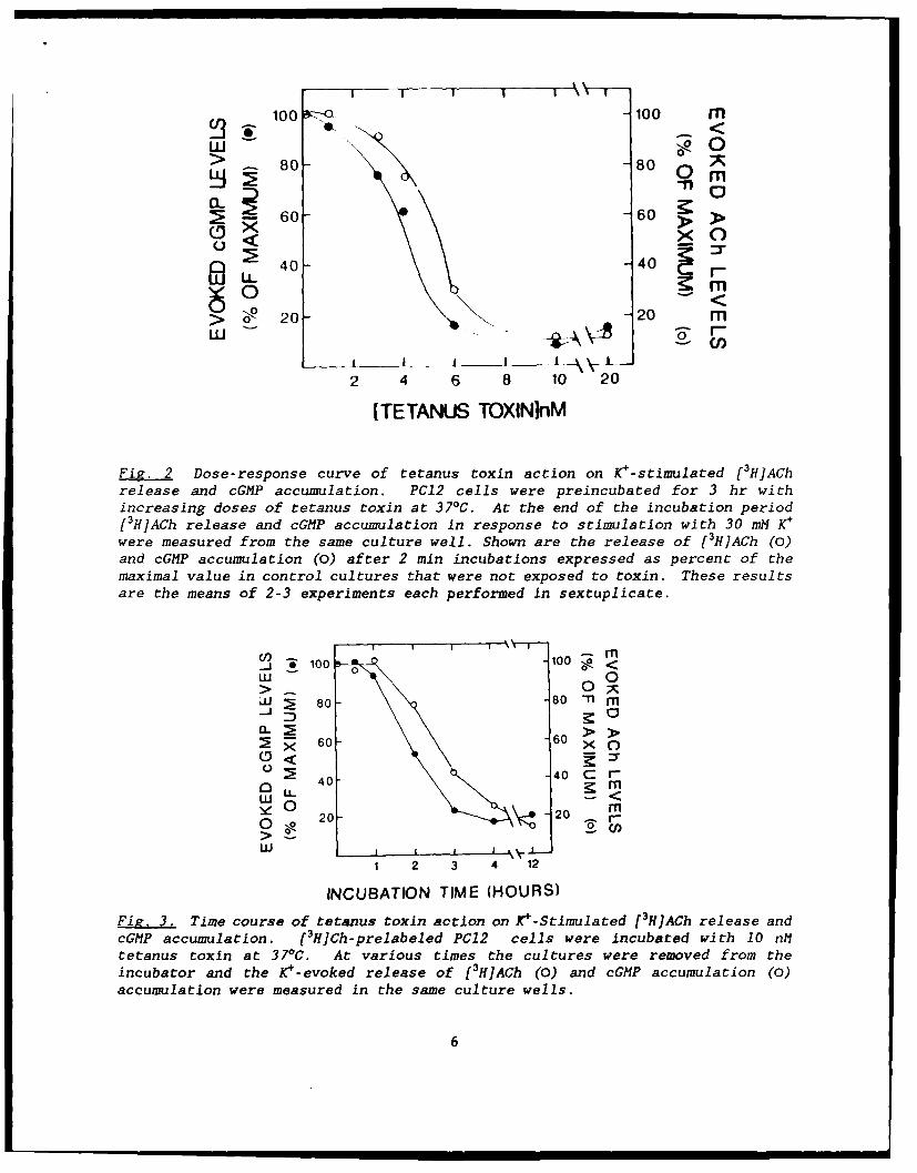
Fig. 2 Dose-response curve of tetanus toxin action on K+-stimulated [3H]AChrelease and cGMP accumulation. PC12 cells were preincubated for 3 hr withincreasing doses of tetanus toxin at 371C. At the end of the incubation period[3H]ACh release and cGMP accumulation in response to stimulation with 30 mM K+
were measured from the same culture well. Shown are the release of [3H]ACh (0)and cGMP accumulation (0) after 2 min incubations expressed as percent of themaximal value in control cultures that were not exposed to toxin. These resultsare the means of 2-3 experiments each performed in sextuplicate.
S100'100 0<
W 0W 80- 801 -nM
60 60 > o
W <
0 20 20 Fg
1 2 3 4 12
INCUBATION TIME (HOURS)
Fig. 3. Time course of tetanus toxin action on K+-Stimulated [3H]ACh release andcGtIP accumulation. [3H]Ch-prelabeled PC12 cells were incubated with 10 nMtetanus toxin at 37"C. At various times the cultures were removed from theincubator and the K4-evoked release of [3H]ACh (0) and cGMP accumulation (0)accumulation were measured in the same culture wells.
6
These data illustrate that there is a very close relation between the potency
of toxin in inhibiting ACh release and cGMP accumulation. Further, there is a
nearly identical time course for the development of the two effects evoked by
the toxin. Taken together, these results provide strong circumstantial evidence
that the toxin-evoked inhibition of cGMP accumulation and ACh release are
causally related.
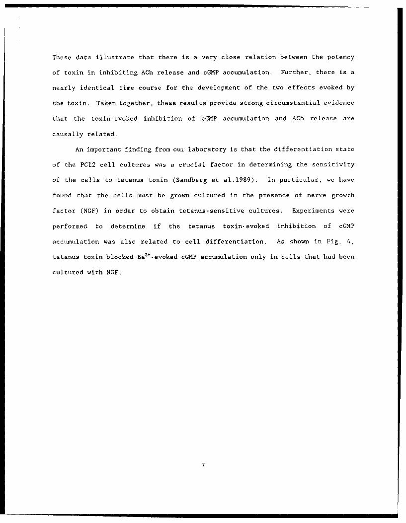
An important finding from our laboratory is that the differentiation state
of the PC12 cell cultures was a crucial factor in determining the sensitivity
of the cells to tetanus toxin (Sandberg et al.1989). In particular, we have
found that the cells must be grown cultured in the presence of nerve growth
factor (NGF) in order to obtain tetanus-sensitive cultures. Experiments were
performed to determine if the tetanus toxin-evoked inhibition of cGMP
accumulation was also related to cell differentiation. As shown in Fig. 4,
tetanus toxin blocked Ba2+-evoked cGMP accumulation only in cells that had been
cultured with NGF.
7
AB
Wo 60 < 160
-j CL W 0
> 2. 80
020 WE
SPRS ENS WE F[ ? T0~ 40
W
SPARSE DENSE DEX NGFDIFFERENTIATION CONDITIONS SAS ES EDIFFERENTIATION CONDITIONS
Fg. Effect of tetanus toxin on Ca2+-evoked [3H]ACh release andcGMP accumulation from PC12 cells grown under variousdifferentiation conditions. Ba2+-evoked cGMP accumulation (Panel A)or [3H]ACh release (Panel B) were measured. Evoked [3H]ACh releaseand cGMP accumulation were measured in the presence (hatched bars)and absence (open bars) )f tetanus toxin (10 nM 16-18 hpreincubations at 37°C) from PCl2 cells grown under a variety ofconditions: 14 days at 5 x 10⁴ cells/10 cm2 , in the presence of 1 x10⁻⁶ M dexamethasone (DEX); 14 days at 5 x 10⁴ cells/lO cm2 , in thepresence of 100 ng/ml nerve growth factor (NGF); 7 days, at highdensity (5 x 10⁵ cells/ 10 cm2) (Dense); or at low density (5 x 10⁴
cells/ 10 cm2) (Sparse). The results are the means of 2-3experiments each performed in sextuplet (± SEM).
These data show that tetanus' effects on ACh release and cGMP accumulation depend
on the differentiation state of PC12 cells in an identical manner. Detailed
examination of the development of the toxin sensitivity in NGF-treated cultures
revealed that the cells became sensitive to tetanus toxin only after culturing
in NGF for at least 8 days. These results are shown in Fig. 5.
8

w BT) 160
-J I -J -
w o 6o 1-12_ CE
*Q -I
0 QL3 2 w"'E .....
T OL>> 40 I3 6 3 10 12 3 6 8 10 12
DAYS IN NGF DAYS IN NGF
Fig. 5. Effect of tetanus toxin on Ba 2+_evoked L3 HJA~h release andcGMP accumulation from PC12 cells as a function of days in NGF.Evoked [3HIACh release (Panel B) and cGMP (Panel A) accumulationwere measured as a function of culture days in NGF (100 ng/ml) inthe presence (hatched bars) and absence (open bars) of tetanus toxin(10 nM; 16-18 h incubation at 370C). The results are the means of2-3 experiments each performed in sextuplet (± SEM).
In s.:mmary, it is clear that the differentiation state of the cells is a crucial
factor in determining the sensitivity of the cells to tetanus toxin as assessed
either at the biochemical of functional level. The factors responsible for the
expression of tetanus toxin sensitivity are intriguing but not known at present.
This will be the subject of future studies.
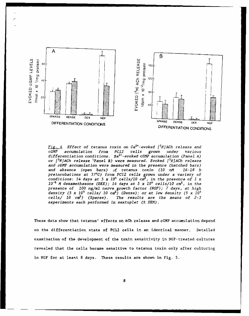
STUDIES WITH PERMEABTLIZED PC12 CELLS
During the past year considerable effort has been devoted to the
9
development and utilization of a permeabilized preparation of PCI2 cells. The
goal of this phase of the work was to use such a preparationi in order to further
characterize the mechanism of action of tetanus toxin. We have utilized a pore-
forming exotoxin, a-toxin, obtained from Staph. aureus. This toxin has been
utilized effectively to examine neurosecretion in several neural preparations
(Ahnert-Hilger et al.1985; Thelestam, and Blomqvist, 1988). We have purified
this toxin and have examined its effects on NGF-treated PC12 cells. As shown
in Fig.6, this toxin is very effective in permeabilizing the cells to small ions
such as Rb , while the cells remain relatively impermeant to larger molecules
such as LDH.
80 86 Rb+
I 3So H DA
60
o 40
-0---
20- o t
0
00
5 10 100lot-toxin] (Units/mil)
Fiz. 6. Release of 86Rb +, ['Hldopamine and LDH as a function of [a-toxin).Differentiated PC12 cells we, ,L preincubated (2h/370 C) with either 1.7Ci 86Rb+/ml(86Rb efflux,e), 1.5 uCi [H] dopamine/ml (dopamine release,O) or in the absenceof radioactivity (LDH release,A). Cells were washed in the same buffer prior tofurther incubation (30min/340C) with increasing concentrations of a-toxin (0-260 Units/ml). Supernatants were collected and aliquots were a,;sayed for releaseof 8 6Rb , [3H]dopamine or LDH. Cells were solubilized and the remaining activitymeasured. Values for release are expressed as % of total activity prior to
10
permeabilisation.
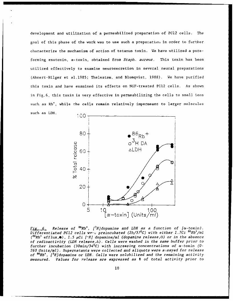
This figure also shows that it was possible to evoke dopamine release in the
presence of low CaZ+ (20 pM) when the cells were permeabilized. In the next
series of experiments the Ca2+-dependency of release of DA and ACh from
permeabilized PC12 cells was examined. The results are shown in Fig. 7.
Ca 2 dependent transmitter re!easefrom permeabilised PC12 cells
0T
0 0-0 H Docnn
0 12- 1-3 H Acetylcholine
Cc---
C
cL 4'"C,'
-0U
_ 0 I -.... - -4 I_ __ _ _ _ _ _
E-1 1 10 100[Free Co2 + ] (AM)
Fig. 7. Ca2+ -dependent release of [3H]dopamine and ACh from permeabilized PC12cells. Cells were loaded with radiolabelled transmitter and washed. Cells werepermeabilized with ct-toxln (30min/340C) in buffer containing the free Ca2
concentrations shown. Amount of label released was assessed after centrifugationof the supernatant and is expressed as a % of the total. Values for release inthe absence of added Ca2 have been subtracted to yield Ca2+ -dependent release of[3H]dopamine (0) and [3H]ACh (0). Data points are averages ± SEM (n-9, DA; n-3,ACh).
11
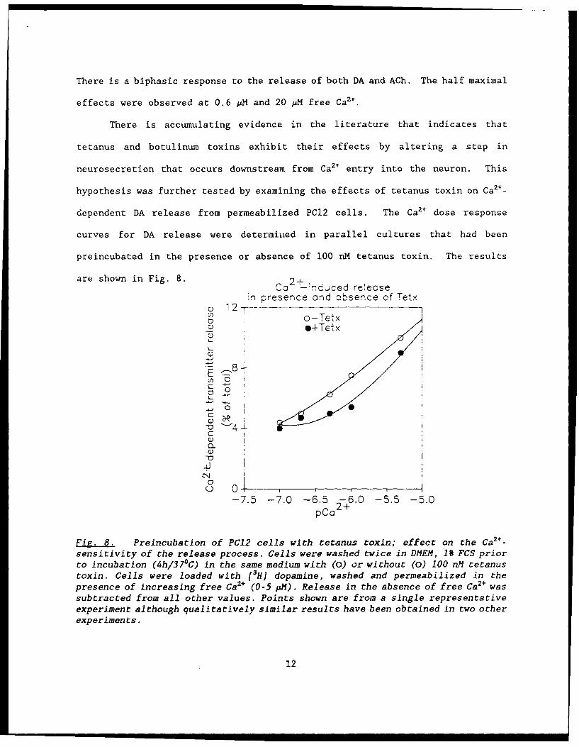
There is a biphasic response to the release of both DA and ACh. The half maximal
effects were observed at 0.6 pM and 20 MM free Ca2 +.
There is accumulating evidence in the literature that indicates that
tetanus and botulinum toxins exhibit their effects by altering a step in
neurosecretion that occurs downstream from Ca2+ entry into the neuron. This
hypothesis was further tested by examining the effects of tetanus toxin on Ca2+ _
dependent DA release from permeabilized PC12 cells. The Ca2+ dose response
curves for DA release were determined in parallel cultures that had been
preincubated in the presence or absence of 100 nM tetanus toxin. The results
are shown in Fig. 8. 2+- ,Ca i nduced release
in presence and absence of Tetx12 ----
o-Tetx_ *+Tetx
8 )
..
U)0
- 0 0 I
C) '4
a_
-c
-4J
-7.5 -7.0 -6.5 -6.0 -5.5 -5.0pCo
2 -
Fig. 8. Preincubation of PC12 cells with tetanus toxin; effect on the Ca2+ -
sensitivity of the release process. Cells were washed twice in DMEM, 19 FCS priorto incubation (4h/37"C) in the same medium with (0) or without (0) 100 nM tetanustoxin. Cells were loaded with [3H] dopamine, washed and permeabilized in thepresence of increasing free Ca2+ (0-5 pM). Release in the absence of free Ca2+ wassubtracted from all other values. Points shown are from a single representativeexperiment although qualitatively similar results have been obtained in two otherexperiments.
12
These results show that tetanus toxin shifts the Ca2+ dose-response curve to the
right. That is, it lowers the sensitivity of the DA release process for Ca2+.
Further, these results also demonstrate that the effects of tetanus in these
permeabilized cells are only seen when neurotransmitter release is evoked at low
a 2+.
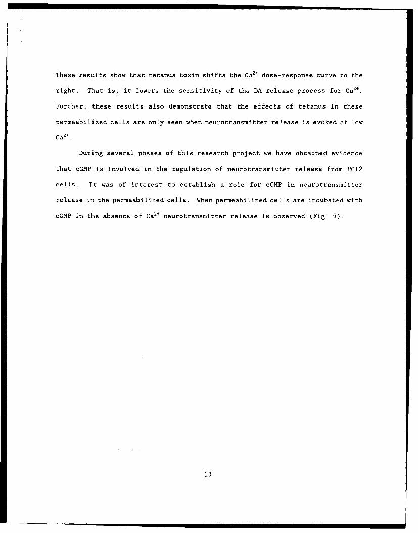
During several phases of this research project we have obtained evidence
that cGMP is involved in the regulation of neurotransmitter release from PC12
cells. It was of interest to establish a role for cGMP in neurotransmitter
release in the permeabilized cells. When permeabilized cells are incubated with
cGMP in the absence of Ca2+ neurotransmitter release is observed (Fig. 9).
13
Cyclic nucleotide-mediated increasein Dopamine release
+cOMR_-E+cAMP=o
o40-
0 .-
Q) 30,.C >E o0
I C
T
5 10 100 1000[Cyclic nucleotide] (AM)
Fig. 9. Cyclic nucleotide-mediated increase in Ca2+-independenttransmitter release. Cells were incubated for 5min/340 C in Ca 2+-freemedium containing cyclic nucleotide (O-1mM). Values are shown (+/-sem; n-6-12) for the 9 increase in release over controls (-
nucleotide) mediated by cGMP (0) and cAMP (a) in Ca2+ free medium.Control release was 5.8-8.1% of total.
These results show that cGMP evokes DA release from permeabilized cell in the
absence of Ca2+ with a half maximal dose of 100 AM. Further these effects are
specific since cAMP, even at concentrations as high as 1 mM, fails to evoke DA
release. These results are very intriguing yet the significance of these
observations and the interactions of cGMP and tetanus toxin in this system remain
to be identified. These will be goals of future studies.
14

CONCLUSIONS
During the early phase of this project we were successful in establishing
a cultured cell model system, the PC12 pheochromocytoma cell line, to study the
mechanism of action of tetanus toxin. We have established that these cells
contain a high density of high affinity tetanus toxin receptors and are sensitive
to intoxication by exposure to low doses of tetanus toxin (Sandberg et al.1989;
Walton et al.1988). Further we have studied the characteristics of the
intoxication pathway (Sandberg et al.1989) and have found that it is analogous
to that which has been characterized, to some extent, in vivo (Simpson, 1986;
Habermann, and Dreyer, 1986). The major thrust during the past year was to
exploit this well characterized model system to gain insight into the molecular
mechanism of action of tetanus toxin.
Our results demonstrate that intracellular cGMP levels increase when PCI2
cells are depolarized. The time courses for cGMP accumulation in response to
various stimuli were similar. The evoked levels of cGMP peaked within 20-40 sec
which is corqistent with the time course of neurotransmitter release under these
conditions (Sandberg et al.1989). Further support for a link between function
and cGMP levels was provided by the observation that there was a proportional
relation between evoked cGMP levels and ACh release under a variety of conditions
of depolarization and cell growth. It has been recognized for many years that
cGMP levels in neural tissues increase in response to depolarizing stimuli
(Nathanson, 1977; Goldberg, and Haddox, 1977). However the functional
significance of this effect has not been elucidated. Our studies with a
homogeneous cell line provide strong circumstantial evidence for a role of this
cyclic nucleotide in neurosecretion.
15
The relationship between cGMP accumulation and neurotransmitter release
was further underscored by the observation that tetanus toxin inhibited both
processes. There is a remarkable correlation between the toxin dose-inhibition
curves and the time course for the development of the inhibitory responses of
cGMP accumulation and ACh release in PC12 cells. We have previously reported
that only NGF-treated PC12 cells were sensitive to tetanus toxin. The same
relation was found for the toxin inhibition of evoked-cGMP accumulation
Previously, the molecular mechanisms of tetanus toxin have remained elusive.
Taken together these results provide the first biochemical evidence for the
underlying mechanism of action for this toxin.
Another major advancement during the past year has been the development
and utilization of a permeabilized PC12 preparation. Permeabilized cells allow
for control of the intracellular environment by direct application of a variety
of agents into this compartment. The use of a-toxin as the permeabilizing agent
has a number of significant advantages over the use of detergents, including
minimal damage to the intracellular organelles. The fact that a large Ca2+-
dependent release of neurotransmitter was observed in permeabilized cells
supports this conclusion.
The permeabilized preparation has been exploited to examine the effects
of tetanus toxin on the role of Ca2+ in neurosecretion. The results reported
here support the hypothesis that part of the action of tetanus toxin is due to
a lowering of the Ca2+ sensitivity of the release process. If this is true, then
it should be possible to override the effects of toxin by applying high levels
of Ca2+ . This was in fact observed. These results are consistent with
observations made in studies with botulinum toxin at the neuromuscular junction
(Simpson, 1986). The precise mechanism whereby tetanus brings about the reduced
16
sensitivity to Ca2+ remains to be identified. The permeabilized cell preparation
represents a valuable system in which to examine this problem.
cGMP was also found to play a role in secretion in permeabilized cells.
Novel results reported here indicate that cGMP evokes neurotransmitter secretion
in a Ca2+ independent manner in permeabilized cells. This is in contrast to the
effects of this nucleotide on ACh release in intact cells, where it did not
stimulate neurotransmitter release by itself or upon depolarization (Sandberg
et al. 1989). The relation between this phenomenon and tetanus action is not
known at this time but will be a focus of future studies.
17
BIBLIOGRAPHY OF PUBLISHED WORK
1. Sandberg, K., Berry, C.J. and Rogers, T.B. (1989) Studies on the
Intoxication pathway of tetanus toxin in the Rat Pheochromocytoma (PCI2)
Cell Line. J. Biol. Chem. 264, 5679 - 5686.
2. Sandberg, K., Berry, C.J., and Rogers, T.B. (1989) A Role for cGMP
During Tetanus Toxin Blockade of Acetylcholine Release in the Rat
Pheochromocytoma (PC12) Cell Line. J. Neurosci., revised manuscript
submitted.
18

PERSONNEL INVOLVED IN CONTRACT WORK
1. Terry B. Rogers, PhD -- Principal Investigator, 25% time
2. David Evans, PhD -- Research Associate, 100% time
3. Andrea Grandin, MS -- Research Assistant, 100% time
19
REFERENCES
Ahnert-Hilger, G., S. Bhakdi, and M. Gratzl (1985) Minimal requirements for
exocytosis. J.Biol.Chem. 260:12730-12734.
Bergey, G.K., R.L. MacDonald, W.H. Habig, M.C. Hardegree, and P.G. Nelson
(1983) Tetanus toxin convulsant action on mouse spinal cord neurons in
culture. J.Neurosci. 3:2310-2323.
Ceccarelli, B., and F. Clementi (1979) Neurotoxins: tools in neurobiology.
Adv. Cytopharmacol. 3:
Collingridge, G.L., G.G.S. Collins, J. Davies, T.A. James, M.J. Neal, and
P. Tongroach (1980) Effect of tetanus toxin on transmitter release from
substantia nigra and striatum in vitro. J.Neurochem. 34:540-547.
Dreyer, F., A. Mallart, and J.L. Brigant (1983) Botulinum A toxin and
tetanus toxin do not affect presynaptic membrane currents in mammalian motor
nerve endings. Brain Res. 270:373-375.
Goldberg, N.D., and M.K. Haddox (1977) Cyclic GMP metabolism and involvement
in biological regulation. Ann.Rev.Biochem. 46:823-896.
Habermann, E., and F. Dreyer (1986) Clostridial neurotoxins: handling and
action at the cellular and molecular level. Curr.Topics Microbiol.Immunol.
129:93-179.
20
Mellanby, J., and J. Green (1981) How does tetanus toxin act?. Neurosci.
6:281-300.
Nathanson, J.A. (1977) Cyclic nucleotides and nervous system function.
Physiol.Revs. 57:158-256.
Osborne, R.H., and H.F. Bradford (1973) Tetanus toxin inhibits amino acid
release from nerve endings in vitro. Nature (New Biol.) 244:157-158.
Sandberg, K., C. Berry, and T.B. Rogers (1989) Studies on the intoxication
pathway of tetanus toxin in the rat pheochromocytoma (PCI2) cell line.
J.Biol.Chem. 264:5679-5686.
Sandberg, K., Berry, C.J., and Rogers, T.B. (1989) A Role for cCMP Tetanus
During Toxin Blockade of Acetylcholine Release in the Rat Pheochromocytoma
kPCI2) Cell Line. J. Neurosci., revised manuscript submitted.
Schmitt, A., F. Dreyer, and C. John (1981) At least three sequential steps
are involved in the tetanus toxin-induced block of neuromuscular
transmission. Nauyn-Scmeid.Arch.Pharmacol. 317:326-330.
Schwab, M.E., and H. Thoenen (1976) Electron microscopic evidence for a
transsynaptic migration of tetanus toxin in spinal cord motoneurons: an
autoradiographic and morphometric study. Brain Res. 105:213-224.
21
Simpson, L.L. (1986) Molecular pharmacology of botulinum toxin and tetanus
toxin. Ann.Rev.Pharmacol.Toxicol. 26:427-454.
Staub, G.C., K.M. Walton, R.L. Schnaar, T. Nichols, R. Baichwal, K.
Sandberg, and T.B. Rogers (1986) Characterization of the binding and
internalization of tetanus toxin in a neuroblastoma hybrid cell line.
J.Neurosci. 6:1443-1451.
Thelestam, M., and L. Blomqvist (1988) Staphylococcal alpha toxin - recent
advances. Toxicon 26:51-65.
Walton, K.M., K. Sandberg, T.B. Rogers, and R.L. Schnaar (1988) Complex
ganglioside expression and tetanus toxin binding by PC12 pheochromoc"toma
cells. J.Biol.Chem. 263:2055-2063.
22
DISTRIBUTION LIST
5 copies Commander
US Army Medical Research Instituteof Infectious Diseases
ATTN: SGRD-UIZ-M
Fort Detrick
Frederick, MD 21701-5011
1 copy Commander
US Army Medical Research andDevelopment Comand
ATTN: SGRD-RMI-SFort Detrick
Frederick, MD 21701-5012
2 copies Defense Technical Information Center
ATTN: DTIC-FDAC
Cameron Station
Alexandria, VA 22304-6145
1 copy Dean
School of MedicineUniformed Services University of the
Health Sciences4301 Jones Bridge RoadBethesda, MD 20814-4799
23