mechanism of creaming down based on chemical
TRANSCRIPT
676 Vol. 64, No. 7
© 2016 The Pharmaceutical Society of Japan
Chem. Pharm. Bull. 64, 676–686 (2016)
Mechanism of Creaming Down Based on Chemical Characterization of a Complex of Caffeine and Tea Catechins
Takashi Ishizu,* Hiroyuki Tsutsumi, and Takashi SatoFaculty of Pharmacy and Pharmaceutical Sciences, Fukuyama University; Sanzo Gakuen-cho 1,
Fukuyama, Hiroshima 729–0292, Japan.Received February 9, 2016; accepted April 21, 2016
The component of a precipitate resulting from creaming down, which was made from caffeine and a catechin mixture, was determined by an integrated value of H2 proton signals of tea catechins in the quan-titative 1H-NMR spectrum. The results showed that gallate-type catechins formed a precipitate by creaming down more predominantly than non-gallate-type catechins. X-ray crystallographic analysis showed that the gallate-type catechin (−)-epigallocatechin-3-O-gallate (EGCg), (−)-epicatechin-3-O-gallate (ECg) formed 2 : 2 and 2 : 4 complexes with caffeine, respectively, and the non-gallate-type catechin (−)-epicatechin (EC) and caffeine formed a 1 : 1 complex. The 2 : 2, 2 : 4 complexes of caffeine and EGCg, ECg formed a hydropho-bic space with three aromatic A, B, and B′ rings of two EGCg, ECg molecules, and one caffeine molecule was captured in this hydrophobic space. However, no such hydrophobic space in the 1 : 1 complex of caffeine and EC formed. It was thought that the hydrophobicity of the 2 : 2, 2 : 4 complexes of caffeine and EGCg, ECg was stronger than that of the 1 : 1 complex of caffeine and EC, with the result that the 2 : 2, 2 : 4 complexes of caffeine and EGCg, ECg precipitated by creaming down more predominantly than the 1 : 1 complex of caf-feine and EC in an aqueous solution. Furthermore, the molecular capture of various heterocyclic compounds by formation of the 2 : 2 complex of EGCg from the aqueous solution was investigated using the quantitative 1H-NMR spectrum.
Key words creaming down; (−)-epigallocatechin-3-O-gallate; (−)-epicatechin-3-O-gallate; (−)-epicatechin; caffeine; X-ray crystallographic analysis
1. IntroductionTea has been drunk in many countries throughout the world
since ancient times for its taste, and to maintain and improve health. It is well known that tea protects against lifestyle-related diseases such as cancer, high blood pressure, diabetes, obesity, and arteriosclerosis.1) Tea is prepared using leaves of the tea plant, Camellia sinensis, Theaceae, which include caf-feine, tannins, vitamins and theanine. Catechins are a kind of tannin that show various physiologically modulating effects, including anti-carcinogenic, anti-metastatic, and anti-oxidative effects.2–7) The eight main tea catechins are classified into four categories by the existence of a galloyl group on the oxygen atom at the C3 position and the relative stereochemistry be-tween the C2 and C3 positions: 2,3-cis-gallate-type, 2,3-trans-gallate-type, 2,3-cis-non-gallate-type, and 2,3-trans-non-gal-late-type8) (Fig. 1). Generally speaking, gallate-type catechins show higher physiological activities than non-gallate-type catechins.9–12)
When a hot tea beverage cools down, brown-white turbidity and precipitation occur in the tea. This phenomenon is called “creaming” or “creaming down (reaction)”. Since the cream-ing down is a trigger that reduces the original taste and flavor of tea, it is one of the most serious problems when making a tea beverage. It has been suggested that caffeine forms a cream with tea polyphenols such as catechins, theaflavins and
thearubigins.13)
Ina and colleagues reported that all the main signals were assigned to tea catechins such as 2,3-cis-gallate-type catechins (−)-epigallocatechin-3-O-gallate (EGCg) and (−)-epicatechin-3-O-gallate (ECg) and caffeine in the 13C-NMR spectrum ofa hot water solution of a precipitate formed by the creamingdown of a tea infusion.14) Thus, we investigated what kind ofcatechins predominantly precipitated using caffeine and a cat-echin mixture by quantitative 1H-NMR. The catechin mixture(purchased from Nagara Science Co., Ltd., Japan) was a frac-tion of extracted catechins included in green tea.
Ina and colleagues also reported that creaming down even-tually occurs when an aqueous caffeine solution is poured into an aqueous solution of EGCg, which is most abundant in tea catechins.14) However, creaming down in tea beverages has not been adequately chemically elucidated. In a previous review we reported that a suspension of an equimolecular amount of caffeine and 2,3-trans-gallate-type catechins (−)-gallo-catechin-3-O-gallate (GCg) in water afforded two kinds of crystals, which were 1 : 2 and 2 : 2 complexes of caffeine and GCg.15) Therefore, we attempted crystallization of a precipitate formed by the creaming down of caffeine and 2,3-cis-type tea catechins, and studied the mechanism of the creaming down. The stereochemical structures of the complexes of caffeine and tea catechins EGCg, ECg, (−)-epicatechin (EC) were de-
* To whom correspondence should be addressed. e-mail: [email protected]
Special Collection of Papers
This article is dedicated to Professor Satoshi Ōmura in celebration of his 2015 Nobel Prize.
Review

Vol. 64, No. 7 (2016) 677Chem. Pharm. Bull.
termined by X-ray crystallographic analysis. The mechanism of creaming down was investigated using molecular interac-tions formed between caffeine and tea catechins.
2. Analysis of the Precipitate Formed by Creaming Down Using Quantitative 1H-NMR Spectra
First, the content of tea catechins contained in the catechin mixture was investigated by the integrated value of the proton signal of the quantitative 1H-NMR spectrum. No overlap of the methine H2 proton signal of each tea catechin in acetone-d6 was observed in the 1H-NMR spectrum.16) Then, the inte-grated value of the H2 proton signal of each tea catechin in-cluded in the catechin mixture was measured using the methyl proton signal (singlet) of Boc-glycine as an internal standard (Fig. 2). The content of the catechin mixture is shown in Table 1, indicating that it included a large amount of 2,3-cis-type catechins, such as EGCg, ECg, and EC. Thus, a simple and easy method for quantitative analysis of a sample containing many kinds of catechins using the 1H-NMR spectrum was developed. The reported contents of catechins in green tea are listed in Table 1.17)
As shown in Fig. 2, the H2 proton signal of 2,3-cis cat-echins of EC, ECg, ECG, and EGCg was observed to be sin-glet-like, whereas those of 2,3-trans catechins of (+)-catechin (CA) and (+)-gallocatechin (GC) were observed as a doublet in the 1H-NMR spectrum. Such a difference in coupling pat-terns resulted from differences in configuration. Judging from the coupling constant J2,3 of EC, ECg, ECG, and EGCg being ca. 0 Hz, it was thought that their dihedral angles ∠H2–C2–C3–H3 were approximately 90°, based on the Kurplus equa-tion.19) On the other hand, since J2,3 of CA and GC were 7.8 and 7.4 Hz, respectively, their dihedral angles ∠H2−C2−C3−H3 were expected to be considerably larger than 90°.15,19)
Equimolar amounts of caffeine and the catechin mixture were dissolved in D2O at 90°C and left at room temperature for a day. The solution was divided into the supernatant liq-
uid and a sticky precipitate, which is a precipitate formed by creaming down. The contents of the supernatant liquid and the sticky precipitate were determined by a similar analyti-cal method using an integrated value of the H2 proton signal of tea catechins and the H8 proton signal of caffeine in the quantitative 1H-NMR spectra. The contents of various tea cat-echins and caffeine are listed in Table 2.
As shown in Table 2, more than 80% of gallate-type cat-echins ECg and EGCg included in the catechin mixture were present in the precipitate formed by creaming down, whereas only 45.9–64.7% of non-gallate-type catechins, EC, EGC, CA, and GC, were present. This finding suggests that the gallate-type catechins formed a creaming down precipitate more pre-dominantly than the non-gallate-type catechins.17)
Fig. 1. The Eight Main Tea Catechins, Caffeine, and Nicotinamide
Fig. 2. The H2 Methine Proton Signal of Each Catechin of the Cat-echin Mixture in the 1H-NMR Spectrum

678 Vol. 64, No. 7 (2016)Chem. Pharm. Bull.
3. Crystallization of the Creaming Down Precipitate Made from Caffeine and EGCg
Equimolecular amounts of caffeine and EGCg were dis-solved in water by heating, and the aqueous solution was left at room temperature. It divided into a supernatant liquid and a sticky precipitate formed by creaming down, which were left
at 10°C for about 3 months. As a result, the sticky precipitate crystallized slowly to give a colorless block crystal20,21) (Fig. 3a).
Similarly equimolecular amounts of caffeine and EC were dissolved in water by heating and the aqueous solution was left at room temperature, but no precipitate was afforded and
Table 1. The Content of Tea Catechins in the Catechin Mixture
EGCg EGC EC ECg GC CA
Content (mg) 5.13 2.70 0.99 0.97 0.43 0.17Amount of substance (mmol) 0.011 0.009 0.003 0.002 0.001 0.001Relative content (%) 49.37 25.99 9.53 9.34 4.14 1.64Relative content (%) of green tea18) 59.1 19.3 6.4 13.7 1.6
Relative content of green tea is taken from “Chanokagaku” (Asakura Publishing, 1991, p. 124).
Table 2. The Content of Caffeine and Tea Catechins and in the Supernatant Liquid and the Sticky Precipitate Made from Caffeine and the Catechin Mixture
EGCg EGC EC ECg GC CA Caffeine
Sample Content (mg) 5.13 2.70 0.99 0.97 0.43 0.17 5.36Amount of substance (mmol) 0.0112 0.0088 0.0034 0.0022 0.0014 0.0006 0.0276
Sticky precipitate Content (mg) 4.22 1.24 0.53 0.81 0.24 0.11 4.46Amount of substance (mmol) 0.0092 0.0040 0.0018 0.0018 0.0008 0.0004 0.0230Relative content (%) 82.3 45.9 53.5 83.5 55.8 64.7 83.2
Supernatant liquid Content (mg) 0.88 1.46 0.45 0.13 0.19 0.06 0.90Amount of substance (mmol) 0.0019 0.0048 0.0016 0.0003 0.0006 0.0002 0.0046Relative content (%) 17.2 5.41 45.5 13.4 44.2 35.3 16.8
Fig. 3. Crystal Preparations of Catechins Complexes with Caffeine(a) Complex of caffeine and EGCg; (b) Complex of caffeine and EC.

Vol. 64, No. 7 (2016) 679Chem. Pharm. Bull.
it was still soluble. The mixture was lyophilized to afford a colorless powder, which was recrystallized from methanol to give a colorless block crystal17,22) (Fig. 3b).
4. Crystal Structure of the Complex of Caffeine and EGCg, Caffeine and EC
A single crystal of the colorless block crystal, which was prepared from the aqueous solution of caffeine and EGCg, was determined to be a 2 : 2 complex of caffeine and EGCg by X-ray crystallographic analysis.20,21) An ORTEP drawing of the 2 : 2 complex of caffeine and EGCg is shown in Fig. 4a. The 2 : 2 complex consisted of two caffeines (caffeines A, B) and two crystallographically different EGCgs (EGCgs A, B), and the caffeines faced the B′ rings of each EGCg. One unit cell contained four units of the 2 : 2 complex of caffeine and EGCg, and 60 water molecules as crystal solvent (Fig. 4b).
The torsion angles of the EGCg moieties (EGCgs A, B) of the 2 : 2 complex of caffeine and EGCg indicated that the B rings of EGCgs A and B were both in equatorial positions, while B′ rings of EGCgs A and B were both in axial posi-tions with respect to the C rings of EGCg molecules (Table 3). As shown in Table 4, the torsion angles of caffeine moieties showed that caffeine moieties (caffeines A, B) were both al-most in plane form.
The layer structure of the 2 : 2 complex of caffeine and EGCg consisted of two layers, which were in parallel in the
same direction as the b-axis (Fig. 5a). Caffeine A was stacked between B′ rings of EGCg A and caffeine B was stacked be-tween B′ rings of EGCg B, and caffeines A or B was located almost in the middle of two B′ rings of EGCgs A or B.
In the 2 : 2 complex of caffeine and EGCg, intermolecular interactions forming between caffeine and EGCg moieties were elucidated (Fig. 5b). Offset π–π stacking interactions formed between a six-membered ring of caffeine and the B′ ring of EGCg, and A rings of EGCgs A and B. Furthermore, CH–π interactions formed between N1–CH3, N7–CH3 of caffeine and the B rings of EGCg A and B (Table 5). Two O–H…N and two O–H…O intermolecular hydrogen bonds were also observed between caffeine and EGCg (Table 5).
A single crystal of the colorless block crystal, which was prepared from the aqueous solution of caffeine and EC, was determined to be a 1 : 1 complex of caffeine and EC by X-ray crystallographic analysis.17,22) An ORTEP drawing of a unit of the 1 : 1 complex of caffeine and EC is shown in Fig. 6a. One unit cell contained four units of the 1 : 1 complex of caffeine and EC and 8 water molecules as crystal solvent (Fig. 6b).
The torsion angles of the EC moiety of the 1 : 1 complex (Table 3) indicated that the B ring and 3-OH group of EC were in equatorial and axial positions with respect to the C ring of the EC molecule, respectively. The caffeine molecule has an almost plain and rigid xanthine skeleton.
In the layer structure shown in Fig. 7, units of the 1 : 1 com-
Fig. 4. 2 : 2 Complex of Caffeine and EGCg(a) ORTEP drawing with thermal ellipsoids at 30% probability level; (b) One unit cell. Crystal solvent and hydrogen atoms are omitted for clarity.
Table 3. Torsion Angles of EGCg, EC and ECg Moieties of Complexes of Caffeine and EGCg, Caffeine and EC, Caffeine and ECg
2 : 2 Complex of caffeine and EGCs1 : 1 Complex of caffeine and EC
2 : 4 Complex of caffeine and ECg
EGCg A EGCg B ECg A ECg B
∠C1′–C2–C3–O4 69.2 (8)° 67.1 (8)° 58.4 (8)° 68.7 (6)° 70.3 (8)°∠H2–C2–C3–H3 72° 68° 60° 68° 71°
Table 4. Torsion Angles of Caffeine Moieties of the 2 : 2 Complex of Caffeine and EGCg
Torsion angle of caffeine A Torsion angle of caffeine B
∠N7–C5–C6–O13 0.5 (13)° 3.4 (13)°∠O13–C6–N1–C10 −1.8 (10)° 2.3 (10)°∠O11–C2–N3–C12 −5.1 (10)° 1.0 (11)°∠C14–N2–C8–N9 −179.6 (7)° 178.1 (7)°∠C10–N1–C2–O11 −0.4 (10)° −4.3 (10)°∠C12–N3–C4–N9 6.2 (11)° −0.1 (12)°

680 Vol. 64, No. 7 (2016)Chem. Pharm. Bull.
Fig. 5. Layer Structure (a) and Intermolecular Interactions (b) of the 2 : 2 Complex of Caffeine and EGCgGray dotted and black arrows and gray and black dotted lines indicate offset π–π stacking and CH–π interactions and O–H…O and O–H…N hydrogen bonds, respec-
tively.
Table 5. Intermolecular Interactions of the 2 : 2 Complex of Caffeine and EGCg
CH…π interactions
D–H Aromatic ring D–H (Å) D–H…Aromatic ring (Å)
CH (28A) B ring of EGCg B 0.97 2.53CH (29C) A ring of EGCg A 0.96 3.25CH (58B) B ring of EGCg A 0.95 2.66CH (59B) A ring of EGCg B 0.96 2.80
Hydrogen bonds
D–H A D…A (Å) D–H (Å) H…A (Å) ∠D–H…A
OH (5O) O (30) 2.830 (10) 0.85 2.06 150.40°OH (6O) O (32) 2.646 (9) 0.82 1.89 153.56°OH (7O) N (3) 2.813 (10) 0.83 2.01 160.83°OH (9O) O (40) 2.751 (6) 0.83 1.94 164.83°OH (10O) O (21) 2.974 (6) 0.83 2.18 159.95°OH (11O) O (34) 2.766 (8) 0.83 1.97 162.23°OH (15O) O (33) 2.852 (7) 0.84 2.07 154.65°OH (16O) O (42) 2.711 (6) 0.83 1.96 150.76°OH (18O) O (40) 2.795 (9) 0.82 2.03 155.52°OH (20O) N (7) 2.650 (10) 0.83 1.90 150.36°OH (22O) O (6) 2.888 (6) 0.83 2.09 160.23°OH (23O) O (27) 2.823 (7) 0.83 2.07 150.14°OH (24O) O (37) 2.759 (6) 0.83 1.93 174.64°
Fig. 6. 1 : 1 Complex of Caffeine and EC(a) ORTEP drawing with thermal ellipsoids at 30% probability level; (b) One unit cell. Crystal solvent and hydrogen atoms are omitted for clarity.

Vol. 64, No. 7 (2016) 681Chem. Pharm. Bull.
plex of caffeine and EC were stacked in parallel in the same direction as the b-axis. The six-membered ring of caffeine and the A ring of EC appeared in turn along the b-axis, and the six-membered rings of caffeines were positioned in almost the middle of the A rings of ECs. The six-membered ring of caf-feine faced the A rings of both upper and lower ECs.
In the 1 : 1 complex of caffeine and EC, face-to-face π–π stacking interactions formed between the six-membered ring of caffeine and the A ring of EC. Furthermore, a CH–π in-teraction formed between C(14)–H(14) of the B ring and the B ring (Fig. 7). Two O–H…O intermolecular hydrogen bonds were observed.
5. Properties of the Complex of Caffeine and EGCg, Caffeine and EC in the Aqueous Solution State
The stability constants for the complex formation of caf-feine and EGCg, caffeine and EC Kc at 40–80°C were esti-mated by Eq. 1, which assumed the order of the reaction n.23) Here, CF, CA and CX refer to caffeine, catechins and the complexes of caffeine and EGCg, EC, respectively, and Δδobs and ΔδCX represent (δEGCg or EC−δobs) and (δEGCg or EC−δCX), respectively. δEGCg or EC, δCX, and δobs represent the chemical shift (ppm) of the H2″,6″ proton of EGCg and the H5′ proton of EC in a free state, complexes of caffeine and EGCg, EC, and the mixture of caffeine and EGCg, EC in 1H-NMR spec-tra, respectively. The stability constants Kc of the complexes of caffeine and EGCg, EC at 40°C were 940.7 and 577.2 M−1, respectively (Table 6).
c
1/obs
0CXCX
cobs
CA CF CX
1 Δ[CF] [CA]
ΔΔ1
Δ
+ −
+
=
K
n
n
δnδδ K
δ
(1)
From the dependency of Kc on temperature, the change in free energy ΔG, enthalpy ΔH, and entropy ΔS, of the complex formation were estimated as shown in Table 6. The entropy (ΔS) for the formation of the complex of caffeine and EGCg had a large negative value (−34.6 J mol−1 K−1), suggesting that caffeine was fixed tightly in the complex of caffeine and EGCg. On the other hand, the entropy (ΔS) for the formation of the complex of caffeine and EC had a small negative value (−5.3 J mol−1 K−1), suggesting that caffeine fitted loosely in the complex of caffeine and EC.
Judging from the crystal structure of the 2 : 2 complex of caffeine and EGCg (Fig. 5), caffeine was thought to be fixed tightly in the space formed by the A, B, B′ rings of three EGCg aromatic rings. On the other hand, judging from the crystal structure of the 1 : 1 complex of caffeine and EC (Fig.
Fig. 7. Layer Structure and Molecular Interactions of the 1 : 1 Complex of Caffeine and EC
Gray and black arrows and gray dotted lines indicate face-to-face π–π stacking interactions, CH–π interactions and O–H…O hydrogen bonds, respectively.
Table 6. Stability Constants for the Complex Formation of Caffeine and EGCg, Caffeine and EC Kc, the Changes in Free Energy ΔG, Enthalpy ΔH, and Entropy ΔS of the Complex Formation of Caffeine and EGCg, Caffeine and EC, and the Order of the Reaction n of the Complex Formation of Caffeine and EGCg, Caffeine and EC
Caffeine-EGCg
T (°C) Kc (M−1) ΔG (kJ mol−1) ΔH (kJ mol−1) ΔS (J mol−1 K−1) n
40 940.7 −17.8 0.4945 762.4 −17.5 0.5150 710.9 −17.6 0.5355 568.6 −17.3 −28.7 −34.6 0.6160 482.4 −17.1 0.6270 348.3 −16.7 0.6780 274.8 −16.5 1.01
Caffeine-EC
T (°C) Kc (M−1) ΔG (kJ mol−1) ΔH (kJ mol−1) ΔS (J mol−1 K−1) n
40 577.2 −16.5 0.5945 495.9 −16.4 0.6150 439.8 −16.3 0.6555 413.6 −16.4 −18.1 −5.3 0.9460 351.9 −16.2 0.7170 311.2 −16.4 0.6180 255.9 −16.3 0.65

682 Vol. 64, No. 7 (2016)Chem. Pharm. Bull.
7), caffeine was thought to fit loosely between the A rings of EC.
Next, changes in chemical shifts of proton signals of EGCg and EC in 1H-NMR spectra by adding regular amounts of caf-feine were investigated. The upfield shift of the proton signal of H2″,6″ was most prominent in the EGCg proton signals (Fig. 8a). As shown in Fig. 5, the upfield shift of the signal of H2″,6″ may result from magnetic anisotropic shielding by the ring current from the xanthine skeleton of caffeine, suggesting that caffeine moieties in the complex of caffeine and EGCg were positioned in the vicinity of the B′ ring of EGCg.
As shown in Fig. 8b, the H6 and H8 proton signals of the EC A ring shifted most predominantly upfield in the proton signals of EC. The upfield shifts of H6 and H8 proton signals were considered to result mainly from the magnetic anisot-ropy of the aromatic ring of the caffeine xanthine skeleton, suggesting that caffeine moieties in the complex of caffeine and EC were positioned in the vicinity of the A ring of EC17) (Figs. 7, 9b).
Information regarding the flexibility of caffeine moieties in the complexes with EGCg and EC in D2O can be experi-mentally estimated by the relaxation times of carbon reso-nances (T1). The NT1 values (N=number of attached protons, T1=longitudinal relaxation time) linked directly with molecu-lar mobility.24) The carbon NT1 values of caffeine moieties in complexes of caffeine and EGCg, EC are shown in Table 7. The NT1 values of N1–CH3, N7–CH3, and C8 of caffeine in the complex of caffeine and EGCg were smaller than those in the complex of caffeine and EC, indicating that the mobil-ity of the caffeine moieties in the complex of caffeine and EGCg was restricted more strongly than that in the complex of caffeine and EC. Judging from the crystal structure of the 2 : 2 complex of caffeine and EGCg (Fig. 5) and the 1 : 1 complex of caffeine and EC (Fig. 7), upon complex formation with EGCg, the caffeine xanthine skeleton caffeine was fixed strongly in the hydrophobic space formed by three aromatic A, B, B′ rings of EGCg. Furthermore, the N1–CH3, N7–CH3 mobility of methyl groups of the caffeine moieties in the com-plex of caffeine and EGCg were thought to be restricted by the force of the CH–π interaction with the B rings of EGCg.
Nuclear Overhauser effect (NOE) difference analysis of the aqueous solution containing equimolar amounts of caffeine and EGCg was conducted. Intermolecular NOEs between N1–CH3, N3–CH3 and N7–CH3 of methyl groups of caffeine and H2″, 6″ of EGCg, between H8 of caffeine and H2″, 6″ of EGCg, between N3–CH3 of methyl group of caffeine and H2′, 6′ of EGCg, and between H8 of caffeine and H2′, 6′ of EGCg were observed (Fig. 9a).
As a result, it was concluded that the structures of com-plexes of caffeine and EGCg, EC in the aqueous solution were the same as their crystal structures.21)
6. Stereochemical Structure of the Complex of Caf-feine and ECg
Equimolecular amounts of caffeine and ECg were dissolved in water by heating, and left at room temperature to give a sticky substance formed by creaming down, which crystal-lized slowly over about 3 d at room temperature to give color-less crystals. This was recrystallized from water to afford col-orless blocks, which were determined to be a 2 : 4 complex of caffeine and ECg by X-ray crystallographic analysis.17,22) An ORTEP drawing of a unit cell of the 2 : 4 complex of caffeine and ECg is shown in Fig. 10a. In a unit of the 2 : 4 complex, the B and B′ rings of one ECg (ECg A) faced the caffeines and the A and B′ rings of the other ECg (ECg B) faced the caffeines. One unit cell contained two units of the 2 : 4 com-plex of caffeine and ECg and 12 water molecules as a crystal solvent (Fig. 10b).
The torsion angles of the ECg moieties (ECgs A, B) of the
Fig. 8. The Shifts in 1H-NMR Signals of the Protons of EGCg and EC upon Addition of Caffeine to the Solution of EGCg and EC in D2O
Intial condition: a solution of EGCg (90 mM) and EC (40 mM) in D2O (550 µL) at room temperature.
Table 7. NT1 Values (ms) of Complexes of Caffeine and EGCg, Caffeine and EC
Sample N1–CH3 N3–CH3 N7–CH3 C8
Complex of caffeine and EGCg
4644 8430 4845 574
Complex of caffeine and EC
7200 7383 8088 727
N and T1 indicate number of attached protons and longitudinal relaxation time, respectively.

Vol. 64, No. 7 (2016) 683Chem. Pharm. Bull.
2 : 4 complex indicated that the B rings of ECg A and ECg B were both in equatorial positions and B′ rings of ECg A and ECg B were both in axial positions with respect to the C rings of ECg molecules (Table 3).
In the layer structure shown in Fig. 11, units of the 2 : 4 complex of caffeine and ECg stacked in parallel in the same direction as the a-axis. The caffeine moieties and the B′ rings of ECg appeared in turn along the a-axis, and the caffeine moieties were positioned in almost the middle of the two B′ rings of ECgs. Furthermore, the A rings of ECg faced the caf-feine moieties and the B rings of ECg also faced the caffeine moieties.
In the 2 : 4 complex of caffeine and ECg, face-to-face and offset π–π stacking interactions formed between caffeine and the B′ ring of ECg (Fig. 11). Also, face-to-face π–π stack-ing interactions formed between caffeine and the A ring of ECg, and between caffeine and the B ring of ECg. CH–π interactions observed between the methyl group of N1–CH3 of caffeine and the A ring of ECg, and the methyl group of N7–CH3 of caffeine and the B ring of ECg. Furthermore, three O–H…O and one O–H…N intermolecular hydrogen bonds were formed between caffeine and ECgs moieties.
Fig. 9. Stereochemical Structures of Complexes of Caffeine and EGCg, Caffeine and EC(a) 2 : 2 complex of caffeine and EGCg (Black arrows indicate intermolecular NOEs); (b) 1 : 1 complex of caffeine and EC.
Fig. 10. 2 : 4 Complex of Caffeine and ECg(a) ORTEP drawing with thermal ellipsoids at 30% probability level; (b) One unit cell. Crystal solvent and hydrogen atoms are omitted for clarity.
Fig. 11. Layer Structure and Molecular Interactions of the 2 : 4 Complex of Caffeine and ECg
Gray, gray dotted, and black arrows and gray dotted lines indicate face-to-face π–π stacking, offset π–π stacking, CH–π interactions and hydrogen bonds, respec-tively.

684 Vol. 64, No. 7 (2016)Chem. Pharm. Bull.
7. The Mechanism of Creaming Down by Caffeine and Tea Catechins in Aqueous Solution
As shown in Fig. 12a, the caffeine moieties of the 2 : 2 complex of caffeine and EGCg were positioned in the space surrounding the top and lower walls of B′ rings of EGCg moieties and right and left walls of A and B rings of EGCg moieties. As a result, caffeine molecules were captured in the hydrophobic space formed by the A, B, B′ rings of three aro-matic rings of EGCg in the 2 : 2 complex by hydrophobic in-teraction. In Fig. 12b, water molecules as a crystal solvent are displayed but caffeine molecules are not. The water molecules existed outside the space formed by three aromatic A, B, B′ rings of EGCg and were not observed in the space, suggesting that the space had high hydrophobicity.
The only structural difference between the gallate-type cat-echins EGCg and ECg is the presence of a hydroxyl group in the 5′ position (Fig. 1). However, a large difference occurred in complex formation with caffeine, as shown in Figs. 12a and d. While EGCg formed a 2 : 2 complex with caffeine, ECg formed a 2 : 4 complex with it. Furthermore, π–π interactions were formed between caffeine and B′ rings of EGCg, and A rings of EGCg A in the 2 : 2 complex of caffeine and EGCg, whereas π–π interactions were formed between caffeines and the A, B, B′ rings of all aromatic rings of ECg in the 2 : 4 complex of caffeine and ECg.
The 2 : 4 complex of caffeine and ECg also had a hydropho-bic space formed by three aromatic A, B, B′ rings that existed in the 2 : 2 complex of caffeine and EGCg. Upon formation of the 2 : 2 complex of caffeine with EGCg, and 2 : 4 complex of caffeine with ECg, EGCg and ECg captured caffeine in the hydrophobic space formed by three aromatic A, B, B′ rings of EGCg and ECg. It was therefore thought that the sticky precipitate formed by creaming down was produced from the aqueous solution of EGCg, ECg and caffeine due to its high
hydrophobicity.20,21)
On the other hand, such a hydrophobic space did not exist in the 1 : 1 complex of caffeine and EC, so it was thought that the precipitate formed by creaming down was not produced from the aqueous solution containing equimolar amounts of caffeine and EC17,22) (Fig. 12c).
8. Molecular Capture Using the Precipitate of Cream-ing Down by EGCg
Nicotinamide, which is also a tea ingredient, has an aro-matic ring and a molecular size about the same as caffeine. We investigated whether EGCg formed a 2 : 2 complex with nicotinamide such as the 2 : 2 complex of caffeine and EGCg with a the hydrophobic space formed by three aromatic A, B, B′ rings.
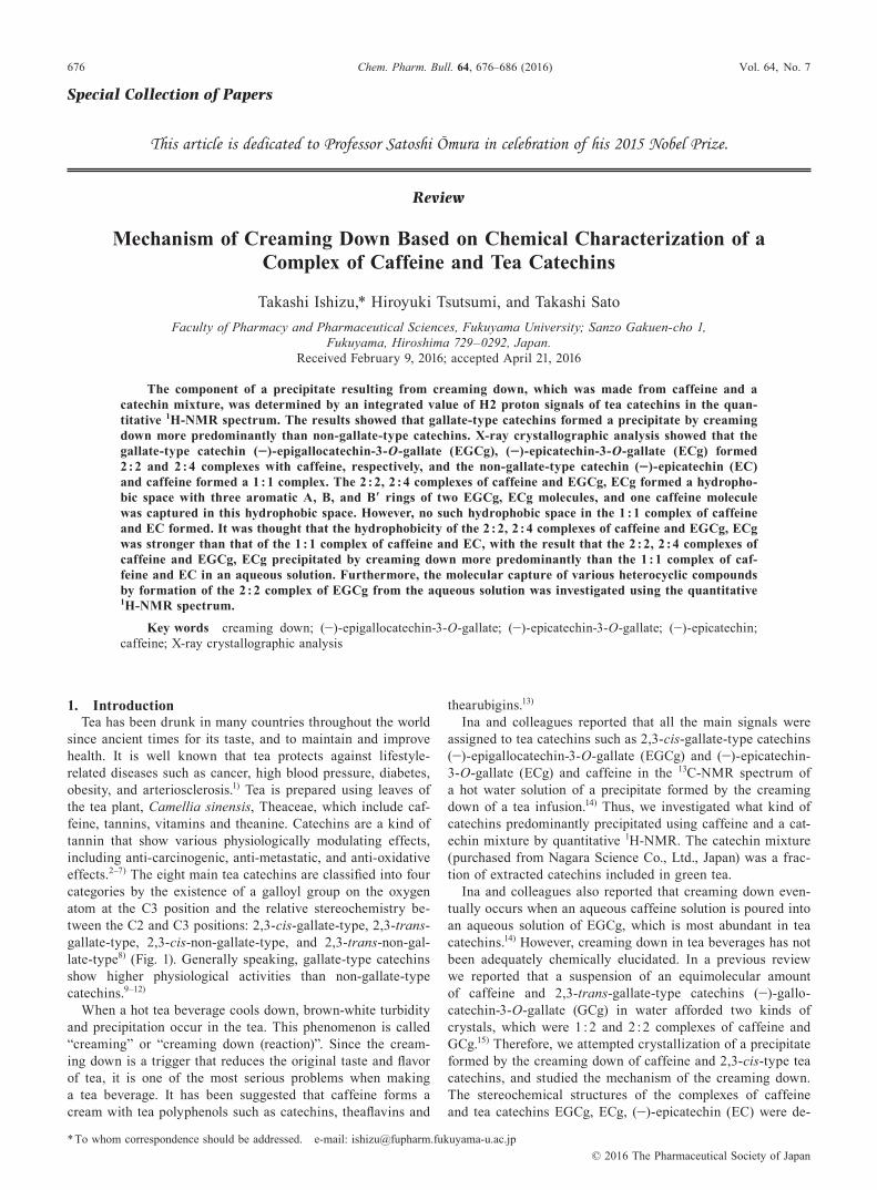
When an aqueous solution of nicotinamide was poured into an equimolecular amount of EGCg in an aqueous solution, a precipitate containing nicotinamide and EGCg at a molar ratio of 1 : 1 was also produced at 4°C. The precipitate was recrystallized from water and CH2Cl2 to afford a colorless block crystal. A single crystal was determined to be a 2 : 2 complex of nicotinamide and EGCg by X-ray crystallographic analysis20) (Fig. 13).
Just like the 2 : 2 complex of caffeine and EGCg, this complex was formed from two crystallographically different nicotinamide and EGCgs (EGCg A, EGCg B), and nicotin-amide was sandwiched between B′ rings of EGCg.
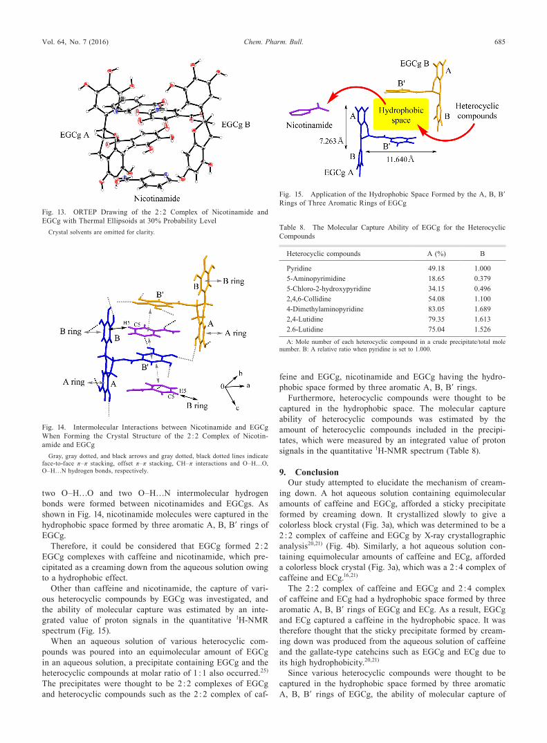
In the 2 : 2 complex of nicotinamide and EGCg, offset π–π stacking interactions formed between nicotinamide and the B′ ring of EGCg A, nicotinamide and the B′ ring of EGCg B, and face-to-face π–π stacking interactions formed between the A rings of EGCg, the B rings of EGCg (Fig. 14). CH–π interactions were observed between the C5–H5 of the pyridine ring of nicotinamide and the B ring of EGCg. Furthermore,
Fig. 12. Layer Structure of the 2 : 2 Complex of Caffeine and EGCg(a) Caffeine molecules are displayed; (b) Water molecules as a crystal solvent are displayed but caffeine molecules are not; (c) 1 : 1 Complex of caffeine and EC; (d) 2 : 4
Complex of caffeine and ECg.
Vol. 64, No. 7 (2016) 685Chem. Pharm. Bull.
two O–H…O and two O–H…N intermolecular hydrogen bonds were formed between nicotinamides and EGCgs. As shown in Fig. 14, nicotinamide molecules were captured in the hydrophobic space formed by three aromatic A, B, B′ rings of EGCg.
Therefore, it could be considered that EGCg formed 2 : 2 EGCg complexes with caffeine and nicotinamide, which pre-cipitated as a creaming down from the aqueous solution owing to a hydrophobic effect.
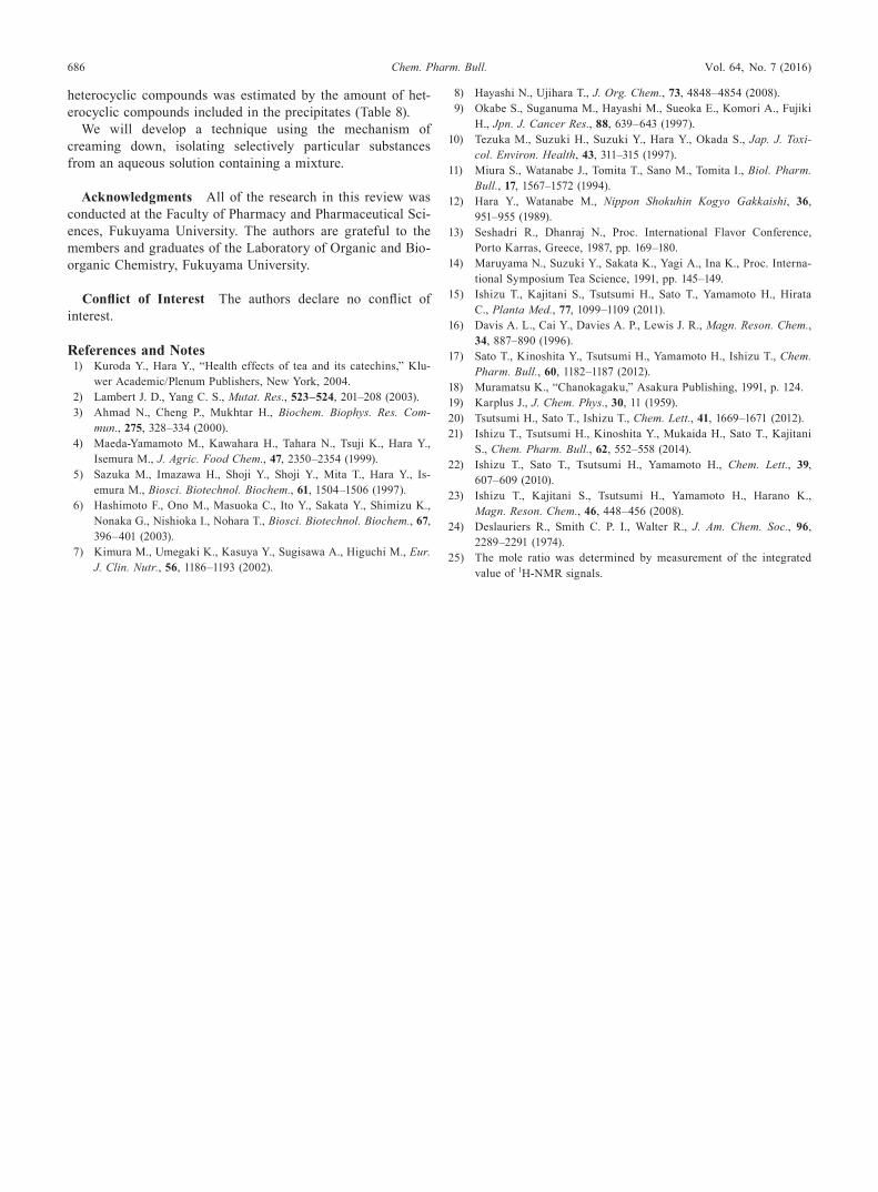
Other than caffeine and nicotinamide, the capture of vari-ous heterocyclic compounds by EGCg was investigated, and the ability of molecular capture was estimated by an inte-grated value of proton signals in the quantitative 1H-NMR spectrum (Fig. 15).
When an aqueous solution of various heterocyclic com-pounds was poured into an equimolecular amount of EGCg in an aqueous solution, a precipitate containing EGCg and the heterocyclic compounds at molar ratio of 1 : 1 also occurred.25) The precipitates were thought to be 2 : 2 complexes of EGCg and heterocyclic compounds such as the 2 : 2 complex of caf-
feine and EGCg, nicotinamide and EGCg having the hydro-phobic space formed by three aromatic A, B, B′ rings.
Furthermore, heterocyclic compounds were thought to be captured in the hydrophobic space. The molecular capture ability of heterocyclic compounds was estimated by the amount of heterocyclic compounds included in the precipi-tates, which were measured by an integrated value of proton signals in the quantitative 1H-NMR spectrum (Table 8).
9. ConclusionOur study attempted to elucidate the mechanism of cream-
ing down. A hot aqueous solution containing equimolecular amounts of caffeine and EGCg, afforded a sticky precipitate formed by creaming down. It crystallized slowly to give a colorless block crystal (Fig. 3a), which was determined to be a 2 : 2 complex of caffeine and EGCg by X-ray crystallographic analysis20,21) (Fig. 4b). Similarly, a hot aqueous solution con-taining equimolecular amounts of caffeine and ECg, afforded a colorless block crystal (Fig. 3a), which was a 2 : 4 complex of caffeine and ECg.16,21)
The 2 : 2 complex of caffeine and EGCg and 2 : 4 complex of caffeine and ECg had a hydrophobic space formed by three aromatic A, B, B′ rings of EGCg and ECg. As a result, EGCg and ECg captured a caffeine in the hydrophobic space. It was therefore thought that the sticky precipitate formed by cream-ing down was produced from the aqueous solution of caffeine and the gallate-type catehcins such as EGCg and ECg due to its high hydrophobicity.20,21)
Since various heterocyclic compounds were thought to be captured in the hydrophobic space formed by three aromatic A, B, B′ rings of EGCg, the ability of molecular capture of
Fig. 13. ORTEP Drawing of the 2 : 2 Complex of Nicotinamide and EGCg with Thermal Ellipsoids at 30% Probability Level
Crystal solvents are omitted for clarity.
Fig. 14. Intermolecular Interactions between Nicotinamide and EGCg When Forming the Crystal Structure of the 2 : 2 Complex of Nicotin-amide and EGCg
Gray, gray dotted, and black arrows and gray dotted, black dotted lines indicate face-to-face π–π stacking, offset π–π stacking, CH–π interactions and O–H…O, O–H…N hydrogen bonds, respectively.
Fig. 15. Application of the Hydrophobic Space Formed by the A, B, B′ Rings of Three Aromatic Rings of EGCg
Table 8. The Molecular Capture Ability of EGCg for the Heterocyclic Compounds
Heterocyclic compounds A (%) B
Pyridine 49.18 1.0005-Aminopyrimidine 18.65 0.3795-Chloro-2-hydroxypyridine 34.15 0.4962,4,6-Collidine 54.08 1.1004-Dimethylaminopyridine 83.05 1.6892,4-Lutidine 79.35 1.6132.6-Lutidine 75.04 1.526
A: Mole number of each heterocyclic compound in a crude precipitate/total mole number. B: A relative ratio when pyridine is set to 1.000.
686 Vol. 64, No. 7 (2016)Chem. Pharm. Bull.
heterocyclic compounds was estimated by the amount of het-erocyclic compounds included in the precipitates (Table 8).
We will develop a technique using the mechanism of creaming down, isolating selectively particular substances from an aqueous solution containing a mixture.
Acknowledgments All of the research in this review was conducted at the Faculty of Pharmacy and Pharmaceutical Sci-ences, Fukuyama University. The authors are grateful to the members and graduates of the Laboratory of Organic and Bio-organic Chemistry, Fukuyama University.
Conflict of Interest The authors declare no conflict of interest.
References and Notes 1) Kuroda Y., Hara Y., “Health effects of tea and its catechins,” Klu-
wer Academic/Plenum Publishers, New York, 2004. 2) Lambert J. D., Yang C. S., Mutat. Res., 523–524, 201–208 (2003). 3) Ahmad N., Cheng P., Mukhtar H., Biochem. Biophys. Res. Com-
mun., 275, 328–334 (2000). 4) Maeda-Yamamoto M., Kawahara H., Tahara N., Tsuji K., Hara Y.,
Isemura M., J. Agric. Food Chem., 47, 2350–2354 (1999). 5) Sazuka M., Imazawa H., Shoji Y., Shoji Y., Mita T., Hara Y., Is-
emura M., Biosci. Biotechnol. Biochem., 61, 1504–1506 (1997). 6) Hashimoto F., Ono M., Masuoka C., Ito Y., Sakata Y., Shimizu K.,
Nonaka G., Nishioka I., Nohara T., Biosci. Biotechnol. Biochem., 67, 396–401 (2003).
7) Kimura M., Umegaki K., Kasuya Y., Sugisawa A., Higuchi M., Eur. J. Clin. Nutr., 56, 1186–1193 (2002).
8) Hayashi N., Ujihara T., J. Org. Chem., 73, 4848–4854 (2008). 9) Okabe S., Suganuma M., Hayashi M., Sueoka E., Komori A., Fujiki
H., Jpn. J. Cancer Res., 88, 639–643 (1997).10) Tezuka M., Suzuki H., Suzuki Y., Hara Y., Okada S., Jap. J. Toxi-
col. Environ. Health, 43, 311–315 (1997).11) Miura S., Watanabe J., Tomita T., Sano M., Tomita I., Biol. Pharm.
Bull., 17, 1567–1572 (1994).12) Hara Y., Watanabe M., Nippon Shokuhin Kogyo Gakkaishi, 36,
951–955 (1989).13) Seshadri R., Dhanraj N., Proc. International Flavor Conference,
Porto Karras, Greece, 1987, pp. 169–180.14) Maruyama N., Suzuki Y., Sakata K., Yagi A., Ina K., Proc. Interna-
tional Symposium Tea Science, 1991, pp. 145–149.15) Ishizu T., Kajitani S., Tsutsumi H., Sato T., Yamamoto H., Hirata
C., Planta Med., 77, 1099–1109 (2011).16) Davis A. L., Cai Y., Davies A. P., Lewis J. R., Magn. Reson. Chem.,
34, 887–890 (1996).17) Sato T., Kinoshita Y., Tsutsumi H., Yamamoto H., Ishizu T., Chem.
Pharm. Bull., 60, 1182–1187 (2012).18) Muramatsu K., “Chanokagaku,” Asakura Publishing, 1991, p. 124.19) Karplus J., J. Chem. Phys., 30, 11 (1959).20) Tsutsumi H., Sato T., Ishizu T., Chem. Lett., 41, 1669–1671 (2012).21) Ishizu T., Tsutsumi H., Kinoshita Y., Mukaida H., Sato T., Kajitani
S., Chem. Pharm. Bull., 62, 552–558 (2014).22) Ishizu T., Sato T., Tsutsumi H., Yamamoto H., Chem. Lett., 39,
607–609 (2010).23) Ishizu T., Kajitani S., Tsutsumi H., Yamamoto H., Harano K.,
Magn. Reson. Chem., 46, 448–456 (2008).24) Deslauriers R., Smith C. P. I., Walter R., J. Am. Chem. Soc., 96,
2289–2291 (1974).25) The mole ratio was determined by measurement of the integrated
value of 1H-NMR signals.