mechanism of the jak2/stat3-cav-1-nr2b signaling pathway ... · cellular signal transduction...
TRANSCRIPT

Endocrine (2019) 64:55–66https://doi.org/10.1007/s12020-019-01880-6
ORIGINAL ARTICLE
Mechanism of the JAK2/STAT3-CAV-1-NR2B signaling pathway inpainful diabetic neuropathy
Chuan-Da Li1 ● Jia-Yi Zhao1● Jia-Li Chen1
● Jia-Hui Lu1● Mao-Biao Zhang1
● Qi Huang1● Yan-Nan Cao1
● Gai-Li Jia1 ●
Yuan-Xiang Tao2● Jun Li1 ● Hong Cao1
Received: 24 October 2018 / Accepted: 22 February 2019 / Published online: 4 March 2019© The Author(s) 2019
AbstractPurpose The aim of the present study was to further elucidate the role of JAK2/STAT3-CAV-1-NR2B on painful diabeticneuropathy.Methods In vivo, the mechanical withdrawal threshold and thermal withdrawal latency were measured to evaluate neu-ropathic pain behaviors (n= 8), while western blot (n= 5) and an immunofluorescence double staining experiment (n= 6)were performed to understand the molecular mechanism. In vitro, the individual culture of BV2 mouse microglia cell lines,the co-culture of BV2 mouse microglia cell lines and PC12 rat neuron cell lines, and western blot analysis were performed tounderstand the molecular mechanism between microglia and neurons.Results The expression of p-JAK2, p-STAT3, t-CAV-1, and p-NR2B was upregulated in the dorsal horn of DNP ratsthroughout the experiment. Through the immunofluorescence double staining experiment, it was found that p-STAT3 wasmainly expressed in activated microglia, and this condition can be stably maintained for approximately 2 weeks after theestablishment of the DNP model. The intrathecal injection of JAK2 inhibitor AG490 can relieve the abnormal expression ofp-JAK2, p-STAT3, t-CAV-1, and p-NR2B, and relieve pain. The remission of AG490 began on the third day, and it couldbe stably sustained for 14 days. In vitro high-glucose induced the activation of p-STAT3 in microglia, thereby upregulatingthe expression of p-CAV-1 and p-NR2B in neurons in the co-culture system. JAK2 inhibitor AG490 can alleviate theabnormal expression of these proteins in the JAK2/STAT3-CAV-1-NR2B signaling pathway in vitro.Conclusions Microglial JAK2/STAT3 signaling probably contributes to neuropathic pain by activating the CAV-1-NR2Bpathway.
Keywords Painful diabetic neuropathy ● JAK2/STAT3 ● CAV-1 ● NR2B
AbbreviationsT2DM Type-2 diabetic mellitusDNP Painful diabetic neuropathyNMDA N-methyl-D-aspartate receptorDRG Dorsal root ganglion
JAK2 Janus Kinase 2STAT3 Signal transducers and activators of
transcriptionCAV-1 Caveolin-1NR2B NMDA receptor 2BHFSD High-fat-sugar dietSTZ StreptozocinMWT Mechanical withdrawal thresholdTWL Thermal withdrawal latencyDMSO DimethylsulfoxideAG-490 Tyrphostin AG490CCI Chronic constrictive injuryISI Insulin sensitivity indexBDNF Brain-derived neurotrophic factor
* Jun [email protected]
* Hong [email protected]
1 Department of Anesthesiology, Second Affiliated Hospital ofWenzhou Medical University, Pain Medicine Institute of WenzhouMedical University, 325035 Zhejiang, China
2 Department of Anesthesiology, New Jersey Medical School,Rutgers, The State University of New Jersey, Newark, NJ 07103,USA
1234
5678
90();,:
1234567890();,:

Introduction
Type-2 diabetic mellitus (T2DM) has reached a pandemicstatus, and has shown no signs of abatement. Painful dia-betic neuropathy (DNP) has been generally considered to beone of the most common complications of T2DM with a30–50% incidence, and it has also been recognized as oneof the most difficult types of pain to treat [1]. Glycemiccontrol and the use of analgesic drugs are the primarytreatments for DNP. However, these are accompanied byunobvious curative effects. Exploring the pathogenesis ofDNP would be helpful for developing novel therapeuticstrategies.
Spinal cord dorsal horn glial cell signal transduction-induced central sensitization is one of the importantpathogeneses of DNPs. The activation of microglia pro-duces and releases a variety of cytokines and excitatorysubstances of neurons or glial cells [2–4], promotes NR2Bactivation on neuronal cells, NR2B is a subunit on NMDA,and increases NMDA-mediated current. NMDA receptoractivation can further induce the sensitization of the centralnervous system, which is one of the important pathogenesesof DNP [5, 6].
JAK2/STAT3 signal transduction, as a classical intra-cellular signal transduction pathway, participates in manypathophysiological processes, such as cell differentiation,proliferation, inflammation, and pain formation. The acti-vation of microglia caused by JAK2/STAT3 has a sig-nificant effect on neuropathic pain. After nerve injuryinduces vigorous IL-6 production in dorsal root ganglion(DRG), the cytokines may be transported to central term-inals of primary afferents. The released cytokines stimulatethe JAK/STAT3 signaling pathway in spinal microglia andpromote genesis of neuropathic pain. Meanwhile, thesecytokines further activate glial cells and neurons to releasemore activating substances such as ATP, pro-inflammatoryfactors, reactive oxygen species (ROS), nitric oxide (NO),prostaglandins (PGs), etc. These activating substances fur-ther enhance neuropathic pain [7]. In addition, JAK2/STAT3 signal transduction has an effect on spinal NMDA-induced currents, causing neuropathic pain [8]. However,the relationship between the JAK2/STAT3 pathway andactivation of microglia in DNP remains unclear.
Caveolae are a specialized type of lipid raft that arestabilized by oligomers of the caveolin protein. Caveolin-1(CAV-1), a major protein component of caveolae, is animportant gene targeting for STAT3. The overexpression ofp-STAT3 can cause CAV-1 promoter activation andincrease gene expression [9]. CAV-1 regulates neuronalplasticity and receptor transport to regulate NR2B-NMDAR, which is closely correlated with pathologic painand central sensitization [10].
However, no studies on DNP have focused on the JAK2/STAT3 signaling pathway. Therefore, the present studyfurther explored the correlation between DNP and micro-glial activation by combining in vivo and in vitro experi-ments to explore the role of this signaling pathway in thepathogenesis of DNP, and explore its regulation of thedownstream of CAV-1 and NR2B.
Materials and methods
Animals
The study protocol was approved by the Animal ResearchCommittee of Wenzhou Medical University. All animalexperiments were performed in accordance with theNational Institutes of Health Guidelines for the Care andUse of Laboratory Animals, and in accordance with theAnimal Research: Reporting In Vivo Experiments(ARRIVE) guidelines. Sprague-Dawley (SD) rats, weighing120–160 g, were provided by the Center for LaboratoryAnimals of Wenzhou Medical University (License No.WYDW2014-0015). These rats were housed in room tem-perature, which was maintained within 23–25 °C, allowedfree access to food and water, and placed under a 12-hour/12-hour dark/light cycle. The detailed design schemes weredepicted in Figs 1 and 2.
Fig. 1 In vivo experiment design scheme. After 3 days of injection,conditions with blood glucose levels≧ 16.7 mmol/L were consideredas T2DM. Rats in the AG490 group and dimethylsulfoxide (DMSO)group were injected, respectively, with 10 μL (1 mmol/L) of AG490and 10 μL of 3.5% DMSO once a day for 14 days. Mechanicalwithdrawal threshold (MWT) and thermal withdrawal latency (TWL)were measured before streptozocin (STZ) injection, on day 3 after STZinjection (as a reference for successful modeling) and on days 3, 7, and14 after intrathecal injection (n= 5)
56 Endocrine (2019) 64:55–66

Induction of T2DM and DNP
These T2DM models were established, as previouslydescribed [11, 12]. After 2 weeks of adaptive feeding, theserats were randomized into two groups: Con group andT2DM group. Rats in the T2DM group were fed with ahigh-fat-sugar diet (HFSD) for 8 weeks, while rats in theCon group were fed with a normal-diet. After 8 weeks, theinsulin sensitivity trail was performed by testing for fastingblood glucose and insulin on blood samples collected fromthe tail vein. Insulin sensitivity index (ISI)= 1/(blood glu-cose × insulin). These results revealed that there was a sig-nificant difference in ISI between these two groups (P <0.05), implying that insulin resistance was successfullyinduced in the T2DM group. Subsequently, rats in theT2DM group were intraperitoneally injected with strepto-zocin (STZ) (Sigma Co., St. Louis, MO, USA) at a dose of
35 mg/kg. As a control, rats in the Con group were intra-peritoneally injected with the same dose of citrate buffer.After three days of injection, rats in the T2DM group had acaudal vein fasting blood glucose of ≥16.7 mmol/L, andwere considered as T2DM rats. After measuring themechanical withdrawal threshold (MWT) and thermalwithdrawal latency (TWL) of these rats, rats with MWT andTWL values ≤ 85% of baseline (before the high-fat-sugardiet) were considered as a successful model of type-2painful diabetic neuropathy, and was redefined as the T-DNP group. The remaining rats in the T2DM group wereredefined as the No-DNP group.
Subarachnoid catheterization and AG490administration
AG490, a specific inhibitor of JAK2, inhibits the activation ofSTAT3 by selectively blocking JAK2. AG490 (1 mmol/L)was dissolved in 3.5% dimethylsulfoxide (DMSO), and 3.5%DMSO was used as the control vehicle (DMSO group). Ratsin the T-DNP group were randomly divided into three groups:DNP group, AG490 group, and DMSO group. Rats in theAG490 and DMSO groups received subarachnoid catheter-ization, as previously described. Under anesthesia with chloralhydrate (0.3 g/kg), a PE-10 silastic tubing (Ningbo Scienceand Technology Park to the Software Technology Co., Ltd,China) was intrathecally inserted between the L4 and L5vertebrae, and advanced 2 cm into the lumbar enlargement ofthe spinal cord. The external end of the intrathecal catheterwas tunneled under the skin to the neck area, and the outerpart of the catheter was exposed, carefully plugged and fixedonto the skin. The animals were allowed to recover for5–6 days after the surgery. In order to verify the location ofthe catheter, 10 μL of 2% lidocaine was given through thecatheter. Limb paralysis response without neurological defi-cits indicated that the insertion was successful. Then, the ratsin the AG490 group and DMSO group were injected,respectively, with AG490 10 μL (1 mmol/L) and 3.5%DMSO 10 μL once a day for 14 days.
Behavioral tests
MWT and TWL were measured before STZ injection, onday 3 after STZ injection (as a reference for successfulmodeling) and on days 3, 7, and 14 after intrathecal injec-tion. The detained behavioral tests and model preparationwere described in the Supplementary Methods.
Cell cultures and treatments
BV2 immortalized murine microglia cell line was purchasedfrom MX Biotechnology Company (Shanghai, China).PC12 neuronal cell line cells were provided by the College
Fig. 2 In vitro experiment design scheme. A Step 1 BV2 cells wereseeded at 1 × 10⁶ cells/well in a 6-well plate. Con-G: BV2 cells treatedwith low glucose (5.5 mM D-glucose); HG-G: BV2 cells treated withhigh glucose (33.3 mM D-glucose); AG490-G: BV2 cells treated withAG490 (50 μM)+ high glucose; DMSO-G: BV2 cells treated withdimethylsulfoxide (DMSO)+ high glucose. After 24 h of culture,western blot was used to detect the expression of p-STAT3 in BV2microglia cells. B Step 2 Transwell was used to establish a co-culturesystem of BV2 cells and PC12 cells. Con-N/G: BV2 cells and PC12cells treated with low glucose (5.5 mM D-glucose); HG- N/G: BV2cells PC12 cells treated with high glucose (33.3 mM D-glucose);AG490- N/G: BV2 cells PC12 cells treated with AG490 (50 μM)+high glucose; DMSO-N/G: BV2 cells PC12 cells treated with DMSO+ high glucose. HG-N: PC12 cells treated with low glucose. After 24 hof culture, western blot was used to detect the expression of p-Cav-1and p-NR2B in PC12 cells
Endocrine (2019) 64:55–66 57

of Pharmacy of Wenzhou Medical University. The cellswere cultured in Dulbecco’s modified eagle’s medium(DMEM) supplemented with 10% fetal bovine serum(FBS), 100 U/mL of penicillin, 100 μg/mL of streptomycin,and 5.5 mmol/L of glucose.
BV2 microglia cell line cultures and treatments
BV2 cells were seeded at 1 × 10⁶ cells/well in a 6-well plate.BV2 microglia cells were randomly divided into fourgroups: control group (Con-G group), high-glucose group(HG-G group), JAK2 inhibitor group (AG490-G group),and solvent control group (DMSO-G group). BV2 cellswere cultured with low-glucose medium (5.5 mM D-glu-cose) to the exponential phase. Then, BV2 cells werereplaced with low-glucose medium (5.5 mM D-glucose),high-glucose medium (33.3 mM D-glucose), AG490(50 μM) plus high-glucose medium (33.3 mM D-glucose),and DMS plus high-glucose medium (33.3 mM D-glucose),respectively. After 24 h of culture, western blot was per-formed to detect the expression of p-STAT3 in BV2microglia cells.
BV2 microglia cell co-culture with PC12 neuron cells
Transwell was used to build the co-culture system of BV2microglia cells and PC12 neuron cells. These cells wererandomly divided into five groups: control group (Con-N/Ggroup), high-glucose group (HG-N/G group), JAK2 inhi-bitor group (AG490-N/G group), solvent control group(DMSO-N/G group), and neuron high-glucose group (HG-N group). In the HG-N group, PC12 cells were seeded in asix-well plate with no cells seeded in the transwell, whichwas inside the six-well plate. For the other four groups,PC12 cells were seeded in a six-well plate, and BV2 cellswere seeded in the transwell, which was inside the six-wellplate. Cells in the Con-N/G group, HG-N/G group, AG490-N/G group, DMSO-N/G group, and AG490-N/G groupwere cultured with low-glucose medium (5.5 mM D-glu-cose) to the exponential phase, and this was subsequentlyreplaced with low-glucose medium (5.5 mM D-glucose),high-glucose medium (33.3 mM D-glucose), AG490(50 μM) plus high-glucose medium (33.3 mM D-glucose),DMSO plus high-glucose medium (33.3 mM D-glucose),and high glucose (33.3 mM D-glucose), respectively. After24 h, western blot analysis was carried out to detect theexpression of p-CAV-1 and p-NR2B in PC12 cells.
Western blot analysis
Protein samples were separated using 10% sodium dodecylsulfate polyacrylamide gel electrophoresis (SDS-PAGE,60 g of total protein per lane) and transferred onto a
polyvinylidene fluoride membrane (Merck Millipore,Temecula, CA, USA). In addition, gels stained with Coo-massie Blue were used to confirm the equal amounts ofprotein loaded on each lane. The membranes were incu-bated overnight at 4 °C with primary polyclonal rabbit anti-p-CAV-1 (1:500; Santa Cruz Biotechnology, Santa Cruz,CA, USA), anti-t-CAV-1 antibody (1:500; Santa CruzBiotechnology, Santa Cruz, CA, USA), anti-p-JAK2(1:500; Abcam, Cambridge, MA, USA), anti-p-STAT3(1:1000; Cell Signaling Technology, Danvers, MA, USA),or anti-p-NR2B antibody (1:500; Millipore, Billerica, MA,USA). The membranes were extensively applied with Trisbuffered saline Tween 20 (TBST) and incubated for 2 hwith horseradish peroxidase conjugated secondary antibody(1:3000; Abcam, UK)) at room temperature. The immunecomplexes were detected using a nitro blue tetrazolium/5-bromo-4-chloro-3-indolyl phosphate assay kit (Sigma Co.,St. Louis, MO, USA), or chemiluminescence (Pierce,Waltham, MA, USA). The density of the specific bands wasanalyzed using NIH ImageJ software, and the expressionlevels of these proteins were normalized to β-actin.
Immunohistochemistry
Rats were deeply anaesthetized with 400 mg/kg of 5%chloral hydrate, transcardially perfused with 200mL ofphosphate buffered saline (PBS, 0.01M; Solarbio Science &Technology, Beijing, China), followed by 300mL of freshlyprepared 4% paraformaldehyde in 0.01M PBS. The fourthto sixth lumbar segments of the spinal cord were removed,post-fixed with the same fixative for 12 h at 4 °C, and placedin 10%, 20 and 30% (w/v) sucrose solutions, one by one, for12 h at 4 °C. Then, the dehydration spinal cord tissue wasembedded with OTC, 20 μm was cut by a frozen sectionmachine at a constant temperature of −20 °C, washed with0.01M PBS solution at 4 °C three times for 20 min eachtime, and incubated in a blocking solution (0.2% Trition; 3%goat serum; 0.01M PBS) for 1 h at 26 °C. Then, the tissuewas incubated for 24 h at 4 °C with the following primaryantibodies: p-STAT3 (1:200; Pierce, Waltham, MA, USA),GFAP (1:1000; Merck Millipore, Temecula, CA, USA),OX-42 (1:200; Abdserotec, Hercules, CA, USA), and Neu(1:800; Merck Millipore, Temecula, CA, USA). Afterincubation, the sections were placed in room temperature for1 h, washed three times (10 min each time) with 0.01M PBSsolution at 4 °C, and incubated for 2 h at 26 °C with thefollowing secondary antibodies: Alexa Fluor® 546 GoatAnti-Mouse IgG, and Alexa Fluor® 488 Goat Anti-RabbitIgG (1:1,000; Invitrogen, Waltham, MA USA). Then, thetissues were washed with 0.01M PBS solution at 4 °C threetimes at 20 min each time. The slices were observed byfluorescence microscopy and analyzed by Image-Pro Plussoftware.
58 Endocrine (2019) 64:55–66
Data analysis
Data were presented as mean ± standard deviation (SD).The results were statistically analyzed using one-way ana-lysis of variance (ANOVA), or paired or unpaired Student’st-test. When the ANOVA results revealed a significantdifference, pairwise comparisons between means were tes-ted by the least significant difference method (LSD). Thesedata were analyzed by SPSS 19.0 (SPSS Inc., Chicago, IL,USA). P < 0.05 was considered statistically significant.
Results
Changes in blood glucose level, insulin level, andinsulin sensitivity index
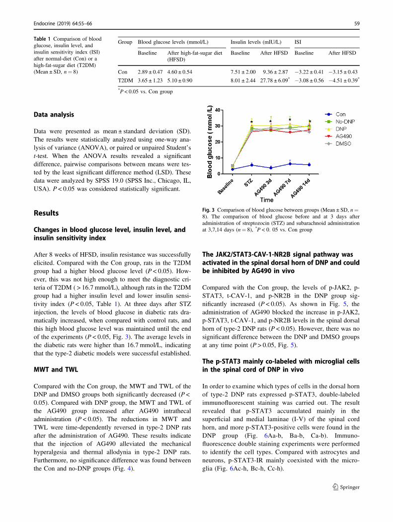
After 8 weeks of HFSD, insulin resistance was successfullyelicited. Compared with the Con group, rats in the T2DMgroup had a higher blood glucose level (P < 0.05). How-ever, this was not high enough to meet the diagnostic cri-teria of T2DM ( > 16.7 mmol/L), although rats in the T2DMgroup had a higher insulin level and lower insulin sensi-tivity index (P < 0.05, Table 1). At three days after STZinjection, the levels of blood glucose in diabetic rats dra-matically increased, when compared with control rats, andthis high blood glucose level was maintained until the endof the experiments (P < 0.05, Fig. 3). The average levels inthe diabetic rats were higher than 16.7 mmol/L, indicatingthat the type-2 diabetic models were successful established.
MWT and TWL
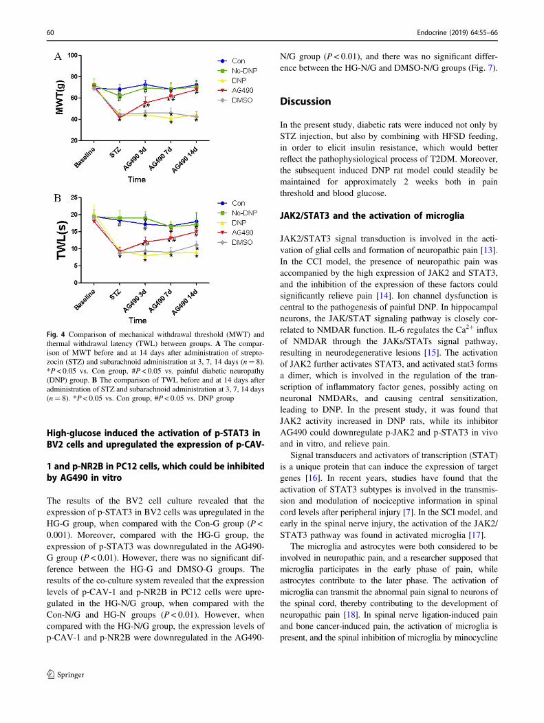
Compared with the Con group, the MWT and TWL of theDNP and DMSO groups both significantly decreased (P <0.05). Compared with DNP group, the MWT and TWL ofthe AG490 group increased after AG490 intrathecaladministration (P < 0.05). The reductions in MWT andTWL were time-dependently reversed in type-2 DNP ratsafter the administration of AG490. These results indicatethat the injection of AG490 alleviated the mechanicalhyperalgesia and thermal allodynia in type-2 DNP rats.Furthermore, no significance difference was found betweenthe Con and no-DNP groups (Fig. 4).
The JAK2/STAT3-CAV-1-NR2B signal pathway wasactivated in the spinal dorsal horn of DNP and couldbe inhibited by AG490 in vivo
Compared with the Con group, the levels of p-JAK2, p-STAT3, t-CAV-1, and p-NR2B in the DNP group sig-nificantly increased (P < 0.05). As shown in Fig. 5, theadministration of AG490 blocked the increase in p-JAK2,p-STAT3, t-CAV-1, and p-NR2B levels in the spinal dorsalhorn of type-2 DNP rats (P < 0.05). However, there was nosignificant difference between the DNP and DMSO groupsat any time point (P > 0.05, Fig. 5).
The p-STAT3 mainly co-labeled with microglial cellsin the spinal cord of DNP in vivo
In order to examine which types of cells in the dorsal hornof type-2 DNP rats expressed p-STAT3, double-labeledimmunofluorescent staining was carried out. The resultrevealed that p-STAT3 accumulated mainly in thesuperficial and medial laminae (I-V) of the spinal cordhorn, and more p-STAT3-positive cells were found in theDNP group (Fig. 6Aa-b, Ba-b, Ca-b). Immuno-fluorescence double staining experiments were performedto identify the cell types. Compared with astrocytes andneurons, p-STAT3-IR mainly coexisted with the micro-glia (Fig. 6Ac-h, Bc-h, Cc-h).
Table 1 Comparison of bloodglucose, insulin level, andinsulin sensitivity index (ISI)after normal-diet (Con) or ahigh-fat-sugar diet (T2DM)(Mean ± SD, n= 8)
Group Blood glucose levels (mmol/L) Insulin levels (mIU/L) ISI
Baseline After high-fat-sugar diet(HFSD)
Baseline After HFSD Baseline After HFSD
Con 2.89 ± 0.47 4.60 ± 0.54 7.51 ± 2.00 9.36 ± 2.87 −3.22 ± 0.41 −3.15 ± 0.43
T2DM 3.65 ± 1.23 5.10 ± 0.90 8.01 ± 2.44 27.78 ± 6.09* −3.08 ± 0.56 −4.51 ± 0.39*
*P < 0.05 vs. Con group
Fig. 3 Comparison of blood glucose between groups (Mean ± SD, n=8). The comparison of blood glucose before and at 3 days afteradministration of streptozocin (STZ) and subarachnoid administrationat 3,7,14 days (n= 8), *P < 0. 05 vs. Con group
Endocrine (2019) 64:55–66 59
High-glucose induced the activation of p-STAT3 inBV2 cells and upregulated the expression of p-CAV-
1 and p-NR2B in PC12 cells, which could be inhibitedby AG490 in vitro
The results of the BV2 cell culture revealed that theexpression of p-STAT3 in BV2 cells was upregulated in theHG-G group, when compared with the Con-G group (P <0.001). Moreover, compared with the HG-G group, theexpression of p-STAT3 was downregulated in the AG490-G group (P < 0.01). However, there was no significant dif-ference between the HG-G and DMSO-G groups. Theresults of the co-culture system revealed that the expressionlevels of p-CAV-1 and p-NR2B in PC12 cells were upre-gulated in the HG-N/G group, when compared with theCon-N/G and HG-N groups (P < 0.01). However, whencompared with the HG-N/G group, the expression levels ofp-CAV-1 and p-NR2B were downregulated in the AG490-
N/G group (P < 0.01), and there was no significant differ-ence between the HG-N/G and DMSO-N/G groups (Fig. 7).
Discussion
In the present study, diabetic rats were induced not only bySTZ injection, but also by combining with HFSD feeding,in order to elicit insulin resistance, which would betterreflect the pathophysiological process of T2DM. Moreover,the subsequent induced DNP rat model could steadily bemaintained for approximately 2 weeks both in painthreshold and blood glucose.
JAK2/STAT3 and the activation of microglia
JAK2/STAT3 signal transduction is involved in the acti-vation of glial cells and formation of neuropathic pain [13].In the CCI model, the presence of neuropathic pain wasaccompanied by the high expression of JAK2 and STAT3,and the inhibition of the expression of these factors couldsignificantly relieve pain [14]. Ion channel dysfunction iscentral to the pathogenesis of painful DNP. In hippocampalneurons, the JAK/STAT signaling pathway is closely cor-related to NMDAR function. IL-6 regulates the Ca2+ influxof NMDAR through the JAKs/STATs signal pathway,resulting in neurodegenerative lesions [15]. The activationof JAK2 further activates STAT3, and activated stat3 formsa dimer, which is involved in the regulation of the tran-scription of inflammatory factor genes, possibly acting onneuronal NMDARs, and causing central sensitization,leading to DNP. In the present study, it was found thatJAK2 activity increased in DNP rats, while its inhibitorAG490 could downregulate p-JAK2 and p-STAT3 in vivoand in vitro, and relieve pain.
Signal transducers and activators of transcription (STAT)is a unique protein that can induce the expression of targetgenes [16]. In recent years, studies have found that theactivation of STAT3 subtypes is involved in the transmis-sion and modulation of nociceptive information in spinalcord levels after peripheral injury [7]. In the SCI model, andearly in the spinal nerve injury, the activation of the JAK2/STAT3 pathway was found in activated microglia [17].
The microglia and astrocytes were both considered to beinvolved in neuropathic pain, and a researcher supposed thatmicroglia participates in the early phase of pain, whileastrocytes contribute to the later phase. The activation ofmicroglia can transmit the abnormal pain signal to neurons ofthe spinal cord, thereby contributing to the development ofneuropathic pain [18]. In spinal nerve ligation-induced painand bone cancer-induced pain, the activation of microglia ispresent, and the spinal inhibition of microglia by minocycline
Fig. 4 Comparison of mechanical withdrawal threshold (MWT) andthermal withdrawal latency (TWL) between groups. A The compar-ison of MWT before and at 14 days after administration of strepto-zocin (STZ) and subarachnoid administration at 3, 7, 14 days (n= 8).*P < 0.05 vs. Con group, #P < 0.05 vs. painful diabetic neuropathy(DNP) group. B The comparison of TWL before and at 14 days afteradministration of STZ and subarachnoid administration at 3, 7, 14 days(n= 8). *P < 0.05 vs. Con group, #P < 0.05 vs. DNP group
60 Endocrine (2019) 64:55–66

effectively reduced allodynia and hyperalgesia [19, 20]. DNPis associated with a slower rate of deafferentation comparedwith traumatic neuropathy, but, recently, microglia cells alsoplay a role in the development of neuropathic pain in DNP[21]. In the present study, the activation of microglia andastrocytes were both observed in DNP in vivo.
STAT3 mainly expressed in microglia is one of themarker proteins of central nervous system injury. It plays arole in regulating the cytokine-mediated signaling pathway
[22]. In the context of maladaptive plasticity, abnormalsignaling between glia and neurons also has a role in pain.After traumatic nerve injury, microglia in the dorsal horn ofthe spinal cord release factors such as brain-derived neu-rotrophic factor (BDNF), leading to the amplification ofnociceptive synaptic processing, resulting in “gating” ofneuropathic pain [23]. In the CCI model, and in the early-stage of pain, the double-label immunofluorescence assaysrevealed that p-STAT3 was mainly co-localized with
Fig. 5 The representative immunoblotting bands (bottom) and thequantitative data (top) showed following points. (Bottom of A–D): Theexpression of p-JAK2, p-STAT3, t-Cav-1 and p-NR2B in spinal cordof group Con, No-DNP, DNP, AG490 and DMSO animals by western
blotting experiment. (Top of A–D): The western blots analysis for thep-JAK2, p-STAT3, t-CAV-1, and p-NR2B protein (n= 5/group) in thedifferent group at different time point. *P < 0.05 vs. Con group; #P <0.05 vs. Painful diabetic neuropathy (DNP) group
Endocrine (2019) 64:55–66 61

microglia [7]. However, some studies have indicated thatSTAT3 co-exists with astrocytes, and participates in theactivation of astrocytes [13, 24, 25]. In the present study,the double-label immunofluorescence assays revealed thatp-STAT3 was mainly expressed in microglia in the spinalcord, which was not consistent with some of the conclu-sions above. This may be due to the different model ofneuropathic pain, such as the particularity of the hypergly-cemia state in DNP model. Moreover, it was found that the
high-glucose environment upregulated the expression of p-STAT3 in microglia, which was consistent with what wasfound in vitro.
The correlation between CAV-1 and STAT3
Caveolae is a specialized type of lipid raft stabilized byoligomers of caveolin protein. Caveolae has a large numberof membrane-bound proteins, which are mostly signal
Fig. 6 The expression of p-STAT3-IR and localization of p-STAT3mainly with microglia, but astrocytes and neuron, in the spinal corddorsal horn of the type-2 painful diabetic neuropathy (DNP) rats. AThe third day after AG490 intrathecal administration. B The seventhday after AG490 intrathecal administration. C The fourteenth day afterAG490 intrathecal administration. There are numerous p-STAT3-IRexists in the superficial and medial laminae (I-V) of spinal cord horn atgroup DNP in picture Aa-b, Ba-b, Ca-b (the arrows to the bright greenin the pictures). The microglia cells (OX-42, red arrows highlight) andthe co-label of p-STAT3-IR with microglial cells (arrow highlights in
yellow), astrocytes cell markers GFAP (the arrows to the red highlightof GFAP, yellow highlights for the co-label) and neurons markersNeuN (the arrows to the red highlight of NeuN, yellow highlights forco-label) by double immunofluorescence table experiments, respec-tively (pictures Ac-f, Bc-f, Cc-f). Neither astrocytes nor neurons inspinal cord of DNP animals are co-labeled with p-STAT3-IR at thethird, seventh, and fourteenth days. Three independent experimentswere performed with different animals from each experimental groupfor immunohistochemical experiments (n= 6). Tissue slices, 20 μm.Scale bar, 100 μm
62 Endocrine (2019) 64:55–66

molecules with lipid modification. Caveolae is a platformfor the exchange of these signaling molecules, allowing thesignal pathways to interact with each other. CAV-1 is thecrucial adjustment of the signal pathways in each platform.In general, CAV-1 has a negative regulatory signal trans-duction pathway for signaling molecules in Caveolae, and italso has the function of enhancing the signal [26–28].
CAV-1 has been shown to directly interact with STAT3on lipid rafts [29, 30]. Activated STATs leave a lipid raftinto the cell, and the subsequent cross-cytoplasm transportrequires a specific chaperone protein, in which CAV-1 isone of the chaperone proteins [31]. Research has shown thatthe JAK/STAT signaling pathway locates on the caveolaecontaining CAV-1 [32, 33]. CAV-1 is an important genetargeting of STAT3, and STAT3 directly affects the tran-scription of CAV-1 by directly binding to the CAV-1 pro-moter. The overexpression of p-STAT3 causes CAV-1promoter activation and the increase in gene expression [9].
In the present study, in the dorsal horn of the spinal cord,the expression of t-CAV-1 was upregulated in vivo. Owingto laboratory conditions, the expression and distribution ofp-CAV-1 in the dorsal horn of the spinal cord were notdetected in the present study. In the high-glucose environ-ment, the expression of p-STAT3 was upregulated in BV2,and the expression of p-CAV-1 was upregulated in the co-culture of BV2 and PC12. In contrast, the expression of p-CAV-1 was not upregulated in PC12 alone. Hence, it can bespeculated that p-CAV-1 may be a downstream target of p-STAT3. In addition, in vivo and in vitro experiments haveconfirmed that AG490 can relieve the abnormal expressionof CAV-1. In view of the exact relationship with the JAK/STAT signaling pathway, the above-mentioned inferencecan be further confirmed. However, since the present studydid not directly interpret the STAT3-CAV-1 signalingpathway, further experiments were needed to confirm
whether CAV-1 acts as an important downstream target ofthe JAK/STAT pathway in the pathogenesis of DNP.
The correlation between CAV-1 and NR2B
The central sensitization caused by NMDAR plays animportant role in the formation and development of DNP. Anumber of studies have shown that NR2B activation andneuropathic pain are closely correlated to its production andmaintenance [34, 35].
HEAD BP found a close association between CAV-1 andNR2B in the primary culture of rat cortical neurons [31].CAV-1 regulates neuronal plasticity and receptor transport,regulating NR2B-NMDAR, which is closely correlated toneuropathic pain and central sensitization. In the CCI model,the expression levels of CAV-1 and NR2B in the anteriorcingulate cortex were significantly higher than those innormal rats. CAV-1 promoted the expression of NR2B onthe membrane, and opened the ion channel, thereby alteringneuronal synaptic plasticity, and causing central sensitiza-tion. In addition, the increase in NR2B expression can beinhibited by CAV-1 siRNA or NR2B inhibitors. The co-immunoprecipitation and two-hybrid assay also demon-strated a direct interaction between CAV-1 and NR2B.CAV-1 regulated chronic neuropathic pain by modulatingNR2B in the anterior cingulate gyrus [10].
In the present study, it was found that the expression ofp-NR2B in the dorsal horn of DNP rats was upregulated. Inthe in vitro experiments, it was also found that the high-glucose environment could increase the expression of p-NR2B in neurons. As the present study did not intervenewith the CAV-1-NR2B pathway, it was difficult to elucidatethe correlation between these two in the pathogenesis ofDNP. In addition, the in vivo and in vitro experimentsconfirmed that AG490 can alleviate the abnormal
Fig. 7 The results of cell culture in vitro. A The expression of p-STAT3 in BV2 cells under high glucose. Mean ± SD. n= 3. ***P <0.001. B The expression of p-CAV-1 in PC12 cells under co-culture
system. Mean ± SD. n= 3. **P < 0.01. C The expression of p-NR2Bin PC12 cells under co-culture system. Mean ± SD. n= 3. **P < 0.01,***P < 0.001
Endocrine (2019) 64:55–66 63
expression of p-NR2B, in view of the important position ofNMDA in the pathogenesis of neuropathic pain. Further-more, to a certain extent, it can reflect the significance of theJAK2/STAT3 signaling pathway in DNP treatment.
Influence of high-glucose on the JAK2/STAT3-CAV-1-NR2B signaling pathway
Persistent high blood glucose level in diabetes plays aninitiation role in the change of voltage gated calciumchannel and voltage gated sodium channel in the cyto-membrane of neuron axons, and the release of neuralgrowth active substances and P substances [36]. Thisthereby upregulates the excitability of peripheral sensorynerve fibers and neurons in the spinal dorsal horn, leadingto the spontaneous discharge activities of neurons andsensitization to stimulation, which may be the foundationof neuropathic pain. In the present study, it was found thathigh glucose may have an effect on the JAK2/STAT3-CAV-1-NR2B signal pathway, and the cell experimentindicated that it may affect p-CAV-1 and p-NR2B inPC12 cells through the upregulation of p-STAT3 on BV2cells.
Several observations have confirmed that high glucosehas a major role in the upregulation of p-JAK2 and p-STAT3 in mesangial cells [37]. Mao et al. [38] found thatthe high-glucose-induced JAK/STAT signaling pathway isactivated in human glomerular mesangial cells. In addition,under high-glucose conditions, the phosphorylation ofCAV-1 was significantly increased in podocytes [39]. Fur-thermore, CAV-1 expression has been observed in mono-layer ECs exposed to high glucose [40]. Moreover, theexpression of NR2B is also upregulated in retinal ganglioncells-5 under high-glucose conditions [41]. A positive cor-relation between poor control of blood glucose and severityof neuropathy or the risk and intensity of neuropathic painwas also detected [42]. Nevertheless, this correlation is not alinear relationship as some patients have severe neuropathybut do not develop neuropathic pain.
In the present in vivo experiments, 56% of T2DM ratssuccessfully progressed to DNP, while the remainingT2DM rats did not present with neuropathic pain. Com-pared with the DNP group, the expression levels of p-JAK2,p-STAT3, CAV-1, and p-NR2B in the spinal dorsal horn inthe no-DNP group were not significantly different, and thiswas the same with the Con group. Moreover, there was nodifference in blood glucose levels between the DNP groupand no-DNP group at all time points, and all remained at ahigh level. Therefore, in contradiction with the results of thein vitro experiments, the in vivo experiments confirmed thathyperglycemia did not affect the expression of p-JAK2, p-STAT3, CAV-1, and p-NR2B.
The following reasons may contribute to the differencesobserved in vivo and in vitro: (1) The expression of proteinswas detected in certain cells in vitro, while this was detectedin all cells in the spinal dorsal horn in vivo. (2) The con-centration of glucose in the high-glucose group (33.3 mM)in vitro was pretty approximate to the blood glucosedetected in T2DM rats (30.2 ± 4.8 mM). However, com-pared with the simple environment in vitro experiment, theresults induced by high glucose in vitro may not be con-sistent with the results under the complex organism envir-onment, involving multiple systems, organs and multiplecells in the in vivo experiment. It was also considered thatthe high blood glucose level under the complex environ-ment in vivo may have some effects on the expression ofsome proteins in the JAK2/STAT3-CAV-1-NR2B signalpathway in certain cells of the spinal cord. However, thiswas not significant, and this pathway may be more corre-lated to the pathogenesis of neuropathic pain, rather thanmerely high blood glucose.
In vitro, the high glucose environment can significantlyaffect the expression of related proteins of the JAK2/STAT3-CAV-1-NR2B pathway. In vivo, high blood glu-cose levels have no significant effect on the expression ofthe protein, but each protein of the DNP rat spinal dorsalhorn were abnormally expressed, and the JAK2 specificinhibitors could inhibit the above abnormal expressionsin vivo and in vitro. Each protein of the JAK2/STAT3-CAV-1-NR2B pathway plays an important role in thepathogenesis of DNP. However, the specific signal trans-duction between the upstream and downstream of CAV-1needs to be further confirmed.
Funding This study was supported by National Natural ScienceFoundation of China (Project No. 81771487) and Zhejiang ProvincialNatural Science Foundation of China (Project No. LY17H070006).
Compliance with ethical standards
Conflict of interest The authors declare that they have no conflict ofinterest.
Ethical approval The study protocol was approved by the AnimalResearch Committee of Wenzhou Medical University. All applicableinternational, national, and/or institutional guidelines for the care anduse of animals were followed. This article does not contain any studieswith human participants performed by any of the authors.
Publisher’s note: Springer Nature remains neutral with regard tojurisdictional claims in published maps and institutional affiliations.
Open Access This article is distributed under the terms of the CreativeCommons Attribution 4.0 International License (http://creativecommons.org/licenses/by/4.0/), which permits use, duplication,adaptation, distribution, and reproduction in any medium or format, aslong as you give appropriate credit to the original author(s) and the
64 Endocrine (2019) 64:55–66

source, provide a link to the Creative Commons license, and indicate ifchanges were made.
References
1. S. Tesfaye, A.J. Boulton, P.J. Dyck, R. Freeman, M. Horowitz, P.Kempler, G. Lauria, R.A. Malik, V. Spallone, A. Vinik, L. Ber-nardi, P. Valensi; Toronto Diabetic Neuropathy Expert Group,Diabetic neuropathies: update on definitions, diagnostic criteria,estimation of severity, and treatments. Diabetes Care 33,2285–2293 (2010)
2. M. Tsuda, Microglia in the spinal cord and neuropathic pain. J.Diabetes Investig. 7, 17–26 (2016)
3. J. Molet, A. Mauborgne, M. Diallo, V. Armand, D. Geny, L.Villanueva, Y. Boucher, M. Pohl, Microglial Janus kinase/signaltransduction and activator of transcription 3 pathway activitydirectly impacts astrocyte and spinal neuron characteristics. J.Neurochem. 136, 133–147 (2016)
4. Y. Lei, Y. Sun, C. Lu, Z. Ma, X. Gu, Activated glia increased thelevel of proinflammatory cytokines in a resiniferatoxin-inducedneuropathic pain rat model. Reg. Anesth. Pain. Med. 41, 744–749(2016)
5. S.R. Chen, G. Samoriski, H.L. Pan, Antinociceptive effects ofchronic administration of uncompetitive NMDA receptorantagonists in a rat model of diabetic neuropathic pain. Neuro-pharmacology 57, 121–126 (2009)
6. R.R. Ji, T. Berta, M. Nedergaard, Glia and pain: is chronic pain agliopathy? Pain 154(Suppl 1), S10–S28 (2013)
7. E. Dominguez, C. Rivat, B. Pommier, A. Mauborgne, M. Pohl,JAK/STAT3 pathway is activated in spinal cord microglia afterperipheral nerve injury and contributes to neuropathic paindevelopment in rat. J. Neurochem. 107, 50–60 (2008)
8. S. Liu, W.L. Mi, Q. Li, M.T. Zhang, P. Han, S. Hu, Q.L. Mao-Ying,Y.Q. Wang, Spinal IL-33/ST2 signaling contributes to neuropathicpain via neuronal CaMKII-CREB and astroglial JAK2-STAT3cascades in mice. Anesthesiology 123, 1154–1169 (2015)
9. W.T. Chiu, H.T. Lee, F.J. Huang, K.D. Aldape, J. Yao, P.S. Steeg,C.Y. Chou, Z. Lu, K. Xie, S. Huang, Caveolin-1 upregulationmediates suppression of primary breast tumor growth and brainmetastases by stat3 inhibition. Cancer Res. 71, 4932–4943 (2011)
10. J.X. Yang, L. Hua, Y.Q. Li, Y.Y. Jiang, D. Han, H. Liu, Q.Q.Tang, X.N. Yang, C. Yin, L.Y. Hao, L. Yu, P. Wu, C.J. Shao, H.L. Ding, Y.M. Zhang, J.L. Cao, Caveolin-1 in the anterior cin-gulate cortex modulates chronic neuropathic pain via regulation ofNMDA receptor 2B subunit. J. Neurosci. 35, 36–52 (2015)
11. J.K. Dang, Y. Wu, H. Cao, B. Meng, C.C. Huang, G. Chen, J. Li,X.J. Song, Q.Q. Lian, Establishment of a rat model of type IIdiabetic neuropathic pain. Pain Med. 15, 637–646 (2014)
12. B. Meng, L.L. Shen, X.T. Shi, Y.S. Gong, X.F. Fan, J. Li, H. Cao,Effects of curcumin on TTX-R sodium currents of dorsal rootganglion neurons in type 2 diabetic rats with diabetic neuropathicpain. Neurosci. Lett. 605, 59–64 (2015)
13. J.E. Herrmann, T. Imura, B. Song, J. Qi, Y. Ao, T.K. Nguyen, R.A. Korsak, K. Takeda, S. Akira, M.V. Sofroniew, STAT3 is acritical regulator of astrogliosis and scar formation after spinalcord injury. J. Neurosci. 28, 7231–7243 (2008)
14. D. Li, Y. Yan, L. Yu, Y. Duan, Procaine attenuates pain behaviorsof neuropathic pain model rats possibly via inhibiting JAK2/STAT3. Biomol. Ther. (Seoul.) 24, 489–494 (2016)
15. D.I. Orellana, R.A. Quintanilla, C. Gonzalez-Billault, R.B. Mac-cioni, Role of the JAKs/STATs pathway in the intracellular cal-cium changes induced by interleukin-6 in hippocampal neurons.Neurotox. Res. 8, 295–304 (2005)
16. T.J. Mitchell, S. John, Signal transducer and activator of tran-scription (STAT) signalling and T-cell lymphomas. Immunology114, 301–312 (2005)
17. P. Calmels, G. Mick, B. Perrouin-Verbe, M. Ventura, SOFMER(French Society for Physical Medicine and Rehabilitation), Neu-ropathic pain in spinal cord injury: identification, classification,evaluation. Ann. Phys. Rehabil. Med. 52, 83–102 (2009)
18. A.K. Clark, D. Gruber-Schoffnegger, R. Drdla-Schutting, K.J.Gerhold, M. Malcangio, J. Sandkühler, Selective activation ofmicroglia facilitates synaptic strength. J. Neurosci. 35, 4552–4570(2015)
19. Y. Liang, Y. Qiu, J. Du, J. Liu, J. Fang, J. Zhu, J. Fang, Inhibitionof spinal microglia and astrocytes contributes to the anti-allodyniceffect of electroacupuncture in neuropathic pain induced by spinalnerve ligation. Acupunct. Med. 34, 40–47 (2016)
20. Y. Yang, H. Li, T.T. Li, H. Luo, X.Y. Gu, N. Lü, R.R. Ji, Y.Q.Zhang, Delayed activation of spinal microglia contributes to themaintenance of bone cancer pain in female Wistar rats via P2X7receptor and IL-18. J. Neurosci. 35, 7950–7963 (2015)
21. M. Tsuda, H. Ueno, A. Kataoka, H. Tozaki-Saitoh, K. Inoue,Activation of dorsal horn microglia contributes to diabetes-induced tactile allodynia via extracellular signal-regulated proteinkinase signaling. Glia 56, 378–386 (2008)
22. X. Yang, G. He, Y. Hao, C. Chen, M. Li, Y. Wang, G. Zhang, Z.Yu, The role of the JAK2-STAT3 pathway in pro-inflammatoryresponses of EMF-stimulated N9 microglial cells. J. Neuroin-flamm. 7, 54 (2010)
23. S. Beggs, T. Trang, M.W. Salter, P2X4R+microglia drive neu-ropathic pain. Nat. Neurosci. 15, 1068–1073 (2012)
24. M. Tsuda, Y. Kohro, T. Yano, T. Tsujikawa, J. Kitano, H. Tozaki-Saitoh, S. Koyanagi, S. Ohdo, R.R. Ji, M.W. Salter, K. Inoue,JAK-STAT3 pathway regulates spinal astrocyte proliferation andneuropathic pain maintenance in rats. Brain 134, 1127–1139(2011)
25. D.S. Aaronson, C.M. Horvath, A road map for those who don’tknow JAK-STAT. Science 296, 1653–1655 (2002)
26. B. Razani, J.A. Engelman, X.B. Wang, W. Schubert, X.L. Zhang,C.B. Marks, F. Macaluso, R.G. Russell, M. Li, R.G. Pestell, D. DiVizio, H. Hou Jr, B. Kneitz, G. Lagaud, G.J. Christ, W. Edel-mann, M.P. Lisanti, Caveolin-1 null mice are viable but showevidence of hyperproliferative and vascular abnormalities. J. Biol.Chem. 276, 38121–38138 (2001)
27. K. Abdelmohsen, Y. Kuwano, H.H. Kim, M. Gorospe, Post-transcriptional gene regulation by RNA-binding proteins duringoxidative stress: implications for cellular senescence. Biol. Chem.389, 243–255 (2008)
28. Y. Jin, S.J. Lee, R.D. Minshall, A.M. Choi, Caveolin-1: a criticalregulator of lung injury. Am. J. Physiol. Lung Cell. Mol. Physiol.300, L151–L160 (2011)
29. P.B. Sehgal, G.G. Guo, M. Shah, V. Kumar, K. Patel, Cytokinesignaling: STATS in plasma membrane rafts. J. Biol. Chem. 277,12067–12074 (2002)
30. G. Shen-Tu, D.B. Schauer, N.L. Jones, P.M. Sherman, Detergent-resistant microdomains mediate activation of host cell signaling inresponse to attaching-effacing bacteria. Lab. Invest. 90, 266–281(2009)
31. B.P. Head, H.H. Patel, Y.M. Tsutsumi, Y. Hu, T. Mejia, R.C.Mora, P.A. Insel, D.M. Roth, J.C. Drummond, P.M. Patel,Caveolin-1 expression is essential for N-methyl-D-aspartatereceptor-mediated Src and extracellular signal-regulated kinase1/2 activation and protection of primary neurons from ischemiccell death. FASEB J. 22, 828–840 (2008)
32. N. Ariotti, R.G. Parton, SnapShot: caveolae, caveolins, andcavins. Cell 154, 704–704.e1 (2013)
33. Y. Liu, Z. Liang, J. Liu, W. Zou, X. Li, Y. Wang, L. An,Downregulation of caveolin-1 contributes to the synaptic
Endocrine (2019) 64:55–66 65

plasticity deficit in the hippocampus of aged rats. Neural Regen.Res. 8, 2725–2733 (2013)
34. W. Zhang, C.X. Shi, X.P. Gu, Z.L. Ma, W. Zhu, Ifenprodilinduced antinociception and decreased the expression of NR2Bsubunits in the dorsal horn after chronic dorsal root gangliacompression in rats. Anesth. Analg. 108, 1015–1020 (2009)
35. M. Zhuo, Plasticity of NMDA receptor NR2B subunit in memoryand chronic pain. Mol. Brain 2, 4 (2009)
36. L. Manni, F. Florenzano, L. Aloe, Electroacupuncture counteractsthe development of thermal hyperalgesia and the alteration ofnerve growth factor and sensory neuromodulators induced bystreptozotocin in adult rats. Diabetologia 54, 1900–1908 (2011)
37. Y.H. Shi, S. Zhao, C. Wang, Y. Li, H.J. Duan, Fluvastatin inhibitsactivation of JAK and STAT proteins in diabetic rat glomeruli andmesangial cells under high glucose conditions. Acta Pharmacol.Sin. 28, 1938–1946 (2007)
38. T. Mao, H. Chen, L. Hong, J. Li, Pigment epithelium-derivedfactor inhibits high glucose-induced JAK/STAT signalling
pathway activation in human glomerular mesangial cells. Saudi.Med. J. 34, 793–800 (2013)
39. L.N. Sun, Z.X. Chen, X.C. Liu, H.Y. Liu, G.J. Guan, G. Liu,Curcumin ameliorates epithelial-to-mesenchymal transition ofpodocytes in vivo and in vitro via regulating caveolin-1. Biomed.Pharmacother. 68, 1079–1088 (2014)
40. C. Tian, R. Zhang, X. Ye, C. Zhang, X. Jin, Y. Yamori, L. Hao,X. Sun, C. Ying, Resveratrol ameliorates high-glucose-inducedhyperpermeability mediated by caveolae via VEGF/KDR path-way. Genes Nutr. 8, 231–239 (2013)
41. W. Qu, B. Zhang, Z.F. Zuo, X.Z. Liu, [The role of NR2B in theapoptosis of retinal ganglion cells-5 induced by glucose]. Chin. J.Anat. 38, 576–578 (2015). [Article in Chinese]
42. A.C. Themistocleous, J.D. Ramirez, P.R. Shillo, J.G. Lees, D.Selvarajah, C. Orengo, S. Tesfaye, A.S. Rice, D.L. Bennett, ThePain in Neuropathy Study (PiNS): a cross-sectional observationalstudy determining the somatosensory phenotype of painful andpainless diabetic neuropathy. Pain 157, 1132–1145 (2016)
66 Endocrine (2019) 64:55–66