mechanisms by which exercise promotes hippocampal function
TRANSCRIPT
Mechanisms by which exercise promotes hippocampal function in both
depressed and non-depressed individuals: A feasibility study
Joanne Caulfield Gourgouvelis BSc
A thesis submitted in fulfilment of the requirements for the degree of
Master of Science in Health Science,
The University Ontario Institute of Technology, 2012.
ii
Abstract
Depression is one of the top ten health problems in the world, affecting millions of
Canadians. Research indicates that exercise is an effective treatment for depression but it
is not clear on exactly how and why it works. Animal studies show that exercise
improves the ability of the brain to function. It can even lead to new cell formation in a
part of the brain called the hippocampus, which is important for memory processing. This
study is investigating whether exercise may also improve hippocampal function in
depressed humans. One way exercise may improve brain function is by normalizing
levels of the hormone cortisol, and its toxic effects on the hippocampus. Exercise may
also normalize levels of biochemical markers called cytokines involved in inflammation,
while improving levels of growth factors important to brain cell function. This feasibility
study aimed to develop protocols to investigate changes in hippocampal activity while
participants are performing memory tests involving association of images and words in a
functional magnetic resonance scanner before and after a 12 week exercise program. It
also aimed to develop and validate protocols to measure changes in cortisol, cytokines
and growth factors which are likely to be affected by exercise. Our preliminary imaging
results revealed hippocampal dysregulation in the depressed brain, and biomarker
analysis revealed abnormal concentrations of interleukin-6, vascular endothelial growth
factor and salivary cortisol when compared to normal healthy controls. However,
following the 12-week exercise program a more normalized pattern of hippocampal
activation associated with successful memory encoding was observed. Additionally,
biomarker concentrations either resembled or were closer to normal healthy values. Over
the long term, the project arising from this feasibility study has the potential to provide a

iii
tool to improve exercise prescription, to predict exercise responders and to guide
development of combined treatment approaches related to biochemical markers in order
to optimize depression outcomes for Canadians.
iv
Acknowledgements
First and foremost, to my supervisors Dr. Bernadette Murphy and Dr. Paul Yielder –
Thank you for all your guidance, encouragement and patience throughout this process.
Thank you also to:
Dr. John Samis, for all his time, patience, mentorship and diligence when
conducting immunoassays in addition to graciously offering his laboratory to
perform biomarker testing
Dr. Scott Fairhall, for his expert consultation on EPrime, SPM and fMRI data
processing
Joanne Free, for all her time and enthusiasm during the blood collection process
Dr. Nancy Wilkinson, for her hospitality during my visits at Lakeridge Health and
her expertise conducting the SCID to ensure participants met eligibility
Ian Barker, for his knowledge and time conducting the fitness assessments
Lakeridge Health for providing the resources and support that made this project
possible
Lydia Carmen for her dedication in recruiting participants
Annette Weekes Holder and Baycrest staff for their technical assistance
All our participants, who volunteered themselves and their time, and followed
instructions diligently – this study would not exist without your contributions!
The Flex Centre gym staff, for being so accommodating and allowing access to
their facility
The UOIT undergraduate students who spent countless patient hours supervising
exercise groups
Finally, a special thank-you goes out to my family, friends and most importantly my
husband for all their love and support. Without their continual encouragement this
research project would not have been possible!
v
Table of contents
ABSTRACT ................................................................................................................................................. II
ACKNOWLEDGEMENTS ....................................................................................................................... IV
TABLE OF CONTENTS ............................................................................................................................. V
LIST OF ABBREVIATIONS ................................................................................................................. VIII
LIST OF TABLES ...................................................................................................................................... XI
LIST OF FIGURES ................................................................................................................................... XII
CHAPTER 1 INTRODUCTION........................................................................................................... 13
1.1 Background to the study ....................................................................................................................... 13
1.2 Experimental objectives and hypotheses ............................................................................................. 16 1.2.1 Original Objectives .......................................................................................................................... 16 1.2.2 Original Hypotheses ........................................................................................................................ 17
1.3 Revised Thesis objectives ...................................................................................................................... 18
1.4 Significance of the study ........................................................................................................................ 20
1.5 Limitations ............................................................................................................................................. 21
CHAPTER 2 LITERATURE REVIEW............................................................................................... 24
2.1 Depression ........................................................................................................................................... 24 2.2.1 Depression and memory ............................................................................................................. 25
2.2 The role of the hippocampus ............................................................................................................. 26 2.3.1 Hippocampus and depression ..................................................................................................... 27 2.3.2 Hippocampus, depression and memory ........................................................................................... 29
2.4 Neurogenesis ....................................................................................................................................... 30
2. 5 The stress response ........................................................................................................................ 32 2.5.1 Stress and the HPA axis .................................................................................................................. 33
2.6 Cortisol ................................................................................................................................................... 34 2.6.1 Cortisol, stress and depression.................................................................................................... 36 2.6.2 Cortisol and memory .................................................................................................................. 37 2.6.3 Cortisol and the hippocampus .................................................................................................... 38 2.6.4 Cortisol and neurogenesis ........................................................................................................... 39
2.7 Inflammation and cytokines .............................................................................................................. 41 2.7.1 Cytokines and depression ................................................................................................................ 42 2.7.2 HPA axis and cytokines ................................................................................................................... 44 2.7.3 Cytokines, neuroplasticity and memory ........................................................................................... 46
2.8 Neurotropic growth factors .................................................................................................................. 47
vi
2.9 Current therapies for depression ...................................................................................................... 49
2. 10 Exercise and depression ................................................................................................................ 51 2.10.1 Exercise, neurogenesis and the hippocampus ............................................................................... 53 2.10.2 Exercise, inflammation and growth factors ................................................................................. 55 2.10.3 Further evidence for exercise and neural function ....................................................................... 56
CHAPTER 3 METHODS ..................................................................................................................... 59
3.1 Experimental design for future pilot study ......................................................................................... 59
3.2 Selection of outcome measures and protocols ..................................................................................... 62 3.2.1 Variables .......................................................................................................................................... 62 3.2.2 Protocols .......................................................................................................................................... 63
3.3 Participants ............................................................................................................................................ 71 3.3.1 Recruitment ...................................................................................................................................... 71 3.3.2 Inclusion/exclusion criteria .............................................................................................................. 75 3.3.3 Informed consent ............................................................................................................................. 76
3.4 Procedure ............................................................................................................................................... 76 3.4.1 Testing ............................................................................................................................................. 78 3.4.2 Intervention ...................................................................................................................................... 84
3.5 Analysis ................................................................................................................................................... 87 3.5.1 fMRI analysis ................................................................................................................................... 87
CHAPTER 4 PRELIMINARY RESULTS ......................................................................................... 87
4.1 Sample .................................................................................................................................................... 87 4.1.1 Psychometric evaluation .................................................................................................................. 88 4.1.2 Biomarker data ................................................................................................................................. 88 4.1.3 Fitness scores ................................................................................................................................... 90
4.2 Change – Participant one ...................................................................................................................... 92 4.2.1 Psychometric changes ...................................................................................................................... 92 4.2.2 Biomarker changes .......................................................................................................................... 92 4.2.3 Fitness change .................................................................................................................................. 93 4.2.4 fMRI analysis ................................................................................................................................... 94
CHAPTER 5 DISCUSSION ................................................................................................................. 97
5.1 Conclusion and recommendations ................................................................................................. 104
LIST OF REFERENCES .......................................................................................................................... 106
APPENDIX 1: PARTICIPANT INFORMATION SHEET ................................................................... 128
APPENDIX 2: CONSENT FORM ........................................................................................................... 129
APPENDIX 3: PAR-Q & YOU ................................................................................................................ 140
APPENDIX 4: BAYCREST SCREENING FORM ................................................................................ 141
APPENDIX 5: BDI .................................................................................................................................... 142
vii
APPENDIX 6: HADS ................................................................................................................................ 145
APPENDIX 9: MOCA .............................................................................................................................. 146
APPENDIX 7: PRE-SCREENING FORM ............................................................................................. 148
APPENDIX 8: PSQI .................................................................................................................................. 149
APPENDIX 9: SALIVA COLLECTION INSTRUCTIONS ................................................................. 151
APPENDIX 10: 11-ITEM EXERCISE SELF-EFFICACY ................................................................... 152
APPENDIX 11: 6-ITEM EXERCISE SELF-EFFICACY ..................................................................... 154
APPENDIX 12: LETTER TO GO WITH PARMED-X ........................................................................ 155
APPENDIX 13: PARMED-X ................................................................................................................... 156
APPENDIX 14: BLOOD COLLECTION LOG ..................................................................................... 160
APPENDIX 15: RESISTANCE EXERCISE OPTIONS ........................................................................ 161
viii
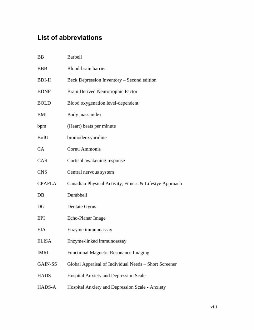
List of abbreviations
BB Barbell
BBB Blood-brain barrier
BDI-II Beck Depression Inventory – Second edition
BDNF Brain Derived Neurotrophic Factor
BOLD Blood oxygenation level-dependent
BMI Body mass index
bpm (Heart) beats per minute
BrdU bromodeoxyuridine
CA Cornu Ammonis
CAR Cortisol awakening response
CNS Central nervous system
CPAFLA Canadian Physical Activity, Fitness & Lifestye Approach
DB Dumbbell
DG Dentate Gyrus
EPI Echo-Planar Image
EIA Enzyme immunoassay
ELISA Enzyme-linked immunoassay
fMRI Functional Magnetic Resonance Imaging
GAIN-SS Global Appraisal of Individual Needs – Short Screener
HADS Hospital Anxiety and Depression Scale
HADS-A Hospital Anxiety and Depression Scale - Anxiety

ix
HADS-D Hospital Anxiety and Depression Scale - Depression
HC High-confidence
HRmax Maximum heart rate
HPA Hypothalamic-pituitary-adrenal
HRP Horseradish peroxidase
ID Identification
IGF Insulin Growth Factor
IL-1 Interleukin 1
IL-6 Interleukin 6
IL-10 Interleukin 10
IFN Interferon
Kcals Kilocalories
LMHDT Lakeridge Mental Health Day Treatment
MHDT Mental Health Day Treatment
MHR Maximum heart rate
MoCA Montreal Cognitive Assessment
MRI Magnetic Resonance Imaging
PAR-Q Physical Activity Readiness Questionnaire
PARmed-X Physical Activity Readiness Medical Examination
PSQI Pittsburgh Sleep Quality Index
REB Research Ethics Board
RPE Rating of Perceived Exertion
RPM Revolutions per minute

x
SCID-I Structural Clinical Interview for DSM-IV Axis I Disorders
SF-36 36-item Short-form questionnaire
SOS Salimetrics® Oral Swab
SSRI Selective serotonin reuptake inhibitor
TNF Tumor Necrosis Factor
VEGF Vascular Endothial Growth Factor
VO2 max Maximal oxygen uptake
xi
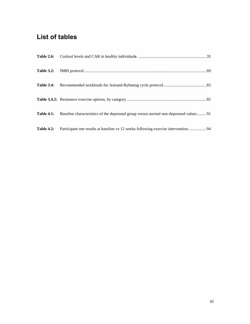
List of tables
Table 2.6: Cortisol levels and CAR in healthy individuals .................................................................. 35
Table 3.2: fMRI protocol ...................................................................................................................... 69
Table 3.4: Recommended workloads for Astrand-Ryhming cycle protocol ......................................... 83
Table 3.4.2: Resistance exercise options, by category ............................................................................. 85
Table 4.1: Baseline characteristics of the depressed group versus normal non-depressed values ......... 91
Table 4.2: Participant one results at baseline vs 12 weeks following exercise intervention. ................ 94
xii
List of figures
Figure 1.1: Overview of the effects of depression and exercise on dependant variables ...................... 15
Figure 2.5: The Hypothalamic–Pituitary–Cortisol System in Depression .............................................. 34
Figure 3.1: Overview of study design for the depressed exercise group, depressed control group
and non-depressed exercise group. ...................................................................................... 61
Figure 3. 2: Associative memory task ..................................................................................................... 68
Figure 3.3: Participant recruitment process ............................................................................................ 74
Figure 3.4: Data collection process. ....................................................................................................... 77
Figure 3.4.1: fMRI and associative memory task process. ........................................................................ 82
Figure 4.1: Baseline cortisol levels and CAR of the depressed group versus normal
non-depressed values............................................................................................................ 89
Figure 4.2.1: Participant one change in depression scores following 12 week exercise intervention........ 92
Figure 4.2.2: Participant one change in cortisol following 12 week exercise intervention vs. mean normal
values. ................................................................................................................................. 93
Figure 4.2.4: A comparison of fMRI results from Fairhall et al. (2010) and participant one.. .................. 96
13
Chapter 1 Introduction
1.1 Background to the study
The effects of depression are profound and far-reaching, being the 3rd largest contributor
to the burden of disease in high income and 7th in middle and low income countries
(Lopez et al. 2006) and affecting millions of Canadians (Canada 2006) . Exercise has
proven benefits in the treatment of depression (Blumenthal et al. 1999; Dunn et al. 2005),
but there are several competing hypotheses in our understanding of its mechanism of
action. Major Depressive Disorder (MDD) is associated with a wide array of
physiological, psychological and cognitive symptoms particularly impairments in
memory (Burt et al. 1995; Ilsley et al. 1995; Zakzanis et al. 1998; Airaksinen et al. 2004).
Exercise has been shown to improve memory in people with MDD (Ernst et al. 2006;
Cotman et al. 2007), suggesting an underlying mechanism that improves neural function
however it has been difficult to ascribe the improvement to the physiological effects of
exercise because of a lack of a physiological test that is clearly linked to improved neural
function (Sperling et al. 2003; Fairhall et al. 2010). Recently, Fairhall (2010) and
colleagues were the first to demonstrate decreased activation of the hippocampus during
the encoding phase of a memory task in depressed individuals compared to control
subjects using functional magnetic resonance imaging (fMRI)(Fairhall et al. 2010).
Researchers compared differences in hippocampal function (as opposed to volume
differences) between eight people with MDD and eight age and gender matched controls.
An associative memory task was designed based on work that showed robust modulation
14
of the right anterior hippocampus during the encoding of events that are subsequently
successfully remembered (Sperling et al. 2003). Likewise, pilot results confirmed robust
activation in the right hippocampus during encoding. Controls showed the predicted
pattern of increased activation in the right anterior hippocampus during the associative
encoding of stimuli that are successfully remembered with confidence compared to those
that are not remembered. However, this pattern was absent in the MDD group, thus
supporting the hypothesis that the normal modulation of right hippocampal activation by
encoding strength is dysregulated in MDD. An additional analysis showed that this
dysfunction was specific to the hippocampus and that a double dissociation between
groups in the hippocampus and intraparietal sulcus suggest that compensatory
mechanisms may exist. These findings provide a cutting edge tool linking memory
impairments and hippocampal changes in MDD and to integrate several current threads
of research to differentiate those factors which are able to be mediated by exercise and
are correlated with improved neural function.
One way that exercise may improve brain function in depression is by restoring normal
hypothalamic-pituitary-adrenal (HPA) axis and immune functioning by reducing cortisol
and pro-inflammatory cytokines levels and their toxic effects on the hippocampus in
addition to increasing neurotrophic growth factors known to enhance neural plasticity,
and new cell formation (neurogenesis). However the relationship between new cell
growth, increased hippocampal volume and more efficient functional activity in the
hippocampus has only been presumed thus far.
With the ability to demonstrate improved hippocampal activation concomitant with
improved memory, it is possible to determine which of the biochemical markers
15
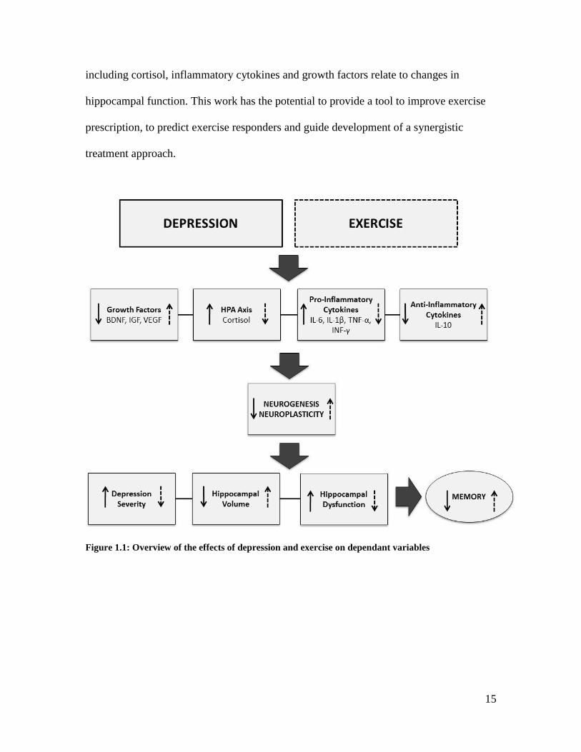
including cortisol, inflammatory cytokines and growth factors relate to changes in
hippocampal function. This work has the potential to provide a tool to improve exercise
prescription, to predict exercise responders and guide development of a synergistic
treatment approach.
Figure 1.1: Overview of the effects of depression and exercise on dependant variables
16
1.2 Experimental objectives and hypotheses
1.2.1 Original Objectives
The objectives of the proposed study are to investigate the effect of a moderate-intensity
structured, supervised 12 week exercise program combined with a mental health day
treatment (MHDT) program, as compared to the MHDT on its own, and exercise in a
non-depressed group. The outcome measures will include: Intensity of depressive
symptoms, sleep quality, overall cognitive function, plasma IL-1β, IL-6, IFN-γ, TNF-α
and IL-10, VEGF, BDNF and IGF, salivary cortisol, performance on an associative
memory task and concomitant fMRI hippocampal activation. A non-depressed exercise
group will be included to determine if the effects of exercise on the outcome measures are
generic. All outcome measures will be assessed at baseline and 12 weeks.
The specific objectives of this research are:
a) To determine whether depressed individuals who perform moderate exercise
perform better on cognitive memory tasks and demonstrate a return towards
the profile of hippocampal activation found in non-depressed individuals:
Hippocampal activation will be assessed using functional magnetic resonance
imaging (fMRI) and measuring activation in the hippocampus during the
encoding phase of a cognitive memory task.
b) To determine whether the depressed exercise group demonstrates lower
levels of cortisol, IL-1β, IL-6, IFN-γ,TNF-αand higher levels of IL-10,
DHEA,VEGF, BDNF and IGF after 12 weeks compared to their own

17
baseline and to any changes in the control group, and the baseline levels in
the non-depressed group: Participant will be sampled mid-week at waking and
30 minutes post-awakening samples for saliva which will be brought to the
investigators when they present for their fMRI scan. Both waking cortisol and the
awakening response (waking – 30 minutes post awakening) will be assayed along
with growth factors and cytokines which will be measured from plasma and
assayed using ELISA test kits for all three groups.
c) To investigate the relationship between changes in biochemical markers and
changes in hippocampal function with exercise: Regression analysis will be
used to determine whether there is any relationship between biochemical markers
and hippocampal function.
d) Determine whether there is relationship between improved fitness and
improved hippocampal function: Aerobic fitness will be measured at 0 and 12
weeks using submaximal tests of oxygen uptake (V02 max). Improvement in fitness
will be compared to improvement in memory and hippocampal activation to
determine if there is any correlation between the two for both exercise groups.
1.2.2 Original Hypotheses
The overall goal of this research is to investigate whether a structured, supervised 12
week exercise program can promote changes in hippocampal function in depressed
individuals and whether this is associated with improvements in depression, memory and
the status of biochemical markers known to be altered in MDD. This study will be
comparing depressed individuals who perform moderate exercise for 12 weeks in
18
addition to attending an outpatient day program to a control group who attend the day
program only. This study will also have a matched non-depressed exercise group to
provide normative data for comparison and to determine whether exercise also improves
hippocampal activation and biomarker status in non-depressed individuals.
It is hypothesized that:
a) Improved cardiovascular fitness as measured by change in maximal oxygen
consumption (VO2max) will be correlated with decreased cortisol levels in both
exercise groups.
b) Participants who receive exercise in addition to the MHDTP will show greater
improvements in depression and hippocampal function as well as lower levels of
cortisol than those in the MHDT only group.
c) Performance on cognitive memory tasks with concomitant hippocampal activation
(assessed using fMRI) will be positively correlated with improvements in
depression scores following the 12 week exercise intervention.
d) Changes in cortisol, IL-1β, IL-6, IFN-γ,TNF-α will be negatively correlated and
changes in IL-10, VEGF, BDNF and IGF will be positively correlated with
changes in hippocampal function following the 12 week exercise intervention.
1.3 Revised Thesis objectives
As the planning for the study began in collaboration with our partners at the Lakeridge
MHDT (LMHDT) program it became clear that there were a large number of preliminary
steps that would need to be completed prior to the initiation of the original aims.
These preliminary steps included:
19
1. Completion of an in depth literature review to ensure the most relevant
biomarkers relating to both exercise and depression would be assayed and the
most up to date evidence based protocols for exercise and depression are utilized.
2. Applying for grant funding to cover the costs of the fMRI scans and biomarker
assays.
3. Obtaining Ethical approval from three different Ethics committees (Lakeridge
Health where patients will be sampled, UOIT and Rotman Baycrest where the
fMRI scans would take place).
4. Validating and piloting the planned fMRI protocol previously piloted in New
Zealand on a 1.5 Tesla General Electric MRI scanner at the 3.0 Tesla Siemans
MRI scanner at the Rotman Baycrest hospital.
5. Developing a recruitment protocol from the LMHDT program to minimize the
number of steps required for staff and potential participants.
6. Developing protocols for blood collection, storage and biomarker assays.
7. Preliminary testing of the baseline samples of the initial recruits to determine the
sensitivity of the assays.
8. Development of an organizational plan to match exercise supervisors to
participants as each participant will be performing three one hour exercise
sessions per week.
Additionally, because it is clear that in order to differentiate the effects of exercise
specific to depression from the general effects of exercise it is important to include a non-
depressed exercise group in the final study which would have been beyond the timeframe
of a Masters‟ thesis. Therefore it was decided that that this Masters‟ thesis would become
20
a feasibility study for the larger study with the aims of completing the preliminary steps
described above, writing an in depth methods chapter to be used in the larger study, and
summarizing and discussing the results of the feasibility study and any implications that
this would have for the larger study.
1.4 Significance of the study
The effects of depression are far-reaching, having become one of the leading contributors
to the global burden of disease (WHO, 2008). Depression affects 1 in 20 Canadians
(Canada 2006) with an economic burden, estimated at over 14 billion dollars annually in
2001 (Stephens and Joubert 2001). The poor remission rates of existing antidepressant
therapies as well as the social stigma associated with seeking medical help leaves many
individuals undertreated (Shelton 2006). With a recent meta-analysis suggesting that
there are minimal benefits of medication vs placebo for mild to moderate depression
(Fournier et al. 2010), the role of non-pharmacologic treatment for depression becomes
increasingly important. Exercise is a therapeutic intervention with proven benefits in the
treatment of depression, but there is minimal research on its mechanism of action.
Therefore it is fundamental to understand why and how exercise works, in order to better
focus the exercise prescription and identify those most likely to benefit from exercise in
place of, or in addition to, other therapies.
It is well established that impaired episodic memory and reduced hippocampal volume is
associated with depression, however there is controversy about the meaning and
mechanism of these findings. Previous pilot data suggest that hippocampal fMRI can
distinguish between depressed and non-depressed individuals and may prove to be

21
sensitive marker of the effect of an intervention on neural function. A potential candidate
mechanism for hippocampal and memory changes are the elevated cortisol and pro-
inflammatory cytokine levels and decreased growth factor levels observed in depressed
individuals. This planned study will significantly advance knowledge as it is the first to
use fMRI to measure changes in hippocampal activation following an exercise
intervention in people with depression. By correlating changes in hippocampal activation
with changes in cortisol, pro-inflammatory cytokines and growth factors it is possible to
determine which of the biochemical markers is most predictive of improved neural
function. This study will also differentiate which markers change in MHDT plus exercise
vs MHDT alone as well as measuring whether these same markers change with exercise
in a non-depressed group. This will create a much more solid rationale for combined
approaches in the future. In the future it may be possible to use a person‟s “biochemical
signature” to determine the combination of exercise, pharmacotherapy and/or
psychological interventions are needed for synergistic effects and optimize treatment
outcomes for an individual. This work has the potential to provide a tool to improve
exercise prescription, to predict exercise responders and to guide development of
synergistic treatment approaches to optimize depression outcomes for Canadians.
1.5 Limitations
Study design
The proposed research will utilize a cohort control design. Participants will be recruited
in blocks. Depressed Group 1 will undertake a 12 week structured, supervised exercise
program in addition to the MHDT and they will be recruited in the first year of the study.
Group 2 from the MHDT will be recruited in the second year so that they can be matched

22
by age, gender, pharmacotherapy and baseline fitness level to the exercise group using
the same inclusion criteria. The reason for running the study as a cohort control rather
than a randomized controlled trial is that the LMHDT is a group program and it might
affect group cohesion if some participants were offered the exercise program and others
were not. This way everyone in the first cohort who meets the inclusion criteria will be
given the opportunity to have the exercise program, and this will not interfere with the
program in the way that it is currently run. It also allows to age and gender match the
control group in the non-exercising LMHDT cohort.
Testing and instrumentation
Cortisol secretion is associated with several variables such as life stressors. Saliva
sampling at baseline and 12 weeks took place midweek on a Wednesday, due to the
significant cortisol secretion differences on week days, versus weekends (Kunz-Ebrecht
et al. 2004).
Although indirect submaximal cardiorespiratory fitness tests estimating VO2max are less
accurate than direct maximal measurements, they are more appropriate test to use when
evaluating sedentary populations. The validity of VO2max tests can be questioned with
variables such as motivation, however workload and durations for the Astrand-Rhyming
cycle ergometer protocol are determined by fixed guidelines and do not allow for
subjectivity. Additionally, provide an accurate estimation of VO2max once adjusted to the
Astrand age correction factors (Cink and Thomas 1981).

23
Participant compliance
To prevent errors associated with self-sampling, participants will be provided with clearly
written instructions in conjunction with a verbal explanation, at baseline and at 12 weeks.
If a participant forgets to sample or a sampling error occurs, an alternative midweek day
(Thursday) will be used to repeat sampling.
To ensure that exercise attendance between-group differences could be accounted for,
individual records were kept of session attendance so that attendance rate could be
calculated. To remain eligible for the study, a minimum of 75% attendance is required,
which translates to a minimum of 27 sessions out of the total 36 allotted.
Controlling for the effects from social contact
It is important to differentiate the benefits of group exercise which are specific to exercise
as opposed to the psychosocial benefits due to the social interaction that occurs within the
group. A meta-analysis of 14 randomised controlled trials in this area, while finding a
positive overall effect for exercise, identified a lack of randomisation concealment,
blinding and intention to treat analysis in the research to date (Lawlor and Hopker 2001).
A point raised is that positive social interactions occurring during exercise may be more
important to the treatment effect than exercise per se, and most research is lacking the
inclusion of contact only groups to control for the psychosocial benefits of exercise, an
issue will be addressed as the LMHDT program provides an ideal solution to this.

Chapter 2 Literature Review
Lack of activity destroys the good condition of every human being, while
movement and methodical physical exercise save it and preserve
it. - Plato (423 BC- 347 BC)
2.1 Depression
Major depressive disorder (MDD) is a global public-health problem and is among the most
prevalent and burdensome of all psychiatric disorders. In Canada, it is estimated that 1 in 20
Canadians are affected by depression and its economic burden is estimated at over 14 billion
dollars annually (Stephens and Joubert 2001). Depression is the fourth leading cause of disease
burden globally and is projected to be the second leading cause by the year of 2030 (WHO,
2004). It has been estimated that 12% of men and 25% of women will experience a clinically
significant depressive episode at least once in their lives (Shelton 2006; Gadalla 2009).
Depression is also a severely undertreated and under recognized disorder in the primary care
setting (Diverty and Beaudet 1997; Insel and Charney 2003; Patten and Beck 2004). The
majority of individuals with mental illness do not consult health professionals (Insel and Charney
2003; Gadalla 2009) and it is estimated that only 56% of depressed Canadians seek medical help.
Of those patients who seek medical help and receive treatment with antidepressant drug therapy,
only half achieve full remission of symptoms (i.e. absence of symptoms and return to normal
functioning) (Fava and Ruini 2002). It seems that many patients feel the stigma of seeking
treatment is greater than the stigma of living with the disorder (Mojtabai and Olfson 2006). The
Diagnostic and Statistical Manual of Mental Disorders, Fourth Edition, Text Revision (DSM-IV-
25
TR) defines a depressive episode as a period greater than 2 weeks that is characterized by five or
more symptoms ranging from depressed mood, anhedonia, feelings of worthlessness or guilt,
suicidal ideation, changes in body weight or appetite, sleep disturbance, lack of energy,
psychomotor agitation or retardation, decreased ability to think or concentrate (APA and DSM-
IV 2000). These symptoms are often associated with higher suicide rates and higher mortality
rates (Kalia, 2005; Wulsin, Vaillant, & Wells, 1999). There is also strong evidence implicating
depression in the later development of other medical illnesses such as increased abdominal fat,
decreased bone density, hypertension, peptic ulcers and diabetes (Brown, Varghese, & McEwen,
2004). Therefore, the need for non-pharmacologic treatments for depression is becoming
increasingly important.
2.2.1 Depression and memory
Of the wide array of cognitive deficits associated with depression, memory impairment is the
most frequently reported (Airaksinen et al. 2004). However, research in this area has presented
ambiguous findings in terms of the type, severity and specificity of memory deficits. One
finding that has consistently been replicated is that of impaired episodic memory (memory for a
specific past experience in one‟s life) with a sparing of semantic memory (present knowledge of
universal truths such as “the sky is blue”), and other facets of memory such as short-term
memory (Ilsley et al. 1995; Tulving and Markowitsch 1998; Sweeney et al. 2000). However,
some studies have found equivalence between depressed and control individuals for both
semantic and episodic memory (Danion et al. 1995). These conflicting findings may be due
somewhat to differences in the sampling populations, and methodological weaknesses. A meta-
analysis of 147 studies examining memory dysfunction in depression (Burt et al. 1995) found a
significant, stable association between depression and memory impairment. A more recent meta-
26
analysis of 22 studies supports the relationship between depression and memory impairment, and
furthermore reveals that depression has the greatest effect on episodic memory as compared to
semantic and short-term memory (Zakzanis et al. 1998). Research identifying the stage of the
memory deficit, in encoding or retrieval, has been equivocal (Ilsley et al. 1995; Airaksinen et al.
2004). A meta-analysis of studies on depression and memory suggests that both encoding and
retrieval processes are impaired somewhat in depression (Zakzanis et al. 1998). More recent,
behavioural and neuroimaging studies have found that both the encoding and retrieval processes
in memory are impaired in depression (Zakzanis et al. 1998; Airaksinen et al. 2004; Behnken et
al. 2010; Fairhall et al. 2010; Milne et al. 2012). Although the neural underpinnings of impaired
memory in MDD are not completely understood, the majority of evidence implicates abnormal
activity in the hippocampal region critical for normal memory formation (Heckers et al. 2002;
Sperling et al. 2003).
2.2 The role of the hippocampus
The role of the hippocampus in the formation of memories has long been recognized. The
hippocampus is located in the medial temporal lobe and is critical for learning and the formation
of stable declarative memory in humans. However, the exact function of the hippocampus in
forming successful memories remains unknown (Scoville and Milner 2000; Kim et al. 2006).
The laminae that comprise the hippocampal complex include the dentate gyrus (DG) and
hippocampal proper, four regions of the cornu Ammonis (CA) termed CA1–CA4 which are
based on pyramidal neuron morphology and sensitivity to anoxia (Sperling et al. 2003; Campbell
and MacQueen 2004; Duvernoy 2005). The critical functions of the hippocampus were first
27
discovered over 50 years ago when severe amnesia and the inability to form new memories
occurred in a patient following removal of this brain region (Scoville and Milner 2000).
At the time of memory formation a connection must be made between the to-be-remembered
stimulus and its context. It is the process of encoding that forms new associations between
previously unrelated items of information and the ability to retain relational information across
time that is considered the heart of declarative memory and the function of the hippocampal
region (Zola et al. 2000; Sperling et al. 2003). Although the exact function of the hippocampus in
creating successful memories is not yet fully understood, converging results from animal models
of amnesia (Zola et al. 2000) and neuroimaging studies in humans have implicate the
hippocampus in the process of encoding episodic memories (Köhler et al. 2000; Heckers et al.
2002; Sperling et al. 2003). It has been assumed that long-term potentiation (LTP), a form of
synaptic plasticity within the hippocampus, contributes to the acquisition and retention of
memories although the exact mechanism remains unknown (Bliss and Collingridge 1993;
Lamprecht and LeDoux 2004; Leuner et al. 2006; Fairhall et al. 2010).
2.3.1 Hippocampus and depression
Brain imaging studies demonstrate that psychiatric disorders such as MDD are associated with
structural alterations such as reduced brain volume to structures that control mood and contribute
to stress related psychiatric illnesses (Pittenger and Duman 2007). Combining evidence from
animal models and post-mortem studies investigating brain tissue from depressed subjects
further detail alterations at the cellular level, such as atrophy of dendrite processes and reduced
neurons and glial elements (Pittenger and Duman 2007; Krishnan and Nestler 2008). However
the specific mechanisms underlying these structural changes have not been determined (Banasr
et al. 2011).
28
The hippocampus is particularly vulnerable to structural, functional, and neurogenic change in
response to stress and stress-related diseases, such as depression. (Conrad 2008). To date, MRI
studies have revealed compelling evidence that the hippocampus undergoes selective volume
reduction in stress-related neuropsychiatric disorders such as schizophrenia and MDD (Sheline et
al. 1996; Duman et al. 1999; Bremner et al. 2000; Czéh et al. 2001; Brown et al. 2004; Videbech
and Ravnkilde 2004; Hickie et al. 2005; McKinnon et al. 2009; Pajonk et al. 2010). Likewise,
several meta analyses confirm that individuals with MDD have hippocampal volumes that are
approximately 5-8% smaller than healthy controls (Videbech and Ravnkilde 2004; McKinnon et
al. 2009). Importantly, hippocampal volume is negatively correlated with depression duration
and morphological changes in the hippocampus are not evident in patients with a first episode of
depression, suggesting cumulative damage to this area. (MacQueen et al. 2003). This work has
recently been extended to indicate that duration of illness plays an important role as a predictor
in hippocampal volume reduction if left untreated by antidepressant drug therapy since
medication status may also play a central role in modulating hippocampus volume in MDD
(McKinnon et al. 2009). Hence, treatment early in disease progression may minimize the
volumetric reductions that are associated with multiple episodes of depression. Hippocampus
volumes are also associated with clinical outcome (MacQueen et al. 2008). Recently, MacQueen
et al. (2008) examined hippocampal volumes in 63 depressed participants who had baseline MRI
scans and then completed eight weeks of first-line pharmaceutical treatment. Researchers
compared the hippocampal volumes between patients who met the criteria for clinical remission
to those who did not meet the criteria for remission. Overall, patients who entered remission had
larger hippocampal volumes than those who did not enter remission.
29
Reduced hippocampal volume is also associated with several other neuropsychiatric conditions,
such as schizophrenia, dementia and posttraumatic stress disorder (MacQueen 2009; Pajonk et al.
2010) . It still has not been determined whether the pathological mechanisms that result in
reduced hippocampal volumes in patients with MDD, psychosis or dementia are unique for each
condition or involve the same pathophysiological processes (MacQueen 2009). For instance, it
has been difficult to determine whether there are reliable differences between individuals with
bipolar disorder and healthy controls due to the treatment effects of lithium, comorbidity, course
of illness and variability in subtypes of patients which may partly explain the mixed finding
reported (MacQueen 2009).
2.3.2 Hippocampus, depression and memory
Memory impairments often accompany depression and are considered to be a direct result from
structural changes in the hippocampus (Hickie et al. 2005). Hippocampal recruitment is linked
with greater hippocampal activation as indicated by regional cerebral blood flow within the
hippocampus (Heckers et al. 2002). In healthy individuals, fMRI revealed that there is an
expected pattern of increased activation in the anterior hippocampus during the encoding of
stimuli that are subsequently remembered with confidence compared to items that are not
remembered. However, this hippocampal activation pattern is absent in depressed individuals
suggesting a dysregulation of hippocampal function (Fairhall et al. 2010). Similarly, abnormal
hippocampal activation was found during the memory retrieval process in depressed individuals
(Milne et al. 2012). Given that most studies investigating the hippocampus in depression use
volumetric imaging analysis there is limited literature regarding hippocampal function.
Therefore, it is still unclear how or whether structural abnormalities of the hippocampus turn into
functional abnormalities. A combination of functional and structural imaging techniques may be
30
helpful in understanding whether structural brain abnormalities translate into function
abnormalities as well as the underlying neural basis of cognitive processes such as memory.
2.4 Neurogenesis
The hippocampus is a highly reactive brain structure that exhibits rapid plasticity at the
molecular, cellular, structural and functional levels in response to a specific stimuli (DeCarolis
and Eisch 2010). One specific feature of hippocampal plasticity noteworthy is the ability of the
DG in the hippocampus to generate new neurons throughout life (Gould et al. 1999; Leuner et al.
2006). In healthy brains, neurogenesis occurs mainly in two distinct areas: the subventricular
zone, which generates new neurons in the olfactory bulb; and the subgranular zone, which
generates new neurons in the DG of the hippocampus (Ming and Song 2005).
Rodent studies are among the first to demonstrate the ability of neurogenesis in specific areas in
the brain (Gould et al. 1999; van Praag et al. 1999; Deng et al. 2010). For instance,
environmental enrichment such as larger living space, physical exercise and social interaction
with other rodents helped to stimulate cell proliferation in the adult hippocampus as well as
significantly increase survival time for newly generated hippocampal neurons (van Praag et al.
1999). Behavioural studies in rodents in which neurogenesis has been experimentally
interrupted support the idea that the newly generated cells play a critical role in some forms of
hippocampus-dependent learning (Winocur et al. 2006).
Neurogenesis can also be regulated by several factors associated with an organism‟s behavioural
states. For instance, learning of hippocampal-dependent tasks such as maze-learning not only
increased the number of adult-generated hippocampal granule cells (Gould et al. 1999) but also
31
increased network activity while also improving cognition (Deng et al. 2010). However, whether
increases neurogenesis is responsible for cognitive improvements remains to be investigated.
In the rodent hippocampus, it has been well established that exposure to chronic stress alters the
number and morphology of neurons; however, very little is known about the likely changes that
occur in the hippocampal vasculature. Importantly, stress-induced reductions in hippocampal
neurogenesis occurs mainly near capillaries suggesting that decreased blood flow and capillary
density in the hippocampus results in the onset of depression (Heine et al. 2005; Kiuchi et al.
2012). Furthermore, the local microvasculature of hippocampus in chronically stressed mice
show decreased capillary density in the DG (Czéh et al. 2010). Together these findings suggest
that the development and improvement of depressive symptoms may be closely associated to
alterations in hippocampal neurogenesis and angiogenesis (Heine et al. 2005; Kiuchi et al. 2012).
Neurogenesis in humans is affected by a variety neurological disorders and mental illnesses, such
as epilepsy, Alzheimer‟s disease, Parkinson‟s disease, cerebral ischemia, schizophrenia and
depression (Zhao et al. 2008; Pajonk et al. 2010). In depression, researchers suggest that
hippocampal atrophy is, in part, a result of suppressed neurogenesis (Sapolsky 2000; McEwen
2005). While antidepressant pharmaceutical agents have been found to promote hippocampal
neurogenesis and improve depressive symptom (McEwen 2005; Sahay and Hen 2007) the
mechanism of action is unknown. However increased hippocampal neurogenesis is tightly
correlated with other variables presumed to promote hippocampal health, such as angiogenesis,
blood flow, growth factor production and pro-inflammatory cytokine reduction (Pereira et al.
2007). Hippocampal neurogenesis has been proposed as a target mechanism for the amelioration
and prevention of mental illness (DeCarolis and Eisch 2010).
32
Together, these findings implicate the hippocampus in the aetiological, rather than purely
symptomatic, role in MDD (Fairhall et al. 2010). However, the relationship between these
findings of macro and microscopic changes and evidence for alterations in hippocampal function
has remained elusive (Werner et al. 2009). Therefore, a better understanding of how newly
generated neurons function and integrate into existing hippocampal circuitry is needed
(DeCarolis and Eisch 2010). Assessing hippocampal function by neuroimaging techniques such
as fMRI during a behavioural task will help fill this gap in research.
2. 5 The stress response
The stress response is a multifaceted biochemical cascade of diverse chemicals that can affect
brain structures, physiological processes and memory (Kim and Diamond 2002). The secretion
of glucocorticoids hormones, cortisol (in humans) or corticosterone (in rodents) is the most
important endocrine component of the response to stress and the one that is most necessary for
successful adaptation (Checkley 1996). Brief episodes of stress are beneficial for an organism as
it increases focus, alertness and enhances memory and learning in order to cope with threatening
situations (McEwen and Sapolsky 1995; Krugers and Hoogenraad 2009). Although the stress
response is an essential survival mechanism, the inability to respond appropriately to intense or
prolonged periods of stress can have deleterious physiological and psychological consequences
(Kim et al. 2006). For instance, elevated cortisol has been associated with increased risk of
several diseases such as cardiovascular disease, Type 2 diabetes, reduced immune functioning
and psychiatric illnesses such as depression (Lundberg 2005).
33
2.5.1 Stress and the HPA axis
A characteristic feature of MDD that has been found over the past several years is the
disturbance in the HPA functionality (Herbert et al. 2006). Glucocorticoids have been associated
with the regulation of neuronal survival, neurogenesis, the acquisition of new memories, the
emotional appraisal of events and the immune response to stress (Herbert et al., 2006).
Consequently, the role of glucocorticoids in both the stress response and brain functioning could
partly elucidate the HPA abnormalities associated with psychiatric disorders such as depression.
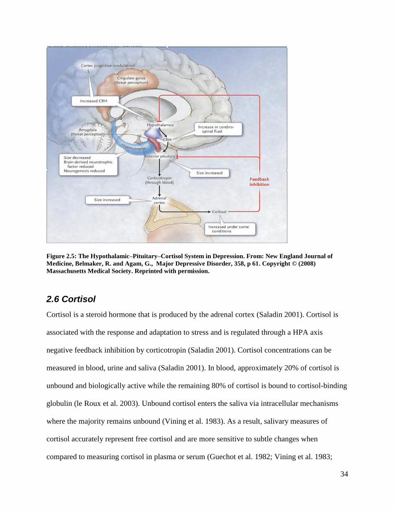
HPA axis activation results in the increased secretion of adrenal glucocorticoids such as cortisol.
Stress is perceived by the brain cortex and the amygdala and is transmitted to the hypothalamus
to release corticotropin-releasing hormone (CRH). CRH then stimulates the anterior pituitary
gland to secrete corticotropin into the bloodstream. Corticotropin then stimulates the adrenal
cortexes to secrete the glucocorticoid hormone cortisol. Cortisol sequentially induces feedback
inhibition in the hypothalamus and the pituitary, suppressing the production of CRH and
corticotropin, respectively (Figure 2.5) (Belmaker and Agam 2008). Under normal conditions,
increased circulating cortisol inhibits HPA axis activity via a negative feedback mechanism by
binding to glucocorticoid receptors at the levels of the hypothalamus and the pituitary (Blume et
al. 2011). However, in chronic stress and psychiatric disorders such as depression, it has been
proposed that this feedback mechanism may be impaired (Belmaker and Agam 2008).
34
Figure 2.5: The Hypothalamic–Pituitary–Cortisol System in Depression. From: New England Journal of
Medicine, Belmaker, R. and Agam, G., Major Depressive Disorder, 358, p 61. Copyright © (2008)
Massachusetts Medical Society. Reprinted with permission.
2.6 Cortisol
Cortisol is a steroid hormone that is produced by the adrenal cortex (Saladin 2001). Cortisol is
associated with the response and adaptation to stress and is regulated through a HPA axis
negative feedback inhibition by corticotropin (Saladin 2001). Cortisol concentrations can be
measured in blood, urine and saliva (Saladin 2001). In blood, approximately 20% of cortisol is
unbound and biologically active while the remaining 80% of cortisol is bound to cortisol-binding
globulin (le Roux et al. 2003). Unbound cortisol enters the saliva via intracellular mechanisms
where the majority remains unbound (Vining et al. 1983). As a result, salivary measures of
cortisol accurately represent free cortisol and are more sensitive to subtle changes when
compared to measuring cortisol in plasma or serum (Guechot et al. 1982; Vining et al. 1983;

35
Gozansky et al. 2005). Cortisol production has a distinct circadian rhythm with levels peaking in
the early morning and dropping to the lowest concentration at night (Stone et al. 2001).
Typically, a secretory burst in cortisol, referred to as the cortisol awakening response (CAR),
occurs 30 minutes post awakening in which free cortisol concentration increases by
approximately 50-75% (Schmidt-Reinwald et al. 1999; Wust et al. 2000b; Wilhelm et al. 2007).
The CAR is considered a reliable biomarker of HPA axis activity (Pruessner et al. 1999; Wust et
al. 2000a) and can be altered with psychological disorders such as depression (Stetler and Miller
2005; Adam et al. 2006; Foley 2006; Huber et al. 2006). Similarly, chronic elevations of cortisol
are associated with a wide array of health consequences such as cardiovascular disease, bone
loss, immune dysfunction and metabolic syndrome (Brunner et al. 2002; Lundberg 2005;
Reynolds et al. 2005). .
Cortisol secretion is a highly variable function that is associated with variables such as sex,
socioeconomic status, sleep disturbance and increased perceived stress particularly on workdays
compared to weekends (van Eck et al. 1996; Wust et al. 2000b; Backhaus et al. 2004; Kunz-
Ebrecht et al. 2004). Table 2.6 illustrates the wide range of values that represent cortisol
concentrations in normal healthy individuals which will be used as normative data in this study
(Clow et al. 2004).
Table 2.6 Cortisol levels and CAR in healthy individuals (Clow et al. 2004)
Mean SD Range
0 min post-waking (nmol/L) 11.6 4.6 4.7 – 18.5
30 min post-waking (nmol/L) 20 5.9 8.6 - 28
Percentage increase 0-30 min (%) 91.4 42.4 50 - 156
Change 0-30 min (nmol/L) 9.3 3.1 3.9 - 15
36
2.6.1 Cortisol, stress and depression
Chronic stress is known to be a strong causal factor in the onset of depression (Kendler et al.
1999). Too much stress and increased cortisol is a major risk factor for mental illness such as
depression and other psychotic disorders (Brown et al. 2004; Phillips et al. 2006). It has been
well established that inventories of stressful events predict subsequent depression (Kessler 1997)
and stressful events are often associated with the onset of MDD (Kendler et al. 2000). The
glucocorticoid cortisol is a major mediator of the physiological stress response. Excess cortisol
secretion is reported to be one of the risk factors for subsequent depression (Herbert et al. 2006).
Not surprisingly, elevated cortisol is a hallmark finding in patients with MDD (Davis et al. 1987;
Newcomer et al. 1999; Sapolsky 2000; Young et al. 2001; Brown et al. 2004; Krishnan and
Nestler 2008).
HPA axis dysregulation is commonly implicated in the course of depressive illness. The
hypothalamic-pituitary-cortisol hypothesis of depression (see Figure 2.6) postulates that
abnormalities in the cortisol response to stress may underlie depression (Belmaker and Agam
2008). The hippocampus participates in the termination of the stress response through the
glucocorticoid-mediated negative feedback that inhibits the HPA axis (Kim and Diamond 2002;
Sapolsky et al. 2002). In depression, the negative feedback mechanism is impaired resulting in
an increased activity of the HPA axis and elevated cortisol secretion. (Belmaker and Agam 2008;
Hinkelmann et al. 2009). It is still disputed whether these phenomena are a consequence of
MDD or whether they represent a vulnerability marker existing before the illness onset (Dedovic
et al. 2010). However, it has been shown that normalization of circulating cortisol levels with
antiglucocorticoid therapy alleviates depressive symptomatology in both patients with Cushing‟s
disease and hypercortisolemic depressed patients and has been associated with successful clinical
37
treatment outcomes (Murphy and Beverley 1997; Reus et al. 1997; Herbert et al. 2006).
Furthermore, the diurnal rhythm of cortisol secretion is also blunted in depression; pulses tend to
be longer and more frequent, with levels remaining consistently high over the day (Stokes 1995).
However findings have been inconsistent across studies (Chida and Steptoe 2009). A recent
meta-analysis found that the magnitude of the CAR is related to a number of psychosocial
factors. Specifically, researchers found that the CAR was positively associated with job stress
and general life stress and was negatively associated with fatigue, burnout, and exhaustion
(Chida and Steptoe 2009).
2.6.2 Cortisol and memory
Stress-induced elevated cortisol levels have well-known effects on cognition. Increasing
evidence has found that exposures to stress and/or stress hormones impair hippocampal-
dependent forms of memory in both humans and animals. Both chronic and acute elevations in
cortisol due to either prolonged stress or experimental injections have consistently been linked
with cognitive deficits such as effortful processing and episodic memory impairment (Rubinow
et al. 1984; Bemelmans et al. 1996; Lupien et al. 1998; Newcomer et al. 1999; Dominique et al.
2003; Sauro et al. 2003; Buss et al. 2004; Herbert et al. 2006; Hinkelmann et al. 2009). This
pattern of increased cortisol and memory deficits has been replicated in Lupien‟s work in
hypercortisolemic aged populations (Lupien et al. 1998), and in those with Cushing‟s Disease, a
state characterised by hypercortisolemia (Starkman et al. 2001). Chronically high levels of
cortisol have been associated with memory deficits in those with depression, and successful
treatment has been associated with decreases in cortisol secretion and subsequent improvement
in episodic memory function (Vythilingam et al. 2004). Also, cortisol is known to reduce
hippocampal long-term potentiation (LTP), a form of synaptic plasticity essential for

38
hippocampal-dependent memory formation (McEwen and Sapolsky 1995; Joels 2008;
Hinkelmann et al. 2009; Krugers and Hoogenraad 2009).
2.6.3 Cortisol and the hippocampus
The biological mechanisms leading to hippocampal volume reduction in depressive disorders are
currently unclear. One possible mechanism of injury to the hippocampus is corticosteroid
exposure (Brown et al. 1999). Chronic stress, hyperactivation of the HPA axis and elevated
cortisol levels have all been suggested to play a role in the down-regulation of neurogenesis, the
volumetric changes of the hippocampus and the deficits in functional capacity of the
hippocampus (Starkman et al. 1992; Sheline et al. 1996; Brown et al. 1999; Duman et al. 1999;
McEwen 2000; Sapolsky 2000; Herbert et al. 2006; Mondelli et al. 2010).
The hippocampus also plays a role in terminating the HPA stress response, however, injury to
the hippocampus impairs this negative feedback mechanism resulting in a more prolonged HPA
response to psychological stressors (Brown et al. 1999; McEwen 2007). The “glucocorticoid
cascade hypothesis” (Sapolsky et al. 2002) suggests that there is an association between
cumulative exposure to high cortisol levels and hippocampal atrophy. Increases in cortisol levels
produce hippocampal damage resulting in even greater increases in cortisol secretion due to
impaired feedback mechanisms to suppress cortisol release. The “neurotoxicity hypothesis”
(Gilbertson et al. 2002), suggests that chronic secretion of elevated glucocorticoids results in the
neurotoxic effects on the hippocampus, such as disruptions in glucose metabolism, dendrite
branching and neurogenesis which ultimately leads to hippocampal atrophy (Lupien et al. 2009).
The hippocampus is one of the main glucocorticoid target sites in the brain, with copious
amounts of corticosteroid receptors (Sapolsky 2000; McEwen 2005; Hinkelmann et al. 2009)
39
making it extremely sensitive to increased glucocorticoid levels. Stress hormones such as cortisol
modulate function within the hippocampus by plasticity (adaptation, changing or remodelling the
structure of neurons) to possibly avoid over-exposure to elevated glucocorticoid levels (Duman
et al. 1999; McEwen 2000; Lee et al. 2002; Campbell and MacQueen 2004; McEwen 2005;
Conrad 2008). As a result, excessive exposure to glucocorticoids have profound effects on the
hippocampus such as volume loss, dendritic atrophy, inhibiting neurogenesis and neuron death
(Brown et al. 1999; Duman et al. 1999; Newcomer et al. 1999; Bremner et al. 2000; Sapolsky
2000; Lee et al. 2002; Brown et al. 2004; McEwen 2005; Pittenger and Duman 2007; Mondelli et
al. 2010).
To test the hypothesis that dysregulation of the HPA axis and hippocampal volume abnormalities
would represent vulnerability factors for depression, Dedovic et al., (2010) compared the CAR
and hippocampal volume in healthy control subjects and individuals at risk for depression.
Supporting their hypothesis, a dysregulated CAR and smaller hippocampal volume was observed
in the subclinical high risk group (Dedovic et al. 2010).
2.6.4 Cortisol and neurogenesis
In rodents, exposure to high levels of glucocorticoids decreases neurogenesis of DG granule
neurons in the hippocampus (Gould et al. 1998; Ekstrand et al. 2008). Therefore, it has been
proposed that the down-regulation of neurogenesis is a potential mechanism for the observed
decrease in hippocampal size as seen with neuroimaging of depressed individuals (MacQueen et
al. 2003; Belmaker and Agam 2008). Likewise, the down-regulation of neurogenesis in response
to increased levels of glucocorticoids could contribute to deficits in the functional capacity of the
hippocampus resulting in learning and memory impairments (Kim et al. 2006).

40
Although there is a relationship between high cortisol levels and reduced hippocampal volume,
this does not suggest that cortisol alone is the only cause but instead the underlying mechanisms
may be associated to the activation of the stress response (Dedovic et al. 2010; Mondelli et al.
2010). For example, glucocorticoids increase the release, as well as decrease the clearance of
glutamate. Glutamate is the major excitatory neurotransmitter in the brain and at elevated levels
becomes neurotoxic leading to the neuronal atrophy associated with chronic stress and
depression (Banasr et al. 2011).
The causal relationship between hypercortisolism and depressive symptoms such as memory
impairment has not been established and is it likely to dissociate the effects of depression and
hypercortisolism on memory impairment in MDD (Egeland et al. 2005). For instance, findings of
memory deficits in other clinical groups suffering from hypercortisolism such as Cushing‟s
disease implies a direct relationship between cortisol and memory dysfunction that is not
facilitated by depression (Egeland et al. 2005). Additionally, depression is only seen in 50% of
Cushing‟s disease patients indicating that depression is secondary to hypercortisolism (Checkley
1996). However, conclusive evidence linking hippocampal dysfunction with elevated cortisol to
determine whether hippocampal alterations are secondary to increased cortisol and if these
changes are reversible for early intervention therapy levels has not yet been determined
(Sherwood 1997). The hippocampus may be a possible candidate for mediating the effects of
cortisol on episodic memory however there is a lack of understanding about the differences
between rodent and human hippocampal neurogenesis.

41
2.7 Inflammation and cytokines
Previously, the majority of research on depression, neurodegeneration and decreased
neurogenesis investigated the effects of elevated glucocorticoids. More recently, MDD has been
suggested to be a psychoneuroimmunological disorder, in which peripheral immune activation,
via cytokine release, is responsible for the array of behavioural, neuroendocrine and
neurochemical changes that occur with this disorder (Schiepers et al. 2005).
Cytokines are proteins that are released by a variety of cells and serve as intercellular signals
regulating immune responses. Cytokines encompass a heterogeneous group of messenger
molecules that are produced by immunocompetent cells, such as lymphocytes and macrophages,
to regulate immune responses (Schiepers et al. 2005). Similar to hormones of the endocrine
system, cytokines transmit messages by interacting with receptors on cell surfaces to
communicate throughout the body (Robles et al. 2005). Cytokines can be divided into two broad
classes based on their effects on the immune response: pro-inflammatory (stimulating
inflammation) and anti-inflammatory (reducing inflammation) (Robles et al. 2005; Schiepers et
al. 2005). Pro-inflammatory cytokines such as interleukin-1 (IL-1), IL-6, tumor necrosis factor-α
(TNF-α) and interferon (IFN), are produced by cells at the site of infection or injury to attract
other immune cells and signal them to activate and respond. Anti-inflammatory cytokines reduce
this immune response, inhibiting immune-cell activities, such as replication, activation, and
synthesis of other cytokines (Robles et al. 2005).
Since peripheral cytokines are relatively large molecules and do not freely pass through the
blood-brain barrier (BBB) research has focussed on the pathways by which peripheral cytokine
signals reach the brain (Raison et al. 2009). Animal research indicates that cytokines
administered peripherally can access the brain and affect function through several pathways via:
42
vagal nerve activation, a leaky or compromised BBB, active transport across the BBB, or
binding to cell-surface proteins on brain endothelial cells. (Dantzer et al. 2008; Miller et al. 2009;
Loftis et al. 2010). Pro-inflammatory cytokines such as IL-1, IL-6, and TNF-α that sub serve
inflammation in the periphery have complex and contrasting functional roles in the CNS (Miller
et al. 2009). Under normal physiological conditions, these cytokines are important for providing
trophic support to neurons, enhancing neurogenesis, and contribute to cognitive functioning such
as memory (Miller et al. 2009). However, in the milieu of excessive and/or prolonged activation,
cytokine networks in the CNS can encourage an interconnected group of abnormalities that are
thought to be partly responsible for the pathophysiology of depression, such as reduced
neurotrophic support, decreased neurogenesis, increased glutamatergic activation, oxidative
stress, induction of cell apoptosis and impaired cognitive function (Miller et al. 2009).
Overall, cytokines have been found to access the brain and interact with nearly every
pathophysiologic domain relevant to depression, including neuroendocrine function,
neurotransmitter metabolism, and neural plasticity (Miller et al. 2009). There is a growing body
of evidence that activation of pro-inflammatory cytokines and/or inhibition of anti-inflammatory
factors cytokines have been reported to modulate CNS functions and contribute to the changes
involved in psychiatric and neurodegenerative diseases such as depression (Song et al. 2003;
Hayley et al. 2005; Irwin and Miller 2007; Song and Wang 2010).
2.7.1 Cytokines and depression
Depression is a multifaceted disorder and the pathogenesis of the disease likely involves
alterations in several systems which interact with one another. Findings that cytokine-mediated
inflammatory processes play a critical role in the development of depression is quite robust
(Zunszain et al. 2011). Depression is highly prevalent among patients suffering from infectious,

43
autoimmune and neurodegenerative diseases, and this co-morbidity cannot be attributed solely to
the psychological distress of the initial disease (Pollak and Yirmiya 2002; Rook and Lowry
2008).
The „„Cytokine Hypothesis of Depression‟‟ proposes that inflammation may be a causal
mechanism in the development of depressive symptomatology (Dantzer et al. 2008; Miller et al.
2009). Research indicates that individuals with depression and other psychiatric disorders
express elevated immune markers (Archer et al. 2010). An experimental model for assessing the
acute immune response to infection in humans is the administration of the endotoxin
lipopolysaccharide (LPS) (Reichenberg et al. 2001). Both acute and chronic administration of
cytokines (or cytokine inducers such as LPS or vaccination) can result in behavioural symptoms
similar to those found in major depression. Likewise, data in humans are consistent with animal
findings, acute administration of LPS to humans leads to a host of behavioural changes,
including depressed mood, anhedonia, sleep disturbance, fatigue, and cognitive dysfunction
(Reichenberg et al. 2001; Dantzer et al. 2008; Harrison et al. 2009; Eisenberger et al. 2010;
Loftis et al. 2010; Haroon et al. 2011). Interestingly, the concentration of these cytokines in the
blood correlates with the severity of depression and the administration of antidepressant
medication normalizes the elevated pro-inflammatory cytokine levels (Miller et al. 2009; Janssen
et al. 2010)
The main pro-inflammatory cytokines IL-1β, IL-6, IFN-γ and TNF-α are considered to be one of
the most reliable peripheral biomarkers in MDD (Mössner et al. 2007) as they consistently
appear to be elevated in depressive states or in response to psychological stress (O'Brien et al.
2004; Schiepers et al. 2005; Raison et al. 2006; Piletz et al. 2009; Dowlati et al. 2010). For
instance, depressed patients who had attempted suicide have shown increased IL-6 in
44
cerebrospinal fluid (Lindqvist et al. 2009) and chronic administration of the inflammatory
cytokine IFN-α induces depressive symptoms in up to 30–50% of IFN-α treated patients.
(Musselman et al. 2001; Raison et al. 2005; Asnis and De La Garza 2006; Raison et al. 2009).
Recent meta-analyses exclusively that examined pro-inflammatory markers determined that
circulating peripheral C-reactive protein (CRP), TNF-α, IL-6, and IL-1β were all significantly
elevated in association with a diagnosis of MDD (Howren et al. 2009; Dowlati et al. 2010). A
prospective study found that an increased inflammatory state at baseline (defined as elevated
levels of CRP and increased capacity of leukocytes to produce IL-1), predicted a later onset of
depression in elderly individuals with no previous history of depression indicating that excess
inflammation precedes depression (van den Biggelaar et al. 2007; McNally et al. 2008).
The ratio of pro-inflammatory to anti-inflammatory cytokines may also be disrupted in
depression and result in a net increase in inflammatory activity (Dhabhar et al. 2009). Decreased
IL-10 levels, a higher IL-6/IL-10 ratio, and the lack of a counter-balancing, immunoregulatory
increase in IL-10 in response to increased IL-6 concentrations underlie the pro-inflammatory
physiological environment that is found to be associated with MDD. As a result, a reduction in
IL-10 that would normally dampen pro-inflammatory cytokine actions and resolve inflammation,
may contribute to the depressogenic, in addition to the inflammatory disease-promoting effects
of chronic, low-level elevations in pro-inflammatory cytokines (Dhabhar et al. 2009).
2.7.2 HPA axis and cytokines
The human stress response has a several checks and balances built in place to ensure that various
components do not become over or underactive (Kendall-Tackett 2009). The stress response
system is intricately linked with pro-inflammatory signalling. In the event of severe stress and

45
disrupted glucocorticoid signals, the result may be reduced restraint on the immune system and
the overproduction of pro-inflammatory cytokines (Raison and Miller 2003).
For example, cortisol is normally anti-inflammatory and keeps pro-inflammatory cytokines in
check however under severe stress cortisol can change function and potentiate the actions of IL-1
and IL-6 rather than inhibiting them. As a result, pro-inflammatory cytokines may themselves
disrupt cortisol signaling and negative feedback regulation of the HPA axis, further increasing
risk for depression (Robles et al. 2005)
Particularly, IL-1, IL-6, TNF- α and IFN- γ induce a cascade of behavioural, neuroendocrine and
central neurotransmitter changes including direct stimulation of the HPA axis and secretion of
cortisol (Song 2002; O'Brien et al. 2004; Hayley et al. 2005; Schiepers et al. 2005; Dantzer et al.
2008). Increased glucocorticoid levels found in depression are partly a result of increased
production of pro-inflammatory cytokines, such as IL-1β and IL-6 (Maes et al. 2009). For
example, chronic administration of IFN-α to humans led to increased cortisol levels and
flattening of the diurnal cortisol slope which was also associated with depression and fatigue
(Raison et al. 2008). These findings parallel the correlations between flattening of the cortisol
curve and plasma IL-6 in patients with advanced cancer (Capuron et al. 2003; Bower et al. 2005;
Rich et al. 2005)
As previously mentioned, it is becoming increasingly evident that in certain disorders, such as
depression, these inhibitory feedback loops become impaired, allowing for chronic immune
activation and the persistence of sickness symptoms that present from this activation (Irwin and
Miller 2007). In depression, both cortisol and pro-inflammatory cytokine levels are elevated,
46
which also indicates a failing or dysregulation of HPA-axis feedback mechanism (Raison and
Miller 2003; O'Brien et al. 2004; Pace et al. 2007; Leonard and Myint 2009).
2.7.3 Cytokines, neuroplasticity and memory
Accumulating evidence indicates that cytokines are critical to the regulation of neuronal
plasticity and survival, and that prolonged disruption of the balance of these cytokines can lead
to alterations in brain structures and function (Loftis et al. 2010). In clinical populations, a
consistent finding is that hepatitis patients treated with IFN therapy often develop a wide array of
cognitive impairments (Reichenberg et al. 2005; Lieb et al. 2006). It has been proposed that
inflammation impacts cognitive function through effects on neurogenesis and synaptic plasticity
(Bernardino et al. 2008; Dantzer et al. 2008; Maes et al. 2009; McAfoose and Baune 2009;
Dantzer 2012). A possible mechanism for the negative effects of pro-inflammatory cytokines is
their ability to modulate hippocampal neurogenesis through their activity on cytokine pathways
and messenger systems (Dowlati et al. 2010). Specifically, elevations in pro-inflammatory
cytokines influence neuronal functioning via cell apoptosis, oxidative stress, metabolic
derangement, and by impairing processes of neuronal branching (Hayley et al. 2005).
Cognitive-behavioural studies in animals have shown that inflammatory manipulations which
substantially increase IL-1 in the hippocampus (either endogenously or exogenously) result in
impairments of hippocampal-dependent memory consolidation, inhibit LTP and cell proliferation
and promote cell atrophy in the DG and CA regions of the hippocampus (Vereker et al. 2000;
Song 2002; Avital et al. 2003; Barrientos et al. 2006; Goshen et al. 2007; Hennigan et al. 2007;
Pickering and O‟Connor 2007; Chen et al. 2008).

47
The involvement of IL-1 in hippocampal-dependent memory processes follows an inverted U-
shape pattern, such that basal levels of IL-1 are required for memory formation, and any
deviation from this physiological range either by an excessive increase in IL-1 levels or by
blockade of IL-1 signaling, impairs memory (Avital et al. 2003; Goshen et al. 2007). What‟s
more, the memory impairments following central administration of IL-1 may be associated with
hypercortisolemia, as the cognitive disturbances were reversed not only by IL-1 receptor agonist,
but also by a glucocorticoid receptor antagonist (Song 2002). Therefore, cortisol alone may
provide an incomplete estimate of the effects of hypercortisolemia (Bauer et al. 2009).
2.8 Neurotropic growth factors
Neurotrophic growth factors such as BDNF, IGF and VEGF contribute to a variety of neural
processes including neural development, neuronal survival, synaptic signalling and synaptic
consolidation making them attractive candidates for the cellular mechanisms underlying both
depression-induced changes and antidepressant responses (Allen and Dawbarn 2006; Warner-
Schmidt and Duman 2008). Stress has been found to decrease levels of VEGF and BDNF in the
hippocampus, as well as produce neuronal atrophy and reduce neurogenesis, which may
contribute to a depressogenic state (Duman and Monteggia 2006). Several studies that have
assessed BDNF levels in MDD found important correlations between stress, MDD and
neurotrophic growth factor levels. A post-mortem study of depressed patients who had
committed suicide found reduced hippocampal BDNF (Karege et al. 2005) and several
biochemical studies have found low BDNF levels in depressed subjects (Shimizu et al. 2003; Lee
et al. 2007; Bocchio-Chiavetto et al. 2010) as well as a negative correlation with the depression
severity (Shimizu et al. 2003).
48
The „„neurotrophic model‟‟ of depression (Duman and Monteggia 2006) postulates that reduced
hippocampal BDNF activity, caused by stress or elevated glucocorticoids, impairs the ability of
stem cells in the subgranular zone of the DG to proliferate into mature cells that remain viable
(Duman and Monteggia 2006; Wolkowitz et al. 2010). There is also evidence that the
downregulation of BDNF by acute stress is mediated by the pro-inflammatory cytokine IL-1β
(Barrientos et al. 2003).
However, antidepressants have been shown to increase levels of VEGF, BDNF, IGF-1, restore
neuronal atrophy, and increase neurogenesis (Warner-Schmidt and Duman 2006; Wolkowitz et
al. 2010). BDNF has anti-inflammatory and antioxidant effects as well as attenuating
glucocorticoid-induced neuronal death by promoting the growth of developing neurons
(Wolkowitz et al. 2010). The suggested role of BDNF in antidepressant mechanisms is supported
by research that hippocampal neurogenesis (in animals) and serum BDNF concentrations (in
depressed humans) increase with antidepressant therapy (Hashimoto et al. 2004). In rodents,
BDNF administration to the DG of adult rats leads to increased neurogenesis of granule cells
(Scharfman et al. 2005) in addition to having antidepressant-like effects (Shirayama et al. 2002) .
In depressed humans, antidepressants increase BDNF levels (Shimizu et al. 2003) as well as
improve depression scores (Brunoni et al. 2008). Research has also found that stress-induced
decreases in neurogenesis and the expression of relevant nerve growth factors which support
neurogenesis, can be reversed by the administration of an IL-1 receptor antagonist (Barrientos et
al. 2003; Duman 2009).
It has been hypothesized that the regulation of neuroplasticity is mediated by various growth
factors (Duman et al. 2009; Paslakis et al. 2012). Similar to BDNF, IGF-I, produces
antidepressant-like behavioural effects (Duman et al. 2009) as well as increases adult
49
hippocampal neurogenesis in the rodent brain (Trejo et al. 2001). Interestingly, researchers
suggest that at BDNF plus IGF-I act synergistically producing a more robust effect than either
neurotrophin alone in activating neurotrophic cascades that alter ongoing neuroplasticity
mechanisms which promote the survival of new and existing hippocampal neurons (Cotman et
al. 2007; Paslakis et al. 2012) . In support of this, researchers have shown that the maximal
survival of new neurons in the rat hippocampus is achieved when both growth factors are present
(Johnson-Farley et al. 2006).
Astroglia also play a part in neuroplasticity through the secretion of neurotrophic factors
(McNally et al. 2008). Reduced astroglia numbers, another feature of depression, disrupts the
balance of anti- and pro-inflammatory mediators and further impairs the elimination process of
excitatory amino acids. Microglia activated by increased inflammation, astroglial loss, and
abnormal glutamate receptor activation disrupt the delicate balance of neuroprotective vs
neurotoxic effects in the brain, possibly leading to depression (McNally et al. 2008).
The majority of research to date found that BNDF levels increase following antidepressant
treatment, however the results are mixed. An important research question is whether changes in
BDNF levels are specific to certain types of antidepressant treatments or whether BDNF levels
are associated with an overall improvement in depressive symptomatology (Brunoni et al. 2008).
2.9 Current therapies for depression
In order to determine whether antidepressant therapy has been successful, the desired outcome
must be clearly defined. Response rate is defined as > 50% improvement from baseline on a
recognized rating scale for depression such as the HADS or the BDI. Nonetheless, many patients
with a 50% reduction in HADS score continue to experience significant residual symptoms and
50
may still meet diagnostic criteria for MDD (Shelton 2006). As a result, remission rate (i.e.,
recovery) has mostly replaced treatment response (improvement) as the standard for successful
therapy. Therefore, patients are considered to be in remission when symptoms are absent and
there is a full restoration with no impairments in psychosocial functioning (Shelton 2006).
The mainstream interventions for treating MDD are pharmacological therapy, electroconvulsive
therapy (ETC) and psychotherapy. However the effect of these treatments is far from
satisfactory. For instance, a placebo-controlled trial compared Cognitive Behavioural Therapy
(CBT), a form of psychotherapy, and antidepressant medication in moderately to severely
depressed individuals who were randomly assigned to 16 weeks of CBT or pharmaceutical
therapy or 8 weeks of pill placebo. At eight weeks, response rates were 50% in the medication
group and 43% in the CBT group and both superior to the placebo group (25%). When
researchers reassessed patients eight week later they found remission rates were only 46% for the
medication group and 40% for the CBT group (DeRubeis et al. 2005).
Generally, ECT is currently considered to be the most effective treatment for major depression
(Shelton 2006). Approximately 60% to 80% of all depressed patients who receive a course of
ECT respond to this treatment (Remick 2002). Although the mechanism of efficacy of ECT
remains unclear and the use of ECT is uncommon and mainly considered a last resort treatment
option (Remick 2002; Shelton 2006).
The low remission rates associated with current therapies are mainly a result of poor patient
compliance to treatment due to the wide range of side-effects accompanied by antidepressant
medication and ECT (Remick 2002; Shelton 2006). For that reason, these trends indicate a need
for more effective treatment options and the role of non-pharmacologic treatments for depression
51
becomes increasingly important. As well, researchers must consider strategies to address the
needs of patients with failed initial treatments as well as target individuals who are reluctant to
seek medical help.
2. 10 Exercise and depression
Due to the unsatisfactory mainstream antidepressant therapies, newer non-pharmacologic
therapeutic options are needed for the treatment of MDD. Research has found that higher
cardiorespiratory fitness levels and increased habitual physical activity are associated with lower
depressive symptomatology and greater emotional well-being (Martinsen 1990; Galper et al.
2006; Boettger et al. 2009). Additionally, lower cardiovascular fitness in adolescents is
associated with increased risk of serious depression in adulthood (Åberg et al. 2012) and MDD
patients who are moderately physically active are more likely to seek treatment than inactive
MDD patients (Diverty and Beaudet 1997). Exercise alone or in combination with other
treatment options, such as pharmacotherapy or cognitive behavioural therapy, have all been
effective in treating depression (Blumenthal et al. 1999; Perraton et al. 2010). For this reason, the
potential use of exercise as an alternative or complementary therapy for depression has received
considerable attention recently.
Both aerobic and anaerobic forms of exercise are equally effective in alleviating symptoms
associated with depression (Doyne et al. 1987; Martinsen et al. 1989). However, a higher
exercise dose leads to greater reductions in depression scores than a low dose (Dunn et al. 2005),
indicative of a possible dose-response for exercise, though further replication of this work is
needed. Overall, the general consensus from studies that report the aerobic intensity and
frequency suggest that 60–80% of maximum heart rate, three 30-minute sessions per week, with

52
an overall duration of 8 weeks is required for a significant improvement in depressive symptoms
(Perraton et al. 2010). It is likely that exercise-mediated amelioration of depression may be
explained by synergistically acting psychological and physiological mechanisms (Backhaus et al.
2004). Physical activity reduces self-reported and clinical depressive symptoms (Koehl et al.
2008), improves learning and memory (Ernst et al. 2006) alters physiological function of the
hippocampus as demonstrated by increased neurogenesis (Cotman and Berchtold 2002; Ernst et
al. 2006; Cotman et al. 2007; Pereira et al. 2007; Koehl et al. 2008; Praag 2008; Wu et al. 2008)
and hippocampal BDNF (Ploughman et al. 2007), reduces cortisol secretion (Nabkasorn et al.
2006; Foley et al. 2008) and even has increased brain volume in elderly individuals (Colcombe et
al. 2006). Mild group jogging reduced urine cortisol and epinephrine excretions, alleviated
MDD, and improved cardio-respiratory fitness (Nabkasorn et al. 2006).
Research in both animal and human subjects suggest that aerobic fitness has a strong biological
basis in the role of maintaining and enhancing central nervous system health as well as
substantial antidepressant effects in psychiatric patients (Martinsen et al. 1985). In fact, the
response rates for exercise were generally comparable to antidepressant medication (Martinsen et
al. 1985; Blumenthal et al. 1999; Babyak et al. 2000; Blumenthal et al. 2007) and both tend to
be better than placebo (Blumenthal et al. 2007). An earlier study conducted by Martinsen et al.
(1985) found that the combination of exercise with tricyclic antidepressants was not more
effective than exercise as a monotherapy. In a similar study, the efficacy of 16 weeks of aerobic
exercise training was comparable to standard pharmacotherapy in treating MDD. Blumenthal et
al., (1999) randomly assigned 156 patients with MDD to 4 months of aerobic physical exercise
alone, 4 months of antidepressant treatment (sertraline) or 4 months of combination treatment in
the form of both medication and physical exercise. All three groups exhibited significant

53
reductions in depressive symptoms and no significant differences were observed between groups
at 4 months, although, the onset of effect was more rapid with the drug treatment. Remarkably,
when researchers reassessed the patients 6 months after completion of treatment they found that
the 10-month relapse rate was significantly lower in the exercise group (8%), when compared to
the sertraline (38%) or the combination group (31%). Combining exercise with medication
adding no additional gain over either treatment alone, suggesting that exercising on one‟s own
initiative during the follow-up period was associated with a reduced probability of depression
diagnosis at the end of that period (Babyak et al. 2000). Consistent with previous findings, a
meta-analysis using 11 studies yielded a large combined effect size for the benefit of exercise
over control conditions when treating depression (d = 1.42) indicating that exercise can be a
powerful intervention for clinical depression (Stathopoulou et al. 2006). A recent review of
studies investigating the relationship between physical activity and depressive symptoms reveal
an inverse relationship between physical activity and depression and that even a low dose of
exercise can reduce the probability of developing depression (Teychenne et al. 2008).
It has been well established that exercise is a therapeutic intervention with proven benefits in the
treatment of depression however its mechanisms of action remain unclear.
2.10.1 Exercise, neurogenesis and the hippocampus
There are broad speculations as to the possible mechanism which exercise might impact aspects
of cognition such as episodic memory. Adult hippocampal neurogenesis has been suggested as a
target for the amelioration or prevention of mental illness (DeCarolis and Eisch 2010). Voluntary
exercise increases cerebral blood flow in the DG of the hippocampus and has been associated
with increased adult hippocampal neurogenesis (van Praag et al. 1999; Pereira et al. 2007). For
instance, in mice the number of proliferating cells detected by bromodeoxyuridine (BrdU)

54
injections over 12 consecutive days increases significantly in runners compared to sedentary
controls (van Praag et al. 1999). The positive association between voluntary exercise and
increased adult hippocampal neurogenesis in rodents has been consistently replicated (van Praag
et al. 1999; Pereira et al. 2007; Ekstrand et al. 2008; Fuss et al. 2010). Running also increases the
volume of the rodent hippocampus after 8-10 weeks of voluntary exercise (Rhodes et al. 2003).
Rhodes et al., (2003) suggest that increases in hippocampal neurogenesis might underlie the 20%
hippocampal volume increase observed after 8-10 weeks of exercise. A possible mechanism by
which exercise may increase hippocampal neurogenesis is increased cerebral blood volume.
Cerebral blood volume, an in vivo correlate of neurogenesis and a potential mechanism, is also
increased in the hippocampus and correlated with improved memory after a 3 month exercise
intervention in mice (Pereira et al. 2007).
Hippocampal neurogenesis has been linked to learning and memory improvements (Leuner et al.
2006; Winocur et al. 2006) and it facilitates LTP in the hippocampus of adult rats (Farmer et al.
2004). In rodents, exercise increases synaptic plasticity and improved retention on tasks
mediated by the hippocampus (van Praag et al. 1999; Kramer and Erickson 2007) and enhanced
the rate of learning on hippocampal mediated spatial navigation tasks such as the Morris Water
maze swimming task that require the use of cues to determine the location of submerged escape
platforms (Adlard et al. 2004).
A recently published three month exercise study with schizophrenic patients and healthy controls
found significant increases in hippocampal volume in both normal and schizophrenic patients
which correlated with improvements in aerobic fitness and no change in a non-exercising
schizophrenic group. In the schizophrenia exercise group only, hippocampal volume increase
was associated with a 35% increase in the N-acetylaspartate to creatine ratio
(considered a marker
55
of healthy neurons) in the hippocampus. Short-term memory improvement correlated with
change in hippocampal volume across all groups (Pajonk et al. 2010).
2.10.2 Exercise, inflammation and growth factors
Compelling evidence suggests that exercise training diminishes inflammation (Beavers et al.
2010). Exercise promotes an increase in the systemic levels of a number of cytokines with anti-
inflammatory properties and therefore protects against chronic medical disorders associated with
low-grade systemic inflammation (Petersen and Pedersen 2007). Specifically, studies have found
that exercise not only decreased the levels of pro-inflammatory cytokines IL-6, TNF-α, and CRP
but also simultaneously elevated the concentrations of anti-inflammatory cytokines IL-4, IL-10
and TGF-β when compared with controls. IL-4, IL-10 and TGF-β are not only anti-
inflammatory in nature but also inhibit the production of pro-inflammatory cytokines IL-1, IL-2,
and TNF-α (Das 2004; Das 2006) . Exercise also sets into action an interactive cascade of
growth factor signalling that has the net effect of stimulating neuroplasticity, enhancing
cognitive function, attenuating the mechanisms promoting depression, up-regulating
neurogenesis and increasing cerebrovascular perfusion (Cotman et al. 2007). For example, IGF-1
signalling converges on BDNF signalling, which may explain the positive effects of exercise on
both memory and depression. Exercise may also improve growth factor signalling by reducing
pro-inflammatory cytokines and directly increasing growth factor levels (Cotman et al. 2007;
Petersen and Pedersen 2007). BDNF, IGF-1 and VEGF are the main growth factors known to
mediate the effects of exercise on the brain (Trejo et al. 2001; Vaynman et al. 2004; Cotman et
al. 2007). The effects of exercise on memory, learning and depression are primarily regulated by
IGF-1 and BDNF, whereas exercise-dependent stimulation of angiogenesis and hippocampal
neurogenesis appear to be regulated by IGF-1 and VEGF (Cotman et al. 2007).
56
Supporting the critical role of the VEGF signaling pathway in neurogenesis, researchers
compared depression-like behaviour, blood vessel density, and neurogenesis in hippocampal DG
between stressed and exercising mice with or without administration of VEGF receptor inhibitor.
Chronic stress led to the development of depression-like behaviour, decreases in blood vessel
density and neurogenesis in the hippocampus, whereas regular exercise training improved
depression-like behaviour, limited the decrease of hippocampal blood vessel density and
enhanced the survival of newly generated neurons in chronic stress. Interestingly the
combination of exercise and VEGF receptor inhibitor cancelled the exercise-induced
antidepressant effect (Kiuchi et al. 2012).
2.10.3 Further evidence for exercise and neural function
Exercise enhances cognition and brain function, protects against the development of
neurodegenerative diseases and aging (Dishman et al. 2006; Kramer and Erickson 2007) and has
been shown to improve sleep quality (Atlantis et al. 2006). Descriptive studies have found that
elderly adults with greater levels of physical activity are less likely to develop dementia or other
cognitive function impairments compared to those who do not exercise (Kramer and Erickson
2007; Rosano et al. 2010). For instance, aerobic exercise improves cognitive function in aging
humans (Colcombe et al. 2003) and has been associated with the sparing of brain tissue in
humans (Colcombe et al. 2006). Also, physical activity and higher fitness levels in elderly
individuals are associated with increased hippocampal volume and better memory function
(Erickson et al. 2009). Importantly, physically fit individuals are reported to have larger left and
right hippocampal volumes than unfit individuals. Furthermore, hippocampal volume increases
following an exercise protocol are partially mediated the relationship between higher fitness
levels and enhanced spatial memory in older adults (Erickson et al. 2009). In elderly humans,
57
persistent physical activity has also shown to increase psychomotor speed as well as increased
brain activation (Rosano et al. 2010). Moreover, aerobic exercise has also been associated with
delaying the onset of and decline in several neurodegenerative diseases (Cotman et al. 2007) and
known to facilitate neurocognitive recovery from traumatic brain injury(Devine and Zafonte
2009; Mossberg et al. 2010).
In addition to the observed structural changes in the brain, research has found that exercise can
alter brain activity and functional connectivity associated with cognitive tasks (Voss et al. 2010).
For instance, Voss et al., (2010) used fMRI to examine cognitive relevant brain networks before
and after a 12 month aerobic exercise intervention in older adults. Researchers found that aerobic
fitness training improved the aging brain‟s resting functional efficiency in higher-level cognitive
networks. Additionally, changes in functional connectivity was associated with a behavioural
change such that increased functional connectivity was correlated with greater improvement in
executive function (Voss et al. 2010).
It is quite clear that exercise sets into motion an interactive cascade of biological events that has
a net effect of stimulating neural plasticity and neurogenesis, improving cognitive function, and
attenuating the mechanisms that drive depression (Cotman et al. 2007). Both human and animal
findings suggest that exercise targets many areas of brain function and has a wide array of
positive effects on overall brain health, resilience, memory and learning, and depression (Cotman
et al. 2007).
Exercise is a new and promising low-cost approach to preserve and protect CNS health. Exercise
may be a viable treatment option in treating depression because it can be recommended to most
individuals and does not carry a negative social stigma as the other treatment therapies do (Dunn

58
et al. 2005) . Research must identify treatment targets and directions for future studies to
identify individuals at risk, decrease relapse rates, and increase long-term favourable outcomes
for those suffering from depression.
59
Chapter 3 Methods
3.1 Experimental design for future pilot study
This future study will utilize a cohort control design. Participants will be recruited in blocks.
Group 1 will undertake a 12 week structured, supervised exercise program in addition to the
LMHDT and they will be recruited in the first year of the study. Group 2 from the LMHDT will
be recruited in the second year so that they can be matched by age, gender, pharmacotherapy and
baseline fitness level to the exercise group using the same inclusion criteria.
The reason for running the study as a cohort control rather than a randomized control trial is that
the LMHDT is a group program and it might affect group cohesion if some participants were
offered the exercise program and others were not. This way everyone in the first cohort who
meets the inclusion criteria will be given the opportunity to have the exercise program, and this
will not interfere with the program in the way that it is currently run. It also allows us to age and
gender match the non-exercising LMHDT cohort (group 2), as well as a cohort of non-depressed
individuals who will undergo baseline fMRI scans to provide normative data for comparison for
potential fMRI activation changes in the depressed groups.
Outcome measures will be assessed at baseline and week 12 (see Figure 3.1). The primary
measures are salivary cortisol, hippocampal activation during a memory task, plasma biomarkers
and depression scores. Additional measures will include subjective exercise barriers and self-
efficacy questionnaires, a face recognition task evaluating hippocampal function, a
cardiorespiratory fitness test, sleep quality questionnaire and a neurocognitive screening test.
60
Additionally, a precursory investigation will be carried out in a group of non-depressed, active
participants, to determine „normal‟ values.
A sedentary non-depressed group will be recruited from UOIT and Durham College in Oshawa
Ontario. Advertisement will be placed throughout the campus. Participants will complete the
same assessments as the depressed groups at baseline and week 12. They will receive the same
12 week exercise intervention as the depressed exercise group. The non-depressed control group
will provide background data on the normal values and variability of the measures in an
asymptomatic group, with particular focus on cortisol and hippocampal function in addition to
the effects of exercise on non-depressed sedentary adults. This will enable studies to discriminate
the general effects of exercise on neural function and biomarker status as distinct from those
unique to depression.
For the non-depressed group, respondents to recruitment advertisements received the Participant
Information Sheet (PIS), orientation session, consent form, Par Q, Baycrest Screening Form, and
the following questionnaires: BDI-II, HADS, MoCA, PSQI (section 3.2.2, Appendices 1-8).
Those who met inclusion criteria attended a baseline assessment, exercise self-efficacy
questionnaires, the Astrand-Ryhming fitness test, first set of salivettes accompanied by saliva
sampling instructions (Appendix 9), blood collection and fMRI. At this stage, the participants
will commence the exercise program, completing three sessions per week. In week 12, all
questionnaires, neurocognitive assessment, fMRI, fitness test, saliva samples and blood
collection were repeated.

61
Figure 3.1: Overview of study design for the depressed exercise group, depressed control group and non-
depressed exercise group.
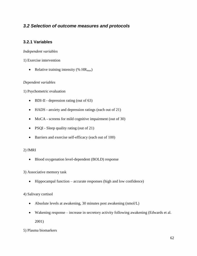
62
3.2 Selection of outcome measures and protocols
3.2.1 Variables
Independent variables
1) Exercise intervention
Relative training intensity (% HRmax)
Dependent variables
1) Psychometric evaluation
BDI-II - depression rating (out of 63)
HADS - anxiety and depression ratings (each out of 21)
MoCA - screens for mild cognitive impairment (out of 30)
PSQI - Sleep quality rating (out of 21)
Barriers and exercise self-efficacy (each out of 100)
2) fMRI
Blood oxygenation level-dependent (BOLD) response
3) Associative memory task
Hippocampal function – accurate responses (high and low confidence)
4) Salivary cortisol
Absolute levels at awakening, 30 minutes post awakening (nmol/L)
Wakening response – increase in secretory activity following awakening (Edwards et al.
2001)
5) Plasma biomarkers

63
IL-6
VEGF
6) Anthropometry
Body mass (kg)
BMI (kg∙m-2
)
7) Astrand-Ryhming protocol
Estimated relative VO2max (mL∙kg-1
∙min-1
)
3.2.2 Protocols
Biomarkers
Presently, immunoassays are the most widely used techniques measuring cytokines and growth
factors in human serum and plasma because of their high specificity, sensitivity, rapid
turnaround time, convenience, the ease of performance and relatively low costs (Sachdeva and
Asthana 2007). Their detection limit is approximately 1-5 pg/ml and they have good
reproducibility with a coefficient of variation between 5-10% (Sachdeva and Asthana 2007). The
basic cytokine sandwich ELISA based method is a monoclonal antibody specific for each
antigen is pre-coated onto a microplate containing 96 wells. Standards, controls and samples are
pipetted into the wells and any biomarker present is bound by the immobilized antibody. After
washing away any unbound substances, an enzyme-linked polyclonal antibody specific for each
antigen is added to the wells. Following the wash to remove an unbound antibody-enzyme
reagent, a substrate solution is added to the wells and color develops in proportion to the amount
of the antigen bound. The color development is stopped and the intensity of the color is
measured.

64
Salivary cortisol
Salimetrics Oral Swab (SOS: Salimetrics®, State College, Pennsylvania) for saliva collection is
an excellent alternative to passive drool because of its ease of use. The SOS is made of a non-
toxic, inert polymer shaped into a 30 x 10 mm cylinder. (Salimetrics 2006; Salimetrics and
Europe 2011).
Enzyme immunoassay (EIA) is an acceptable widely used method for determining salivary
cortisol (Gozansky et al. 2005). This method is advantageous due to the high sensitivity, ease of
use, rapid detection and also avoiding the disposal and lifetime issues associated with using
radioisotopes (Blackburn et al. 1991).
Beck Depression Inventory – Second Edition (BDI-II)
The BDI-II (Appendix 5) is a 21-item self-reported instrument that corresponds to the criteria for
depressive disorders from the Diagnostic and Statistical Manual of Mental Disorders – Fourth
Edition (DSM-IV; American Psychiatric Association, 1994). Each multiple choice question is
rated on a 4-point scale of 0-3, ranging in intensity. Total numerical scores are compared to a
classification key with higher scores indicating greater depression severity. Total depression
scores of 0-13 is minimal, 14-19 is mild, 20-28 is moderate, and 29-63 is severe (Beck et al.
1996). The BDI-II has demonstrated high internal consistency (0.92 and 0.93, respectively), test-
retest reliability of 0.93 (p < .001), and has found to have significant correlations (r = 0.68 - 0.93)
with three other major instruments used in the assessment of depression in samples of psychiatric
outpatients and University students (Beck et al. 1996).
Hospital Anxiety and Depression Scale (HADS)
The HADS (Appendix 6) developed by Zigmond and Snaith (1983), is a 14-item self-report
instrument, that assesses anxiety and depression (Snaith 2003). Seven items measure depression
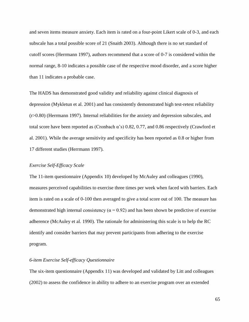
65
and seven items measure anxiety. Each item is rated on a four-point Likert scale of 0-3, and each
subscale has a total possible score of 21 (Snaith 2003). Although there is no set standard of
cutoff scores (Herrmann 1997), authors recommend that a score of 0-7 is considered within the
normal range, 8-10 indicates a possible case of the respective mood disorder, and a score higher
than 11 indicates a probable case.
The HADS has demonstrated good validity and reliability against clinical diagnosis of
depression (Mykletun et al. 2001) and has consistently demonstrated high test-retest reliability
(r>0.80) (Herrmann 1997). Internal reliabilities for the anxiety and depression subscales, and
total score have been reported as (Cronbach α‟s) 0.82, 0.77, and 0.86 respectively (Crawford et
al. 2001). While the average sensitivity and specificity has been reported as 0.8 or higher from
17 different studies (Herrmann 1997).
Exercise Self-Efficacy Scale
The 11-item questionnaire (Appendix 10) developed by McAuley and colleagues (1990),
measures perceived capabilities to exercise three times per week when faced with barriers. Each
item is rated on a scale of 0-100 then averaged to give a total score out of 100. The measure has
demonstrated high internal consistency (α = 0.92) and has been shown be predictive of exercise
adherence (McAuley et al. 1990). The rationale for administering this scale is to help the RC
identify and consider barriers that may prevent participants from adhering to the exercise
program.
6-item Exercise Self-efficacy Questionnaire
The six-item questionnaire (Appendix 11) was developed and validated by Litt and colleagues
(2002) to assess the confidence in ability to adhere to an exercise program over an extended
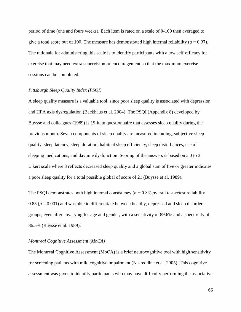
66
period of time (one and fours weeks). Each item is rated on a scale of 0-100 then averaged to
give a total score out of 100. The measure has demonstrated high internal reliability (α = 0.97).
The rationale for administering this scale is to identify participants with a low self-efficacy for
exercise that may need extra supervision or encouragement so that the maximum exercise
sessions can be completed.
Pittsburgh Sleep Quality Index (PSQI)
A sleep quality measure is a valuable tool, since poor sleep quality is associated with depression
and HPA axis dysregulation (Backhaus et al. 2004). The PSQI (Appendix 8) developed by
Buysse and colleagues (1989) is 19-item questionnaire that assesses sleep quality during the
previous month. Seven components of sleep quality are measured including, subjective sleep
quality, sleep latency, sleep duration, habitual sleep efficiency, sleep disturbances, use of
sleeping medications, and daytime dysfunction. Scoring of the answers is based on a 0 to 3
Likert scale where 3 reflects decreased sleep quality and a global sum of five or greater indicates
a poor sleep quality for a total possible global of score of 21 (Buysse et al. 1989).
The PSQI demonstrates both high internal consistency (α = 0.83),overall test-retest reliability
0.85 (p = 0.001) and was able to differentiate between healthy, depressed and sleep disorder
groups, even after covarying for age and gender, with a sensitivity of 89.6% and a specificity of
86.5% (Buysse et al. 1989).
Montreal Cognitive Assessment (MoCA)
The Montreal Cognitive Assessment (MoCA) is a brief neurocognitive tool with high sensitivity
for screening patients with mild cognitive impairment (Nasreddine et al. 2005). This cognitive
assessment was given to identify participants who may have difficulty performing the associative
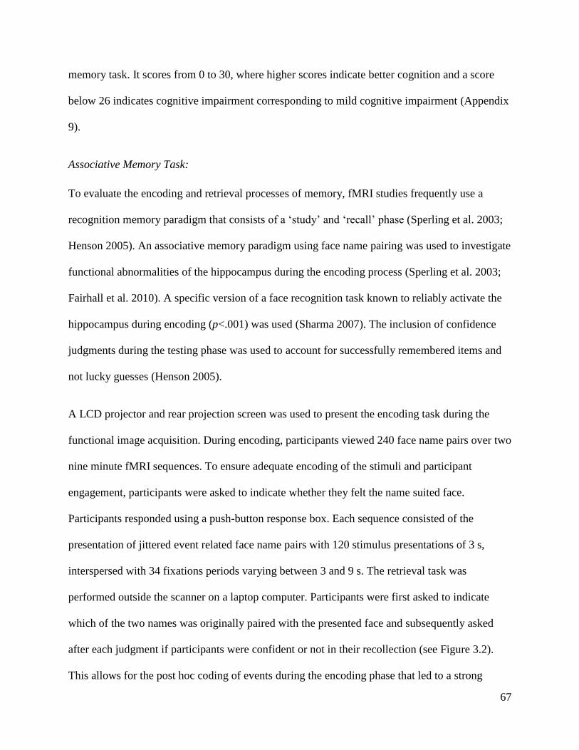
67
memory task. It scores from 0 to 30, where higher scores indicate better cognition and a score
below 26 indicates cognitive impairment corresponding to mild cognitive impairment (Appendix
9).
Associative Memory Task:
To evaluate the encoding and retrieval processes of memory, fMRI studies frequently use a
recognition memory paradigm that consists of a „study‟ and „recall‟ phase (Sperling et al. 2003;
Henson 2005). An associative memory paradigm using face name pairing was used to investigate
functional abnormalities of the hippocampus during the encoding process (Sperling et al. 2003;
Fairhall et al. 2010). A specific version of a face recognition task known to reliably activate the
hippocampus during encoding (p<.001) was used (Sharma 2007). The inclusion of confidence
judgments during the testing phase was used to account for successfully remembered items and
not lucky guesses (Henson 2005).
A LCD projector and rear projection screen was used to present the encoding task during the
functional image acquisition. During encoding, participants viewed 240 face name pairs over two
nine minute fMRI sequences. To ensure adequate encoding of the stimuli and participant
engagement, participants were asked to indicate whether they felt the name suited face.
Participants responded using a push-button response box. Each sequence consisted of the
presentation of jittered event related face name pairs with 120 stimulus presentations of 3 s,
interspersed with 34 fixations periods varying between 3 and 9 s. The retrieval task was
performed outside the scanner on a laptop computer. Participants were first asked to indicate
which of the two names was originally paired with the presented face and subsequently asked
after each judgment if participants were confident or not in their recollection (see Figure 3.2).
This allows for the post hoc coding of events during the encoding phase that led to a strong
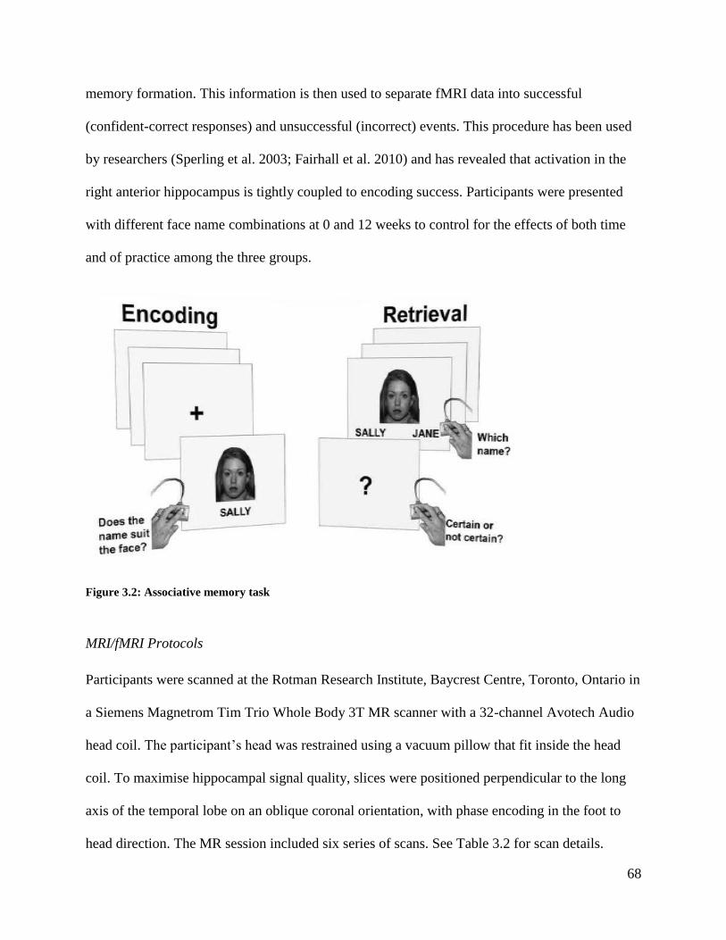
68
memory formation. This information is then used to separate fMRI data into successful
(confident-correct responses) and unsuccessful (incorrect) events. This procedure has been used
by researchers (Sperling et al. 2003; Fairhall et al. 2010) and has revealed that activation in the
right anterior hippocampus is tightly coupled to encoding success. Participants were presented
with different face name combinations at 0 and 12 weeks to control for the effects of both time
and of practice among the three groups.
Figure 3.2: Associative memory task
MRI/fMRI Protocols
Participants were scanned at the Rotman Research Institute, Baycrest Centre, Toronto, Ontario in
a Siemens Magnetrom Tim Trio Whole Body 3T MR scanner with a 32-channel Avotech Audio
head coil. The participant‟s head was restrained using a vacuum pillow that fit inside the head
coil. To maximise hippocampal signal quality, slices were positioned perpendicular to the long
axis of the temporal lobe on an oblique coronal orientation, with phase encoding in the foot to
head direction. The MR session included six series of scans. See Table 3.2 for scan details.

69
For functional scans, 50 oblique coronal 3.5 mm slices with full brain coverage were obtained
(TE=27 ms; TR=2.5 s; flip angle 70⁰; FOV=200 mm; resolution=3.125x3.125x5mm; zero gap
using T2*-weighted EPI sequence). Anatomical images were obtained following the fMRI
consisting of 3D T1-weighted pulse sequence (TE=2.63 ms; TR=2 s; FOV=256 mm; 1 mm
isotropic voxels; 176 slices).
Table 3.2: fMRI protocol
MR Series Data Aquisition Time
1. Auto align scout Slice positioning 00:00:46
2. Localizer Slice positioning 00:01:26
3. fMRI BOLD at rest 00:09:00
4. fMRI BOLD during memory encoding task 00:09:00
5. fMRI BOLD during memory encoding task 00:09:00
6. MRI structural 00:06:26
Instructions between series 00:03:00
Set up 00:15:00
Total scanner time 00:53:38
Stimuli were presented using E-Prime software version 2.0 (Psychology Software Tools). Images
were back-projected onto a screen behind the scanner and shown to the participant using a mirror
mounted on the head coil. The display had a spatial resolution of 1024 x 768 pixels and a visual
angle of approximately 14.8⁰ x 12.1⁰ at a viewing distance of 132 cm.
70
Total time for each scanning session was under one hour (see Table 3.2). Behavioural tasks and
fMRI were synchronized according to trigger pulses sent by the scanner. For vision correction
MR safe glasses made by SafeVision (Webster Groves, MO, USA) were provided. Participants
responded with the right index and middle fingers using buttons on a Fiber-Optic Response Pad
System paddle developed by Current Designs Inc. (Philadelphia, PA, USA).
Astrand-Ryhming cycle protocol
The Astrand-Ryhming cycle ergometer protocol is a six minute submaximal exercise test capable
of estimating VO2max from heart rate (HR) measurements and perceived exertion (Whaley 2006).
It is a single-stage test, with HR measured in the fifth and sixth minute (Whaley 2006). The
average of these values is adjusted for age using age correction factors, and an estimation of
VO2max is made using a specifically-designed nomogram (Astrand 1960; Whaley 2006, cited in
Whaley, 2006).
Cardiovascular fitness was assessed by the RC and final year kinesiology internship students at 0
and 12 weeks. The prescribed intensities of the exercise program in this research are at a level
that should lead to improvements in cardiovascular fitness if undertaken correctly. It is expected
that cardiovascular fitness will improve in the exercise groups, and not in the MHDT control
group.
Body mass index (BMI)
BMI provides a relative anthropometric measurement that is sensitive to changes over time. BMI
values were obtained by dividing body weight in kilograms by height in metres squared (kg∙m-2
),
(Whaley 2006). The „normal‟ BMI range is 18.5 – 24.9 kg∙m-2
(Panel 1998).

71
Exercise intervention
Review of the literature suggests that both aerobic and resistance exercises are effective in the
treatment of depressive disorders (Pedersen and Saltin 2006). For the aerobic component, an
initial intensity of 12-13 on the Borg Scale of Perceived Exertion (RPE) is recommended for a
duration of 10-20 minutes, gradually increasing to 15-16 RPE for 30 minutes (Pedersen and
Saltin 2006). Using maximum heart rate (Whaley 2006), to determine intensity, it is suggested
that a minimum of 70% HRmax is necessary to solicit greater improvements (Dunn et al. 2001). A
higher total weekly energy expenditure, obtained through manipulating frequency, duration and
intensity, has been shown to be significantly more effective in the treatment of mild to moderate
severity depression (Dunn et al. 2005).
Guidelines for resistance training in healthy adults recommend an 8-12 repetition maximum,
with large-muscle and multiple-joint exercises being performed before small-muscle and single-
joint exercises, and increases of 2-10% (equivalent to around 1-2 repetitions) implemented
before load is increased (Kraemer et al. 2002).
3.3 Participants
3.3.1 Recruitment
It is important to differentiate the benefits of group exercise which are specific to exercise as
opposed to the psychosocial benefits due to the social interaction that occurs within the group.
The LMHDT provides an ideal solution to this. Participants are referred into this program by
psychiatrists at Lakeridge Health with a DSM-V axis I diagnosis of depression and sometimes
concomitant anxiety. Upon referral all participants are screened to determine if there is a history
72
of substance abuse, internal mental distress, behaviour complexity or crime and violence using
the GAIN-SS (Dennis et al. 2006), a validated tool designed to identify individuals who are
likely to have a mental health and/or substance use disorder and who should be referred for
further assessment or treatment. Participants with concomitant substance abuse disorders were
not included in this feasibility study and will not be included in either arm of the pilot study.
Participants will also undergo the SCID-I to confirm the clinical diagnosis of major depression.
This will be administered by either the referring psychiatrist or the clinical psychologist. The
SCID-I has excellent inter-rater and longitudinal reliability (Zanarini and Frankenburg 2001).
The LMHDT is a 12 week program which includes a four week transition phase where
participants meet twice per week, followed by six weeks of intensive group work focused on
interpersonal relationships, depression, anxiety and stress management, relaxation, self-esteem
building, communication skills, symptom management, goal setting / problem solving, mental
health and substance use and community linkage. Participants then have a further two week
transition out of the program where they meet once or twice per week.
Participants in this program will be offered the opportunity to participate in a structured 12 week
exercise program in addition to the LMHDT and their results will be compared to a control group
who has undergone the LMHDT but not exercised. The exercise cohort will be recruited in the
first half of the study so that the study may age and gender match the control group in the second
half of the study. This design also maintains the integrity of the LMHDT in that participants
won‟t feel that other people in their therapeutic group are getting something that they aren‟t.
Matched control participants will also be recruited from UOIT and Durham college for the non-
depressed exercise group.

73
An objective for this feasibility study was to develop a recruitment process that minimizes the
number of steps required for both Lakeridge staff and potential participants. Following an
expression of interest, potential participants underwent a Pre-screening questionnaire (see
Appendix 7) administered by Lakeridge staff. Those who were still eligible at this stage were
given an Information Session Invitation (see Appendix 1) and their contact information was
forwarded to the RC for further screening. Additional screening of participants was conducted
over the telephone by the RC which consisted of the Baycrest Screening Form, and Par-Q. All
eligible participants were then invited to a face-to-face screening interview and study orientation
with the RC to ascertain their eligibility for the study. This included reviewing and signing
Baycrest Screening Form and PAR-Q. Based on the official recommendations of the Canadian
Society for Exercise Physiology (CSEP) and Health Canada if participants answer yes to any
questions on the PAR-Q, they will be given a letter addressed to their physician (see Appendix
12) explaining the study along with the PARmed-X (see Appendix 13) and they must receive
clearance by their physician to enrol in the study. Participants satisfying all criteria provided
informed consent and were enrolled in the study. See Figure 3.3 for recruitment process.
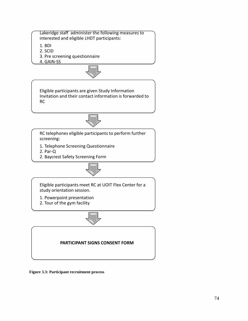
74
Figure 3.3: Participant recruitment process
Lakeridge staff administer the following measures to interested and eligible LHDT participants:
1. BDI 2. SCID 3. Pre screening questionnaire 4. GAIN-SS
Eligible participants are given Study Information Invitation and their contact information is forwarded to RC
RC telephones eligible participants to perform further screening:
1. Telephone Screening Questionnaire 2. Par-Q 2. Baycrest Safety Screening Form
Eligible participants meet RC at UOIT Flex Center for a study orientation session.
1. Powerpoint presentation 2. Tour of the gym facility
PARTICIPANT SIGNS CONSENT FORM
75
3.3.2 Inclusion/exclusion criteria
Inclusion criteria for depressed group
Male or female, age 18-50
DSM-V diagnosis of major depression confirmed by the SCID-I interview
BDI score ≥ 20
Depression symptoms minimum 6 months
Attending LHDT program
Concomitant pharmacological treatment must have been stabilized four weeks prior to
study enrolment
Currently sedentary (exercise less than 20 minutes, three times weekly)
Exclusion criteria for depressed group
Medical contraindications to exercise, without relevant medical clearance
Co-existing DSM-V Axis I apart from anxiety (APA, 1994)
Participants who screened positive for substance abuse
Religious or cultural beliefs that prevent the collection of bodily fluids
Personal decision to withdraw
Inclusion criteria for non-depressed group
Male or female, age 18-50
Currently sedentary (exercise less than 20 minutes, three times weekly)
Exclusion criteria for non-depressed group
Any potentially confounding medical conditions (including, but not limited to, diabetes,
chronic fatigue, any DSM-IV Axis I and II disorders)
76
Evidence of depressive symptoms (BDI score >13), or anxiety (HADS anxiety subscale
score >7)
3.3.3 Informed consent
This study was approved by three ethics committees: University of Ontario Institute of
Technology, Lakeridge Health and The Rotman Brain Research Institute/Baycrest. All three
REB committees approved research ethics for the duration of one year with the option to renew
in the subsequent year. Written consent (Appendix 2) was obtained from the participant at the
study orientation session. They were assured they would not be identifiable from their individual
data, and advised of their right to withdraw at any stage, without giving a reason.
3.4 Procedure
All measures (excluding fMRI and memory encoding and retrieval task) were conducted at
UOIT. Psychometric assessments were administered by RC in the Flex Centre boardroom. Blood
collection was performed in the UOIT laboratory by a Medical Lab Technician. Fitness
assessment was conducted by a Certified Personal Trainer in the UOIT Kinesiology lab. A
polystyrene bin with two saliva collection devices and saliva collection instructions (see
Appendix 9) was given to participants to perform on the subsequent Wednesday morning
immediately upon wakening. fMRI and memory tasks were performed at Baycrest Hospital. For
data collection process (see Figure 3.4)
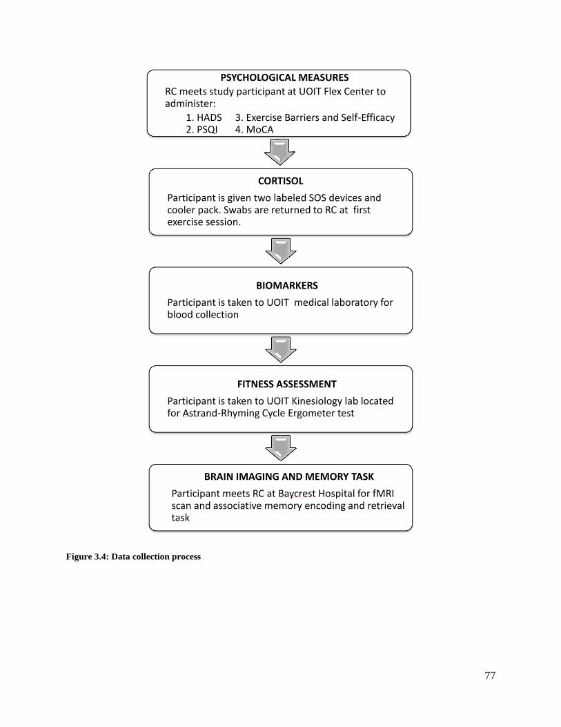
77
Figure 3.4: Data collection process
PSYCHOLOGICAL MEASURES
RC meets study participant at UOIT Flex Center to administer:
1. HADS 3. Exercise Barriers and Self-Efficacy 2. PSQI 4. MoCA
CORTISOL
Participant is given two labeled SOS devices and cooler pack. Swabs are returned to RC at first exercise session.
BIOMARKERS
Participant is taken to UOIT medical laboratory for blood collection
FITNESS ASSESSMENT
Participant is taken to UOIT Kinesiology lab located for Astrand-Rhyming Cycle Ergometer test
BRAIN IMAGING AND MEMORY TASK
Participant meets RC at Baycrest Hospital for fMRI scan and associative memory encoding and retrieval task
78
3.4.1 Testing
Psychometric measures
Questionnaires and neurocognitive assessment were administered by the RC in the UOIT Flex
Centre boardroom. Each form was explained in detail by the RC. The entire process took
approximately 30 minutes.
Salivary cortisol
Saliva samples were procured with an inert polymer non-citric acid collection device (SOS:
Salimetrics®
, State College, Pennsylvania). Participants received a polystyrene bin and a set of
two SOS tubes. The SOS tubes were labelled „waking‟ and ‟30 min‟ to indicate sampling times,
which were immediately upon awakening and 30 minutes after awakening. Instructions were
given to place the cotton swab under the tongue and salivate for two minutes, replace the lid, and
put straight into the freezer. Participants were instructed to follow standard sampling protocol
(Clow et al. 2004; Salimetrics and Europe 2011): nil by mouth, except water, and no cleaning
teeth, steroid inhalers or smoking between the two morning samples. This is all detailed in
written instructions which was provided to participants (Appendix 9). Sampling took place on
the Wednesday following the orientation session. Completed samples were returned in the
polystyrene bin at a subsequent exercise session. Samples were immediately transported to the
UOIT laboratory and stored in the in a -80°C freezer in the UOIT laboratory.
Blood collection
Peripheral venous blood was drawn in the UOIT laboratory by a medical lab technician.
Collection times and other information that could affect biomarker levels such as illness or

79
infection were logged by the technician (see Appendix 14). A total of 35 ml of blood was
collected from each participant by venipuncture into ethylenediaminetetraacetic
acid (EDTA) tubes. Blood samples were prepared within 30 minutes by centrifugation (3000g
for 5 minutes), and the fibrinogen containing plasma supernatant was aliquot into thirty 0.5 mL
labeled tubes and stored at -85°C until assayed. A contingency plan using two freezers for
aliquot storage was followed in the event of a power outage or freezer malfunction.
Biomarker analysis
On the day of analysis, plasma and saliva were thawed and brought to room temperature. Plasma
samples were vortexed and saliva samples were centrifuged at room temperature 2500 rpm for
15 minutes. All assays were conducted according to the manufacturer‟s protocols. The antigen
levels of IL-6 and VEGF were measured using sandwich ELISA kits (Quantikine®HS, R&D
Systems Inc., Minneapolis, Minn., USA). Salivary cortisol was measured using Salimetrics
Expanded Range High Sensitivity Salivary Cortisol Enzyme Immunoassay kit (Salimetrics LLC,
Philadelphia, PA). Briefly, standards, controls, and samples were pipetted into a 96-well
microtiter plate coated with unconjugated antibodies to human IL-6, VEGF and cortisol. Plasma
assays were incubated at room temperature then washed. After washing away any unbound
substances, an enzyme-linked polyclonal antibody specific for each of these components was
added to the wells, incubated at room temperature and washed again accordingly. Since the
cortisol assay utilizes a competitive reaction, a horseradish peroxidase-conjugated cortisol was
immediately added, incubated at room temperature and washed. Following a wash to remove
unbound antibody-enzyme reagents, a substrate solution was added to the wells. Colour
development for all the assays was stopped by a sulfuric acid and the intensity of the colour was
measured by a microtiter plate reader within 10 minutes. Linear regression was performed on
80
plots of corrected absorbance at 450 nm for IL-6, VEGF and cortisol. The concentration of
antigens in patient plasma and enzyme-linked cortisol was then quantified by interpolation from
the standard curves. All assays were performed in duplicate and the average was reported.
fMRI and memory encoding task
MRI suite procedures were followed according to the Baycrest MRI suite procedures. A unique
participant ID was assigned by Baycrest Subject Coordinator for each participant and scan time
was reserved online through the Baycrest Resource Scheduler. Immediately upon arrival,
participants were given a Baycrest parking pass and 25 dollar gas voucher from the RC.
Participants arrived at the MR Suite 15 minutes prior to their scheduled scan time accompanied
by the RC. The MRI Screening Form with participant ID number was filled in and reviewed by
the MR Technologist. Participants were required to remove all makeup, jewellery, metallic and
electronic items and all clothing except for underwear and socks. A hospital gown tied at the
back and a pair of trousers was provided for participants to change into. The MR technologist
escorted participants into the magnet room and the RC in the control area from the testing room.
The MR Technologist gave the participant a brief orientation of what to expect in the scanner.
Participants were given a panic button to press in case they needed to be removed from the
scanner for any reason. During the resting state scan, participants were instructed to close their
eyes but remain awake. During the memory encoding task (see Figure 3.2) participants were
instructed to use their index finger to press the first button if the name suited the face and their
middle finger to press the second response button to indicate if the name did not suit the face. All
instructions were given by the RC and MR Technologist through a microphone/intercom system.
Once the scan was complete the MR Technologist removed the participant from the scanner and
relinquish them to the RC to get changed.

81
Memory retrieval
Immediately following the scan, participants were taken to a Rotman Research Institute (RRI)
testing room located in Baycrest Hospital to perform the retrieval task on a laptop computer
provided by the RC. (see Figure 3.2).
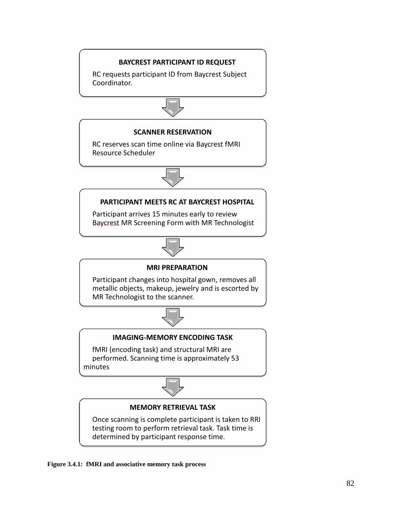
82
Figure 3.4.1: fMRI and associative memory task process
BAYCREST PARTICIPANT ID REQUEST
RC requests participant ID from Baycrest Subject Coordinator.
SCANNER RESERVATION
RC reserves scan time online via Baycrest fMRI Resource Scheduler
PARTICIPANT MEETS RC AT BAYCREST HOSPITAL
Participant arrives 15 minutes early to review Baycrest MR Screening Form with MR Technologist
MRI PREPARATION
Participant changes into hospital gown, removes all metallic objects, makeup, jewelry and is escorted by MR Technologist to the scanner.
IMAGING-MEMORY ENCODING TASK
fMRI (encoding task) and structural MRI are performed. Scanning time is approximately 53 minutes
MEMORY RETRIEVAL TASK
Once scanning is complete participant is taken to RRI testing room to perform retrieval task. Task time is determined by participant response time.
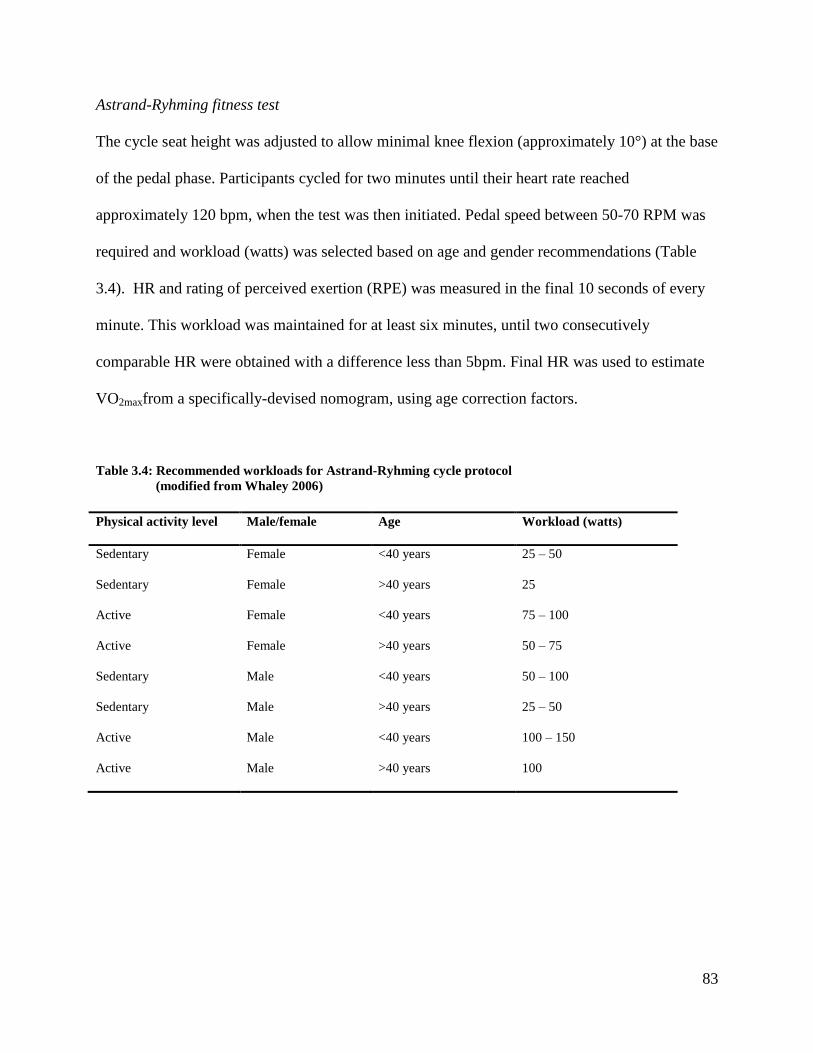
83
Astrand-Ryhming fitness test
The cycle seat height was adjusted to allow minimal knee flexion (approximately 10°) at the base
of the pedal phase. Participants cycled for two minutes until their heart rate reached
approximately 120 bpm, when the test was then initiated. Pedal speed between 50-70 RPM was
required and workload (watts) was selected based on age and gender recommendations (Table
3.4). HR and rating of perceived exertion (RPE) was measured in the final 10 seconds of every
minute. This workload was maintained for at least six minutes, until two consecutively
comparable HR were obtained with a difference less than 5bpm. Final HR was used to estimate
VO2maxfrom a specifically-devised nomogram, using age correction factors.
Table 3.4: Recommended workloads for Astrand-Ryhming cycle protocol
(modified from Whaley 2006)
Physical activity level Male/female Age Workload (watts)
Sedentary Female <40 years 25 – 50
Sedentary Female >40 years 25
Active Female <40 years 75 – 100
Active Female >40 years 50 – 75
Sedentary Male <40 years 50 – 100
Sedentary Male >40 years 25 – 50
Active Male <40 years 100 – 150
Active Male >40 years 100

84
3.4.2 Intervention
Based on the general consensus from studies that report an aerobic intensity of 60–80% of
maximum heart rate and an exercise frequency of three 30-minute sessions per week for 8 weeks,
for a significant improvement in depressive symptoms, the intervention consisted of three
sessions per week of aerobic and resistance exercises over a 12-week period, for a total of 36
sessions. Participants were eligible to be included in analysis if they attended at least 75% of
these sessions. One of the three sessions was aerobic exercise only, and the remaining two were
resistance exercise followed by a shorter aerobic bout. The latter were performed at least 24
hours apart. Participants attended on an individual basis. All sessions began with a 5-10 minute
aerobic warm-up, and were supervised by a Certified Personal Trainer. Participants were advised
to bring a drink bottle and keep well hydrated. Opportunities were provided to make up missed
sessions.
Based on the participant‟s experience and ability, one resistance exercise was selected from each
of the six categories – horizontal push, anterior lower body, vertical pull, posterior lower body,
horizontal pull, and vertical push (Table 3.4.2) – in order to establish a balanced, whole-body
prescription (see photos, Appendix 15).
These exercises were performed as two or three paired supersets, in order to reduce the workout
duration while still promoting adequate rest periods. The sessions utilised an 8-10 repetition
range over three sets. To facilitate progression, participants were encouraged to increase their
weights once they could perform three sets of 10 comfortably, reverting back to eight repetitions
at the higher weight. Exercises were changed after four weeks, to avoid adaptation. The aerobic
bout following resistance sessions was 20 minutes in the first week, and 30 minutes in
subsequent weeks.

85
Table 3.4.2: Resistance exercise options, by category
Category Exercise options
Horizontal push Machine bench press
DB bench press
BB bench press
Anterior lower body Machine leg press
DB split squat
Vertical pull Underhand pull down
Overhand wide-grip pull down
Overhand close-grip pull up
Cable tricep extension
Posterior lower body Machine leg curl
One-leg calf raise
Machine calf raise
Horizontal pull Machine seated row
DB row
Machine preacher curl
BB biceps curl
DB jack-hammer curls
Vertical push DB shoulder press

86
All aerobic exercises were performed on either the cycle or treadmill. Workloads were
determined by HR response, which was prescribed as 70-85% max. The full aerobic sessions was
initially 30 minutes, and progressed in five-minute increments over the 12 weeks, up to 60
minutes. Individuals could also progress their target HR within the prescribed range.
At every session, information was recorded for each participant on average and maximum HR
obtained, exercise duration, workload (watts, for the cycle, or speed and incline for the
treadmill), weights, sets, and repetitions.
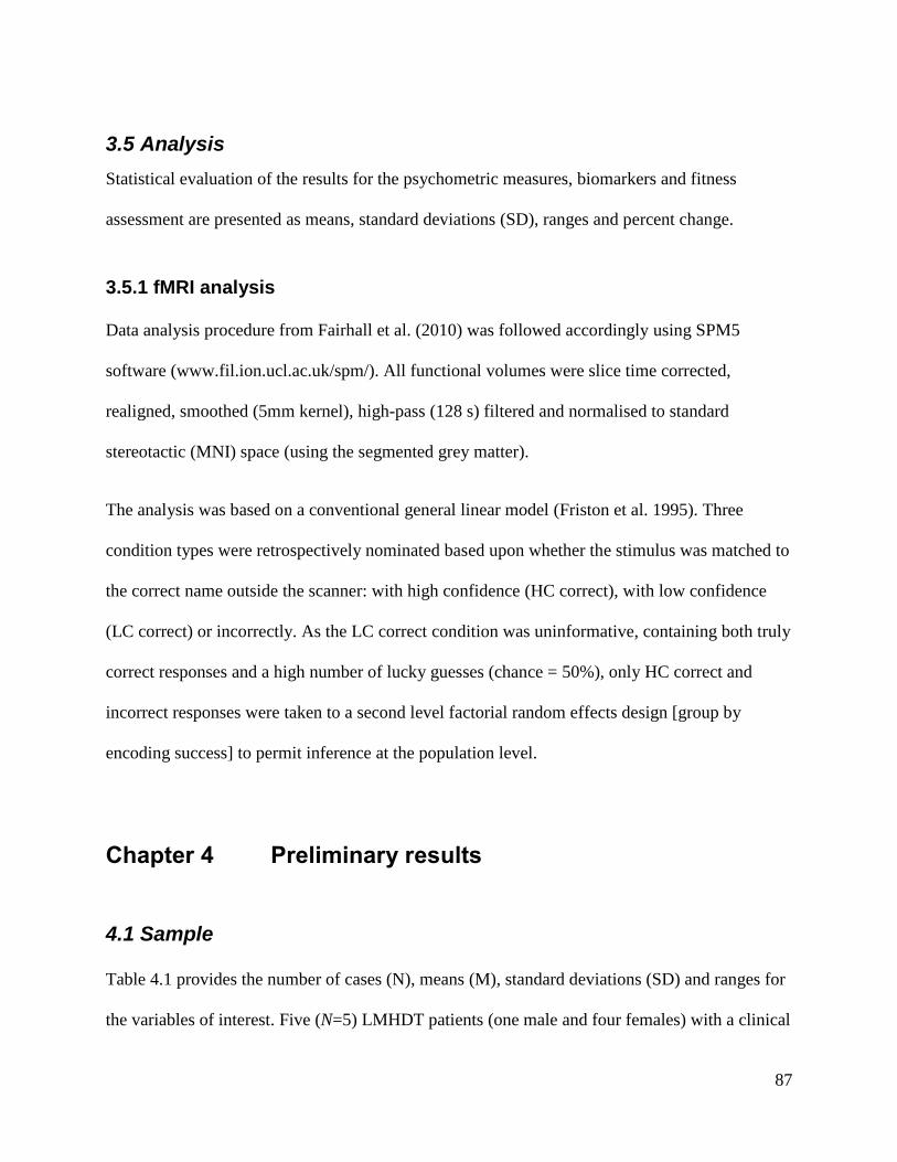
87
3.5 Analysis
Statistical evaluation of the results for the psychometric measures, biomarkers and fitness
assessment are presented as means, standard deviations (SD), ranges and percent change.
3.5.1 fMRI analysis
Data analysis procedure from Fairhall et al. (2010) was followed accordingly using SPM5
software (www.fil.ion.ucl.ac.uk/spm/). All functional volumes were slice time corrected,
realigned, smoothed (5mm kernel), high-pass (128 s) filtered and normalised to standard
stereotactic (MNI) space (using the segmented grey matter).
The analysis was based on a conventional general linear model (Friston et al. 1995). Three
condition types were retrospectively nominated based upon whether the stimulus was matched to
the correct name outside the scanner: with high confidence (HC correct), with low confidence
(LC correct) or incorrectly. As the LC correct condition was uninformative, containing both truly
correct responses and a high number of lucky guesses (chance = 50%), only HC correct and
incorrect responses were taken to a second level factorial random effects design [group by
encoding success] to permit inference at the population level.
Chapter 4 Preliminary results
4.1 Sample
Table 4.1 provides the number of cases (N), means (M), standard deviations (SD) and ranges for
the variables of interest. Five (N=5) LMHDT patients (one male and four females) with a clinical

88
diagnoses of MDD, confirmed by the SCID-I, and no other co-existing DSM-IV Axis I
disorders, have enrolled in this research study thus far. The mean age was 37.4 years (SD=10.2)
with a range of 28 to 49 years. Attrition rate was 60% however since one participant dropped out
at nine weeks it was decided to collect their post exercise measures.
Of these five participants, three were medicated with a selective serotonin reuptake inhibitor
(SSRI) and two were taking a serotonin-norepinephrine reuptake inhibitor (SNRI). In addition to
antidepressant medications one participant was taking anti-inflammatory and
immunosuppressant medications and one participant was taking psychostimulant and
anticonvulsant medications. The mean depressive episode was 11.6 months (SD=10.6) however
four participants reported suffering from depression for several years. The data set did contain
missing data. Of the biomarker variables, one participant failed to provide a waking saliva
sample and one baseline IL-6 data was excluded due to an elevation from a confounding case of
acute otitis media.
4.1.1 Psychometric evaluation
The mean baseline BDI score (M=36, SD=2) indicates severe depression, the HADS-D (M=11,
SD=3.16) indicates definite depression, the HADS-A (M=15.80, SD=2.28) indicates definite
anxiety, the PSQI (M=11, SD=5.53) indicates poor sleep quality, and the MoCA (M=23.6,
SD=2.10) indicates mild cognitive impairment.
4.1.2 Biomarker data
The mean waking salivary cortisol concentration (M=10.73, SD=9.75), the 30 minute post
awakening (M=19.96, SD=13.82) and CAR (M=133.10, SD=164.50) are within the normal
healthy range. Unfortunately, the mean group value is misleading and does not translate the
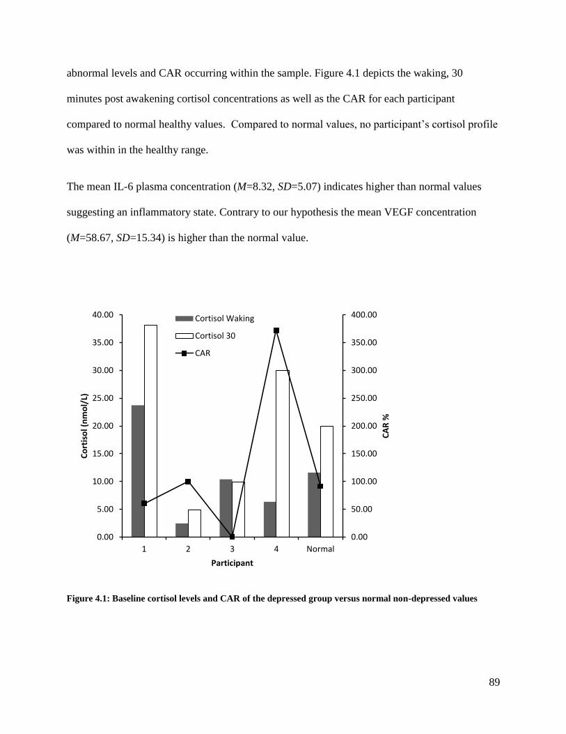
89
abnormal levels and CAR occurring within the sample. Figure 4.1 depicts the waking, 30
minutes post awakening cortisol concentrations as well as the CAR for each participant
compared to normal healthy values. Compared to normal values, no participant‟s cortisol profile
was within in the healthy range.
The mean IL-6 plasma concentration (M=8.32, SD=5.07) indicates higher than normal values
suggesting an inflammatory state. Contrary to our hypothesis the mean VEGF concentration
(M=58.67, SD=15.34) is higher than the normal value.
Figure 4.1: Baseline cortisol levels and CAR of the depressed group versus normal non-depressed values
0.00
50.00
100.00
150.00
200.00
250.00
300.00
350.00
400.00
0.00
5.00
10.00
15.00
20.00
25.00
30.00
35.00
40.00
1 2 3 4 Normal
CA
R %
Co
rtis
ol (
nm
ol/
L)
Participant
Cortisol Waking
Cortisol 30
CAR

90
4.1.3 Fitness scores
Individual fitness scores are allocated based on age and sex. Therefore a mean group score does
not indicate an overall group fitness level. This measure will be used at 12 weeks following the
exercise program to determine the relationship between changes in estimated relative VO2max
with other variables.
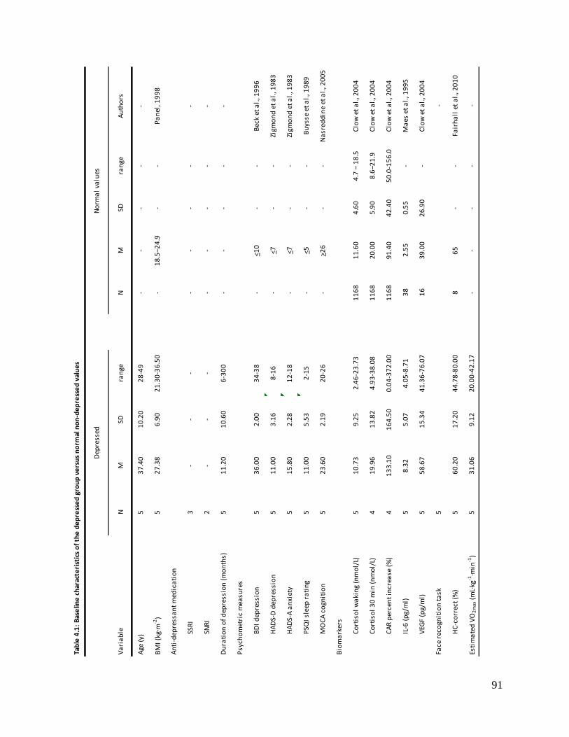
91
Tab
le 4
.1:
Bas
elin
e c
har
acte
rist
ics
of
the
de
pre
sse
d g
rou
p v
ers
us
no
rmal
no
n-d
ep
ress
ed
val
ue
s
Va
ria
ble
NM
SDra
nge
NM
SDra
nge
Au
tho
rs
Age
(y)
53
7.4
01
0.2
02
8-4
9-
--
--
BM
I (k
g∙m
-2)
52
7.3
86
.90
21
.30
-36
.50
-1
8.5
–24
.9-
-P
an
el, 1
99
8
An
ti-d
epre
ssa
nt
med
ica
tio
n
SSR
I3
--
--
--
--
SNR
I2
--
--
--
--
Du
rati
on
of
dep
ress
ion
(m
on
ths)
51
1.2
01
0.6
06
-30
0-
--
--
Psy
cho
met
ric
mea
sure
s
BD
I d
epre
ssio
n5
36
.00
2.0
03
4-3
8-
<10
--
Bec
k et
al.
, 19
96
HA
DS-
D d
epre
ssio
n
51
1.0
03
.16
8-1
6-
<7-
-Zi
gmo
nd
et
al.
, 19
83
HA
DS-
A a
nxi
ety
51
5.8
02
.28
12
-18
-<7
--
Zigm
on
d e
t a
l., 1
98
3
PSQ
I sl
eep
ra
tin
g5
11
.00
5.5
32
-15
-<5
--
Bu
ysse
et
al.
, 19
89
MO
CA
co
gnit
ion
52
3.6
02
.19
20
-26
->2
6-
-N
asr
edd
ine
et a
l., 2
00
5
Bio
ma
rker
s
Co
rtis
ol
wa
kin
g (n
mo
l/L)
51
0.7
39
.25
2.4
6-2
3.7
31
16
81
1.6
04
.60
4.7
– 1
8.5
Clo
w e
t a
l., 2
00
4
Co
rtis
ol
30
min
(n
mo
l/L)
41
9.9
61
3.8
24
.93
-38
.08
11
68
20
.00
5.9
08
.6–2
1.9
Clo
w e
t a
l., 2
00
4
CA
R p
erce
nt
incr
ease
(%
)4
13
3.1
01
64
.50
0.0
4-3
72
.00
11
68
91
.40
42
.40
50
.0-1
56
.0C
low
et
al.
, 20
04
IL-6
(p
g/m
l)5
8.3
25
.07
4.0
5-8
.71
38
2.5
50
.55
-M
aes
et
al.
, 19
95
VEG
F (p
g/m
l)5
58
.67
15
.34
41
.36
-76
.07
16
39
.00
26
.90
-C
low
et
al.
, 20
04
Face
rec
ogn
itio
n t
ask
5-
HC
-co
rrec
t (%
)5
60
.20
17
.20
44
.78
-80
.00
86
5-
-Fa
irh
all
et
al.
, 20
10
Esti
ma
ted
VO
2m
ax (
mL∙
kg-1
∙min
-1)
53
1.0
69
.12
20
.00
-42
.17
--
--
-
Dep
ress
edN
orm
al
valu
es
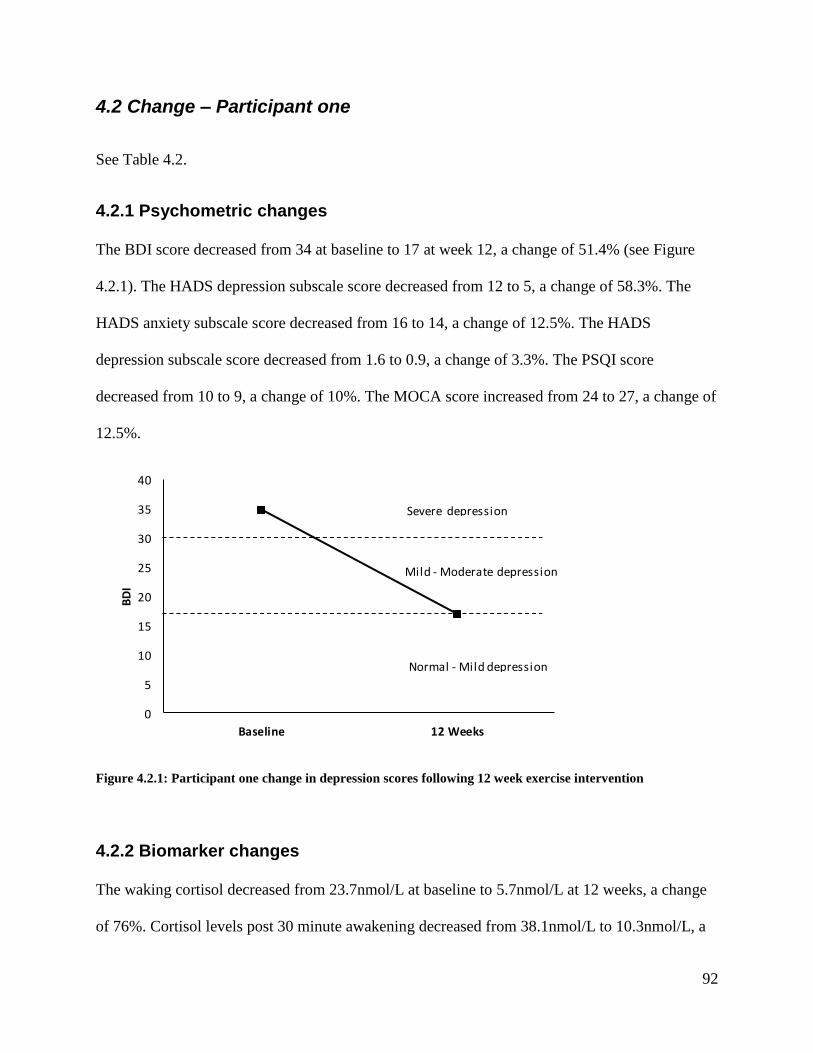
92
4.2 Change – Participant one
See Table 4.2.
4.2.1 Psychometric changes
The BDI score decreased from 34 at baseline to 17 at week 12, a change of 51.4% (see Figure
4.2.1). The HADS depression subscale score decreased from 12 to 5, a change of 58.3%. The
HADS anxiety subscale score decreased from 16 to 14, a change of 12.5%. The HADS
depression subscale score decreased from 1.6 to 0.9, a change of 3.3%. The PSQI score
decreased from 10 to 9, a change of 10%. The MOCA score increased from 24 to 27, a change of
12.5%.
0
5
10
15
20
25
30
35
40
Baseline 12 Weeks
BD
I
Normal - Mild depression
Mild - Moderate depression
Severe depression
Figure 4.2.1: Participant one change in depression scores following 12 week exercise intervention
4.2.2 Biomarker changes
The waking cortisol decreased from 23.7nmol/L at baseline to 5.7nmol/L at 12 weeks, a change
of 76%. Cortisol levels post 30 minute awakening decreased from 38.1nmol/L to 10.3nmol/L, a
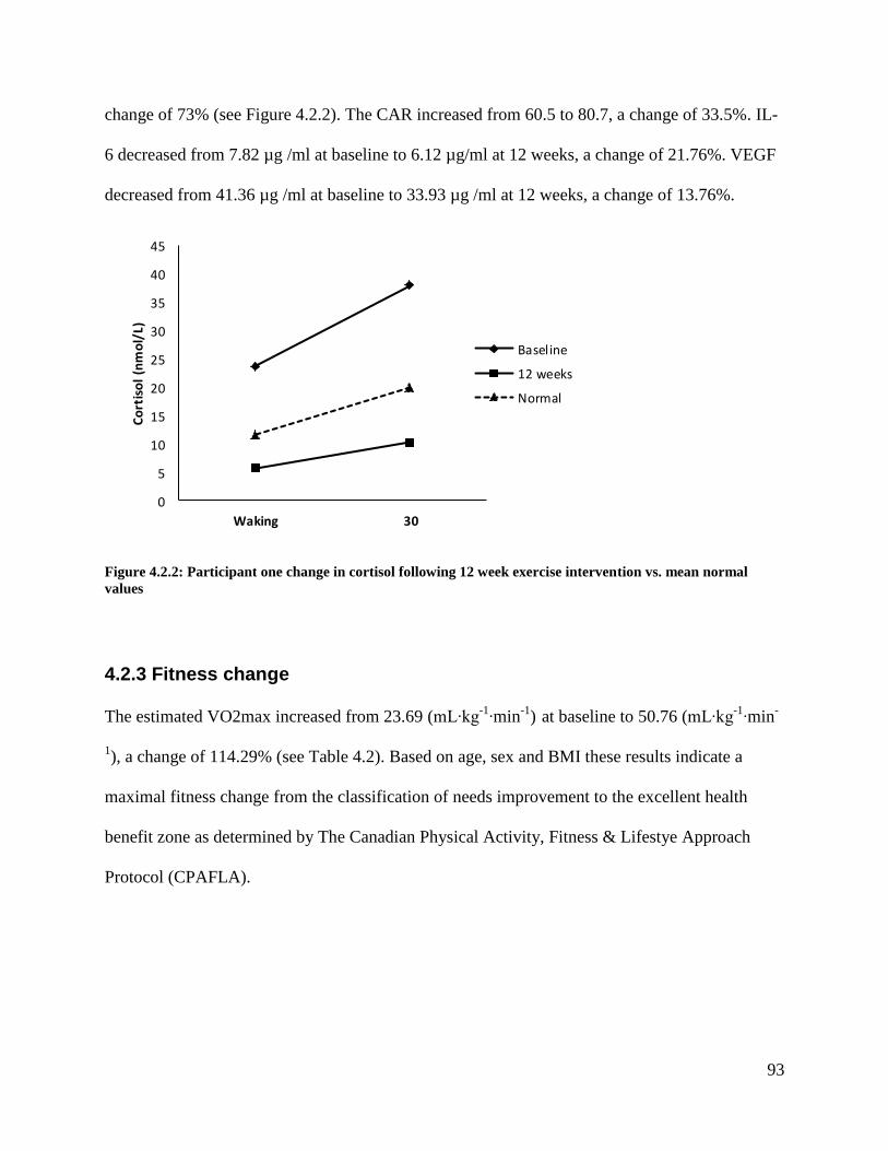
93
change of 73% (see Figure 4.2.2). The CAR increased from 60.5 to 80.7, a change of 33.5%. IL-
6 decreased from 7.82 µg /ml at baseline to 6.12 µg/ml at 12 weeks, a change of 21.76%. VEGF
decreased from 41.36 µg /ml at baseline to 33.93 µg /ml at 12 weeks, a change of 13.76%.
0
5
10
15
20
25
30
35
40
45
Waking 30
Co
rtis
ol
(nm
ol/
L)
Baseline
12 weeks
Normal
Figure 4.2.2: Participant one change in cortisol following 12 week exercise intervention vs. mean normal
values
4.2.3 Fitness change
The estimated VO2max increased from 23.69 (mL∙kg-1
∙min-1
) at baseline to 50.76 (mL∙kg-1
∙min-
1), a change of 114.29% (see Table 4.2). Based on age, sex and BMI these results indicate a
maximal fitness change from the classification of needs improvement to the excellent health
benefit zone as determined by The Canadian Physical Activity, Fitness & Lifestye Approach
Protocol (CPAFLA).
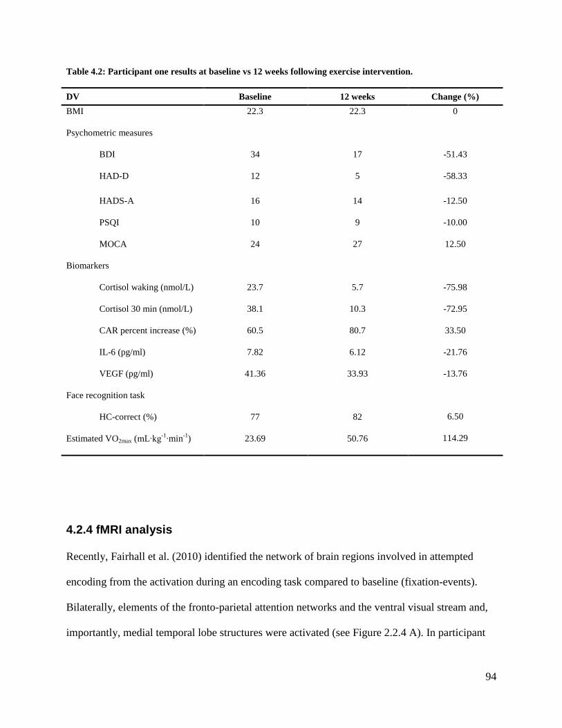
94
Table 4.2: Participant one results at baseline vs 12 weeks following exercise intervention.
DV Baseline 12 weeks Change (%)
BMI
Psychometric measures
BDI
22.3
34
22.3
17
0
-51.43
HAD-D 12 5 -58.33
HADS-A
PSQI
MOCA
Biomarkers
Cortisol waking (nmol/L)
Cortisol 30 min (nmol/L)
CAR percent increase (%)
IL-6 (pg/ml)
VEGF (pg/ml)
Face recognition task
HC-correct (%)
Estimated VO2max (mL∙kg-1
∙min-1
)
16
10
24
23.7
38.1
60.5
7.82
41.36
77
23.69
14
9
27
5.7
10.3
80.7
6.12
33.93
82
50.76
-12.50
-10.00
12.50
-75.98
-72.95
33.50
-21.76
-13.76
6.50
114.29
4.2.4 fMRI analysis
Recently, Fairhall et al. (2010) identified the network of brain regions involved in attempted
encoding from the activation during an encoding task compared to baseline (fixation-events).
Bilaterally, elements of the fronto-parietal attention networks and the ventral visual stream and,
importantly, medial temporal lobe structures were activated (see Figure 2.2.4 A). In participant

95
one (Figure 4.2.4 C) a similar activation pattern in the medial temporal lobe structures, weighted
towards the hippocampal complex rather than the parahippocampal gyrus, indicates that this task
mainly recruited hippocampal structures. Activation has been colour coded into cortical (orange),
hippocampal (red), parahippocampal (blue) and amygdalic regions (green) using anatomical
templates (Tzourio-Mazoyer et al. 2002). All activations are thresholded at p < .001 and
corrected for multiple comparisons at the cluster level. The yellow dot in the hippocampus
represents the region extracted for further analysis (see Fig. 4.2.4 A).
Previous pilot data (Fairhall et al. 2010) formally demonstrates that the strong relationship
between hippocampal activation and encoding success present in normal subjects is absent in
individuals experiencing MDD (Fairhall et al. 2010). Figure 4.2.4 (B) shows a significant group
by group (Controls vs MDD) interaction within the bilateral anterior hippocampus. This
interaction was driven by a positive association between hippocampal activity and successful
encoding in non-depressed controls (left: t(7) = 2.83, p < .05; right: t(7) = 3.00, p < .05) that was
not present in individuals with MDD (left: t(7) =−0.46, ns; right: t(7) =−0.44, ns.). To measure
the difference in the relationship between encoding success and bilateral hippocampal activation
in control and MDD groups, Cohen‟s d was calculated separately for each group. A large effect
size (d= 1.11) was found for controls while depressed individuals showed no relationship
between anterior hippocampus activity and stimulus encoding success (d=−0.19).
For this feasibility study, a region-of-interest (ROI) approach was adopted from previous pilot
work by Fairhall and colleagues. The peak voxel in left and right anterior hippocampi were
selected from the contrast of task versus fixation-events irrespective of encoding success (see
Figure 4.2.4 C). This comparison to the loose fixation-baseline allowed for the identification of
eloquent-voxels in a way orthogonal to the effects of interest (i.e. condition, group). Results also

96
show that we were able to detect differences pre/post intervention with the data set from
participant one. Figure 4.2.4 D illustrates a baseline result similar to the „MDD response‟ found
in Fairhall et al. (2010) and the predicted pattern of increased activation in the anterior
hippocampus during the associative encoding of stimuli that are later remembered with
confidence following the 12 week exercise program.
Figure 4.2.4: A comparison of fMRI results from Fairhall et al. (2010) and participant one.
97
Chapter 5 Discussion
Despite the attempts of several theoretical models delineating the causes of depression, the
pathogenesis remains elusive preventing the availability of treatments that target the causal
factors. It has been well established that exercise is an effective treatment for depression
however it is not clear on exactly how and why it works. The overall goal of this research is to
investigate whether the mechanism by which exercise may improve depression is related to
improving hippocampal function and if a supervised 12 week exercise program can promote
changes in hippocampal function and improve memory in people living with depression. The
purpose of this feasibility study was to develop comprehensive methods and validation of
protocols to conduct our future pilot study. These included: 1) MDD participant recruitment from
the LMHDT program; 2) fMRI protocol; 3) specimen collection and storage; 4) biomarker
assays; and 5) an effective exercise program capable of improving depressive symptomatology.
A preliminary results section was also generated based on data collected thus far. Although the
sample size is too small to infer meaningful statistical results, our preliminary findings have
raised several methodological concerns that will be discussed.
Recruitment procedure
To ensure that the sample population is a true representation of the MDD population, all
participants recruited were diagnosed as clinically depressed based on DSM-V criteria confirmed
by the SCID-I. Our partners at the LMHDT program ensured that recruitment staff have been
properly trained to administer the SCID-I. Additionally, to avoid unnecessary meetings between
participants and RC, Lakeridge staff administered a pre-screening questionnaire provided by the
RC to identify eligible sedentary, MRI safe participants. In support of these procedures, all five

98
participants recruited by Lakeridge staff have qualified for the study as confirmed by the
additional screening measures and depression inventories administered by RC.
Methods for specimen collection and immunoassays
Well defined methods for specimen collection, processing and storage were established in
accordance with laboratory procedures. The SOS was an effective device for obtaining copious
saliva for cortisol analysis. Participants followed the saliva collection instructions provided to
them and all samples were returned frozen and packed in their corresponding polystyrene bin.
Given that immunoassays require very small samples volumes, plasma samples were divided
into several small volume aliquots so that each aliquot could be assayed independently. This
procedure proved to be very effective in minimizing plasma waste and avoiding the need for
repeated freeze-thaw cycles. Additionally, the availability of several replicate aliquots will allow
for further biomarker analysis and also increase reliability of assays by providing additional
control samples to test inter- and intra- assay precision. All immunoassays were performed
according to the manufacturers‟ protocols. Standard curves generated from each biomarker
corresponded with the manufacturer values. Optical density values from each standard curve
were also sent to the manufacturers for further verification.
fMRI protocol validation
Four fMRI pilot scans were performed to successfully validate the fMRI protocol previously
piloted in New Zealand on a 1.5 Tesla General Electric MRI scanner using the 3.0 Tesla Siemans
MRI scanner at the Rotman Baycrest hospital. Ideally this experimental paradigm will produce
robust modulation of hippocampal activity with encoding success, separation of true encoding
success from chance success and the selection of a specific brain region-of-interest. Baseline
results from participant one indicates hippocampal dysregulation as data fails to show the
99
positive relationship between hippocampal activity and successful encoding as seen in the non-
depressed controls from Fairhall et al., (2010). Encouragingly, following the 12 week exercise
program a more normalized pattern of hippocampal activation associated with successful
memory encoding was observed. Taken together, these findings support the use of this
experimental paradigm for future pilot work and also suggest that exercise may normalize
hippocampal functioning in depressed individuals. If the study is able to show significant pilot
results this may eventually lead to a potential in vivo biomarker for the functional integrity of the
hippocampus in MDD to explore the relationships between 1) hippocampal dysregulation and
clinical presentation, 2) hippocampal dysregulation and biomarker status and 3) the neurogenic
effects of anti-depressant treatments such as pharmaceutical agents or exercise and the regulation
of abnormal hippocampal function (Fairhall et al. 2010).
Preliminary results
Baseline psychometric data indicates that all participants were experiencing severe depression,
anxiety, mild cognitive impairment and sleep disturbance. However 12 week results from
participant one revealed the following improvements: severe depression to mild depression, mild
cognitive impairment to normal cognitive functioning, and minimal improvements in anxiety
score and sleep disturbance.
The cortisol analysis yielded variable results across the group and no individual cortisol profile
resided in the healthy normal range. Observations ranged from below normal to above normal
levels and an absent CAR to an above normal CAR of 372%. Although these findings do not
support our hypothesis that cortisol would be elevated in MDD, they indicate that depression is
associated with a dysregulation in cortisol secretion. Nonetheless, normal cortisol values were
established in participant one following the 12 week exercise program.
100
Consistent with the literature the results show that depressed participants expressed higher
plasma IL-6 concentrations compared with normal controls suggesting an inflammatory state. In
support of our hypothesis, 12 week results from participant one show a decrease in IL-6
concentration following the exercise program. An additional observation was that the participant
with the highest depression score revealed the highest IL-6 concentration. Although it remains
unknown whether exercise specifically targets pro-inflammatory cytokines, exercise may
actually dampen the inflammatory processes through its immuno-regulatory/anti-inflammatory
effects. An important issue that needs further evaluation may be the absence of counter-
balancing, immunoregulatory relationship between pro- and anti-inflammatory cytokines
(Dhabhar et al. 2009). The future pilot study will include further analysis of additional pro-
inflammatory cytokines (IFN-γ - and TNF-α) and the anti-inflammatory cytokine IL-10 to seek
these relationships.
Compared with normal healthy individuals, VEGF concentrations were increased in depressed
individuals. Results from participant one showed a decrease in VEGF following the 12 week
exercise program. These findings do not support the hypothesis or the neurogenic/neurotrophic
hypothesis of depression which suggests that depression results partly from decreased
neurotrophic growth factors. However the findings correspond with recent research findings that
VEGF levels are higher in depression (Takebayashi et al. 2010) and depression comorbid with
borderline personality disorder (Kahl et al. 2009) when compared to normal healthy controls.
Other studies report no significant difference in plasma VEGF concentration were been found
between medication-free MDD patients and controls (Ventriglia et al., 2009), and no alteration in
VEGF plasma levels in medicated depressed patients during the courses of recovery (Dome et
al., 2011). Studies have also found correlations with hormones and have reported higher levels of
101
VEGF in the follicular phase (Heer et al. 1998), the luteal phase (Agrawal et al. 1999), and
significantly higher levels have been reported in postmenopausal compared with premenopausal
women (Heer et al. 1998; Kim 1998) . Interestingly, the older female participants experiencing
menopause in this study were found to have the highest VEGF levels (M=71.01) compared to
those who were premenopausal (M=41.36). These findings highlight the critical gap in research
that peripheral VEGF levels and brain VEGF levels have not been quantified in parallel in any
psychiatric population. To shed some light, an animal model investigating the possible
correlation of peripheral and central VEGF levels using a genetic rat model of depression, found
that VEGF levels were identical in serum, corpus striatum, and hypothalamus whereas in the
hippocampus and frontal cortex, the VEGF levels were significantly decreased in the depressed
rats compared to the non-depressed rats (Elfving et al. 2010). The authors considered these
findings to be salient since the frontal cortex and hippocampus are considered to be core brain
regions involved in depression (Elfving et al. 2010).
To further explore the relationship between exercise, neurotrophic factors and depression, the
future pilot study will include additional growth factors (BDNF, IGF) known to be implicated in
depression while eliminating VEGF for the reason that peripheral VEFG concentrations are
influenced by several variables not associated with depression and do not correspond to VEGF
levels in the CNS.
Methodological confounds
The high attrition rate of 60% indicates that the 12 week exercise program was difficult to adhere
to for most participants. Two participants dropped out at seven weeks and one participant
dropped out at nine weeks. A primary reason for withdrawal was that the LMHDT program
ended two weeks prior to the exercise study and participants were returning to work. Since
102
research has shown that the benefits of exercise are reaped as early as eight weeks, it is
recommended that the exercise program be reduced to 10 weeks. This will increased the
probability of more participants completing the program.
Disturbingly, a major barrier confronted while conducting this study was obtaining sound
comparative normal and depressed biomarker data due to the extreme variability reported
between studies. This warrants the need to standardize methods of biological sample collection
and assays, otherwise using what is reported in the literature will be equivocal.
Depression is a clinically and biochemically heterogeneous disorder. However, the majority of
research examining the pathophysiology of depression have classified depression dichotomously
either as yes/no or restricted the analyses to MDD when in fact there are several subtypes of
depression which are based on the symptomatology (Baune et al. 2012). Subtypes of depression
include dysthymia (a chronic, low-grade form of depression), melancholic MD (MMD) and MD
with typical or atypical features (American Psychiatric Association 1994). Melancholic
depression is characterized by anhedonia, lack of reactivity to pleasurable stimuli, reduced
appetite, insomnia, and depression that is worse in the morning. In contrast, atypical depression
is defined by appetite and weight increase, hypersomnia, fatigue and mood reactivity for positive
events (Gold and Chrousos 2002). To further distinguish between subtypes of depression
research has found that the principal arousal producing mediators of the stress response, such as
CRH, are hyperactive in melancholic depression but not in non-melancholic depression (Gold
and Chrousos 2002). Conversely, atypical depression and dysthymia (characterized by lethargy,
fatigue, hypersomnia and hyperphagia) and have been associated with down regulated HPA
activity and decreased cortisol levels (Griffiths et al. 2000; Hayley et al. 2005; Kaestner et al.

103
2005). Furthermore, increased inflammation has been found in non-melancholia depression and
dysthymia but not in melancholic depression (Griffiths et al. 2000; Kaestner et al. 2005).
In short, the majority of antidepressant medications target monoamine systems and have
ultimately fallen short in successfully treating depression. There is now sufficient evidence in the
literature to support the exploration of alternative therapeutic areas to target mechanisms
associated with depression such as neuroendocrine function, immune regulation and
neurotrophic factors (Berton and Nestler 2006; Kennedy and Rizvi 2009). In order to delineate
specific mechanisms, biomarker or combination of biomarkers, several challenges in this field
the following must be addressed: 1) the clinical variability within the syndrome of MDD; 3) the
need to standardize methods of biological sample collection and assay; 4) longitudinal studies to
better illustrate the relationships between depressive symptoms, neuroendocrine, inflammatory
and neurotrophic markers. (Maes et al. 2009; Loftis et al. 2010; Blume et al. 2011).
Personal observations
Overall, participant compliance was excellent for all data collection methods. Participant
feedback from the study was very positive indicating that saliva collection instructions were easy
to follow and no barriers (lack of transportation) were associated with commuting to the Rotman
Baycrest hospital for the fMRI scans. On the other hand, participant compliance was poor for
exercise attendance due to frequent tardiness for appointments and last minute cancellations and
rescheduling which led to reduced trainer availability. Exercise attendance is a critical aspect of
this study and trainer availability is essential. Therefore, a larger trainer pool is required for
future pilot work to ensure trainer availability in the event of an inevitable schedule change.
104
Although participants were eager to participate in the study in hopes of curing their depression,
participant anxiety was experienced during the initial exercise sessions. Feelings reported were
poor body image and low self-efficacy for their exercise capabilities. To alleviate these anxiety
provoking feelings associated with the initial exercise sessions, special attention must be given
towards scheduling initial training sessions during the non-busy gym times or conducting initial
training sessions in a private fitness room with only the participant and trainer.
Finally, each personal trainer was assigned a participant for the entire exercise program.
Consequently, it was noted that participant-trainer attachment bonds were formed by the
participant during the 12 weeks. To prevent the formation of bonds and separation anxiety
experienced by the participants following study completion, future pilot work must assign and
rotate several trainers to each participant throughout the 12 week exercise program.
5.1 Conclusion and recommendations
Depression is a multifaceted, biochemically and clinically heterogeneous disorder that may
account for some of the variability and unpredictability associated with treatments. The future of
successful treatments for MDD is contingent on the ability to produce a „personalized medicine‟
paradigm (Kennedy and Rizvi 2009). Research must generate an integrative model illustrating
how all the various targets interact. This will allow for a more precise exploration of
neurobiological indicators of dysfunction and ultimately enable clinicians to identify and treat
patients with specific dysfunctions based on their biochemical signature and clinical profile
(Kennedy and Rizvi 2009).
105
Contrary to antidepressant medications, exercise is a treatment option that is linked to a myriad
of therapeutic indications. Exercise sets into action an interactive cascade of biochemical
reactions that has an overall net effect of stimulating neuroplasticity, enhancing cognitive
function, normalizing HPA functioning, stimulating neurogenesis, improving cerebrovascular
perfusion and improving immune function (Cotman et al. 2007). For researchers, this provides an
optimal research setting to explore the broad assortment of biomarkers associated with the
biological systems known to be altered in depression. However, before any relationship findings
can be inferred, methodological limitations must be addressed.
First and foremost, the need to standardize methods of biological sample collection and assays
must be established to minimize the variability between studies so that definite conclusions can
be made. To increase assay reliability, it is recommended to prepare a normal human plasma
bank from 15 normal healthy volunteers to create high volume plasma pools that can be used to
test intra- and inter- assay precision. To ensure reproducibility between labs it is recommended
that a plasma sample of known concentration be obtained from another institution using the same
ELISA kits.
As a final point, symptoms of depression differ markedly across individuals and subtypes of
depression exist that may implicate different neurochemical substrates. Future studies must
consider these differences and recruit individuals with a specific subtype and/or assign
participants into homogeneous groups based on specific criteria. Indubitably, future pilot work
must categorize participants into depression subtypes and consider these subtypes during the
hypotheses formulation and data interpretation. Otherwise specific relationships and significant
findings will be lost in the compilation of heterogeneous results.

106
List of References
Åberg, M. A. I., Waern, M., et al. (2012). "Cardiovascular fitness in males at age 18 and risk of
serious depression in adulthood: Swedish prospective population-based study." The
British Journal of Psychiatry.
Adam, E. K., Hawkley, L. C., et al. (2006). "Day-to-day dynamics of experience--cortisol
associations in a population-based sample of older adults." Proceedings of the National
Academy of Sciences of the United States of America 103(45): 17058-17063.
Adlard, P., Perreau, V., et al. (2004). "The timecourse of induction of brain-derived neurotrophic
factor mRNA and protein in the rat hippocampus following voluntary exercise."
Neuroscience letters 363(1): 43-48.
Agrawal, R., Conway, G., et al. (1999). "Serum vascular endothelial growth factor (VEGF) in
the normal menstrual cycle: association with changes in ovarian and uterine Doppler
blood flow." Clin Endocrinol (Oxf) 50(1): 101-106.
Airaksinen, E., Larsson, M., et al. (2004). "Cognitive functions in depressive disorders: evidence
from a population-based study." Psychological medicine 34(01): 83-91.
Allen, S. and Dawbarn, D. (2006). "Clinical relevance of the neurotrophins and their receptors."
Clinical Science 110: 175-191.
American Psychiatric Association (1994). Diagnostic and statistical manual of mental disorders.
Washington, DC, Author.
APA and DSM-IV, A. P. A. T. F. o. (2000). Diagnostic and statistical manual of mental
disorders: DSM-IV-TR, American Psychiatric Publishing, Inc.
Archer, T., Fredriksson, A., et al. (2010). "Influence of Physical Exercise on
Neuroimmunological Functioning and Health: Aging and Stress." Neurotoxicity
Research: 1-15.
Asnis, G. M. and De La Garza, R. (2006). "Interferon-induced depression in chronic hepatitis C:
a review of its prevalence, risk factors, biology, and treatment approaches." Journal of
clinical gastroenterology 40(4): 322-335.
Astrand, P. (1960). "Aerobic work capacity in men and women with special reference to age."
Acta Physiol Scand(49): 45-60.
Atlantis, E., Chow, C. M., et al. (2006). "Worksite intervention effects on sleep quality: A
randomized controlled trial." Journal of Occupational Health Psychology 11(4): 291-304.
Avital, A., Goshen, I., et al. (2003). "Impaired interleukin‐1 signaling is associated with deficits
in hippocampal memory processes and neural plasticity." Hippocampus 13(7): 826-834.
107
Babyak, M., Blumenthal, J., et al. (2000). "Exercise treatment for major depression: maintenance
of therapeutic benefit at 10 months." Psychosom Med 62(5): 633-638.
Backhaus, J., Junghanns, K., et al. (2004). "Sleep disturbances are correlated with decreased
morning awakening salivary cortisol." Psychoneuroendocrinology 29(9): 1184-1191.
Banasr, M., Dwyer, J. M., et al. (2011). "Cell atrophy and loss in depression: reversal by
antidepressant treatment." Current Opinion in Cell Biology: 730-737.
Barrientos, R., Sprunger, D., et al. (2003). "Brain-derived neurotrophic factor mRNA
downregulation produced by social isolation is blocked by intrahippocampal interleukin-
1 receptor antagonist." Neuroscience 121(4): 847-853.
Barrientos, R. M., Higgins, E. A., et al. (2006). "Peripheral infection and aging interact to impair
hippocampal memory consolidation." Neurobiology of aging 27(5): 723-732.
Bauer, M. E., Jeckel, C. M. M., et al. (2009). "The role of stress factors during aging of the
immune system." Annals of the New York Academy of Sciences 1153(1): 139-152.
Baune, B., Stuart, M., et al. (2012). "The relationship between subtypes of depression and
cardiovascular disease: a systematic review of biological models." Translational
Psychiatry 2(3): e92.
Beavers, K. M., Brinkley, T. E., et al. (2010). "Effect of exercise training on chronic
inflammation." Clinica Chimica Acta 411(11-12): 785-793.
Beck, A. T., Steer, R. A., et al. (1996). BDI-II Beck Depression Inventory Manual. San Antonio,
TX, Harcourt Brace & Company.
Behnken, A., Ger, J., et al. (2010). "Persistent non-verbal memory impairment in remitted major
depression--Caused by encoding deficits?" Journal of affective disorders 122(1-2): 144-
148.
Belmaker, R. and Agam, G. (2008). "Major depressive disorder." New England Journal of
Medicine 358(1): 55-70.
Bemelmans, K., Goekoop, J., et al. (1996). "Recall performance in acutely depressed patients
and plasma cortisol." Biological Psychiatry 39(8): 750-752.
Bernardino, L., Agasse, F., et al. (2008). "Tumor Necrosis Factor‐α Modulates Survival,
Proliferation, and Neuronal Differentiation in Neonatal Subventricular Zone Cell
Cultures." Stem cells 26(9): 2361-2371.
Berton, O. and Nestler, E. J. (2006). "New approaches to antidepressant drug discovery: beyond
monoamines." Nature Reviews Neuroscience 7(2): 137-151.
108
Blackburn, G. F., Shah, H. P., et al. (1991). "Electrochemiluminescence detection for
development of immunoassays and DNA probe assays for clinical diagnostics." Clin
Chem 37(9): 1534-1539.
Bliss, T. and Collingridge, G. (1993). "A synaptic model of memory: long-term potentiation in
the hippocampus." Nature 361(6407): 31-39.
Blume, J., Douglas, S. D., et al. (2011). "Immune suppression and immune activation in
depression." Brain, behavior, and immunity 25(2): 221-229.
Blumenthal, J., Babyak, M., et al. (2007). "Exercise and pharmacotherapy in the treatment of
major depressive disorder." Psychosom Med 69(7): 587.
Blumenthal, J., Babyak, M., et al. (1999). "Effects of exercise training on older patients with
major depression." Archives of internal medicine 159(19): 2349.
Bocchio-Chiavetto, L., Bagnardi, V., et al. (2010). "Serum and plasma BDNF levels in major
depression: a replication study and meta-analyses." World Journal of Biological
Psychiatry 11(6): 763-773.
Boettger, S., Wetzig, F., et al. (2009). "Physical fitness and heart rate recovery are decreased in
major depressive disorder." Psychosom Med 71(5): 519-523.
Bower, J. E., Ganz, P. A., et al. (2005). "Diurnal cortisol rhythm and fatigue in breast cancer
survivors." Psychoneuroendocrinology 30(1): 92-100.
Bremner, J., Narayan, M., et al. (2000). "Hippocampal volume reduction in major depression."
American Journal of Psychiatry 157(1): 115-118.
Brown, E., Rush, A., et al. (1999). "Hippocampal Remodeling and Damage by Corticosteroids::
Implications for Mood Disorders." Neuropsychopharmacology 21(4): 474-484.
Brown, E., Varghese, F., et al. (2004). "Association of depression with medical illness: does
cortisol play a role?" Biological Psychiatry 55(1): 1-9.
Brunner, E., Hemingway, H., et al. (2002). "Adrenocortical, autonomic, and inflammatory causes
of the metabolic syndrome." Circulation 106(21): 2659-2665.
Brunoni, A. R., Lopes, M., et al. (2008). "A systematic review and meta-analysis of clinical
studies on major depression and BDNF levels: implications for the role of neuroplasticity
in depression." The International Journal of Neuropsychopharmacology 11(08): 1169-
1180.
Burt, D., Zembar, M., et al. (1995). "Depression and memory impairment: a meta-analysis of the
association, its pattern, and specificity." Psychological Bulletin 117(2): 285-305.
Buss, C., Wolf, O., et al. (2004). "Autobiographic memory impairment following acute cortisol
administration." Psychoneuroendocrinology 29(8): 1093-1096.
109
Buysse, D. J., Reynolds, C. F., 3rd, et al. (1989). "The Pittsburgh Sleep Quality Index: a new
instrument for psychiatric practice and research." Psychiatry Research 28(2): 193-213.
Campbell, S. and MacQueen, G. (2004). "The role of the hippocampus in the pathophysiology of
major depression." Journal of Psychiatry and Neuroscience 29(6): 417-426.
Canada, G. o. (2006). The Human Face of Mental Health and Mental Illness in Canada G. o.
Canada: 59.
Capuron, L., Raison, C. L., et al. (2003). "Association of exaggerated HPA axis response to the
initial injection of interferon-alpha with development of depression during interferon-
alpha therapy." American Journal of Psychiatry 160(7): 1342-1345.
Checkley, S. (1996). "The neuroendocrinology of depression and chronic stress." British medical
bulletin 52(3): 597-617.
Chen, J., Buchanan, J. B., et al. (2008). "Neuroinflammation and disruption in working memory
in aged mice after acute stimulation of the peripheral innate immune system." Brain,
behavior, and immunity 22(3): 301-311.
Chida, Y. and Steptoe, A. (2009). "Cortisol awakening response and psychosocial factors: a
systematic review and meta-analysis." Biological Psychology 80(3): 265-278.
Cink, R. E. and Thomas, T. R. (1981). "Validity of the Astrand-Ryhming nomogram for
predicting maximal oxygen intake." British Journal of Sports Medicine 15(3): 182-185.
Clow, A., Thorn, L., et al. (2004). "The awakening cortisol response: methodological issues and
significance." Stress 7(1): 29-37.
Colcombe, S., Erickson, K., et al. (2003). "Aerobic fitness reduces brain tissue loss in aging
humans." The Journals of Gerontology Series A: Biological Sciences and Medical
Sciences 58(2): M176-180.
Colcombe, S., Erickson, K., et al. (2006). "Aerobic exercise training increases brain volume in
aging humans." The Journals of Gerontology Series A: Biological Sciences and Medical
Sciences 61(11): 1166-1170.
Conrad, C. (2008). "Chronic stress-induced hippocampal vulnerability: The glucocorticoid
vulnerability hypothesis." Reviews in the neurosciences 19(6): 395-411.
Cotman, C. and Berchtold, N. (2002). "Exercise: a behavioral intervention to enhance brain
health and plasticity." TRENDS in Neurosciences 25(6): 295-301.
Cotman, C., Berchtold, N., et al. (2007). "Exercise builds brain health: key roles of growth factor
cascades and inflammation." TRENDS in Neurosciences 30(9): 464-472.
Cotman, C. W., Berchtold, N. C., et al. (2007). "Exercise builds brain health: key roles of growth
factor cascades and inflammation." TRENDS in Neurosciences 30(9): 464-472.

110
Crawford, J. R., Henry, J. D., et al. (2001). "Normative data for the HADS from a large non-
clinical sample." Br J Clin Psychol 40(Pt 4): 429-434.
Czéh, B., Abumaria, N., et al. (2010). "Quantitative changes in hippocampal microvasculature of
chronically stressed rats: no effect of fluoxetine treatment." Hippocampus 20(1): 174-
185.
Czéh, B., Michaelis, T., et al. (2001). "Stress-induced changes in cerebral metabolites,
hippocampal volume, and cell proliferation are prevented by antidepressant treatment
with tianeptine." Proceedings of the National Academy of Sciences of the United States
of America 98(22): 12796-12801.
Danion, J.-M., Kauffmann-Muller, F., et al. (1995). "Affective valence of words, explicit and
implicit memory in clinical depression." Journal of affective disorders 34(3): 227-234.
Dantzer, R. (2012). "Depression and inflammation: an intricate relationship." Biological
Psychiatry 71(1): 4-5.
Dantzer, R., O'Connor, J. C., et al. (2008). "From inflammation to sickness and depression: when
the immune system subjugates the brain." Nature Reviews Neuroscience 9(1): 46-56.
Das, U. N. (2004). "Anti-inflammatory nature of exercise." Nutrition 20(3): 323-326.
Das, U. N. (2006). "Exercise and inflammation." European heart journal 27(11): 1385-1385.
Davis, M., Greenblatt, D., et al. (1987). "The dexamethasone suppression test: an overview of its
current status in psychiatry. The APA Task Force on Laboratory Tests in Psychiatry."
American Journal of Psychiatry 144(10): 1253.
DeCarolis, N. and Eisch, A. (2010). "Hippocampal neurogenesis as a target for the treatment of
mental illness: A critical evaluation." Neuropharmacology 58(6): 884-893.
Dedovic, K., Engert, V., et al. (2010). "Cortisol Awakening Response and Hippocampal
Volume: Vulnerability for Major Depressive Disorder?" Biological Psychiatry.
Deng, W., Aimone, J., et al. (2010). "New neurons and new memories: how does adult
hippocampal neurogenesis affect learning and memory?" Nature Reviews Neuroscience
11(5): 339-350.
Dennis, M., Chan, Y., et al. (2006). "Development and validation of the GAIN Short Screener
(GSS) for internalizing, externalizing and substance use disorders and crime/violence
problems among adolescents and adults." American Journal on Addictions 15(s1): 80-91.
DeRubeis, R., Hollon, S., et al. (2005). "Cognitive therapy vs medications in the treatment of
moderate to severe depression." Archives of general psychiatry 62(4): 409-416.

111
Devine, J. and Zafonte, R. (2009). "Physical exercise and cognitive recovery in acquired brain
injury: a review of the literature." PM & R: the journal of injury, function, and
rehabilitation 1(6): 560-575.
Dhabhar, F. S., Burke, H. M., et al. (2009). "Low serum IL-10 concentrations and loss of
regulatory association between IL-6 and IL-10 in adults with major depression." Journal
of psychiatric research 43(11): 962-969.
Dishman, R., Berthoud, H., et al. (2006). "Neurobiology of Exercise&ast." Obesity 14(3): 345-
356.
Diverty, B. and Beaudet, M. (1997). "Depression: an undertreated disorder." Health Rep 8(4): 9-
18.
Dominique, J., Henke, K., et al. (2003). "Glucocorticoid-induced impairment of declarative
memory retrieval is associated with reduced blood flow in the medial temporal lobe."
European Journal of Neuroscience 17(6): 1296-1302.
Dowlati, Y., Herrmann, N., et al. (2010). "A meta-analysis of cytokines in major depression."
Biological Psychiatry 67(5): 446-457.
Doyne, E. J., Ossip-Klein, D. J., et al. (1987). "Running versus weight lifting in the treatment of
depression." Journal of Consulting & Clinical Psychology 55(5): 748-754.
Duman, C. H., Schlesinger, L., et al. (2009). "Peripheral insulin-like growth factor-I produces
antidepressant-like behavior and contributes to the effect of exercise." Behavioural brain
research 198(2): 366-371.
Duman, R., Malberg, J., et al. (1999). "Neural plasticity to stress and antidepressant treatment."
Biological Psychiatry 46(9): 1181-1191.
Duman, R. S. (2009). "Neuronal damage and protection in the pathophysiology and treatment of
psychiatric illness: stress and depression." Dialogues in clinical neuroscience 11(3): 239.
Duman, R. S. and Monteggia, L. M. (2006). "A neurotrophic model for stress-related mood
disorders." Biological Psychiatry 59(12): 1116-1127.
Dunn, A. L., Trivedi, M. H., et al. (2005). "Exercise treatment for depression: efficacy and dose
response." Am J Prev Med 28(1): 1-8.
Dunn, A. L., Trivedi, M. H., et al. (2001). "Physical activity dose-response effects on outcomes
of depression and anxiety." Med Sci Sports Exerc 33: S587-597.
Duvernoy, H. M. (2005). The human hippocampus: functional anatomy, vascularization, and
serial sections with MRI, Springer Verlag.
Edwards, S., Clow, A., et al. (2001). "Exploration of the awakening cortisol response in relation
to diurnal cortisol secretory activity." Life Sciences 68(18): 2093-2103.

112
Egeland, J., Lund, A., et al. (2005). "Cortisol level predicts executive and memory function in
depression, symptom level predicts psychomotor speed." Acta Psychiatrica Scandinavica
112(6): 434-441.
Eisenberger, N. I., Inagaki, T. K., et al. (2010). "Inflammation and social experience: an
inflammatory challenge induces feelings of social disconnection in addition to depressed
mood." Brain, behavior, and immunity 24(4): 558-563.
Ekstrand, J., Hellsten, J., et al. (2008). "Environmental enrichment, exercise and corticosterone
affect endothelial cell proliferation in adult rat hippocampus and prefrontal cortex."
Neuroscience letters 442(3): 203-207.
Elfving, B., Plougmann, P. H., et al. (2010). "Differential brain, but not serum VEGF levels in a
genetic rat model of depression." Neuroscience letters 474(1): 13-16.
Erickson, K., Prakash, R., et al. (2009). "Aerobic fitness is associated with hippocampal volume
in elderly humans." Hippocampus 19(10): 1030-1039.
Ernst, C., Olson, A. K., et al. (2006). "Antidepressant effects of exercise: evidence for an adult-
neurogenesis hypothesis." Journal of Psychiatry and Neuroscience 31: 84-92.
Ernst, C., Olson, A. K., et al. (2006). "Antidepressant effects of exercise: Evidence for an adult-
neurogenesis hypothesis?" Journal of psychiatry & neuroscience 31(2): 84.
Fairhall, S. L., Sharma, S., et al. (2010). "Memory related dysregulation of hippocampal function
in major depressive disorder." Biol Psychol 85(3): 499-503.
Farmer, J., Zhao, X., et al. (2004). "Effects of voluntary exercise on synaptic plasticity and gene
expression in the dentate gyrus of adult male Sprague-Dawley rats in vivo." Neuroscience
124(1): 71-79.
Fava, G. and Ruini, C. (2002). "Long-term treatment of depression: there is more than drugs."
Recenti progressi in medicina 93(6): 343-345.
Foley, L. (2006). The effect of an eight week exercise program on depression intensity, self-
efficacy and cortisol secretion in a clinically depressed individual and an examination of
the salivary cortisol method. Sportsci 702 project. Auckland, University of Auckland.
Foley, L., Prapavessis, H., et al. (2008). "An examination of potential mechanisms for exercise
as a treatment for depression: A pilot study." Mental Health and Physical Activity 1: 69-
73.
Fournier, J., DeRubeis, R., et al. (2010). "Antidepressant drug effects and depression severity: a
patient-level meta-analysis." Jama 303(1): 47-53.
Friston, K. J., Holmes, A. P., et al. (1995). "Analysis of fMRI time-series revisited." NeuroImage
2(1): 45-53.
113
Fuss, J., Nada, M., et al. (2010). "Voluntary exercise induces anxiety-like behavior in adult
C57BL/6J mice correlating with hippocampal neurogenesis." Hippocampus 20(3): 364-
376.
Gadalla, T. (2009). "Association between mood and anxiety disorders and self-reported
disability: Results from a nationally representative sample of Canadians." Journal of
Mental Health 18(6): 495-503.
Galper, D. I., Trivedi, M. H., et al. (2006). "Inverse association between physical inactivity and
mental health in men and women." Medicine & Science in Sports & Exercise 38(1): 173-
178.
Gilbertson, M., Shenton, M., et al. (2002). "Smaller hippocampal volume predicts pathologic
vulnerability to psychological trauma." Nature neuroscience 5(11): 1242-1247.
Gold, P. and Chrousos, G. (2002). "Organization of the stress system and its dysregulation in
melancholic and atypical depression: high vs low CRH/NE states." Molecular Psychiatry
7(3): 254-275.
Goshen, I., Kreisel, T., et al. (2007). "Brain interleukin-1 mediates chronic stress-induced
depression in mice via adrenocortical activation and hippocampal neurogenesis
suppression." Molecular Psychiatry 13(7): 717-728.
Goshen, I., Kreisel, T., et al. (2007). "A dual role for interleukin-1 in hippocampal-dependent
memory processes." Psychoneuroendocrinology 32(8-10): 1106-1115.
Gould, E., Beylin, A., et al. (1999). "Learning enhances adult neurogenesis in the hippocampal
formation." Nature neuroscience 2(3): 260-265.
Gould, E., Tanapat, P., et al. (1998). "Proliferation of granule cell precursors in the dentate gyrus
of adult monkeys is diminished by stress." Proceedings of the National Academy of
Sciences of the United States of America 95(6): 3168-3171.
Gozansky, W. S., Lynn, J. S., et al. (2005). "Salivary cortisol determined by enzyme
immunoassay is preferable to serum total cortisol for assessment of dynamic
hypothalamic--pituitary--adrenal axis activity." Clin Endocrinol (Oxf) 63(3): 336-341.
Griffiths, J., Ravindran, A., et al. (2000). "Dysthymia: neurochemical and behavioral
perspectives." Mol. Psychiatry 5: 242-261.
Guechot, J., Fiet, J., et al. (1982). "Physiological and pathological variations in saliva cortisol."
Horm Res 16(6): 357-364.
Haroon, E., Raison, C. L., et al. (2011). "Psychoneuroimmunology meets
neuropsychopharmacology: translational implications of the impact of inflammation on
behavior." Neuropsychopharmacology 37(1): 137-162.
114
Harrison, N. A., Brydon, L., et al. (2009). "Inflammation causes mood changes through
alterations in subgenual cingulate activity and mesolimbic connectivity." Biological
Psychiatry 66(5): 407-414.
Hashimoto, K., Shimizu, E., et al. (2004). "Critical role of brain-derived neurotrophic factor in
mood disorders." Brain Research Reviews 45(2): 104-114.
Hayley, S., Poulter, M., et al. (2005). "The pathogenesis of clinical depression: stressor-and
cytokine-induced alterations of neuroplasticity." Neuroscience 135(3): 659-678.
Heckers, S., Weiss, A., et al. (2002). "Hippocampal and brain stem activation during word
retrieval after repeated and semantic encoding." Cerebral Cortex 12(9): 900-907.
Heer, K., Kumar, H., et al. (1998). "Vascular endothelial growth factor in premenopausal
women--indicator of the best time for breast cancer surgery?" British journal of cancer
78(9): 1203.
Heine, V. M., Zareno, J., et al. (2005). "Chronic stress in the adult dentate gyrus reduces cell
proliferation near the vasculature and VEGF and Flk-1 protein expression." European
Journal of Neuroscience 21(5): 1304-1314.
Hennigan, A., Trotter, C., et al. (2007). "Lipopolysaccharide impairs long-term potentiation and
recognition memory and increases p75NTR expression in the rat dentate gyrus." Brain
research 1130: 158-166.
Henson, R. (2005). "A mini-review of fMRI studies of human medial temporal lobe activity
associated with recognition memory." Quarterly Journal of Experimental Psychology B
58(3-4): 340-360.
Herbert, J., Goodyer, I., et al. (2006). "Do corticosteroids damage the brain?" Journal of
neuroendocrinology 18(6): 393-411.
Herrmann, C. (1997). "International experiences with the Hospital Anxiety and Depression
Scale--a review of validation data and clinical results." J Psychosom Res 42(1): 17-41.
Hickie, I., Naismith, S., et al. (2005). "Reduced hippocampal volumes and memory loss in
patients with early-and late-onset depression." The British Journal of Psychiatry 186(3):
197-202.
Hinkelmann, K., Moritz, S., et al. (2009). "Cognitive impairment in major depression:
association with salivary cortisol." Biological Psychiatry 66(9): 879-885.
Howren, M. B., Lamkin, D. M., et al. (2009). "Associations of depression with C-reactive
protein, IL-1, and IL-6: a meta-analysis." Psychosom Med 71(2): 171-186.
Huber, T. J., Issa, K., et al. (2006). "The cortisol awakening response is blunted in psychotherapy
inpatients suffering from depression." Psychoneuroendocrinology 31(7): 900-904.
115
Ilsley, J. E., Moffoot, A. P. R., et al. (1995). "An analysis of memory dysfunction in major
depression." Journal of the Affective Disorders 35: 1-9.
Insel, T. and Charney, D. (2003). "Research on major depression: strategies and priorities." Jama
289(23): 3167.
Irwin, M. R. and Miller, A. H. (2007). "Depressive disorders and immunity: 20 years of progress
and discovery." Brain, Behavior, and Immunity 21(4): 374-383.
Janssen, D., Caniato, R., et al. (2010). "A psychoneuroimmunological review on cytokines
involved in antidepressant treatment response." Human Psychopharmacology: clinical
and experimental 25(3): 201-215.
Joels, M. (2008). "Functional actions of corticosteroids in the hippocampus." European journal
of pharmacology 583(2-3): 312-321.
Johnson-Farley, N. N., Travkina, T., et al. (2006). "Cumulative activation of akt and consequent
inhibition of glycogen synthase kinase-3 by brain-derived neurotrophic factor and
insulin-like growth factor-1 in cultured hippocampal neurons." Journal of Pharmacology
and Experimental Therapeutics 316(3): 1062-1069.
Kaestner, F., Hettich, M., et al. (2005). "Different activation patterns of proinflammatory
cytokines in melancholic and non-melancholic major depression are associated with HPA
axis activity." Journal of affective disorders 87(2-3): 305-311.
Kahl, K. G., Bens, S., et al. (2009). "Angiogenic factors in patients with current major depressive
disorder comorbid with borderline personality disorder." Psychoneuroendocrinology
34(3): 353-357.
Karege, F., Vaudan, G., et al. (2005). "Neurotrophin levels in postmortem brains of suicide
victims and the effects of antemortem diagnosis and psychotropic drugs." Molecular
Brain Research 136(1-2): 29-37.
Kendall-Tackett, K. (2009). "Psychological trauma and physical health: A
psychoneuroimmunology approach to etiology of negative health effects and possible
interventions." Psychological Trauma: theory, research, practice, and policy 1(1): 35-48.
Kendler, K., Thornton, L., et al. (2000). "Stressful life events and previous episodes in the
etiology of major depression in women: an evaluation of the" kindling" hypothesis."
American Journal of Psychiatry 157(8): 1243-1251.
Kendler, K. S., Karkowski, L. M., et al. (1999). "Causal relationship between stressful life events
and the onset of major depression." American Journal of Psychiatry 156(6): 837-848.
Kennedy, S. H. and Rizvi, S. J. (2009). "Emerging drugs for major depressive disorder." 3: 439-
453.
116
Kessler, R. (1997). "The effects of stressful life events on depression." Annual review of
psychology 48: 191-214.
Kim, J. and Diamond, D. (2002). "The stressed hippocampus, synaptic plasticity and lost
memories." Nature Reviews Neuroscience 3(6): 453-462.
Kim, J., Song, E., et al. (2006). "Stress effects in the hippocampus: synaptic plasticity and
memory." Stress: The International Journal on the Biology of Stress 9(1): 1-11.
Kim, J. J. (1998). "Changes of Vascular Endothelial Growth Factor Levels During Menstruation
Cycle." Korean Journal of Clinical Pathology 18(2): 134-138.
Kiuchi, T., Lee, H., et al. (2012). "Regular exercise cures depression-like behavior via VEGF-
Flk-1 signaling in chronically stressed mice." Neuroscience 207: 208-217.
Koehl, M., Meerlo, P., et al. (2008). "Exercise-induced promotion of hippocampal cell
proliferation requires b-endorphin." The FASEB Journal 22: 2253-2262.
Köhler, S., Moscovitch, M., et al. (2000). "Episodic encoding and recognition of pictures and
words: role of the human medial temporal lobes." Acta psychologica 105(2-3): 159-179.
Kraemer, W. J., Adams, K., et al. (2002). "American College of Sports Medicine position stand.
Progression models in resistance training for healthy adults." Med Sci Sports Exerc
34(2): 364-380.
Kramer, A. and Erickson, K. (2007). "Capitalizing on cortical plasticity: influence of physical
activity on cognition and brain function." Trends in cognitive sciences 11(8): 342-348.
Krishnan, V. and Nestler, E. (2008). "The molecular neurobiology of depression." Nature
455(7215): 894-902.
Krugers, H. and Hoogenraad, C. (2009). "Hormonal regulation of AMPA receptor trafficking
and memory formation." Frontiers in synaptic neuroscience 1(2).
Kunz-Ebrecht, S. R., Kirschbaum, C., et al. (2004). "Differences in cortisol awakening response
on work days and weekends in women and men from the Whitehall II cohort."
Psychoneuroendocrinology 29(4): 516-528.
Lamprecht, R. and LeDoux, J. (2004). "Structural plasticity and memory." Nature Reviews
Neuroscience 5(1): 45-54.
Lawlor, D. A. and Hopker, S. W. (2001). "The effectiveness of exercise as an intervention in the
management of depression: systematic review and meta-regression analysis of
randomised controlled trials." British Medical Journal 322: 1-8.
le Roux, C. W., Chapman, G. A., et al. (2003). "Free cortisol index is better than serum total
cortisol in determining hypothalamic-pituitary-adrenal status in patients undergoing
surgery." J Clin Endocrinol Metab 88(5): 2045-2048.
117
Lee, A., Ogle, W., et al. (2002). "Stress and depression: possible links to neuron death in the
hippocampus." Bipolar Disorders 4(2): 117-128.
Lee, B. H., Kim, H., et al. (2007). "Decreased plasma BDNF level in depressive patients."
Journal of affective disorders 101(1-3): 239-244.
Leonard, B. E. and Myint, A. (2009). "The psychoneuroimmunology of depression." Human
Psychopharmacology: clinical and experimental 24(3): 165-175.
Leuner, B., Gould, E., et al. (2006). "Is there a link between adult neurogenesis and learning?"
Hippocampus 16(3): 216-224.
Lieb, K., Engelbrecht, M. A., et al. (2006). "Cognitive impairment in patients with chronic
hepatitis treated with interferon alpha (IFNα): results from a prospective study."
European psychiatry 21(3): 204-210.
Lindqvist, D., Janelidze, S., et al. (2009). "Interleukin-6 is elevated in the cerebrospinal fluid of
suicide attempters and related to symptom severity." Biological Psychiatry 66(3): 287-
292.
Litt, M. D., Kleppinger, A., et al. (2002). "Initiation and maintenance of exercise behavior in
older women: predictors from the social learning model." Journal of Behavioral Medicine
25(1): 83-97.
Loftis, J. M., Huckans, M., et al. (2010). "Neuroimmune mechanisms of cytokine-induced
depression: current theories and novel treatment strategies." Neurobiology of disease
37(3): 519-533.
Lopez, A. D., Mathers, C. D., et al. (2006). "Global and regional burden of disease and risk
factors, 2001: systematic analysis of population health data." The Lancet 367(9524):
1747-1757.
Lundberg, U. (2005). "Stress hormones in health and illness: the roles of work and gender."
Psychoneuroendocrinology 30(10): 1017-1021.
Lupien, S., de Leon, M., et al. (1998). "Cortisol levels during human aging predict hippocampal
atrophy and memory deficits." Nature neuroscience 1(1): 69-73.
Lupien, S., McEwen, B., et al. (2009). "Effects of stress throughout the lifespan on the brain,
behaviour and cognition." Nature Reviews Neuroscience 10(6): 434-445.
MacQueen, G., Campbell, S., et al. (2003). "Course of illness, hippocampal function, and
hippocampal volume in major depression." Proceedings of the National Academy of
Sciences of the United States of America 100(3): 1387-1392.
MacQueen, G., Yucel, K., et al. (2008). "Posterior hippocampal volumes are associated with
remission rates in patients with major depressive disorder." Biol Psychiatry 64(10): 880-
883.
118
MacQueen, G. M. (2009). "Magnetic resonance imaging and prediction of outcome in patients
with major depressive disorder." Journal of Psychiatry & Neuroscience: JPN 34(5): 343.
Maes, M., Yirmyia, R., et al. (2009). "The inflammatory & neurodegenerative (I&ND)
hypothesis of depression: leads for future research and new drug developments in
depression." Metabolic brain disease 24(1): 27-53.
Martinsen, E., Medhus, A., et al. (1985). "Effects of aerobic exercise on depression: a controlled
study." British Medical Journal 291(6488): 109.
Martinsen, E. W. (1990). "Benefits of exercise for the treatment of depression." Sports medicine
(Auckland, NZ) 9(6): 380.
Martinsen, E. W., Hoffart, A., et al. (1989). "Comparing aerobic with nonaerobic forms of
exercise in the treatment of clinical depression: a randomized trial." Comprehensive
Psychiatry 30(4): 324-331.
McAfoose, J. and Baune, B. T. (2009). "Evidence for a cytokine model of cognitive function."
Neuroscience & Biobehavioral Reviews 33(3): 355-366.
McAuley, E., Poag, K., et al. (1990). "Attrition from exercise programs: Attributional and
affective perspectives." Journal of Social Behavior and Personality(5): 591-692.
McEwen, B. (2000). "Effects of adverse experiences for brain structure and function." Biological
Psychiatry 48(8): 721-731.
McEwen, B. (2005). "Glucocorticoids, depression, and mood disorders: structural remodeling in
the brain." Metabolism 54(5): 20-23.
McEwen, B. and Sapolsky, R. (1995). "Stress and cognitive function." Current Opinion in
Neurobiology 5(2): 205-216.
McEwen, B. S. (2007). "Physiology and neurobiology of stress and adaptation: central role of the
brain." Physiological reviews 87(3): 873-904.
McKinnon, M., Yucel, K., et al. (2009). "A meta-analysis examining clinical predictors of
hippocampal volume in patients with major depressive disorder." Journal of Psychiatry &
Neuroscience: JPN 34(1): 41-54.
McNally, L., Bhagwagar, Z., et al. (2008). "Inflammation, glutamate, and glia in depression: a
literature review." CNS Spectr 13(6): 501-510.
Miller, A. H., Maletic, V., et al. (2009). "Inflammation and its discontents: the role of cytokines
in the pathophysiology of major depression." Biological Psychiatry 65(9): 732-741.
Milne, A. M. B., MacQueen, G. M., et al. (2012). "Abnormal hippocampal activation in patients
with extensive history of major depression: an fMRI study." Journal of Psychiatry &
Neuroscience: JPN 37(1): 28-36.
119
Ming, G. and Song, H. (2005). "Adult neurogenesis in the mammalian central nervous system."
Annu. Rev. Neurosci. 28: 223-250.
Mojtabai, R. and Olfson, M. (2006). "Treatment seeking for depression in Canada and the United
States." Psychiatric Services 57(5): 631.
Mondelli, V., Pariante, C., et al. (2010). "Higher cortisol levels are associated with smaller left
hippocampal volume in first-episode psychosis." Schizophrenia research 119(1-3): 75-78.
Mossberg, K., Amonette, W., et al. (2010). "Endurance Training and Cardiorespiratory
Conditioning After Traumatic Brain Injury." The Journal of Head Trauma Rehabilitation
25(3): 173-183.
Mössner, R., Mikova, O., et al. (2007). "Consensus paper of the WFSBP Task Force on
Biological Markers: biological markers in depression." World Journal of Biological
Psychiatry 8(3): 141-174.
Murphy, P. and Beverley, E. (1997). "Antiglucocorticoid therapies in major depression: a
review." Psychoneuroendocrinology 22: S125-S132.
Musselman, D. L., Lawson, D. H., et al. (2001). "Paroxetine for the prevention of depression
induced by high-dose interferon alfa." New England Journal of Medicine 344(13): 961-
966.
Mykletun, A., Stordal, E., et al. (2001). "Hospital Anxiety and Depression (HAD) scale: factor
structure, item analyses and internal consistency in a large population." The British
Journal of Psychiatry 179(6): 540-544.
Nabkasorn, C., Miyai, N., et al. (2006). "Effects of physical exercise on depression,
neuroendocrine stress hormones and physiological fitness in adolescent females with
depressive symptoms." The European Journal of Public Health 16(2): 179-184.
Nasreddine, Z. S., Phillips, N. A., et al. (2005). "The Montreal Cognitive Assessment, MoCA: a
brief screening tool for mild cognitive impairment." Journal of the American Geriatrics
Society 53(4): 695-699.
Newcomer, J., Selke, G., et al. (1999). "Decreased memory performance in healthy humans
induced by stress-level cortisol treatment." Archives of general psychiatry 56(6): 527-
533.
O'Brien, S. M., Scott, L. V., et al. (2004). "Cytokines: abnormalities in major depression and
implications for pharmacological treatment." Human Psychopharmacology: clinical and
experimental 19(6): 397-403.
Pace, T. W. W., Hu, F., et al. (2007). "Cytokine-effects on glucocorticoid receptor function:
relevance to glucocorticoid resistance and the pathophysiology and treatment of major
depression." Brain, Behavior, and Immunity 21(1): 9-19.
120
Pajonk, F., Wobrock, T., et al. (2010). "Hippocampal plasticity in response to exercise in
schizophrenia." Archives of general psychiatry 67(2): 133-143.
Panel, E. (1998). "Executive summary of the clinical guidelines on the identification, evaluation,
and treatment of overweight and obesity in adults." Arch Intern Med(158): 1855-1867.
Paslakis, G., Blum, W., et al. (2012). "Intranasal insulin-like growth factor I (IGF-I) as a
plausible future treatment of depression." Medical Hypotheses.
Patten, S. and Beck, C. (2004). "Major depression and mental health care utilization in Canada:
1994 to 2000." Canadian journal of psychiatry 49(5): 303-309.
Pedersen, B. K. and Saltin, B. (2006). "Evidence for prescribing exercise as therapy in chronic
disease." Scand J Med Sci Sports 16 ((Suppl. 1)): 3-63.
Pereira, A., Huddleston, D., et al. (2007). "An in vivo correlate of exercise-induced neurogenesis
in the adult dentate gyrus." Proceedings of the National Academy of Sciences 104(13):
5638.
Perraton, L. G., Kumar, S., et al. (2010). "Exercise parameters in the treatment of clinical
depression: a systematic review of randomized controlled trials." Journal of Evaluation in
Clinical Practice 16(3): 597-604.
Petersen, A. M. W. and Pedersen, B. K. (2007). "The anti-inflammatory effect of exercise." J
Appl Physiol 103(5): 1744-1751.
Phillips, L., McGorry, P., et al. (2006). "Stress, the hippocampus and the hypothalamic pituitary
adrenal axis: implications for the development of psychotic disorders." Australian and
New Zealand Journal of Psychiatry 40(9): 725-741.
Pickering, M. and O‟Connor, J. J. (2007). "Pro-inflammatory cytokines and their effects in the
dentate gyrus." Progress in brain research 163: 339-354.
Piletz, J., Halaris, A., et al. (2009). "Pro-inflammatory biomakers in depression: treatment with
venlafaxine." World Journal of Biological Psychiatry 10(4): 313-323.
Pittenger, C. and Duman, R. (2007). "Stress, depression, and neuroplasticity: a convergence of
mechanisms." Neuropsychopharmacology 33(1): 88-109.
Ploughman, M., Granter-Button, S., et al. (2007). "Exercise intensity influences the temporal
profile of growth factors involved in neuronal plasticity following focal ischemia." Brain
research 1150: 207-216.
Pollak, Y. and Yirmiya, R. (2002). "Cytokine-induced changes in mood and behaviour:
implications for „depression due to a general medical condition‟, immunotherapy and
antidepressive treatment." The International Journal of Neuropsychopharmacology 5(04):
389-399.

121
Praag, H. (2008). "Neurogenesis and exercise: past and future directions." NeuroMolecular
Medicine 10: 128-140.
Pruessner, J. C., Hellhammer, D. H., et al. (1999). "Burnout, perceived stress, and cortisol
responses to awakening." Psychosom Med 61(2): 197-204.
Raison, C., Borisov, A., et al. (2008). "Interferon-α effects on diurnal hypothalamic–pituitary–
adrenal axis activity: relationship with proinflammatory cytokines and behavior."
Molecular Psychiatry 15(5): 535-547.
Raison, C., Capuron, L., et al. (2006). "Cytokines sing the blues: inflammation and the
pathogenesis of depression." Trends in immunology 27(1): 24-31.
Raison, C. L., Borisov, A. S., et al. (2005). "Depression during pegylated interferon-alpha plus
ribavirin therapy: prevalence and prediction." The Journal of clinical psychiatry 66(1):
41-48.
Raison, C. L., Borisov, A. S., et al. (2009). "Activation of central nervous system inflammatory
pathways by interferon-alpha: relationship to monoamines and depression." Biological
Psychiatry 65(4): 296-303.
Raison, C. L. and Miller, A. H. (2003). "When not enough is too much: the role of insufficient
glucocorticoid signaling in the pathophysiology of stress-related disorders." American
Journal of Psychiatry 160(9): 1554-1565.
Reichenberg, A., Gorman, J. M., et al. (2005). "Interferon-induced depression and cognitive
impairment in hepatitis C virus patients: a 72 week prospective study." Aids 19: S174-
178.
Reichenberg, A., Yirmiya, R., et al. (2001). "Cytokine-associated emotional and cognitive
disturbances in humans." Archives of general psychiatry 58(5): 445-452.
Remick, R. (2002). "Diagnosis and management of depression in primary care: a clinical update
and review." Canadian Medical Association Journal 167(11): 1253-1260.
Reus, V., Wolkowitz, O., et al. (1997). "Antiglucocorticoid treatments in psychiatry."
Psychoneuroendocrinology 22: S121-S124.
Reynolds, R. M., Dennison, E. M., et al. (2005). "Cortisol secretion and rate of bone loss in a
population-based cohort of elderly men and women." Calcif Tissue Int 77(3): 134-138.
Rhodes, J., van Praag, H., et al. (2003). "Exercise increases hippocampal neurogenesis to high
levels but does not improve spatial learning in mice bred for increased voluntary wheel
running." Behavioral Neuroscience 117(5): 1006-1016.
Rich, T., Innominato, P. F., et al. (2005). "Elevated serum cytokines correlated with altered
behavior, serum cortisol rhythm, and dampened 24-hour rest-activity patterns in patients
with metastatic colorectal cancer." Clinical cancer research 11(5): 1757-1764.
122
Robles, T. F., Glaser, R., et al. (2005). "Out of Balance A New Look at Chronic Stress,
Depression, and Immunity." Current Directions in Psychological Science 14(2): 111-115.
Rook, G. A. W. and Lowry, C. A. (2008). "The hygiene hypothesis and psychiatric disorders."
Trends in immunology 29(4): 150-158.
Rosano, C., Venkatraman, V., et al. (2010). "Psychomotor Speed and Functional Brain MRI 2
Years After Completing a Physical Activity Treatment." The Journals of Gerontology
Series A: Biological Sciences and Medical Sciences 65(6): 639-647.
Rubinow, D., Post, R., et al. (1984). "Cortisol hypersecretion and cognitive impairment in
depression." Archives of general psychiatry 41(3): 279-283.
Sachdeva, N. and Asthana, D. (2007). "Cytokine quantitation: technologies and applications."
Front Biosci 12: 4682-4695.
Sahay, A. and Hen, R. (2007). "Adult hippocampal neurogenesis in depression." Nature
neuroscience 10(9): 1110-1115.
Saladin, K. S. (2001). The Endocrine System. Anatomy and Physiology: The unity of form and
function. New York, McGraw-Hill Higher Education: 638-679.
Salimetrics (2006). Expanded range high sensitivity salivary cortisol enzyme immunoassay kit.
Pennsylvania, USA.
Salimetrics, L. and Europe, S. (2011). "Saliva collection and handling advice." Available oat
www. salimetrics. com. Accessed 1.
Sapolsky, R. (2000). "The possibility of neurotoxicity in the hippocampus in major depression: a
primer on neuron death." Biological Psychiatry 48(8): 755-765.
Sapolsky, R., Krey, L., et al. (2002). "The neuroendocrinology of stress and aging: the
glucocorticoid cascade hypothesis." Science's SAGE KE 2002(38): 284-301.
Sauro, M., Jorgensen, R., et al. (2003). "Stress, glucocorticoids, and memory: a meta-analytic
review." Stress (Amsterdam, Netherlands) 6(4): 235-245.
Scharfman, H., Goodman, J., et al. (2005). "Increased neurogenesis and the ectopic granule cells
after intrahippocampal BDNF infusion in adult rats." Experimental neurology 192(2):
348-356.
Schiepers, O., Wichers, M., et al. (2005). "Cytokines and major depression." Progress in Neuro-
Psychopharmacology and Biological Psychiatry 29(2): 201-217.
Schmidt-Reinwald, A., Pruessner, J. C., et al. (1999). "The cortisol response to awakening in
relation to different challenge tests and a 12-hour cortisol rhythm." Life Sciences 64(18):
1653-1660.

123
Scoville, W. and Milner, B. (2000). "Loss of recent memory after bilateral hippocampal lesions.
1957." The Journal of neuropsychiatry and clinical neurosciences 12(1): 103-113.
Sharma, S. (2007). The development of a paradigm to test episodic memory that would reliably
activate the hippocampus. Faculty of Science. Auckland, University of Auckland.
Sheline, Y., Wang, P., et al. (1996). "Hippocampal atrophy in recurrent major depression."
Proceedings of the National Academy of Sciences of the United States of America 93(9):
3908-3913.
Shelton, R. (2006). "Management of major depressive disorder following failure of first
antidepressant treatment." Primary Psychiatry 13(4): 73.
Sherwood, L. (1997). Principles of endocrinology; the central endocrine glands. Human
Physiology: From cells to systems. Belmont, CA, Wadsworth Publishing Company: 641-
643.
Shimizu, E., Hashimoto, K., et al. (2003). "Alterations of serum levels of brain-derived
neurotrophic factor (BDNF) in depressed patients with or without antidepressants."
Biological Psychiatry 54(1): 70-75.
Shirayama, Y., Chen, A. C. H., et al. (2002). "Brain-derived neurotrophic factor produces
antidepressant effects in behavioral models of depression." Journal of Neuroscience
22(8): 3251.
Snaith, R. P. (2003). "The Hospital Anxiety And Depression Scale." Health Qual Life Outcomes
1(1): 29.
Song, C. (2002). "The effect of thymectomy and IL-1 on memory: implications for the
relationship between immunity and depression." Brain, Behavior, and Immunity 16(5):
557-568.
Song, C., Phillips, A., et al. (2003). "Ethyl-eicosapentaenoic acid ingestion prevents
corticosterone-mediated memory impairment induced by central administration of
interleukin-1 in rats." Molecular Psychiatry 9(6): 630-638.
Song, C. and Wang, H. (2010). "Cytokines mediated inflammation and decreased neurogenesis
in animal models of depression." Progress in Neuro-Psychopharmacology and Biological
Psychiatry: 760-768.
Sperling, R., Chua, E., et al. (2003). "Putting names to faces::: Successful encoding of
associative memories activates the anterior hippocampal formation." NeuroImage 20(2):
1400-1410.
Sperling, R., Chua, E., et al. (2003). "Putting names to faces: successful encoding of associative
memories activates the anterior hippocampal formation." NeuroImage 20(2): 1400-1410.
124
Starkman, M., Gebarski, S., et al. (1992). "Hippocampal formation volume, memory
dysfunction, and cortisol levels in patients with Cushing's syndrome* 1." Biological
Psychiatry 32(9): 756-765.
Starkman, M. N., Giordani, B., et al. (2001). "Elevated cortisol levels in Cushing's disease are
associated with cognitive decrements." Psychosom Med 63(6): 985-993.
Stathopoulou, G., Powers, M., et al. (2006). "Exercise interventions for mental health: a
quantitative and qualitative review." Clinical Psychology: Science and Practice 13(2):
179-193.
Stephens, T. and Joubert, N. (2001). "The economic burden of mental health problems in
Canada." Chronic Diseases in Canada 22(1): 18-23.
Stetler, C. and Miller, G. E. (2005). "Blunted cortisol response to awakening in mild to moderate
depression: regulatory influences of sleep patterns and social contacts." Journal of
Abnormal Psychology 114(4): 697-705.
Stokes, P. E. (1995). "The potential role of excessive cortisol induced by HPA hyperfunction in
the pathogenesis of depression." European Neuropsychopharmacology 5(Supplement):
77-82.
Stone, A. A., Schwartz, J. E., et al. (2001). "Individual differences in the diurnal cycle of salivary
free cortisol: a replication of flattened cycles for some individuals."
Psychoneuroendocrinology 26(3): 295-306.
Sweeney, J., Kmeic, J., et al. (2000). "Neuropsychologic impairments in bipolar and unipolar
mood disorders on the CANTAB neurocognitive battery." Biological Psychiatry 48: 674-
685.
Takebayashi, M., Hashimoto, R., et al. (2010). "Plasma levels of vascular endothelial growth
factor and fibroblast growth factor 2 in patients with major depressive disorders." Journal
of Neural Transmission 117(9): 1119-1122.
Teychenne, M., Ball, K., et al. (2008). "Physical activity and likelihood of depression in adults: a
review." Prev Med 46(5): 397-411.
Trejo, J. L., Carro, E., et al. (2001). "Circulating insulin-like growth factor I mediates exercise-
induced increases in the number of new neurons in the adult hippocampus." The Journal
of neuroscience 21(5): 1628-1634.
Tulving, E. and Markowitsch, H. (1998). "Episodic and declarative memory: role of the
hippocampus." Hippocampus 8(3): 198-204.
Tzourio-Mazoyer, N., Landeau, B., et al. (2002). "Automated anatomical labeling of activations
in SPM using a macroscopic anatomical parcellation of the MNI MRI single-subject
brain." NeuroImage 15(1): 273-289.
125
van den Biggelaar, A. H. J., Gussekloo, J., et al. (2007). "Inflammation and interleukin-1
signaling network contribute to depressive symptoms but not cognitive decline in old
age." Experimental gerontology 42(7): 693-701.
van Eck, M., Berkhof, H., et al. (1996). "The effects of perceived stress, traits, mood states, and
stressful daily events on salivary cortisol." Psychosom Med 58(5): 447-458.
van Praag, H., Kempermann, G., et al. (1999). "Running increases cell proliferation and
neurogenesis in the adult mouse dentate gyrus." Nature neuroscience 2(3): 266-270.
Vaynman, S., Ying, Z., et al. (2004). "Hippocampal BDNF mediates the efficacy of exercise on
synaptic plasticity and cognition." European Journal of Neuroscience 20(10): 2580-2590.
Vereker, E., Campbell, V., et al. (2000). "Lipopolysaccharide inhibits long term potentiation in
the rat dentate gyrus by activating caspase-1." Journal of Biological Chemistry 275(34):
26252-26258.
Videbech, P. and Ravnkilde, B. (2004). "Hippocampal volume and depression: a meta-analysis
of MRI studies." American Journal of Psychiatry 161(11): 1957-1966.
Vining, R. F., McGinley, R. A., et al. (1983). "Hormones in saliva: mode of entry and
consequent implications for clinical interpretation." Clin Chem 29(10): 1752-1756.
Voss, M., Erickson, K., et al. (2010). "Functional connectivity: A source of variance in the
association between cardiorespiratory fitness and cognition?" Neuropsychologia 48(5):
1394-1406.
Vythilingam, M., Vermetten, E., et al. (2004). "Hippocampal volume, memory, and cortisol
status in major depressive disorder: effects of treatment." Biological Psychiatry 56: 101-
112.
Warner-Schmidt, J. L. and Duman, R. S. (2006). "Hippocampal neurogenesis: opposing effects
of stress and antidepressant treatment." Hippocampus 16(3): 239-249.
Warner-Schmidt, J. L. and Duman, R. S. (2008). "VEGF as a potential target for therapeutic
intervention in depression." Current opinion in pharmacology 8(1): 14-19.
Werner, N., Meindl, T., et al. (2009). "Functional MRI study of memory-related brain regions in
patients with depressive disorder." Journal of affective disorders 119(1-3): 124-131.
Whaley, M. H., Ed. (2006). ACSM's guidelines for exercise testing and prescription.
Philadelphia, Lippincott Williams & Wilkins.
Wilhelm, I., Born, J., et al. (2007). "Is the cortisol awakening rise a response to awakening?"
Psychoneuroendocrinology 32(4): 358-366.
Winocur, G., Wojtowicz, J., et al. (2006). "Inhibition of neurogenesis interferes with
hippocampus-dependent memory function." Hippocampus 16(3): 296-304.
126
Wolkowitz, O., Epel, E., et al. (2010). "Depression gets old fast: do stress and depression
accelerate cell aging?" Depression and Anxiety 27(4): 327-338.
Wu, C., Chang, Y., et al. (2008). "Exercise enhances the proliferation of neural stem cells and
neurite growth and survival of neuronal progenitor cells in dentate gyrus of middle-aged
mice." Journal of Applied Physiology 105: 1585-1594.
Wust, S., Federenko, I., et al. (2000a). "Genetic factors, perceived chronic stress, and the free
cortisol response to awakening." Psychoneuroendocrinology 25(7): 707-720.
Wust, S., Wolf, J., et al. (2000b). "The cortisol awakening response: Normal values and
confounds." Noise & Health 7: 77-85.
Young, E., Carlson, N., et al. (2001). "Twenty-four-hour ACTH and cortisol pulsatility in
depressed women." neuropsychopharmacology 2: 267-276.
Zakzanis, K. K., Leach, L., et al. (1998). "On the nature and pattern of neurocognitive function in
major depressive disorder." Neuropsychiatry, Neuropsychology, & Behavioral Neurology
11(3): 111-119.
Zanarini, M. and Frankenburg, F. (2001). "Attainment and maintenance of reliability of axis I
and II disorders over the course of a longitudinal study." Comprehensive Psychiatry
42(5): 369-374.
Zhao, C., Deng, W., et al. (2008). "Mechanisms and functional implications of adult
neurogenesis." Cell 132(4): 645-660.
Zigmond, A. S. and Snaith, R. P. (1983). "The Hospital Anxiety and Depression Scale." Acta
Psychiatrica Scandinavica 67: 361-370.
Zola, S., Squire, L., et al. (2000). "Impaired recognition memory in monkeys after damage
limited to the hippocampal region." Journal of Neuroscience 20(1): 451-463.
Zunszain, P. A., Anacker, C., et al. (2011). "Glucocorticoids, cytokines and brain abnormalities
in depression." Progress in Neuro-Psychopharmacology and Biological Psychiatry 35(3):
722-729.

127
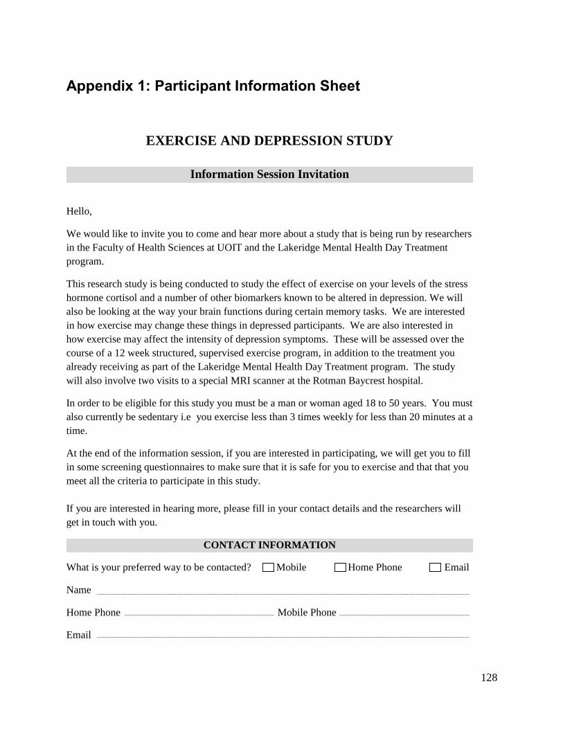
128
Appendix 1: Participant Information Sheet
EXERCISE AND DEPRESSION STUDY
Information Session Invitation
Hello,
We would like to invite you to come and hear more about a study that is being run by researchers
in the Faculty of Health Sciences at UOIT and the Lakeridge Mental Health Day Treatment
program.
This research study is being conducted to study the effect of exercise on your levels of the stress
hormone cortisol and a number of other biomarkers known to be altered in depression. We will
also be looking at the way your brain functions during certain memory tasks. We are interested
in how exercise may change these things in depressed participants. We are also interested in
how exercise may affect the intensity of depression symptoms. These will be assessed over the
course of a 12 week structured, supervised exercise program, in addition to the treatment you
already receiving as part of the Lakeridge Mental Health Day Treatment program. The study
will also involve two visits to a special MRI scanner at the Rotman Baycrest hospital.
In order to be eligible for this study you must be a man or woman aged 18 to 50 years. You must
also currently be sedentary i.e you exercise less than 3 times weekly for less than 20 minutes at a
time.
At the end of the information session, if you are interested in participating, we will get you to fill
in some screening questionnaires to make sure that it is safe for you to exercise and that that you
meet all the criteria to participate in this study.
If you are interested in hearing more, please fill in your contact details and the researchers will
get in touch with you.
CONTACT INFORMATION
What is your preferred way to be contacted? Mobile Home Phone Email
Name
Home Phone Mobile Phone

129
Appendix 2: Consent Form
Professor Bernadette Murphy
University of Ontario Institute of Technology
Faculty of Health Sciences
2000 Simcoe St. North
Oshawa, Ontario
CANADA L0B 1J0
Email: [email protected]
Phone: (905) 721-8668 Fax: (905) 721-3179
The role of exercise in improving brain function: Part A: Exercise MHDTP group You are invited to participate in a research study entitled “The role of exercise in improving brain function”. This study (# REB 10-104) has been reviewed by the University of Ontario Research Ethics Board and has been approved as of July 6, 2011. Please read this form carefully, and feel free to ask any questions you might have. If you have any questions about your rights as a participant in this study, please contact the Compliance Officer at 905 721 8668 ext 3693 or compliance.uoit.ca.
Researchers
Dr. Bernadette Murphy, Professor, Head-Kinesiology Specialization, Faculty of Health Sciences, University of Ontario Institute of Technology, Phone: (905) 721-8668 ext 2778, email: [email protected], Fax: (905) 721-3179
Dr. Paul Yielder, Professor, Director-Bachelor of Health Sciences, Faculty of Health Sciences, University of Ontario Institute of Technology, Phone: (905) 721-8668 ext 2768, email: [email protected], Fax: (905) 721-3179
Dr. Nancy Wilkinson, Client Care Manager/Psychologist, Mental Health and Pinewood Program, Lakeridge Health, Phone: (905) 576-8711 ext 6233, email: [email protected]
Dr. Stephen Strother, Professor, Dept. of Medical Biophysics, University of Toronto, Senior Scientist, Rotman Research Institute, Baycrest, Phone: (416) 785-2500 ext 2956, email: [email protected]

130
Dr. Ron Heslegrave, Chair, Research Ethics Board, Baycrest, Phone: (416) 785-2500 ext. 2440, email: [email protected]
Joanne Gourgouvelis, Masters Student, Faculty of Health Sciences, University of Ontario Institute of Technology, Phone: (905) 550-4055, email: [email protected], Fax: (905) 721-3179
Purpose of the Study This research study is being conducted to study the effect of exercise on your levels of the stress hormone cortisol and a number of other biomarkers known to be altered in depression. We will also be looking at the way your brain functions during certain memory tasks. We are interested in how exercise may change these things in depressed participants. We are also interested in how exercise may affect the intensity of depression symptoms. These will be assessed over the course of a 12 week structured, supervised exercise programme, in addition to the treatment you already receiving as part of the Lakeridge Mental Health Day Treatment Programme. Once this phase of the study is complete we will be recruiting a group of patients in the Mental Health Day Treatment Programme who are of a similar age and gender to investigate the effects of the Day Treatment Programme on its own in order to compare to the results of the exercise group. Potential Benefits to Participants and/or to Society Exercise has been shown in past studies to be an effective treatment for depression. In this study we want to learn more about how and why it works in order to help us design the most effective types of exercise interventions. Following the exercise programme, participants may receive additional relief from their depression. This study offers a structured exercise programme suitable for beginners and may also help you resolve any physical issues you have. If we show a strong effect, it may become a future part of the Day Treatment Programme and you will be making an important contribution to improving treatment options for future patients. Participation and Withdrawal Your participation to take part in this study is voluntary. You may choose not to participate or you may withdraw from the study at any time. However, if you decide to stop participating in the study, we encourage you to talk to the study staff first. The staff here is very flexible about meeting your needs and if there is anything we can do to accommodate your needs, we are more than willing to help. You are still free to withdraw at any time without giving a reason, and your decision not to participate will not affect your care at this center. Rights of Research Participants You are free to ask any questions that you may have about your treatment and your rights as a research participant. You may withdraw your consent at any time and discontinue participation without penalty.
131
If any questions come up during or after the study or if you have a research-related injury, contact the study researchers listed on the first page. Should you have any questions or concerns regarding your rights as a participant in this research study, or if you wish to speak with someone who is not related to this study, you may contact the Chair of the Lakeridge Health Corporation Research Ethics Board at (905) 576-8711 or UOIT Research Ethics Board through the Compliance Office (905) 721-8668 ext 6393. You are one of 20 individuals being asked to participate in this study and have been referred to us by Lakeridge Mental Health Day Treatment programme with a diagnosis of depression and you are between the ages of 18 and 50. Please take your time to review this consent form and discuss any questions you may have with the study staff. You may take your time to make your decision about participating in this study and you may discuss it with your friends, family or your doctor before you make your decision. This consent form may contain words that you do not understand. Please ask the study staff to explain any words or information that you do not clearly understand. Eligibility In order to be eligible for either group, you must not be currently doing regular exercise; that means your levels of exercise should be less than 20 minutes, three times per week. People suffering from mental conditions other than depression, or with physical conditions that would prevent them from doing exercise, such as heart disease, will be excluded from the study. An understanding of English sufficient to answer questionnaires is required.
If you have questions the stated procedures, timeline or exclusion criteria please feel free to discuss them with the study coordinator, Joanne Gourgouvelis or the investigators (contact details provided on page 1). The following sections will outline the various tests and procedures of the study, including: 1) the purpose of each test or procedure; 2) the respective timelines; 3) exclusion criteria (factors that might make you ineligible to participate); If you take part in this study, you will need to participate in several procedures. Please read the following pages and tick the boxes to indicate if any of the exclusion criteria pertain to you: 1) Questionnaire completion (page 3), 2) Saliva samples (page 3), 3) Bloodwork (see page 4), 4) Brainscan (see page 5) 5) Exercise (see page 6) You will also be loaned a special activity monitor to wear for the duration of the study in order to monitor your background activity levels. The monitor straps on to your waist band and the research assistant will show you how to use it.
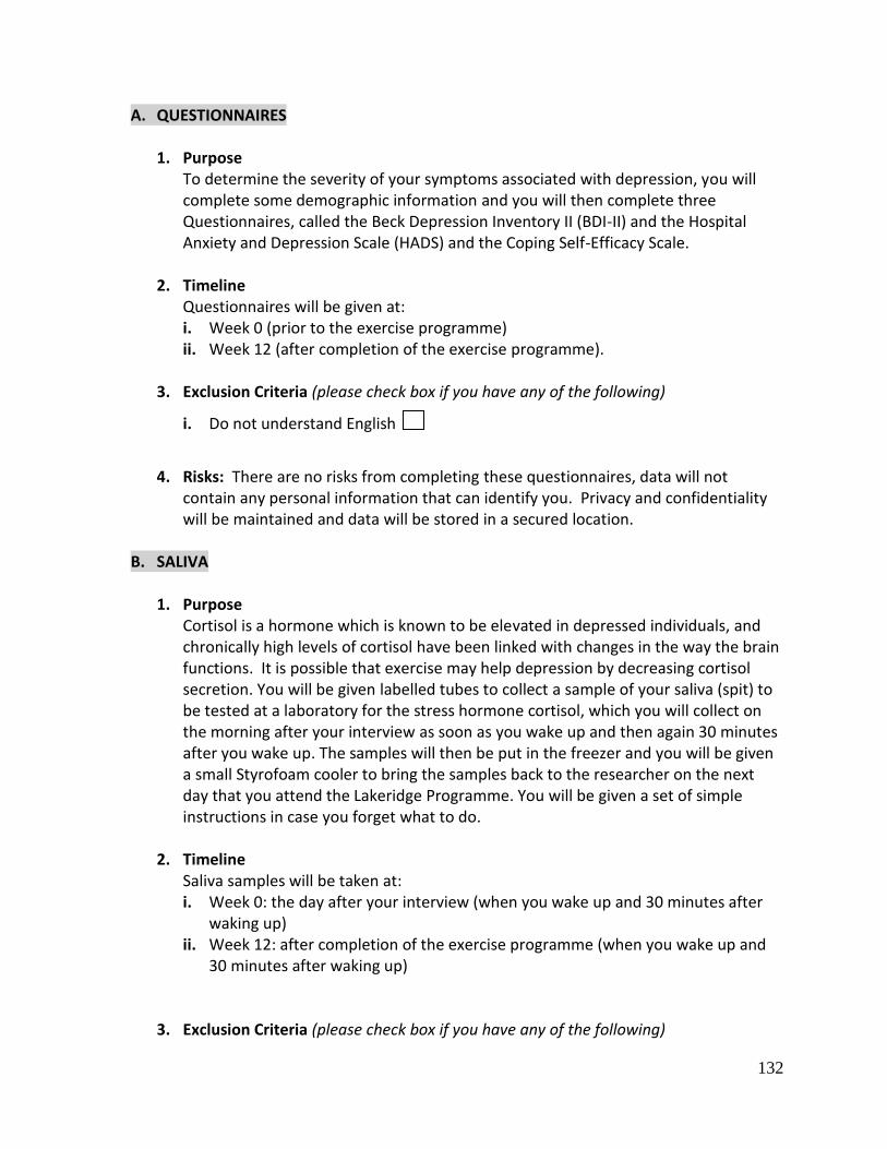
132
A. QUESTIONNAIRES
1. Purpose To determine the severity of your symptoms associated with depression, you will complete some demographic information and you will then complete three Questionnaires, called the Beck Depression Inventory II (BDI-II) and the Hospital Anxiety and Depression Scale (HADS) and the Coping Self-Efficacy Scale.
2. Timeline Questionnaires will be given at: i. Week 0 (prior to the exercise programme) ii. Week 12 (after completion of the exercise programme).
3. Exclusion Criteria (please check box if you have any of the following)
i. Do not understand English
4. Risks: There are no risks from completing these questionnaires, data will not
contain any personal information that can identify you. Privacy and confidentiality will be maintained and data will be stored in a secured location.
B. SALIVA
1. Purpose
Cortisol is a hormone which is known to be elevated in depressed individuals, and chronically high levels of cortisol have been linked with changes in the way the brain functions. It is possible that exercise may help depression by decreasing cortisol secretion. You will be given labelled tubes to collect a sample of your saliva (spit) to be tested at a laboratory for the stress hormone cortisol, which you will collect on the morning after your interview as soon as you wake up and then again 30 minutes after you wake up. The samples will then be put in the freezer and you will be given a small Styrofoam cooler to bring the samples back to the researcher on the next day that you attend the Lakeridge Programme. You will be given a set of simple instructions in case you forget what to do.
2. Timeline Saliva samples will be taken at: i. Week 0: the day after your interview (when you wake up and 30 minutes after
waking up) ii. Week 12: after completion of the exercise programme (when you wake up and
30 minutes after waking up)
3. Exclusion Criteria (please check box if you have any of the following)
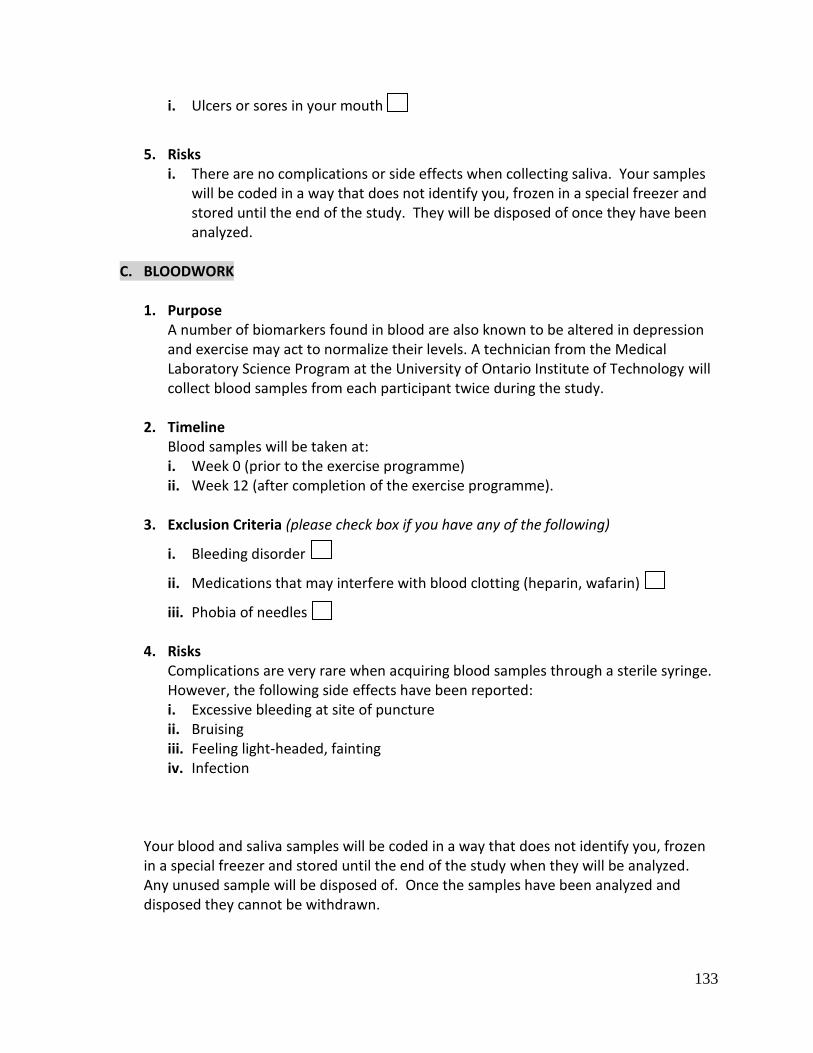
133
i. Ulcers or sores in your mouth
5. Risks
i. There are no complications or side effects when collecting saliva. Your samples will be coded in a way that does not identify you, frozen in a special freezer and stored until the end of the study. They will be disposed of once they have been analyzed.
C. BLOODWORK
1. Purpose A number of biomarkers found in blood are also known to be altered in depression and exercise may act to normalize their levels. A technician from the Medical Laboratory Science Program at the University of Ontario Institute of Technology will collect blood samples from each participant twice during the study.
2. Timeline Blood samples will be taken at: i. Week 0 (prior to the exercise programme) ii. Week 12 (after completion of the exercise programme).
3. Exclusion Criteria (please check box if you have any of the following)
i. Bleeding disorder
ii. Medications that may interfere with blood clotting (heparin, wafarin)
iii. Phobia of needles
4. Risks Complications are very rare when acquiring blood samples through a sterile syringe. However, the following side effects have been reported: i. Excessive bleeding at site of puncture ii. Bruising iii. Feeling light-headed, fainting iv. Infection
Your blood and saliva samples will be coded in a way that does not identify you, frozen in a special freezer and stored until the end of the study when they will be analyzed. Any unused sample will be disposed of. Once the samples have been analyzed and disposed they cannot be withdrawn.

134
D. BRAIN SCAN (functional MRI)
1. Purpose
Prior to commencing your programme, fMRI images of your brain will be obtained to investigate the way that your brain activates when you are performing a task requiring your concentration. We will also look at anatomical details of brain structures known to be involved in depression such as the hippocampus. Once you come out of the scanner, you will go to a separate room and be asked to go through some of the images from the task that you did in the scanner. The scan will take about one hour and will be carried out at Baycrest hospital attached to the Rotman Brain Research Institute. We will then retest you at the end of your programme (12 weeks) to see if exercise has changed the way your brain processes information. There will be no cost to you for the scans and you will be provided with parking and gasoline vouchers to cover travel costs for each session.
The MRI scan being done is designed to answer research questions, not to examine your brain medically. This MRI scan is not a substitute for one that a doctor would order, and it may not show problems that would be picked up by a medical MRI scan. However, in the unlikely event that we note an atypical finding on your MRI scan, we will contact you to help you arrange medical follow-up to interpret the significance of the findings, if any. We may also ask a radiologist, or other health professional, to look at your scan, and by signing this consent form you agree to releasing the scan for review. It is possible that you could be unnecessarily worried if a problem were suspected, but not actually found.
The MRI technique uses magnets and radiowaves to construct a picture of the brain on a computer. Before the scan begins, you will be asked to remove any magnetic metals that you may be wearing. For the procedure, you will be asked to lie on a padded bed that will be moved into a tunnel-like machine for the MRI scan of your brain. Since you will be inside the machine during the scan, and a screen will be in place for viewing the visual images, you may not be able to see the technicians operating the machine or the investigators. However, there is an intercom system that will allow you to talk with them at any time. If you feel uncomfortable during the scan and you wish to discontinue the procedure, you will be taken out of the machine at your request. We will obtain a series of MRI scans, separated by short breaks, and the entire procedure will take approximately one hour. During the scans we will ask you to carry out a variety of tasks. You should try to remain as still as possible during each scan. Movement will not be dangerous to you in any way, but would blur the picture of your brain. You will hear moderately loud knocking or beeping during the scan when the MRI machine is in operation. Although you may find this to be unsettling, the machine cannot hurt you in any way.
135
Please note: Prior to your first scan you will also be required to sign an additional consent form required by Baycrest hospital which provides similar information about fMRI as this consent form.
2. Timeline Brain scans will be taken at: i. Week 0 (prior to the exercise programme) ii. Week 12 (after completion of the exercise programme).
3. Exclusion Criteria (please check box if you have any of the following)
i. Phobia of small confined spaces
i. Metal Implants, pieces of shrapnel, aneurysm clips, or wires in your head. etc.
ii. Implanted Pace Maker
iii. Pregnant
iv. Inability to get to the scanner at Rotman Baycrest*. *We will compensate you for your parking and gasoline costs but you will need to be able to drive yourself or have someone else able to drive you in order to participate.
4. Risks and Discomforts The MRI scan is not associated with any known risks to your health and there is no evidence that there will be either short-term or long-term side effects. Participants undergoing fMRI may feel uncomfortable or claustrophobic in the confined space. Furthermore, the machine makes a loud noise which some people may find distressing. There will be someone with you at all times during the brain imaging, in case you should experience any adverse effects.
E. EXERCISE PROGRAMME
1. Description
You will complete an exercise safety screening questionnaire called the Par-Q. If there are any concerns about the safety of your participation, you will have the option of withdrawing or getting your family doctor to complete the ParMed-X to ensure it safe for you to exercise.
Once enrolled and baseline assessments have been carried out you will commence a 12 week exercise programme as part of the Mental Health Treatment Programme. The exercise sessions will take place at the Durham Family YMCA, located just down the road from the Lakeridge Health Day Programme. All sessions will be in groups of
136
2 to 5 participants and will be supervised by the Exercise Supervisor for three sessions per week and you must be prepared to commit to attending at least 80% of the exercise sessions. The role of the supervisor will be to give you verbal encouragement, monitor you for any possible adverse effects and provide aid as needed, and to help you monitor your heart rate to ensure they you are working at the correct intensity. The exercise prescription will be individualized based on your starting fitness levels. One session per week will be aerobic exercise only, and the remaining two will be resistance exercise followed by a shorter aerobic bout. Based on your experience and ability, one resistance exercise will be selected from each of six categories – horizontal push, anterior lower body, vertical pull, posterior lower body, horizontal pull, and vertical push in order to establish a balanced, whole-body exercise prescription. Exercises will be changed after four weeks, to avoid adaptation. The aerobic bout following resistance sessions will be 20 minutes in the first week, and 30 minutes in subsequent weeks. Sessions containing a resistance component will be performed at least 24 hours apart. All sessions begin with a ten-minute aerobic warm-up, and are supervised by a kinesiologist. Participants are advised to bring a drink bottle and keep well hydrated. Opportunities will be provided to make up missed sessions. In order to provide a transition out of the exercise programme, in weeks 9 and 12, participants will have one supervised session per week.
2. Timeframe i. 3 one hour sessions per week for 12 weeks
3. Exclusion Criteria (please check box if you have any of the following)
i. Already exercise more than 20 minutes, three times per week
ii. Physical conditions such as heart disease, or exclusions identified based on ParQ
or ParMedX questionnaires.
iii. I am unable to attend at least 80% of the exercise sessions
4. Risks and Discomforts
You may experience some physical discomfort both during exercise or stretching and following exercise or stretching, such as muscle soreness. There is a small risk of injury occurring during any exercise programme; however, the kinesiologists involved are trained in modifying exercise programmes to minimize this risk and you will be supervised at all times. There will be someone with you at all times during the exercise sessions, in case you should experience any adverse effects.
RESULTS You will also be given a log to keep track of each procedure required from you. This log is to be completed and given to the researcher at the end of the study (see attached).
137
Once the information has been collected from all participants, the questionnaire responses will be examined to establish the effect of exercise on depression, cortisol secretion, several biomarkers and coping self-efficacy, and fMRI results will be analyzed and a report will be written up. A short version of this report will be available to participants who would like a copy. You can indicate at the bottom of the consent form whether you would like to receive your report by email or by regular mail. If you have depression and you experience any worsening of your depression during your participation in the study, you will be referred immediately to the treatment team at Lakeridge Mental Health. If you are concerned about any aspect of your participation, do not hesitate to contact the researchers at the numbers provided. Compensation for Participation There will be no direct compensation for participating in this research study, although you will be given a free supervised exercise programme and you will be compensated for parking and gasoline costs. Disclosure Despite efforts to keep your personal information confidential, absolute confidentiality cannot be guaranteed. Your personal information may be disclosed if required by law. This legal obligation includes a number of circumstances, such as suspected child abuse and infectious disease, elevated risk or harm to you or others, and a court order by authorized agencies. For your information, the research consent form will be inserted in the patient health record. Medical records that contain your identity will be treated as confidential in accordance with Ontario’s privacy legislation, the Personal Health Information Protection Act, 2004. The only people who will be accessing your patient records are the Lakeridge clinicians involved in your treatment. The other information collected by the researchers is separate and will be seen only by the researchers and all identifying information will be removed in any publications. The Lakeridge Health Corporation Research Ethics Board and the University of Ontario Institute of Technology Research Ethics Board may review records related to the study for quality assurance purposes, as it oversees the conduct of this study at Lakeridge Health.
Please read the following before signing the consent form and remember to keep a copy for your own records.
By signing this form, I agree that: • The study has been explained to me. All my questions were answered to my satisfaction.
138
• The possible harms and discomforts and the possible benefits (if any) of this study have been explained to me.
• I know about the alternatives to taking part in this study. I understand that I have the right not to participate and the right to stop at any time. The decision about whether or not to participate will not affect my health care at Lakeridge Health.
• I am free now, and in the future, to ask any questions about the study.
• I have been told that my medical records will be kept confidential, except where release of information is required by law
• I understand that no information that would identify me, will be released without asking me first, unless the disclosure is required by law
• I hereby consent to participate.
I have completed the individual procedure assessments on pages 3 to 8 of this form and hereby consent to participate in the following procedures (please tick the box): 1) Questionnaire completion
2) Blood samples
3) Saliva samples
4) fMRI
5) Exercise
Dissemination Information gathered in this research study may be published or presented in public forums; however your name and other identifying information will not be used or revealed. We hope the information learned from this study will benefit other people and support the use of exercise as a treatment for depression in future patients. You will be provided with a summary of findings at the end of the study, if you so desire. We will do our best to explain the findings to you in way that you understand and what these changes mean. Often in research we don’t get the findings we expect. This is often because we didn’t study enough participants or the changes are of a different nature then we expected. This is okay and still makes a very important contribution to science and to our understanding of depression. If you are concerned about any of the study findings or you would like more information, please contact Dr. Bernadette Murphy or Dr. Paul Yielder to discuss. Please advise us of your preferable format for communication (check one and provide details in the space provided):
139
Please indicated by ticking the box whether you wish to receive your summary Please provide either the land or email address you would like it sent to:
________________________________________________________________________________________________________________________________________________________________________________________________________________________ Participant Signature_____________________________ Date______________________ Participant Printed Name_________________________________ I, the undersigned, have fully explained the relevant details of this research study to the participant named above and believe that the participant has understood and has knowingly given their consent. ____________________________________________ ________________________ Printed Name Date ____________________________________________ Signature ____________________________________________ Role in the Study (only authorized / qualified member of the research team)

140
Appendix 3: PAR-Q & You

141
Appendix 4: Baycrest screening form
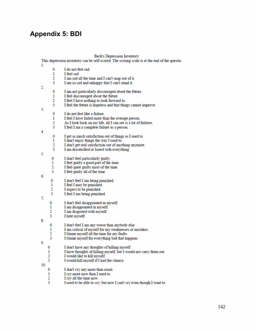
142
Appendix 5: BDI
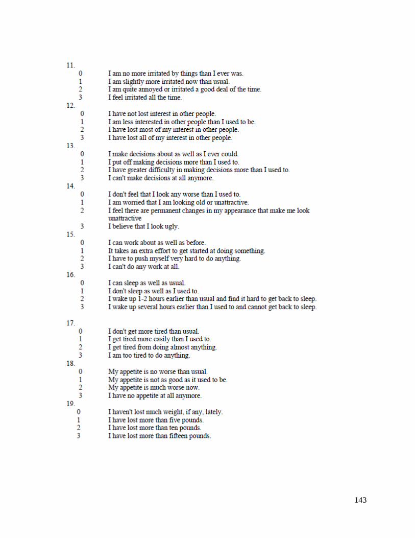
143
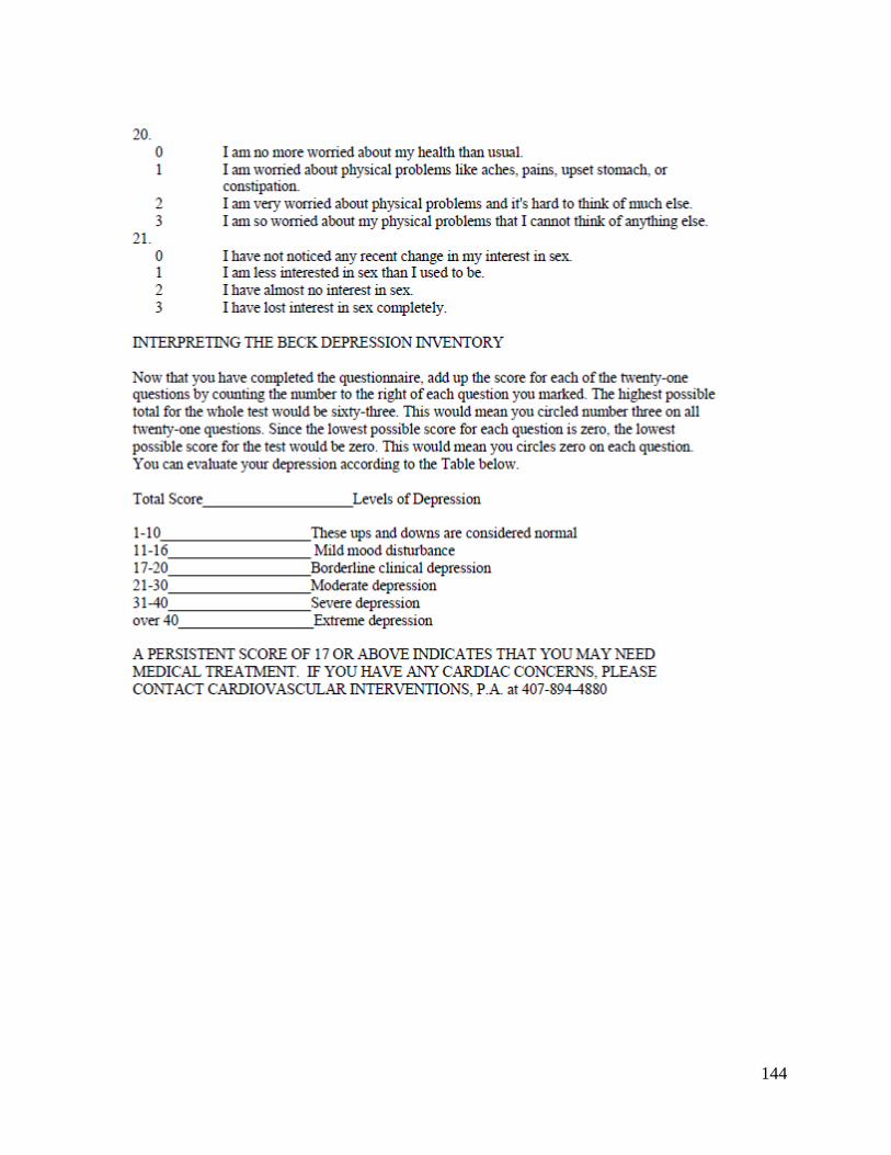
144
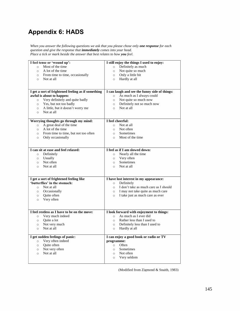
145
Appendix 6: HADS
When you answer the following questions we ask that you please chose only one response for each
question and give the response that immediately comes into your head.
Place a tick or mark beside the answer that best relates to how you feel.
I feel tense or ‘wound up’:
o Most of the time
o A lot of the time
o From time to time, occasionally
o Not at all
I still enjoy the things I used to enjoy:
o Definitely as much
o Not quite so much
o Only a little bit
o Hardly at all
I get a sort of frightened feeling as if something
awful is about to happen:
o Very definitely and quite badly
o Yes, but not too badly
o A little, but it doesn‟t worry me
o Not at all
I can laugh and see the funny side of things:
o As much as I always could
o Not quite so much now
o Definitely not so much now
o Not at all
Worrying thoughts go through my mind:
o A great deal of the time
o A lot of the time
o From time to time, but not too often
o Only occasionally
I feel cheerful:
o Not at all
o Not often
o Sometimes
o Most of the time
I can sit at ease and feel relaxed:
o Definitely
o Usually
o Not often
o Not at all
I feel as if I am slowed down:
o Nearly all the time
o Very often
o Sometimes
o Not at all
I get a sort of frightened feeling like
‘butterflies’ in the stomach:
o Not at all
o Occasionally
o Quite often
o Very often
I have lost interest in my appearance:
o Definitely
o I don‟t take as much care as I should
o I may not take quite as much care
o I take just as much care as ever
I feel restless as I have to be on the move:
o Very much indeed
o Quite a lot
o Not very much
o Not at all
I look forward with enjoyment to things:
o As much as I ever did
o Rather less than I used to
o Definitely less than I used to
o Hardly at all
I get sudden feelings of panic:
o Very often indeed
o Quite often
o Not very often
o Not at all
I can enjoy a good book or radio or TV
programme:
o Often
o Sometimes
o Not often
o Very seldom
(Modified from Zigmond & Snaith, 1983)

146
Appendix 9: MoCA

147

148
Appendix 7: Pre-screening form
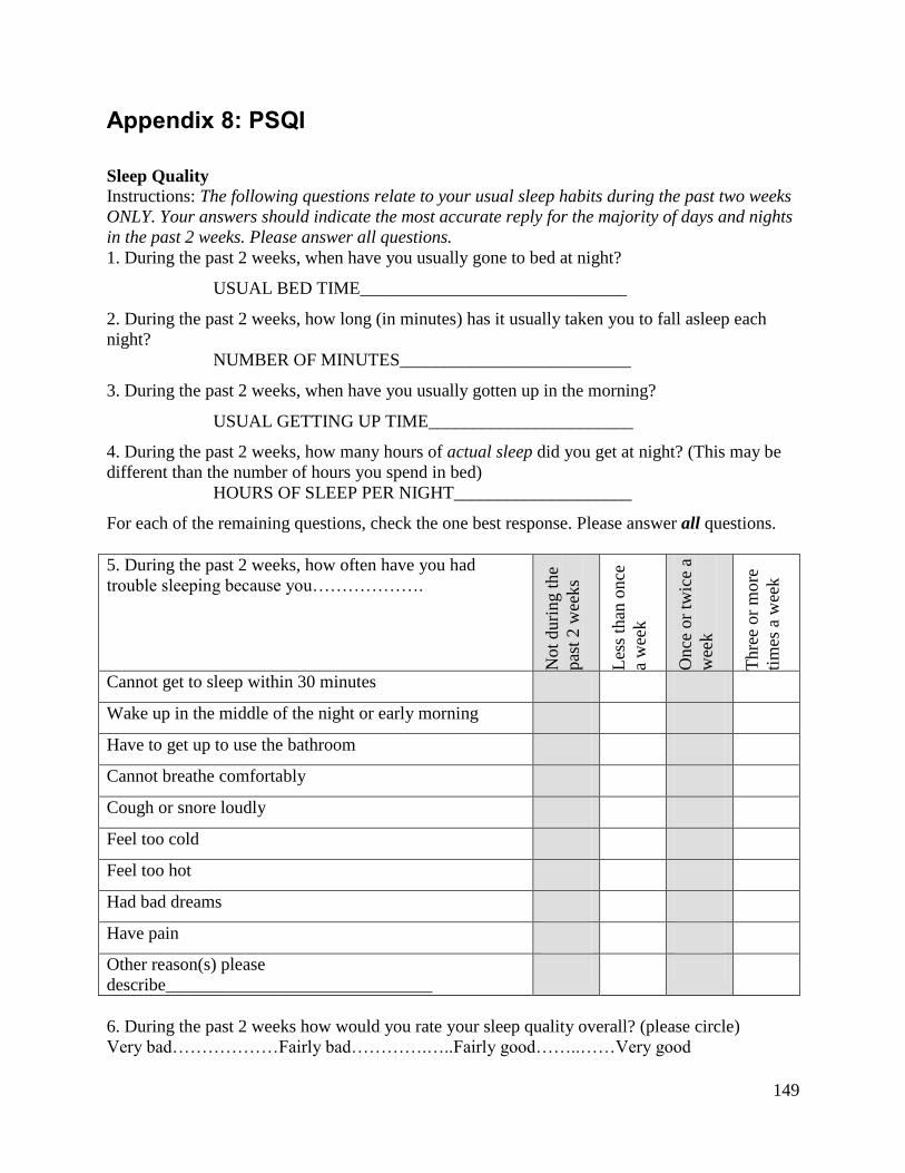
149
Appendix 8: PSQI
Sleep Quality
Instructions: The following questions relate to your usual sleep habits during the past two weeks
ONLY. Your answers should indicate the most accurate reply for the majority of days and nights
in the past 2 weeks. Please answer all questions.
1. During the past 2 weeks, when have you usually gone to bed at night?
USUAL BED TIME______________________________
2. During the past 2 weeks, how long (in minutes) has it usually taken you to fall asleep each
night?
NUMBER OF MINUTES__________________________
3. During the past 2 weeks, when have you usually gotten up in the morning?
USUAL GETTING UP TIME_______________________
4. During the past 2 weeks, how many hours of actual sleep did you get at night? (This may be
different than the number of hours you spend in bed)
HOURS OF SLEEP PER NIGHT____________________
For each of the remaining questions, check the one best response. Please answer all questions.
5. During the past 2 weeks, how often have you had
trouble sleeping because you……………….
Not
duri
ng t
he
pas
t 2 w
eeks
Les
s th
an o
nce
a w
eek
Once
or
twic
e a
wee
k
Thre
e or
mo
re
tim
es a
wee
k
Cannot get to sleep within 30 minutes
Wake up in the middle of the night or early morning
Have to get up to use the bathroom
Cannot breathe comfortably
Cough or snore loudly
Feel too cold
Feel too hot
Had bad dreams
Have pain
Other reason(s) please
describe______________________________
6. During the past 2 weeks how would you rate your sleep quality overall? (please circle)
Very bad………………Fairly bad………….…..Fairly good……..……Very good

150
7. During the past 2 weeks, how often have you taken medicine (prescribed or “over the
counter”) to help you sleep? (Please circle)
Not during the Less than Once or Three or more
past 2 weeks once a week twice a week times a week
8. During the past 2 weeks, how often have you had trouble staying awake while driving, eating
meals, or engaging in social activity? (Please circle)
Not during the Less than Once or Three or more
past 2 weeks once a week twice a week times a week
9. During the past 2 weeks, how much of a problem has it been for you to keep up enough
enthusiasm to get things done? (Please circle)
No problem Only a very Somewhat A very
at all slight problem of a problem big problem
10. Do you have a bed partner or share a room?
No bed partner or do not share a room ______________
Partner/flat mate in other room ______________
Partner in the same room, but not same bed ______________
Partner in the same bed ______________
11. If you have a bed partner or share a room, ask him/her how often in the past 2 weeks you
have had….
Not
duri
ng t
he
pas
t 2 w
eeks
Les
s th
an o
nce
a w
eek
Once
or
twic
e a
wee
k
Thre
e or
mo
re
tim
es a
wee
k
Loud snoring
Long pauses between breaths while asleep
Legs twitching or jerking while you sleep
Episodes of disorientation or confusion during sleep
Other restlessness while you sleep (please describe)
(Modified from (Buysse et al. 1989)
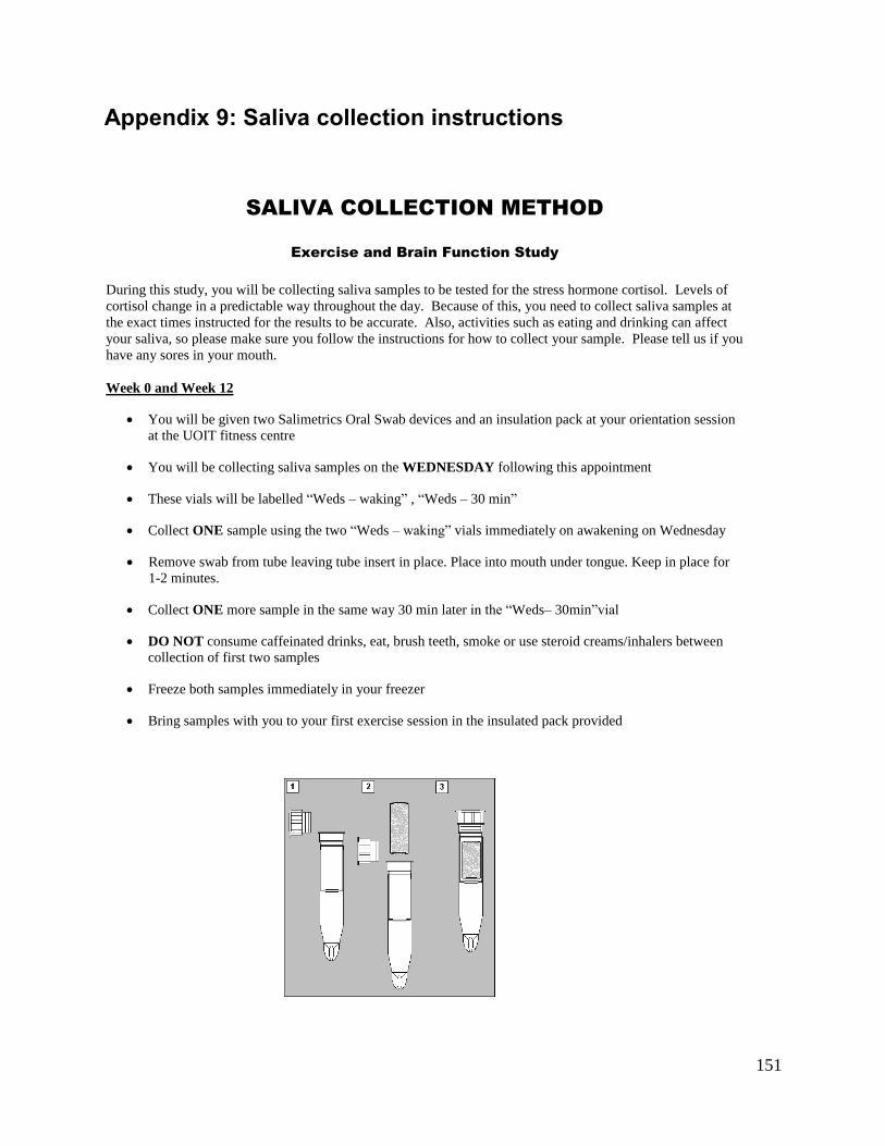
151
Appendix 9: Saliva collection instructions
SALIVA COLLECTION METHOD
Exercise and Brain Function Study
During this study, you will be collecting saliva samples to be tested for the stress hormone cortisol. Levels of
cortisol change in a predictable way throughout the day. Because of this, you need to collect saliva samples at
the exact times instructed for the results to be accurate. Also, activities such as eating and drinking can affect
your saliva, so please make sure you follow the instructions for how to collect your sample. Please tell us if you
have any sores in your mouth.
Week 0 and Week 12
You will be given two Salimetrics Oral Swab devices and an insulation pack at your orientation session
at the UOIT fitness centre
You will be collecting saliva samples on the WEDNESDAY following this appointment
These vials will be labelled “Weds – waking” , “Weds – 30 min”
Collect ONE sample using the two “Weds – waking” vials immediately on awakening on Wednesday
Remove swab from tube leaving tube insert in place. Place into mouth under tongue. Keep in place for
1-2 minutes.
Collect ONE more sample in the same way 30 min later in the “Weds– 30min”vial
DO NOT consume caffeinated drinks, eat, brush teeth, smoke or use steroid creams/inhalers between
collection of first two samples
Freeze both samples immediately in your freezer
Bring samples with you to your first exercise session in the insulated pack provided
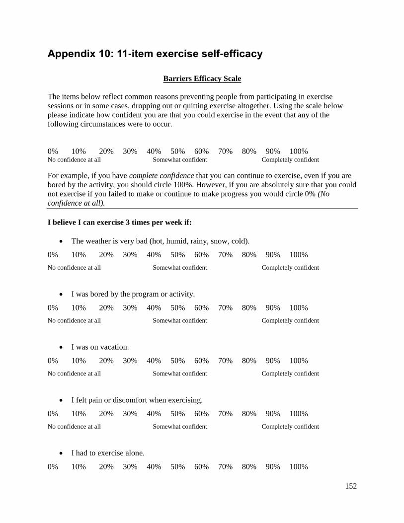
152
Appendix 10: 11-item exercise self-efficacy
Barriers Efficacy Scale
The items below reflect common reasons preventing people from participating in exercise
sessions or in some cases, dropping out or quitting exercise altogether. Using the scale below
please indicate how confident you are that you could exercise in the event that any of the
following circumstances were to occur.
0% 10% 20% 30% 40% 50% 60% 70% 80% 90% 100% No confidence at all Somewhat confident Completely confident
For example, if you have complete confidence that you can continue to exercise, even if you are
bored by the activity, you should circle 100%. However, if you are absolutely sure that you could
not exercise if you failed to make or continue to make progress you would circle 0% (No
confidence at all).
I believe I can exercise 3 times per week if:
The weather is very bad (hot, humid, rainy, snow, cold).
0% 10% 20% 30% 40% 50% 60% 70% 80% 90% 100%
No confidence at all Somewhat confident Completely confident
I was bored by the program or activity.
0% 10% 20% 30% 40% 50% 60% 70% 80% 90% 100%
No confidence at all Somewhat confident Completely confident
I was on vacation.
0% 10% 20% 30% 40% 50% 60% 70% 80% 90% 100%
No confidence at all Somewhat confident Completely confident
I felt pain or discomfort when exercising.
0% 10% 20% 30% 40% 50% 60% 70% 80% 90% 100%
No confidence at all Somewhat confident Completely confident
I had to exercise alone.
0% 10% 20% 30% 40% 50% 60% 70% 80% 90% 100%
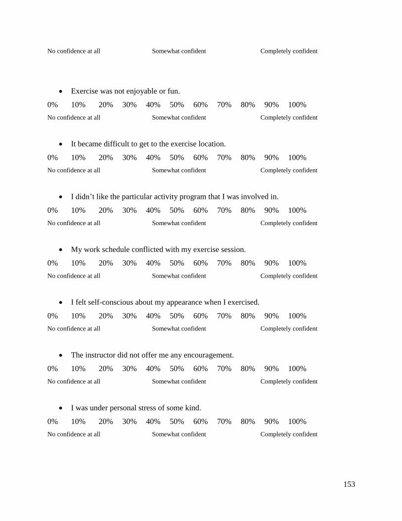
153
No confidence at all Somewhat confident Completely confident
Exercise was not enjoyable or fun.
0% 10% 20% 30% 40% 50% 60% 70% 80% 90% 100%
No confidence at all Somewhat confident Completely confident
It became difficult to get to the exercise location.
0% 10% 20% 30% 40% 50% 60% 70% 80% 90% 100%
No confidence at all Somewhat confident Completely confident
I didn‟t like the particular activity program that I was involved in.
0% 10% 20% 30% 40% 50% 60% 70% 80% 90% 100%
No confidence at all Somewhat confident Completely confident
My work schedule conflicted with my exercise session.
0% 10% 20% 30% 40% 50% 60% 70% 80% 90% 100%
No confidence at all Somewhat confident Completely confident
I felt self-conscious about my appearance when I exercised.
0% 10% 20% 30% 40% 50% 60% 70% 80% 90% 100%
No confidence at all Somewhat confident Completely confident
The instructor did not offer me any encouragement.
0% 10% 20% 30% 40% 50% 60% 70% 80% 90% 100%
No confidence at all Somewhat confident Completely confident
I was under personal stress of some kind.
0% 10% 20% 30% 40% 50% 60% 70% 80% 90% 100%
No confidence at all Somewhat confident Completely confident
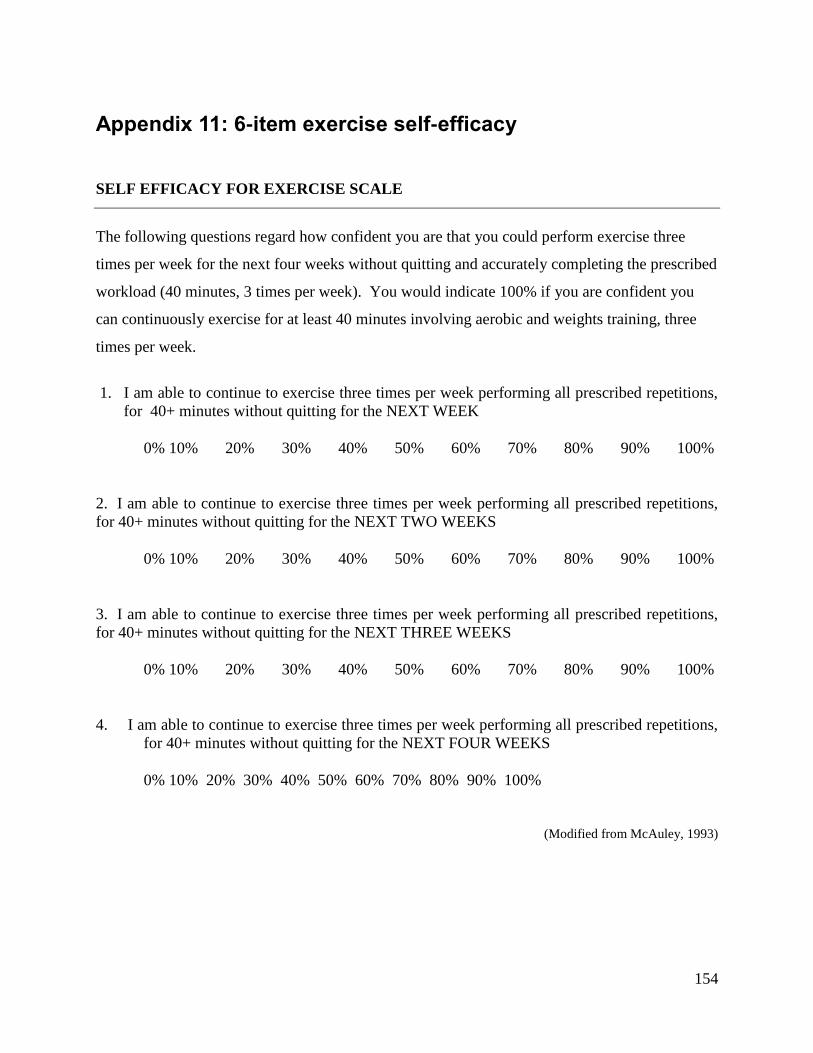
154
Appendix 11: 6-item exercise self-efficacy
SELF EFFICACY FOR EXERCISE SCALE
The following questions regard how confident you are that you could perform exercise three
times per week for the next four weeks without quitting and accurately completing the prescribed
workload (40 minutes, 3 times per week). You would indicate 100% if you are confident you
can continuously exercise for at least 40 minutes involving aerobic and weights training, three
times per week.
1. I am able to continue to exercise three times per week performing all prescribed repetitions,
for 40+ minutes without quitting for the NEXT WEEK
0% 10% 20% 30% 40% 50% 60% 70% 80% 90% 100%
2. I am able to continue to exercise three times per week performing all prescribed repetitions,
for 40+ minutes without quitting for the NEXT TWO WEEKS
0% 10% 20% 30% 40% 50% 60% 70% 80% 90% 100%
3. I am able to continue to exercise three times per week performing all prescribed repetitions,
for 40+ minutes without quitting for the NEXT THREE WEEKS
0% 10% 20% 30% 40% 50% 60% 70% 80% 90% 100%
4. I am able to continue to exercise three times per week performing all prescribed repetitions,
for 40+ minutes without quitting for the NEXT FOUR WEEKS
0% 10% 20% 30% 40% 50% 60% 70% 80% 90% 100%
(Modified from McAuley, 1993)
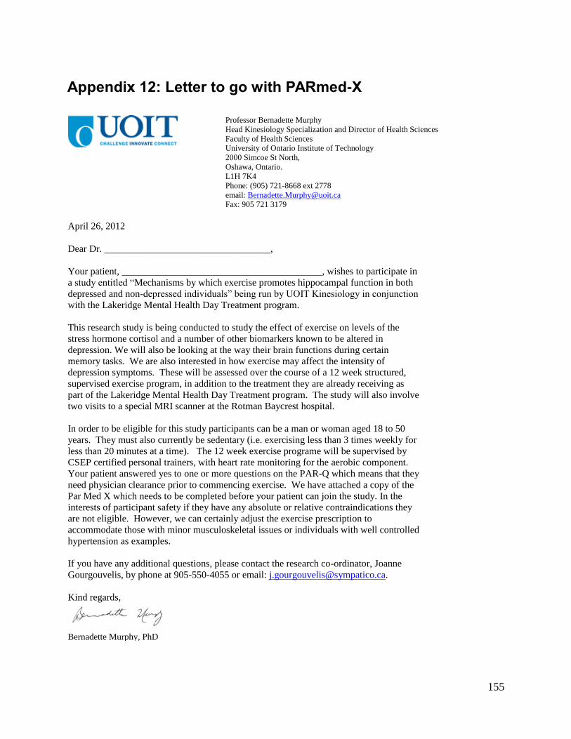
155
Appendix 12: Letter to go with PARmed-X
Professor Bernadette Murphy
Head Kinesiology Specialization and Director of Health Sciences
Faculty of Health Sciences
University of Ontario Institute of Technology
2000 Simcoe St North,
Oshawa, Ontario.
L1H 7K4
Phone: (905) 721-8668 ext 2778
email: [email protected]
Fax: 905 721 3179
April 26, 2012
Dear Dr. __________________________________,
Your patient, _________________________________________, wishes to participate in
a study entitled “Mechanisms by which exercise promotes hippocampal function in both
depressed and non-depressed individuals” being run by UOIT Kinesiology in conjunction
with the Lakeridge Mental Health Day Treatment program.
This research study is being conducted to study the effect of exercise on levels of the
stress hormone cortisol and a number of other biomarkers known to be altered in
depression. We will also be looking at the way their brain functions during certain
memory tasks. We are also interested in how exercise may affect the intensity of
depression symptoms. These will be assessed over the course of a 12 week structured,
supervised exercise program, in addition to the treatment they are already receiving as
part of the Lakeridge Mental Health Day Treatment program. The study will also involve
two visits to a special MRI scanner at the Rotman Baycrest hospital.
In order to be eligible for this study participants can be a man or woman aged 18 to 50
years. They must also currently be sedentary (i.e. exercising less than 3 times weekly for
less than 20 minutes at a time). The 12 week exercise programe will be supervised by
CSEP certified personal trainers, with heart rate monitoring for the aerobic component.
Your patient answered yes to one or more questions on the PAR-Q which means that they
need physician clearance prior to commencing exercise. We have attached a copy of the
Par Med X which needs to be completed before your patient can join the study. In the
interests of participant safety if they have any absolute or relative contraindications they
are not eligible. However, we can certainly adjust the exercise prescription to
accommodate those with minor musculoskeletal issues or individuals with well controlled
hypertension as examples.
If you have any additional questions, please contact the research co-ordinator, Joanne
Gourgouvelis, by phone at 905-550-4055 or email: [email protected].
Kind regards,
Bernadette Murphy, PhD
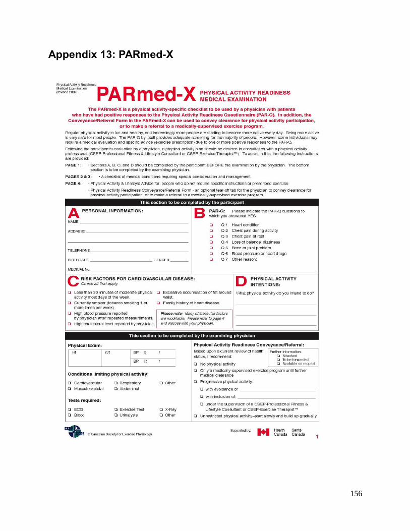
156
Appendix 13: PARmed-X
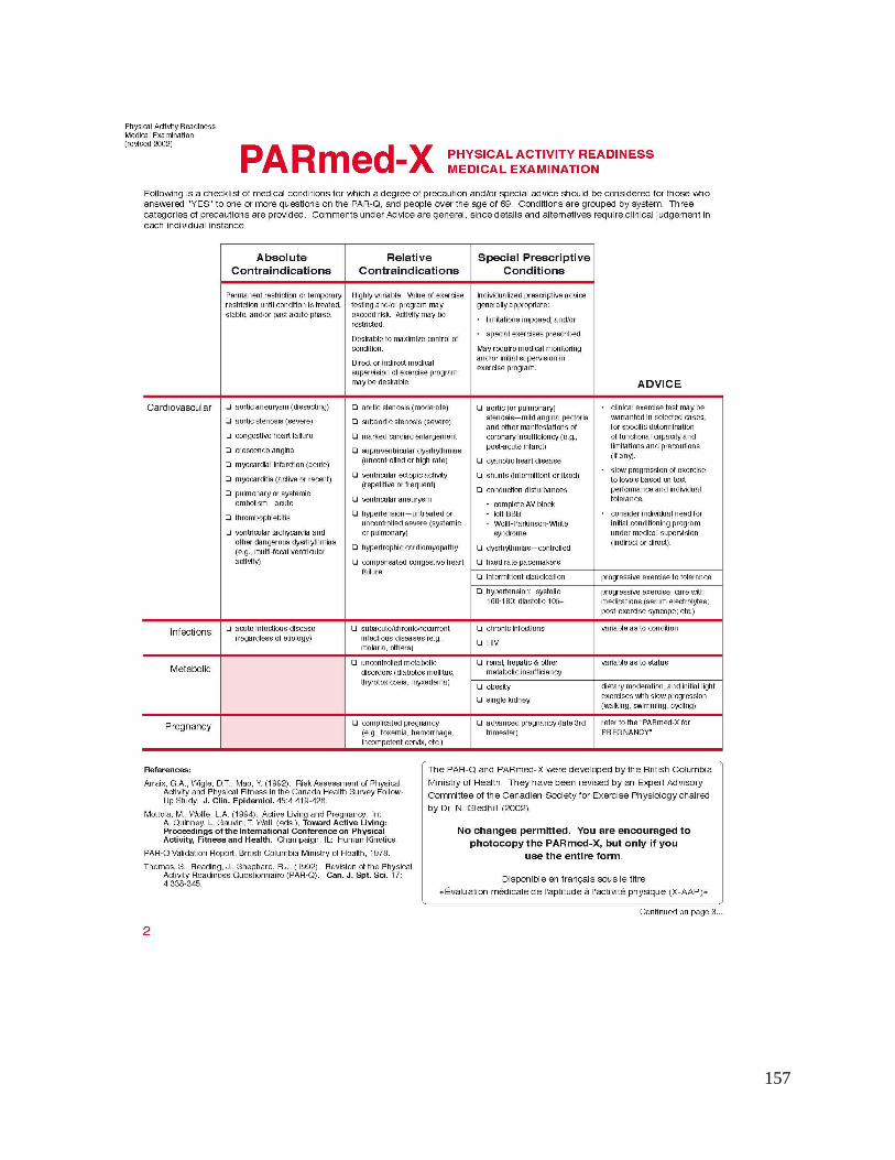
157
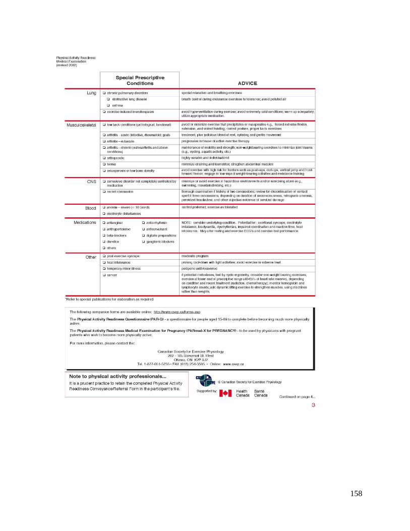
158
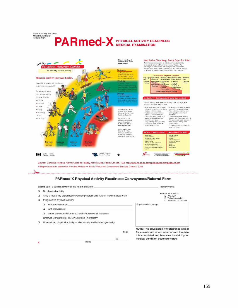
159

160
Appendix 14: Blood collection log
EXERCISE AND DEPRESSION STUDY
Participant Name:
Participant Number:
DATE TIME OF
COLLECTION NO. OF TUBES
TIME OF CENTRIFUGE
TIME PLACE IN FREEZER
EMOTIONAL STATUS
NOTES:

161
Appendix 15: Resistance exercise options
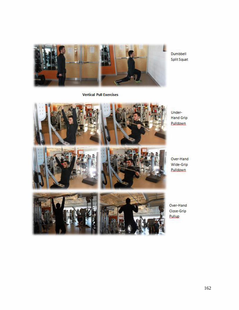
162
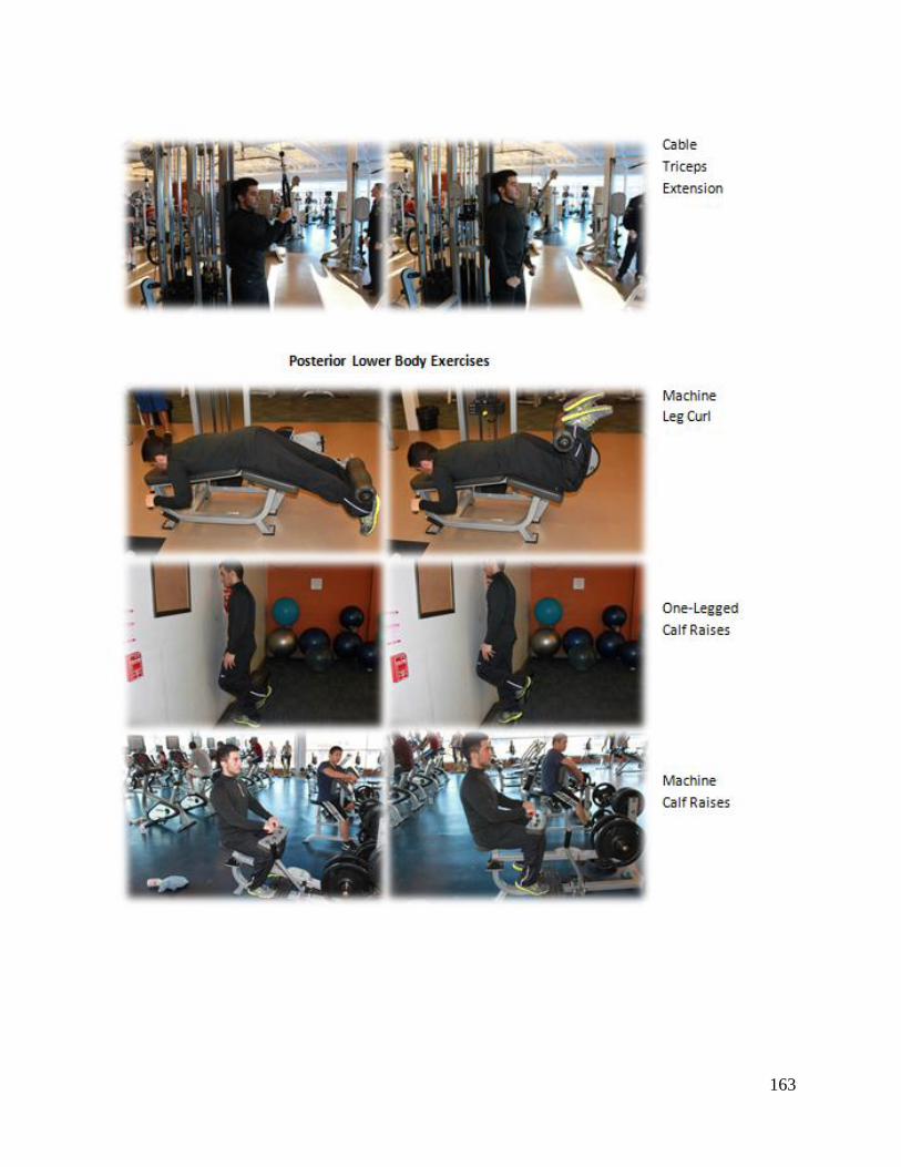
163
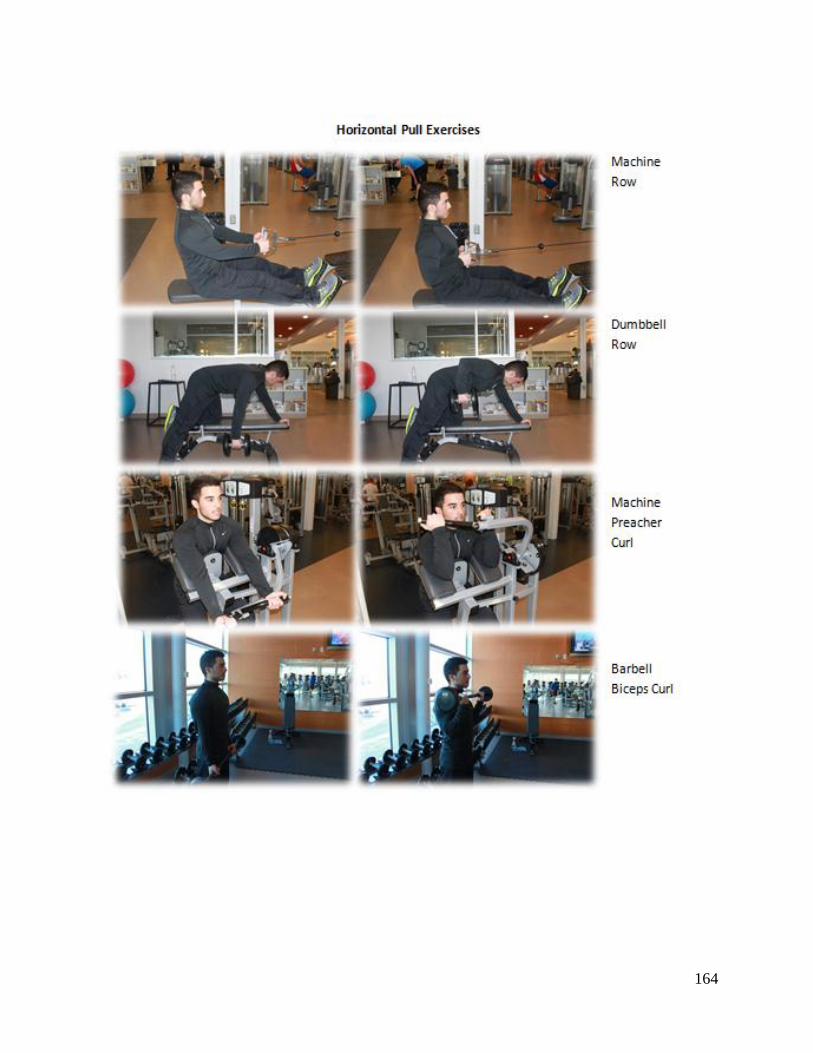
164
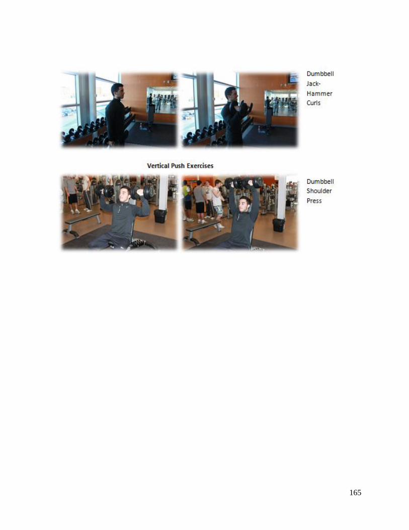
165