mechanisms by which hiv-1 nef disrupts the - deep blue
TRANSCRIPT
Mechanisms by which HIV-1 Nef disrupts the intracellular trafficking of host
proteins
by
Jolie A. Leonard
A dissertation submitted in partial fulfillment
Of the requirements for the degree of
Doctor of Philosophy
(Cellular and Molecular Biology)
In The University of Michigan
2011
Doctoral Committee:
Associate Professor Kathleen L. Collins, Chair
Professor Cheong-Hee Chang
Professor Robert S. Fuller
Associate Professor Kristen J. Verhey
Assistant Professor Akira Ono

Jolie A. Leonard
2011

ii
To Stephen

ii
Acknowledgements
This dissertation is the cumulative result of several years of scientific research. Though it
represents considerable effort on my part, I could never have completed such an
undertaking without an immense amount of help. First, I would like to thank the
members of my thesis committee for their invaluable guidance and technical suggestions
over the last five years. I am especially grateful to my mentor, Dr. Kathleen Collins, for
her knowledge, guidance, and support. She has taught me how to conduct scientific
research in a logical and thoughtful manner, and how to communicate data clearly. I
have appreciated Kathy’s positive attitude and her ability to glean some scrap of
information from any experiment, even in the face of seemingly disappointing results. I
would also like to acknowledge and thank all of the members of the Collins lab. In
addition to endlessly patient technical advice, listening to practice seminars, and editing
early drafts of papers, my coworkers have made the lab environment as enjoyable a place
to conduct science as any I can imagine. In particular I would like to thank the
postdoctoral fellows, Adewunmi Onafuwa-Nuga and Deanna Kulpa, for all of the
invaluable, informal mentorship I have received from them regarding science, career, and
life.
I am eternally grateful to my family, Hoffmans and Leonards alike, but especially to my
parents, Ross and Karen Hoffman. They have been my biggest source of support and
encouragement throughout my life, praying ceaselessly and pushing me to succeed. They
always believed that I was capable of doing exactly this, and, in large part because of
their belief, I am. Most of all, I truly could never have done this without the perpetual

iii
support of my husband, Steve. I am inadequate to the task of expressing how grateful I
am for everything he has done, from playing the roles of chauffeur, housekeeper, editor,
and secretary; to simply understanding the exhausting demands of research science. I am
truly blessed to have a companion and partner such as he.

iv
Preface
This thesis is the compilation of published and unpublished work on the effects of HIV-1
Nef on cellular trafficking pathways. Nef removes a number of host proteins from the
cell surface, interfering with antigen presentation and immune cell activation, resulting in
evasion of the host immune system. Nef directly promotes HIV-1 pathogenesis and is
therefore an appealing target for therapeutic intervention.
Chapter 1 is a discussion of the biological properties of HIV-1 Nef and Nef’s impact on
HIV-1 pathogenesis. We compare two competing models for the mechanism by which
Nef reduces the surface expression of MHC-I and discuss the current evidence for the
downmodulation of a variety of host factors by Nef.
In Chapter 2, we analyze a panel of putative cellular targets of Nef. We compare the
relative impact of Nef on expression of each of these targets and assess the contribution
of cellular trafficking machinery. This work has been published as Leonard JA, Filzen T,
Carter CC, Schaefer M, Collins KL. “HIV-1 Nef Disrupts Intracellular Trafficking of
Major Histocompatibility Complex Class I, CD4, CD8, and CD28 by Distinct Pathways
That Share Common Elements.” J Virol. (2011) Jul;85(14):6867-81.
In Chapter 3, we present data assessing the relative contribution of ARF-1 and ARF-6 in
Nef-induced downmodulation of MHC-I. Expression of a constitutively GTP-bound
ARF-1 mutant revealed a requirement for ARF-1 in Nef-dependant MHC-I trafficking.
v
Conversely, ARF-6 was found to be dispensable for the effects of Nef on MHC-I, as
demonstrated in experiments with ARF-6 mutants or ARF-6 depletion. These data have
been submitted for publication and are currently in revision.
In Chapter 4 we describe methods for a live cell flow-cytometric screen to identify
chemical inhibitors of Nef-mediated MHC-I downmodulation. We present preliminary
findings from a screen of approximately 100,000 small molecules, in which we identified
a small pool of Nef inhibitors for further development.
Finally, Chapter 5 is a discussion of impact of the data presented in the previous chapters,
as well as proposed future directions for additional mechanistic studies of Nef and for
developing small molecule inhibitors of Nef for clinical use.

vi
Table of Contents
Dedication ii
Acknowledgements iii
Preface iv
List of Figures xiii
Abstract xvi
Chapter
1. The effects of HIV-1 Nef on cellular proteins important for immune 1
response
1.1. Overview 1
1.2. Summary of the HIV pandemic 1
1.3. Natural history of the untreated disease 2
1.4. The virus 2
1.4.1. HIV-1 replication 4
1.4.2. Accessory genes 7
1.5. HIV-1 Nef 8
1.6. Nef disrupts antigen presentation to cytotoxic T lymphocytes 9
1.6.1. Natural killer cells 13
1.6.2. Functional domains required for Nef to downmodulate MHC-I 14
1.6.3. Candidate host factors that partner with Nef to downmodulate 17
MHC-I
1.6.3.1. Adaptor protein complexes 17
1.6.3.2. ADP-ribosylation factors 18
vii
1.6.3.3. -COP 21
1.6.4. Downmodulation of MHC-I: endocytic mechanism 21
1.6.5. Evidence against an endocytic mechanism 24
1.6.6. Downmodulation of MHC-I: evidence for targeting of 25
Newly synthesized proteins in the secretory pathway
1.6.6.1. Disruption of MHC-I transport 25
1.6.6.2. AP-1 is required for disruption of antigen presentation 29
by HIV Nef
1.6.6.2.1. Nef stabilizes an interaction between the AP-1 30
tyrosine binding pocket and the MHC-I cytoplasmic tail
1.6.6.2.2. Nef domains and AP-1-dependant MHC-I 31
trafficking
1.6.6.3. A role for -COP in disruption of antigen 32
presentation by Nef
1.6.7. Summary of MHC-I downmodulation 33
1.7. Nef downmodulates CD4 34
1.7.1. In vivo evidence for the importance of CD4 downmodulation 35
1.7.2. Functional domains required for Nef to downmodulate CD4 35
1.7.3. Nef downmodulates CD4 by an endocytic mechanism 39
1.7.4. Host factors that partner with Nef to induce CD4 39
downmodulation and degradation
1.7.4.1. AP-2 mediates CD4 downmodulation by Nef 39

viii
1.7.4.2. -COP is required for Nef-induced CD4 degradation 41
1.8. Nef is reported to disrupt trafficking of a variety of cellular proteins 41
1.8.1. Nef downmodulates CD28 42
1.8.2. Evidence for downmodulation of CD8 by Nef 43
1.8.3. Evidence for CD1d downmodulation by HIV-1 45
1.8.3.1. Reports of CD1d downmodulation by Nef 46
1.8.3.2. Evidence of CD1d downmodulation by HIV-1 Vpu 47
1.8.4. Evidence for downmodulation of CD80 and CD86 by HIV-1 48
Nef
1.8.4.1. Proposed mechanism for CD80/86 downmodulation 50
1.8.4.2. Functional importance of CD80/86 downmodulation 54
1.9. Summary 55
1.10. References 56
2. HIV-1 Nef disrupts intracellular trafficking of MHCV-1, CD4, CD8 72
and CD28 by distinct pathways that share common elements
2.1. Abstract 72
2.2. Introduction 74
2.3. Results 76
2.3.1. Nef targets HLA-A2, CD4, CD8 , and CD28 through their 76
cytoplasmic tail domains
2.3.2. Endogenous CD80 and CD86 are not downmodulated by Nef 81
in primary APCs
ix
2.3.3. Multiple HIV-1 Nef variants downmodulate HLA-A2, CD4, 85
CD8 , and CD28
2.3.4. HIV-1 Nef downmodulates CD4 and CD8 in primary 90
T lymphocytes
2.3.5. Nef recruits AP-1 to the cytoplasmic tail of CD8 and CD28 91
2.3.6. Nef recruits AP-1 to CD8 and CD28 through its dileucine 94
motif
2.3.7. Nef increases CD28 recycling 103
2.3.8. Nef utilizes -COP in CD8 and CD28 downmodulation 105
2.3.9. Combined effects of AP-1 and -COP 107
2.4. Discussion 109
2.5. Experimental methods 116
2.5.1. Cell culture 116
2.5.2. Preparation of T cell lines expressing HLA-A2-chimeric 116
molecules
2.5.3. Primary T cell preparation 116
2.5.4. Preparation of macrophages and dendritic cells 117
2.5.5. DNA constructs 117
2.5.6. Cloning of nef alleles into pMIG 119
2.5.7. shRNA constructs 120
2.5.8. Viral transductions 121
2.5.9. Western blot analyses and immunoprecipitation 121
2.5.10. Flow cytometry 122
x
2.6. References 124
3. ARF-1 activity is required to recruit AP1 to the MHC-I cytoplasmic 132
tail and disrupt MHC-I trafficking in HIV-1 infected primary T cells
3.1. Abstract 132
3.2. Introduction 133
3.3. Results 136
3.3.1. Functional ARF-1 is required for Nef to disrupt the trafficking 136
of MHC-I
3.3.2. Functional ARF-6 is dispensable for Nef-dependent 141
MHC-I downmodulation.
3.3.3. Dominant active ARF-1 increases AP-1 recruitment to HLA-A2 149
A323V and the HLA-A2-Nef complex
3.4. Discussion 157
3.5. Materials and methods 160
3.5.1. Cell lines and primary cell isolation 160
3.5.2. DNA constructs 161
3.5.2.1. Construction of pMSCV IRES GFP vectors expressing 161
ARF-1 and ARF-6
3.5.2.2. Construction of HIV vectors expressing ARF-1 162
and ARF-6
3.5.2.3. shRNA constructs 163
3.5.3. Virus preparation and transductions 163
3.5.3.1. Retrovirus 163
xi
3.5.3.2. Adenovirus 163
3.5.3.3. HIV 164
3.5.4. Flow cytometry and antibodies 164
3.5.5. Immunofluorescence microscopy and antibodies 165
3.5.6. Immunoprecipitations and western blotting 165
3.6. References 167
4. Cell-based high throughput screening methodology to identify small 173
Molecule inhibitors of HIV-1 Nef
4.1. Introduction 173
4.2. Methods and results 179
4.2.1. Assay design 179
4.2.2. Assay optimization 182
4.2.3. Protocol 191
4.2.4. Preliminary results 194
4.3. Discussion 196
4.4. References 200
5. Discussion of results and future directions 204
5.1. Overview 204
5.2. Adaptor protein involvement in Nef-dependant trafficking 205
5.3. -COP is a common element in multiple Nef-induced pathways 207
5.4. CD80 and CD86 208
5.5. Nef has a small effect on CD1d 209
5.6. CD28 recycling may be regulated by Nef 211
xii
5.7. Nef-induced CD8 downmodulation plays an unknown role in 213
HIV-1 pathogenesis
5.8. Nef inhibits antigen presentation in an ARF-1-dependant manner 216
5.9. Methods for the identification of a pharmacologic inhibitor of Nef 217
5.10. Concluding remarks 220
5.11. References 221
xiii
List of Figures
1.1 HIV-1 Genome and Nef structure 3
1.2 HIV Life Cycle 5
1.3 Presentation of Antigen to Cytotoxic T Lymphocytes 11
1.4 Functionally important Nef domains 15
1.5 Endocytic model of MHC-I downmodulation by Nef. 20
1.6 Newly synthesized MHC-I is targeted for degradation by Nef 27
1.7 Cellular proteins targeted by Nef participate in immune activation and 37
signaling
1.8 An endocytic model for CD80/86 downmodulation by Nef 53
2.1 . Diagrammatic representation of chimeric molecules HLA-A2, A2/CD4, 77
A2/CD8 , and A2/CD28 in CEM T cells
2.2 Nef downmodulates HLA-A2, A2/CD4, A2/CD8 , and A2/CD28 in 78
CEM T cells
2.3 Nef downmodulates HLA-A2, A2/CD4, A2/CD8 , and A2/CD28 in 80
T cell lines
2.4 NL43 Nef does not downmodulate CD80, CD86, or CD1d 82
2.5 NL43 Nef does not downmodulate endogenous CD80 or CD86 in 84
primary antigen presenting cells.
2.6 Nef-induced downmodulation of MHC-I, CD4, CD8 , and CD28 is 86
conserved across multiple clades
2.7 CD4 and CD8 are downmodulated by Nef in HIV-1-infected PBMCs 89
xiv
2.8 Nef physically associates with the cytoplasmic domains of HLA-A2, CD4, 94
CD8 , and CD28 and recruits AP-1 to HLA-A2, CD8 , and CD28
2.9 Nef recruits AP-1 for the downmodulation of CD8 and CD28 in a 95
dileucine dependant manner
2.10 Nef recruits AP-1 for the downmodulation of CD8 and CD28 in a 98
tyrosine independent manner
2.11 Nef requires AP-1 and -COP for downmodulation of CD8 and CD28 101
2.12 CD28 is recycled rapidly in Nef-expressing cells. 104
2.13 -COP is required for Nef-mediated downmodulation of HLA-A2/CD8 106
2.14 -COP is required for Nef-mediated degradation of HLA-A2/CD8 108
2.15 Schematic representation of the two AP-1 binding sites that Nef utilizes 114
when bound to the MHC-I cytoplasmic domain
3.1 ARF-1 activity is required for Nef-induced downmodulation of HLA-A2. 137
3.2 Two-color flow cytometry of HLA-A2 or CD4 in CEM T cells transduced 139
with either control or Nef-expressing adenovirus and ARF-1 or
ARF-6 constructs
3.3 ARF-1 but not ARF-6 activity is required for Nef-dependent HLA-A2 and 142
CD4 downmodulation in T cell lines
3.4 ARF-6 Q67L disrupts HLA-A2 localization in a Nef-independent 144
manner in T cell lines
3.5 ARF-1 but not ARF-6 activity is required for Nef-dependent HLA-A2 146
and CD4 downmodulation in PTEN-expressing T cell lines

xv
3.6 Two color flow cytometry of GFP and HLA-A2 or CD4 staining in cells 148
transduced with control or adeno-Nef
3.7 ARF-1 but not ARF-6 activity is required for Nef-dependent endogenous 150
HLA-A2 and CD4 downmodulation in HIV infected primary T cells
3.8 ARF-6 knockdown does not inhibit Nef-induced MHC-I downmodulation 152
3.9 Dominant active ARF-1 stabilizes AP-1 binding to the Nef-MHC-I 153
complex
3.10 MHC-I, Nef and AP-1 co-precipitate with ARF-1 Q71L 156
3.11 Model of Nef-dependent CD4 and MHC-I trafficking 158
4.1 FACS stain protocol development 181
4.2 Schematic representation of experimental controls and potential 184
inhibitor results
4.3 High throughput assay optimization 186
4.4 Schematic representation of possible positive sample outcomes 190
4.5 Flow chart of the high throughput screening protocol 192

xvi
ABSTRACT
Mechanisms by which HIV-1 Nef disrupts the intracellular trafficking of host
proteins
by
Jolie A. Leonard
Chair: Kathleen L. Collins
The Nef protein is an important HIV virulence factor that promotes the degradation of
host proteins to augment virus production and facilitate immune evasion. The best-
characterized targets of Nef are major histocompatibility complex class I (MHC-I)
and
CD4, but Nef also has been reported to target several other proteins, including CD8β,
CD28, CD80, CD86, and CD1d. To compare and contrast the effects of Nef on each
protein, we constructed a panel of chimeric proteins in which the extracellular
and
transmembrane regions of the MHC-I allele HLA-A2 were fused to the cytoplasmic tails
of CD4, CD28, CD8β, CD80, CD86, and CD1d. We found that Nef coprecipitated with
and disrupted the expression of molecules with cytoplasmic tails from MHC-I
HLA-A2,
CD4, CD8β, and CD28, but Nef did not bind to or alter the expression of molecules with
cytoplasmic tails from CD80, CD86, and CD1d. In addition, we used short interfering
RNA (siRNA) knockdown and coprecipitation experiments to implicate adaptor protein-1
(AP-1) as a cellular cofactor for Nef in the downmodulation of both CD28 and CD8β.
The interaction with AP-1 required
for CD28 and CD8β differed from the AP-1
interaction required for MHC-I downmodulation in that it was mediated through the
xvii
dileucine motif within Nef (LL164,165AA) and did not require the tyrosine binding pocket
of the AP-1 µ subunit. In addition, we demonstrated a requirement for COP-I coatomer
subunit β-COP as a cellular cofactor for Nef that was necessary for the degradation of
targeted
molecules HLA-A2, CD4, and CD8. Additionally, we expressed ADP-
ribosylation factor (ARF) mutants to demonstrate a requirement for ARF-1, but not ARF-
6, in Nef-dependent downmodulation of MHC-I. Finally, we developed cell-based, flow
cytometric high throughput screening methods to identify a select group of chemical
compounds which may be potent inhibitors of MHC-I downmodulation by Nef. These
studies provide important new information on the similarities and differences between the
ways in which Nef affects intracellular trafficking of different host proteins and help
focus future research on the best potential pharmaceutical targets.

1
Chapter 1
The effects of HIV-1 Nef on cellular proteins important for immune responses
1.1. Overview
This dissertation primarily pertains to HIV-1 Nef function. Nef removes a variety of host
proteins from the cell surface, disregulating immune signaling and activation. However,
conflicting reports exist regarding which proteins are targeted by Nef, and what
mechanisms are involved. In this chapter, the relevant background and current
understanding of Nef function will be discussed. Subsequent chapters will describe the
development and employment of tools to directly compare the relative effects of Nef on
various putative target proteins, and investigate trafficking factors and adaptor protein
involvement. Furthermore, this dissertation will explore methods for identifying
chemical inhibitors of Nef, an appealing drug target due to the importance of Nef for
HIV-1 pathogenesis.
1.2. Summary of the HIV pandemic
Despite major advances in research and treatment, the human immunodeficiency virus
(HIV) continues to persist as a pandemic. In 2009, an estimated 33.4 million people were
living with HIV, including 2.1 million children. There were 2.7 million new infections

2
and 2 million people died of acquired immunodeficiency syndrome (AIDS) (194). While
great progress has been made in drug therapies that dramatically decrease mortality and
prevent mother to child transmission, a cure for the disease remains an elusive goal and
an effective, prophylactic vaccine is not yet in hand.
1.3. Natural history of the untreated disease
Following initial infection by HIV, a partially effective immune response reduces the
viral load to an equilibrium level, or set point, the magnitude of which has prognostic
significance with respect to how rapidly disease progression occurs (137). During a
period of clinical latency, HIV preferentially infects and destroys activated CD4+ T
lymphocytes, including those T cells that are HIV-specific, which eventually leads to a
defective anti-HIV immune response (54). Once the total CD4+ T cell count reaches
<200 cells per microliter of blood, the clinical definition of AIDS, the immune system is
functionally impaired and HIV infected individuals become susceptible to opportunistic
infections, which are the primary cause of death from AIDS.
1.4. The virus
HIV-I is a retrovirus of the genus Lentivirus, a group of viruses that characteristically
cause a chronic infection with a long incubation period and have the ability to replicate in
nondividing cells. Like all retroviruses, HIV-1 reverse transcribes its single-stranded
3
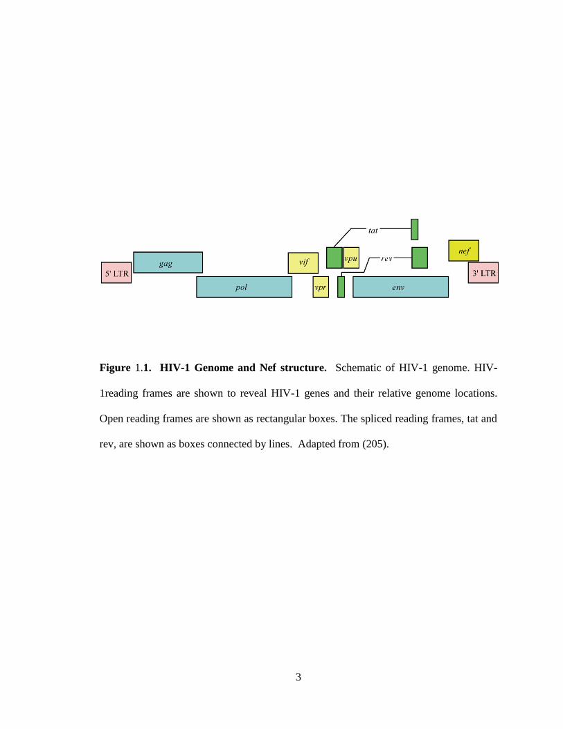
Figure 1.1. HIV-1 Genome and Nef structure. Schematic of HIV-1 genome. HIV-
1reading frames are shown to reveal HIV-1 genes and their relative genome locations.
Open reading frames are shown as rectangular boxes. The spliced reading frames, tat and
rev, are shown as boxes connected by lines. Adapted from (205).
4
RNA (ssRNA) genome into a DNA intermediate prior to integration into the host cell
genomic DNA. Also typical of retroviruses, HIV encodes group-specific antigen (gag),
polymerase (pol), and envelope (env) (Figure 1.1). (for review see (68), (86)). Gag
polyprotein is the major structural component of the viral capsid, and is necessary and
sufficient for virus-like particle assembly (71). Gag is processed into matrix, capsid, and
nucleocapsid by the viral protease within nascent virions in the process of particle
maturation. Pol is also initially expressed as a polyprotein that is cleaved within newly
budding virions into functional enzymes reverse transcriptase (RT), integrase, and
protease. Envelope (Env) is likewise expressed as a 160 kilodalton (kDa) polyprotein
(gp160), which is cleaved by the viral protease into gp41 and gp120 within the Golgi of
the infected cell. gp120 directly interacts with CD4, the viral receptor, while gp41 is
required for virus fusion with the host cell (115). Additionally, HIV-1 Env has been
demonstrated to directly reduce the surface expression of CD4 in order to prevent viral
superinfection and enable viral particle assembly and release (17, 88, 113, 167).
1.4.1 HIV-1 replication
The HIV-1 lifecycle begins with binding of Env to the viral receptor, CD4, and co-
receptors, typically CCR5 or CXCR4 (Figure 1.2) (3, 44, 48, 64). This ligation event
induces a conformation change in Env and allows fusion with the cell membrane and
release of the viral capsid into the cytoplasm (172). In the cytoplasm, RT accesses the
viral genome and begins transcribing the RNA into double-stranded DNA (11). This
DNA genome, along with RT, Vpu, matrix, and integrase form a pre-integration complex

5
Figure 1.2. HIV Life Cycle. 1. HIV Envelope binds to CD4 and co-receptor CXCR4
or CCR5. The virion fuses with the plasma membrane of the target cell and releases the
RNA-containing capsid into the cytoplasm. 2. The viral reverse transcriptase (RT)
transcribes the single-stranded RNA (ssRNA) genome into double-stranded DNA
(dsDNA). 3. The dsDNA genome is transported into the nucleus through a nuclear pore,
where integrase mediates integration of provirus into the cellular genome. The provirus
may remain latent indefinitely. 4. Active infection results from the expression of viral
mRNA and proteins. 5. Viral proteins, along with two strands of viral ssRNA, assemble
at sites of Gag multimerization at the plasma membrane. The nascent virion buds and is
then released from the plasma membrane. 6. Protease cleaves HIV polyproteins within
the immature virion, inducing maturation into fully infectious HIV.

6
Figure 1.2. HIV Life Cycle
7
(PIC) (23, 61, 62, 98, 100). The PIC is translocated into the nucleus of the cell through a
nuclear pore, where integrase mediates the insertion of viral DNA into the cellular
genome, forming a provirus (22, 58). Here, the provirus may remain latent in the absence
of viral gene expression, or may engage in active infection. During active infection, viral
genes are transcribed into RNA for incorporation into virions as full-length viral
genomes, or processed for subsequent translation of viral proteins. Virion assembly
occurs through the accumulation of HIV structural proteins Env and Gag at the plasma
membrane, where Gag also associates with nascent RNA genomes (34, 95, 121, 170).
Gag multimerization induces virion budding from the plasma membrane (191). After
scission of immature virions from producer cells, the HIV protease enzyme cleaves
polyproteins Gag and Pol, which then reassemble into a mature, fully infectious virion
(196).
1.4.2 Accessory genes
HIV is unique among retroviruses in that it has acquired accessory genes tat, rev, vif, vpr,
vpu, and nef, which encode proteins that optimize viral fitness and spread in the host.
HIV tat and rev promote transcription of the viral genome and nuclear export of viral
RNA, respectively (for review see (146, 155)). Tat binds to trans-activating response
element (TAR) in the LTR to promote transcription elongation (99). Rev binds to Rev
Response Elements (RRE) in viral RNA transcripts to promote nuclear export and RNA
stability and utilization. The Vif protein counteracts the intrinsic antiviral factor
apolipoprotein B mRNA editing enzyme, catalytic polypeptide-like 3G (APOBEC3G), a

8
cellular cytosine deaminase which hypermutates the viral genome during reverse
transcription, resulting in attenuated or non-infectious virus. Vif associates with a
cellular ubiquitin ligase complex in order to target APOBEC3G for degradation (179).
Similarly, Vpu also utilizes a cellular ubiquitin ligase complex to degrade cellular targets,
including the viral receptor CD4 and the intrinsic antiviral protein tetherin. Degradation
of these targets allows more efficient budding of nascent virions (145). The role of Vpr
is not entirely clear, but it is known that Vpr induces a G2 mitotic arrest in infected cells
(12), a state which favors transcription from the HIV-1 LTR (73). Recent data indicate
that Vpr associates with a cellular ubiquitin ligase complex to degrade cellular factors
that may otherwise inhibit viral infection and/or spread in the host (for review see (55,
110)). Finally, the viral accessory protein Nef is a multifunctional protein that disrupts
intracellular signaling and trafficking pathways to favor viral infection and spread. Nef
has the well-characterized ability to alter the intracellular trafficking of major
histocompatibility complex proteins class I and II (MHC-I and MHC-II) and CD4, and
has also been reported to affect the trafficking of a variety of other cellular proteins, such
as CD28, and CD8 (69, 120, 178, 183, 184, 187). Nef is important for viral fitness and
persistence, and mediates immune evasion by HIV (139, 140).
1.5 HIV-I Nef
The importance of Nef in viral pathogenesis is highlighted by a cohort of blood
transfusion recipients exposed to an HIV-I strain that contained a large deletion within
the nef gene as well as the long terminal repeat (LTR), but not affecting any of the major
9
promoter elements in the LTR (47, 118). Three decades after exposure, and in the
absence of anti-retroviral treatment, half of the patients demonstrated delayed progression
to CD4 T cell loss or AIDS (19). These patients are termed long term non-progressors
(LTNPs), while the other patients in the cohort maintain undetectable viral loads and are
categorized as elite controllers (74, 75). Thus, Nef is required for maximal pathogenic
potential.
More direct evidence for the requirement for Nef in HIV-induced immune collapse has
been revealed through non-human primate research. Rhesus macaques infected with a
Nef-deleted ( nef) strain of simian immunodeficiency virus (SIV) did not progress to
AIDS (105). The combination of non-human primate research and longitudinal patient
cohort studies has revealed the requirement for Nef in progression from HIV disease to
AIDS.
Nef is a multifunctional adaptor protein which is known to disrupt intracellular
trafficking of host proteins, activate cellular kinases, and enhance virion infectivity. As
mentioned, one of the primary physiological roles of this protein is the downregulation of
cell surface receptors such as MHC-I and CD4 from the cell surface. Nef accomplishes
this by interacting with a multitude of endogenous trafficking and signaling proteins.
This dissertation will focus on the effects of Nef on the trafficking and expression of a
variety of cellular proteins significant to immune recognition and signaling.

10
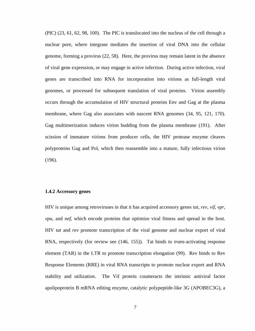
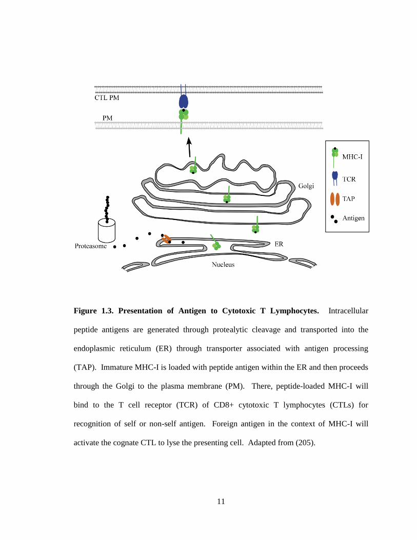
1.6 Nef disrupts antigen presentation to cytotoxic T lymphocytes
CD8+ cytotoxic T lymphocytes (CTLs) are important for the control of chronic viral
infections. The T cell receptor (TCR) in conjunction with CD8, another reported target
of Nef downmodulation, is capable of distinguishing between “self” and “non-self”
peptide antigens presented by MHC-I on the cell surface (Figure 1.3). Normal cellular
peptides typically do not activate a CTL response. However, in a virally infected cell,
MHC-I molecules also present peptides derived from viral proteins (“non-self” peptides).
In response to the recognition of a “non-self” signal presented by MHC-I, the CTL
releases perforins and granzymes that kill the virally infected cell, preventing further
spread of the virus (reviewed in (18)).
There is a great deal of evidence that CTLs play an important role in the control of HIV
infection (for review see (38)). For example, individuals mounting a Gag-specific CTL
response have improved parameters with regard to controlling disease (70, 106). Despite
the efficacy with which CTLs control viral load early in infection, in most individuals,
anti-HIV CTLs ultimately fail to prevent progression of disease. There is evidence that
antigenic variation, viral effects on CTL differentiation, viability, proliferative capacity
and function influence the ability of CTLs to control HIV infection. However, this
chapter will focus on the effect of HIV Nef on the trafficking of cellular proteins that
influence CTL activation.
11
Figure 1.3. Presentation of Antigen to Cytotoxic T Lymphocytes. Intracellular
peptide antigens are generated through protealytic cleavage and transported into the
endoplasmic reticulum (ER) through transporter associated with antigen processing
(TAP). Immature MHC-I is loaded with peptide antigen within the ER and then proceeds
through the Golgi to the plasma membrane (PM). There, peptide-loaded MHC-I will
bind to the T cell receptor (TCR) of CD8+ cytotoxic T lymphocytes (CTLs) for
recognition of self or non-self antigen. Foreign antigen in the context of MHC-I will
activate the cognate CTL to lyse the presenting cell. Adapted from (205).
12
While Nef expression is required for viral spread and pathogenesis in vivo, it is not
necessary for production of infectious virus in vitro and, in fact, is quickly lost in culture.
Therefore, it is clear that Nef functions to antagonize the host immune response. It was
first observed that MHC-I cell surface expression is reduced in HIV-1-infected cells, and
this activity was subsequently attributed to HIV-1 Nef (178). Functional significance
was provided by a study which demonstrated that HIV-1-infected peripheral blood
mononuclear cells (PBMC) expressing Nef were rescued from anti-HIV CTL lysis as
compared to PBMC infected with a nef variant of HIV-1 (39). This work was the first
to directly demonstrate that Nef downmodulates MHC-I molecules to allow HIV-1-
infected cells to evade detection and lysis by CTLs.
Additional studies in which killing of HIV-infected cells was directly compared plus or
minus Nef expression have supported the conclusion that Nef protects infected cells from
CTL-mediated lysis (37, 122, 189, 207). Nef has been shown to protect HIV-infected
primary T cells from CTL lysis using flow cytometric killing assays (37, 122), CTL co-
culture assays (207) and chromium release assays (189). Although Nef limits the ability
of CTLs to recognize and kill infected cells, it does not appear to abrogate the capacity of
CTLs to produce inhibitory cytokines in response to infected cells (189). Recent in vivo
evidence supports the hypothesis that CTLs may control HIV infection in vivo primarily
by the elaboration of inhibitory cytokines, but fail to eradicate the infection because the
CTLs cannot efficiently lyse the infected cell source of new virions (206).
13
Based on in vivo studies, it is known that progression to AIDS is delayed in the absence
of an intact nef gene in most humans and monkeys (46, 105, 111). However, Nef has
multiple functions including CD4 downmodulation and kinase activation; therefore these
studies do not prove an important role for Nef-mediated MHC-I downmodulation
specifically in vivo. To address this point, several studies have used SIV systems to
demonstrate that the capacity to downmodulate MHC-I is selected for in vivo (26, 142,
186). In addition, it was recently demonstrated that the ability of in vivo-derived Nef to
down-regulate MHC-I predicted the resistance of HIV-1 to suppression by CTL in vitro
(123). Taken together, these data demonstrate that the ability of Nef to down-regulate
MHC-I in vivo is maintained by the selective pressure for HIV-1 to evade the antiviral
CTL response.
1.6.1 Natural killer cells
Many viruses, HIV-1 included, have evolved strategies to reduce MHC-I expression in
order to evade CTL recognition by their hosts. As a countermeasure, host natural killer
(NK) cells monitor the overall surface levels of MHC-I. Low expression of MHC-I can
activate NK cells to lyse target cells. In order to evade NK cell detection, Nef selectively
downmodulates some MHC-I allotypes, while allowing others to remain on the cell
surface.
There are three classical MHC-I genes expressed by all nucleated cells in humans; HLA-
A, HLA-B and HLA-C. These genes are highly polymorphic and hundreds of alleles of
14
each have been identified. HLA-A and HLA-B are the primary allotypes that present
antigens to CTLs, whereas HLA-C may function primarily to regulate NK cell function.
In addition, a non-classical MHC-I called HLA-E, which does not commonly present
antigens to CTLs also inhibits NK cell function (reviewed in (144)).
Nef has been shown to directly interact with an amino acid sequence (Y320SQAASS326)
present in the cytoplasmic domain of HLA-A and HLA-B molecules (203). This
interaction is necessary for Nef-dependent downmodulation of MHC-I molecules (35,
117, 203). In contrast, HLA-C and HLA-E have amino acid variations within this domain
(35, 117, 203) and thus remain unaffected by Nef. It has therefore been proposed that
Nef selectively downmodulates a subset of MHC-I molecules to evade CTL killing
without activating NK cell lysis. However, recent evidence demonstrating that HLA-C is
expressed at very low levels on primary T cells suggests that additional mechanisms may
be necessary to fully explain HIV evasion of NK cells (175).
1.6.2. Functional domains required for Nef to downmodulate MHC-I
Though small (25 kDa), Nef is structurally complex, containing numerous protein-protein
interaction domains that mediate associations with a variety of host proteins (Figure 1.4).
Nef can be divided into the N-terminal anchor domain, the core domain, and the C-
terminal flexible loop. The N-terminal anchor domain and C-terminal flexible loop are
relatively disordered, making structural analysis of the full length protein difficult.
However, structures have been solved separately for the core domain by X-ray

15
Figure 1.4. Functionally important Nef domains. (A) Domains in Nef that are
pertinent to reducing the surface expression of cellular targets. Adapted from (205). (B)
Table of Nef domains that are required for the downmodulation of the indicated
molecules. A “+” indicates that a domain is required, while a “-” indicates that a domain
is dispensible.
16
crystallography and nuclear magnetic resonance (NMR), and for the N-terminal anchor
with NMR spectroscopy (7, 67, 79, 119). These structural data were combined to
assemble a model for the conformation of full length Nef. The result predicts that the
flexibility of Nef allows for exposure of numerous protein-protein interaction domains,
regulated by multiple conformations that may depend on intracellular localization and
binding partners (7).
Two sites in Nef are required for most of the functions of Nef. First, Nef is myristoylated
at the glycine residue at position 2, which allows Nef to bind the inner leaflet of the
plasma membrane (60). In addition, an aspartic acid at position 123 (D123) is required to
form homodimers of Nef (126). If either of these sites is mutated (G2A or D123G), Nef is
inactive for nearly all of its functions.
The three structural elements of Nef that are required for MHC-I downmodulation are
contained within the N terminal anchor or core domains. They are an N-terminal a-
helical domain (R17ERM20RRAEPA26 and specifically M20) (2, 133), an acidic cluster
(E62-65) , and a polyproline repeat (P72/75/78). These motifs are required for Nef to bind to
the cytoplasmic tail of MHC-I (202) and for Nef to downmodulate MHC-I (77, 133).
The C-terminal loop of Nef contains a number of trafficking signals capable of binding
adaptor proteins, a coatomer protein, and a vacuolar ATPase (for review see (164)).
However, the C-terminal loop of Nef is only active against other Nef targets, such as
CD4 (133), suggesting that there are structural constraints that limit the ability of the C-

17
terminal loop to recruit trafficking factors when Nef is bound to MHC-I with its natural
conformation.
1.6.3 Candidate host factors that partner with Nef to downmodulate MHC-I
1.6.3.1. Adaptor protein complexes
Nef is reported to interact with a variety of cellular trafficking factors, including the
clathrin-associated adaptor proteins. Clathrin-coated vesicles transport cargo from the
trans-Golgi network (TGN), plasma membrane, or endosomal network. Clathrin-
associated adaptor proteins (APs) are composed of four subunits: two large subunits ( 1
or 2 and AP-1 , AP-2 , or AP-3 ), one medium subunit ( ), and one small subunit ( )
(161, 162, 190). The four subunits combine to function as a heterotetrameric adaptor
complex that recognizes Yxx (Y, tyrosine; , bulky hydrophobic amino acid; x, any
amino acid) and [D/E]xxxLL (D, aspartic acid; E, glutamic acid; L, leucine) sorting
signals and recruits clathrin coats. AP-1 transports proteins between the trans-Golgi
network and endosomes (52, 112, 197). AP-2 localizes to the plasma membrane and is
necessary for internalization of some types of cargo into endosomes (190). AP-3
localizes to endosomes and is thought to transport proteins into acidic, degradative
compartments (151).
Recent structural studies have provided confirmation that clathrin adaptor proteins have
physically separate signal-recognition sites for Yxx and [D/E]xxxLL motifs. The μ

18
subunit contains a tyrosine binding pocket (TBP) and a hydrophobic binding pocket,
which recognize Yxx signals (149). In contrast, a hydrophobic pocket in the 2 subunit
plus a positively charged patch made from residues in both the 2 and subunits
combine to form the recognition site for [D/E]xxxLL motifs (104).
Yeast two-hybrid assays initially revealed that HIV Nef’s C-terminal dileucine motif
(LL164,165) interacts with the subunit of AP-1 and AP-3 (21, 40, 42, 59, 76, 90, 92, 117,
152). However, consistent with the structural analysis described above, a much more
robust interaction occurs between Nef’s dileucine motif and hemicomplexes composed of
and or subunits (30, 53, 92). Recent data suggest that a robust interaction
between the subunit of AP-1 and Nef also occurs, but only when Nef is bound to the
MHC-I cytoplasmic tail. In this case, the MHC-I cytoplasmic tail provides the tyrosine
residue necessary for binding to the AP-1 1 subunit tyrosine binding pocket (147, 165,
181, 204).
1.6.3.2. ADP-ribosylation factors
In addition to the clathrin-associated adaptor proteins, the small GTPases, ADP-
ribosylation factors (ARFs), are important for cellular control of assembly and
disassembly of various intracellular trafficking complexes (10, 97). ARFs are important
for clathrin dependent (50, 148, 150, 163) and clathrin independent (188, 198) trafficking
pathways. ARF activation and recruitment to cellular membranes is regulated by its
guanine nucleotide exchange factors (GEFs), which are required for the recruitment of

19
GTP to ARF and are necessary for the maintenance of overall Golgi structure ((85, 89)
and reviewed in (51)). Conversely, GTPase-activation proteins (GAPs) promote GTP
hydrolysis, thus inactivating ARFs (193).
ARF-1 is a clathrin regulatory protein that recruits AP-1 or coat protein complex-I (COP-
I) coatomers (9). In studying AP-1 involvement in Nef biology, it was observed that
functional ARF-1 is required for recruitment of AP-1 by Nef (36). Additionally, an
interaction between Nef and -COP, which promotes degradation of internalized CD4,
also requires ARF-1. Interestingly ARF-1 does not need to be in the activated GTP bound
state for this interaction to occur (63).
There is also evidence that activation of ARF-6, a regulator of clathrin independent
trafficking, by a Nef-dependent multi-kinase cascade induces MHC-I endocytosis (Figure
1.5) (20, 28, 87). This is consistent with normal ARF-6 biology, as ARF-6 localizes to
the plasma membrane and is involved in clathrin-independent endocytosis and recycling
(158). ARF-6 is regulated by ARF nucleotide binding site opener (ARNO), an ARF-6
GEF that is activated and recruited to the plasma membrane by PI3-kinase (195). There
is evidence that overexpression of ARF-6 and ARNO mutants alters the intracellular
localization of MHC-I in Nef-expressing HeLa cells (20). This mechanism is discussed in
detail below.
20
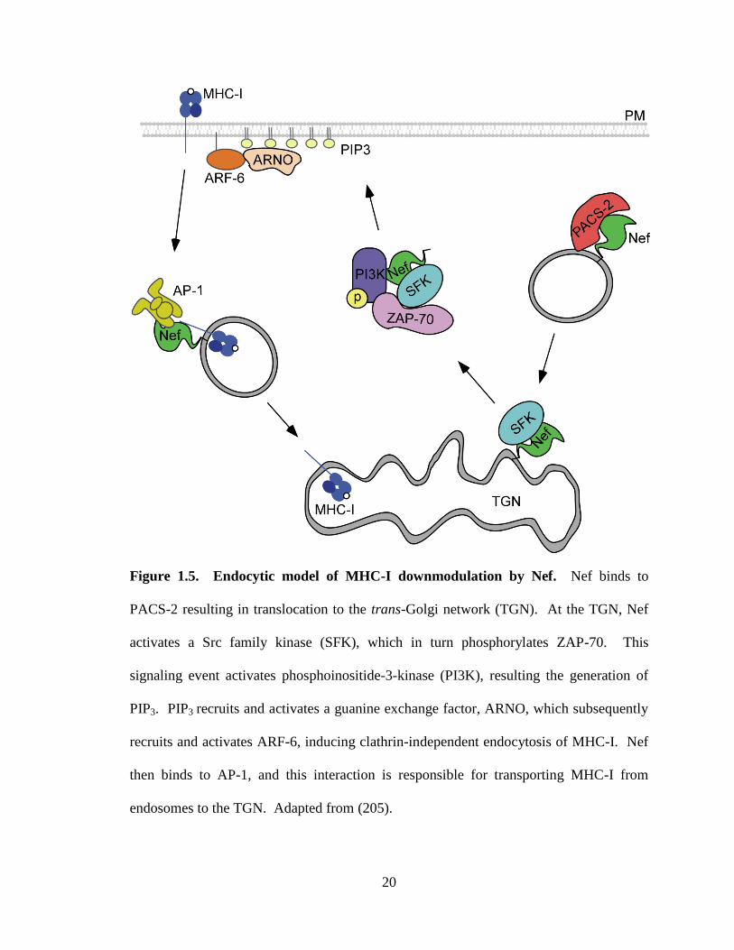
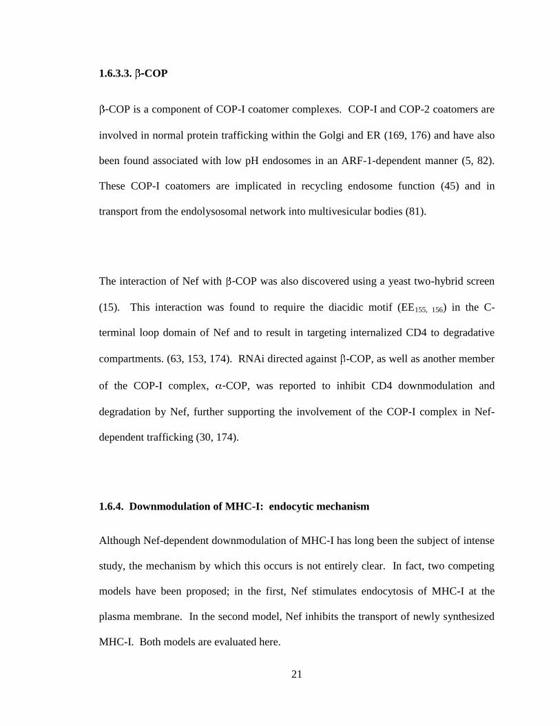
Figure 1.5. Endocytic model of MHC-I downmodulation by Nef. Nef binds to
PACS-2 resulting in translocation to the trans-Golgi network (TGN). At the TGN, Nef
activates a Src family kinase (SFK), which in turn phosphorylates ZAP-70. This
signaling event activates phosphoinositide-3-kinase (PI3K), resulting the generation of
PIP3. PIP3 recruits and activates a guanine exchange factor, ARNO, which subsequently
recruits and activates ARF-6, inducing clathrin-independent endocytosis of MHC-I. Nef
then binds to AP-1, and this interaction is responsible for transporting MHC-I from
endosomes to the TGN. Adapted from (205).
21
1.6.3.3. -COP
-COP is a component of COP-I coatomer complexes. COP-I and COP-2 coatomers are
involved in normal protein trafficking within the Golgi and ER (169, 176) and have also
been found associated with low pH endosomes in an ARF-1-dependent manner (5, 82).
These COP-I coatomers are implicated in recycling endosome function (45) and in
transport from the endolysosomal network into multivesicular bodies (81).
The interaction of Nef with -COP was also discovered using a yeast two-hybrid screen
(15). This interaction was found to require the diacidic motif (EE155, 156) in the C-
terminal loop domain of Nef and to result in targeting internalized CD4 to degradative
compartments. (63, 153, 174). RNAi directed against -COP, as well as another member
of the COP-I complex, -COP, was reported to inhibit CD4 downmodulation and
degradation by Nef, further supporting the involvement of the COP-I complex in Nef-
dependent trafficking (30, 174).
1.6.4. Downmodulation of MHC-I: endocytic mechanism
Although Nef-dependent downmodulation of MHC-I has long been the subject of intense
study, the mechanism by which this occurs is not entirely clear. In fact, two competing
models have been proposed; in the first, Nef stimulates endocytosis of MHC-I at the
plasma membrane. In the second model, Nef inhibits the transport of newly synthesized
MHC-I. Both models are evaluated here.

22
Initial studies examining the effects of Nef on MHC-I trafficking in T cell lines revealed
that the rate of MHC-I synthesis and trafficking through the ER and cis-Golgi is
unaffected by Nef, but that MHC-I stability over time is decreased through lysosomal
degradation (cited as data not shown (177). Furthermore, Nef causes an accumulation of
MHC-I in juxtanuclear and endosomal compartments and enhances the rate of
endocytosis in some cell types (for review see (164)). Normally, T lymphocytes and
macrophages spontaneously internalize and recycle MHC-I back to the plasma membrane
at high rates in an AP-2 dependent manner (131, 192). However, in Nef-expressing cell
lines, over-expression of a dominant negative dynamin, a protein that mediates scission
of clathrin vesicles, (29, 116, 185) or a dominant negative mutant subunit of AP-2 (20)
did not affect Nef-induced MHC-I endocytosis suggesting this process could be clathrin
and AP-2 independent. It has also been demonstrated that Nef does not affect the rate of
MHC-I recycling in HeLa cells or CEM SS T cells (101). More recently it was shown
that a dominant negative dynamin reduced Nef-induced MHC-I downmodulation in
primary T cells from about 50% in this assay system to approximately 25% (208)
indicating that clathrin-dependent endocytosis cannot fully account for MHC-1
downmodulation. Furthermore, Greenberg et al determined that MHC-I co-localizes with
AP-1, but not AP-2 in Nef-expressing cells (77), arguing against an AP-2-dependent
internalization pathway.
23
Alternatively, there is evidence for an ARF-6-dependent, clathrin-independent pathway
by which Nef affects MHC-I in some cell types (Figure 1.5). This mechanism relies on
an interaction between the acidic cluster E62-65 in Nef and phosphofurin acidic cluster
sorting proteins (PACS-1 and PACS-2) (154). Antisense to hPACS-1a increases steady
state MHC-I surface expression in Nef-expressing cells by about 20% and redistributes
the intracellular localization of MHC-I in A7 astrocytic cells. Another group confirmed
that knockdown of PACS-1 inhibited Nef-induced MHC-I downmodulation in HeLa cells
(but not Jurkat T cells) (208). Based on these data a model was proposed in which Nef
physically recruits MHC-I and links it to a PACS-1 based TGN retrieval pathway (154).
This model was later modified to indicate that PACS-2 was more important for
translocation of Nef to the TGN (8). Once at the TGN, Nef is proposed to bind to and
activate a src family kinase (SFK), such as Hck, through Nef’s polyproline motif,
P72/75/78, which is a Src homology 3 (SH3)-binding domain. The active SFK then triggers
a signaling cascade by binding to and phosphorylating tyrosine kinase ZAP70, which in
turn enables ZAP70 to activate phosphoinositide 3-kinase (PI3K) (87). PI3K activity
generates phosphatidylinositol (3,4,5)-triphosphate (PIP3), which results in ARNO
recruitment to the plasma membrane and activation. There is evidence that
overexpression of ARF-6 and ARNO mutants alters the intracellular localization of
MHC-I in Nef-expressing HeLa cells and that overexpression of Nef and PACS-1 in A7
cells increases PI3-kinase-dependent GTP loading of ARF (20). A relatively small effect
(approximately two fold) of a dominant negative ARF-6 mutant was noted in primary T
cells when pan-MHC-I antibodies were used (208). These antibodies recognize all
MHC-I allotypes, including those that are unaffected by Nef and thus small effects of Nef

24
are usually detected. ARF-6 activation is proposed to result in clathrin-independent
endocytosis of MHC-I and accumulation in the Golgi in a mechanism that depends upon
AP-1 and M20 within the alpha helical region of Nef (20, 28).
1.6.5 Evidence against an endocytic mechanism
Arguing against an important role for PACS proteins are data from investigators who
reported no effect of knocking down PACS-1 on Nef-induced downregulation of MHC-I
HLA-A2, or on the localization of other proteins containing acidic cluster motifs, in
HeLa cells (127). Additionally, Baugh et al were unable to demonstrate a significant
interaction between the acidic cluster in Nef and the PACS-1 furin binding region (13),
and mutating three of the four glutamates in the acidic cluster decreased Nef’s effects on
MHC-I by only 50% (13). Chemical inhibitors of SFKs, Src knockdown, and expression
of dominant negative Src all failed to inhibit Nef-dependent downmodulation of MHC-I
in U937 monocytic cells, arguing against an important role for SFKs in this pathway (29).
Overexpression of a GTP-locked ARF-6 mutant (ARF-6 Q67L) is reported to alter the
intracellular localization of MHC-I in Nef-expressing HeLa cells (20). However,
mutation of an additional residue in ARF-6 (ARF-6 N48I,Q67L) prevents phospholipase D
activation, an effector required for endosomal membrane recycling (96), but had no effect
on MHC-I downmodulation in Jurkat T cells (114). Furthermore, inhibition of PI3-
kinase, had no effect on the internalization step in U373mg astrocytoma cells (114).
Instead, other investigators provided evidence that PI3-kinase inhibitors affected
localization of intracellular MHC-I to the TGN in Nef-expressing U373mg astrocytoma
25
cells (114, 185). Therefore, an alternative mechanism of MHC-1 downregulation by Nef
has been proposed, as discussed below.
.
1.6.6. Downmodulation of MHC-I: evidence for targeting of newly synthesized
protein in the secretory pathway
1.6.6.1. Disruption of MHC-I transport
A dramatic effect of Nef on MHC-I is required for HIV-infected primary T cells to
effectively evade anti-HIV CTLs (up to a 300-fold reduction (cited as data not shown
(37)). The degree of MHC-I downmodulation in HeLa cells (2-4 fold reduction (20) is
small relative to the effect of Nef on an endogenous MHC-I allotype (HLA-A2) in HIV-
infected primary T lymphocytes (37). Thus, the internalization pathways described
mainly in HeLa cells may not fully explain the intracellular trafficking required for the
maximal effect of Nef necessary for HIV immune evasion in T cells. Indeed, direct
comparison of Nef activity in HeLa versus T cell lines revealed striking differences in the
degree of MHC-I downmodulation (101).
Most viruses that disrupt antigen presentation target newly synthesized MHC-I rather
than “old” MHC-I at the cell surface because the newly synthesized molecules harbor
viral antigens present at the time of infection. For example, Herpes Simplex Virus,
Human Cytomegalovirus, Epstein-Barr Virus, and Adenovirus all encode proteins that
block peptide translocation into the ER, target nascent MHC-I for degradation, induce ER
26
retention of peptide-loaded MHC-I, or prevent transport of MHC-I to the plasma
membrane (for review see (83)). Older MHC-I molecules are likely to be presenting
cellular antigens, which are present prior to infection, and therefore would not be a threat
to the virus. In fact, MHC-I loaded with cellular antigens would be protective against
NK cell recognition.
In Nef-expressing cells, reports of MHC-I localizing to the trans-Golgi and AP-1-
containing vesicles suggested that Nef could be directly disrupting MHC-I trafficking at
the trans-Golgi network to prevent nascent MHC-1 from presenting new antigens rather
than only affecting MHC-I after it had reached the cell surface (Figure 1.6). The first
study supporting this model examined the effect of Nef on an HLA-A2-GFP fusion
protein in U373mg astrocytoma cells (185). In this series of experiments investigators
utilized a temperature block (20oC ) to prevent TGN exit and to allow accumulation of
MHC-I in the TGN. When cells were subsequently shifted to 37oC, MHC-I could be
detected by microscopy at the cell surface within 15 minutes, whereas in Nef-expressing
cells MHC-I remained within a juxtanuclear compartment (185). Biochemical
experiments examining the transport of newly synthesized MHC-I to the cell surface in T
cell lines confirmed that there was a dramatic effect of Nef on the transport of MHC-I to
the cell surface. Moreover, the effect of Nef on transport of newly synthesized MHC-I
was much greater than its effect on MHC-I internalization (101). An effect of Nef on
intracellular transport of endogenous MHC-I HLA-A2 was confirmed in HIV-infected
primary T cells (101). PI3-kinase inhibitors did not reduce the ability of Nef to disrupt

27
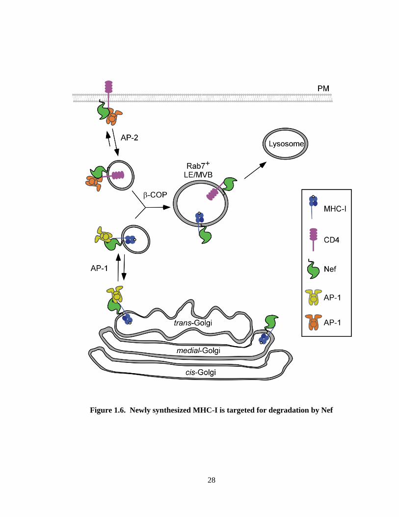
Figure 1.6. Newly synthesized MHC-I is targeted for degradation by Nef. Nef binds
to the cytoplasmic tail domain of MHC-I in the Golgi network. Nef recruits AP-1 to the
complex at the TGN, resulting in routing of MHC-I into endocytic compartments.
Subsequent recruitment of -COP targets MHC-I into endolysosomal compartments. Nef
binds to the cytoplasmic tail of CD4 at the plasma membrane and recruits AP-2, inducing
rapid CD4 internalization. Subsequent recruitment of -COP results in the convergence
of MHC-I and CD4 into Rab7+ late endosomes or multivesicular bodies (MVBs) for
eventual lysosomal degradation. Adapted from (205).
28
Figure 1.6. Newly synthesized MHC-I is targeted for degradation by Nef

29
MHC-I transport to the cell surface as measured by a one hour biochemical assay but the
investigators could not rule out an effect of PI3-kinase on the intracellular localization of
retained MHC-I molecules (101) as was subsequently proposed (114).
Additional evidence in favor of a transport block are studies in which Nef was found to
associate with immature forms of MHC-I (102). The HLA-A2 cytoplasmic tail is
phosphorylated at specific serines upon exiting the TGN (57). Interestingly, Nef
preferentially binds immature, hypophosphorylated forms of HLA-A2 and inhibits
phosphorylation of the MHC-I cytoplasmic tail (102). Based on these data it was
proposed that Nef binds MHC-I very early in the secretory pathway (102). In support of
this model, a recent study was able to observe a Nef-CFP fusion protein in complex with
a subset of HLA-A2-Venus in the ER as well as in the Golgi and at the plasma membrane
of HeLa cells using two photon two color fluorescence cross correlation spectroscopy
(208). However, there was no detectable effect of Nef on MHC-I transport until MHC-I
reached the trans-Golgi apparatus, thus binding to Nef was not sufficient for disruption of
MHC-I trafficking (102, 165) (Figure 1.6).
1.6.6.2. AP-1 is required for disruption of antigen presentation by HIV Nef
Because AP-1 is a clathrin adaptor protein that acts at the TGN and because Nef had been
reported to interact with AP-1, it was hypothesized that Nef might disrupt post-TGN
transport of MHC-I by promoting an interaction between MHC-I and AP-1. Indeed,
RNAi directed against the AP-1 1 subunit inhibited downmodulation of endogenous

30
HLA-A2 in U373mg astrocytoma cells and exogenous HLA-A2 expressed in CEM-SS T
cells (120, 165, 174, 204). Furthermore, siRNA directed against AP-1 1 also inhibited
Nef-induced downmodulation of MHC-I in HeLa and Jurkat cell lines, as well as in
primary T lymphocytes (208).
Consistent with these observations, AP-1 co-precipitated with Nef and endogenous HLA-
A2 from lysates made from HIV infected primary T cells (165). In contrast, complexes
of Nef-MHC-I and AP-1 were not detected in HeLa cells unless the cells were incubated
at room temperature overnight. Further experiments revealed that temperature reduction
decreased the rate of MHC-I trafficking sufficiently to allow the Nef-MHC-I-AP-1
complex to form. For unclear reasons, T cells naturally traffic MHC-I at slower rates and
lower incubation temperatures do not change the ability of Nef to form this complex
(102). These data help explain why investigators that focused on non-T cell lines did not
detect this pathway.
1.6.6.2.1. Nef stabilizes an interaction between the AP-1 tyrosine binding pocket and
the MHC-I cytoplasmic tail
Yeast two-hybrid interaction assays and microscopic analyses provided evidence that
interactions between Nef and the adaptor proteins AP-1 and AP-3 depend on Nef’s
dileucine motif (21, 40, 42, 59, 76, 90, 92, 152). In contrast, MHC-I downmodulation
and AP-1 recruitment in T cell systems do not require these amino acids (77, 165, 202).
Thus, the complex between Nef-MHC-I and AP-1 most likely occurred independently of

31
the dileucine motif and involved a separate AP-1 binding domain. Indeed, it was
demonstrated that the MHC-I cytoplasmic tail mediates a key interaction between the
Nef-MHC-I complex and AP-1 (165). The tyrosine in the MHC-I cytoplasmic tail does
not form a canonical Yxx AP-1 sorting signal and does not bind AP-1 in T cells in the
absence of Nef. Remarkably , Nef binding to the cytoplasmic tail provides the necessary
elements for this non-canonical tyrosine signal to function as a potent AP-1 binding motif
(165). Providing further support for the model that Nef stabilizes an interaction between
the AP-1 tyrosine-binding pocket (TBP) and the tyrosine residue in the MHC-I
cytoplasmic tail, it was shown that a dominant negative mutant of AP-1 1 that had two
amino acid substitutions in the tyrosine binding pocket (TBPM) dramatically and
specifically inhibited Nef-mediated MHC-I downmodulation (204).
1.6.6.2.2. Nef domains and AP-1-dependent MHC-I trafficking
All of the domains of Nef that are required for MHC-I downmodulation are also
required for Nef to interact with the MHC-I cytoplasmic tail (202). To determine
whether some of these domains might also be important for recruitment of AP-1, a fusion
protein between MHC-I and Nef was examined (165). These studies confirmed that the
MHC-I cytoplasmic tail tyrosine was required for AP-1 recruitment and that Nef’s
dileucine motif was dispensable for this interaction (165, 204). In this system, the acidic
cluster (E62-65) and polyproline helix (P72/75/78) of Nef were dispensable for AP-1
recruitment as long as a chemical crosslinker was used (165). However, when a digitonin
detergent based buffer that lacked crosslinker was substituted, a requirement for these
32
domains to optimally stabilize the interaction between AP-1 and MHC-I was noted (204).
In addition, the N-terminal -helix and specifically M20, were required for AP-1
recruitment under all conditions tested (165, 204).
Experiments using purified Nef-MHC-I cytoplasmic tail fusion proteins and either whole
AP-1 complexes from crude lysates or purified 1 subunit support the conclusion that
Nef stabilizes an interaction between the MHC-I cytoplasmic tail and the AP-1 1
subunit. Moreover, these experiments provide evidence that the polyproline helix and the
acidic domain within Nef are needed for Nef to stabilize the interaction between the AP-1
-1 subunit and the MHC-I cytoplasmic tail domain. In the pure protein system,
formation of a complex between the Nef-MHC-I cytoplasmic tail fusion protein and the
AP-1 1 subunit also required an intact tyrosine binding pocket in the AP-1 1 subunit.
However, no role for Nef M20 was identified and thus this amino acid, although required
for Nef-induced MHC-I downmodulation, may not be directly involved in protein-protein
interactions but may serve another role in intact cells (181). Therefore, at least three Nef
domains are required for AP-1 recruitment and subsequent downmodulation of MHC-I in
Nef expressing cells (165, 204).
1.6.6.3. A role for -COP in disruption of antigen presentation by Nef
Although the experiments described above provide evidence that Nef recruits AP-1 to
reroute MHC-I into the endosomal network (165) (Figure 1.6), it remained unclear how
33
Nef promoted accelerated degradation of MHC-I (165, 177). However, prior reports had
determined that Nef accelerated the degradation of internalized CD4 through an
interaction between Nef and -COP. Interestingly, MHC-I and internalized CD4 co-
localize in Rab7+
late endosomes in Nef-expressing cells (174, 208) and RNAi against -
COP disrupts Nef-dependent degradation of both MHC-I and CD4 (174). Recent studies
have shown that two distinct domains in Nef recruit -COP, thus clearing up
discrepancies in binding data found in previously published literature (63, 91, 124, 153).
An arginine rich domain in the N-terminal alpha helix of Nef (R17XR19) mediates -COP
binding and MHC-I degradation, whereas a diacidic motif (EE155, 156) in the C-terminal
flexible loop of Nef mediates -COP binding and CD4 degradation. (153, 174). The
inability of Nef to utilize sequences within the C-terminal loop to affect MHC-I
downmodulation or to use sequences within the N-terminal alpha helix to affect CD4
downmodulation support the notion that there are important structural differences
between Nef molecules bound to the MHC-I cytoplasmic tail and Nef bound to the CD4
tail.
1.6.7. Summary of MHC-I downmodulation
In sum, a consensus is starting to emerge regarding which host factors are required for
Nef to disrupt antigen presentation in HIV infected cells. There is broad agreement
among investigators that the cellular clathrin adaptor protein AP-1 is necessary for Nef to
disrupt MHC-I trafficking in a wide variety of cell types (49, 127, 165, 174, 204, 208).
Additionally, there is agreement that three Nef domains are required (acidic, polyproline
and N-terminal alpha helix, including M20A) for Nef-induced MHC-I downmodulation
34
(20, 77, 133, 147, 165, 181, 202, 204). There are data from two separate groups
indicating that a three-way complex forms, which contains Nef, MHC-I and AP-1
proteins and that this complex can be detected in lysates from HIV infected primary T
cells and in purified protein reactions (165, 181). At least two of the three required Nef
domains plus the MHC-I cytoplasmic tail, including the tyrosine at position 320, are
directly needed for formation of the Nef-MHC-I-AP-1 complex (147, 165, 181, 204).
Moreover, there is a consensus that a functional tyrosine binding pocket in the AP-1 1
subunit is needed for formation of the Nef, AP-1, MHC-I complex and for Nef to disrupt
MHC-I antigen presentation (181, 204). Finally, a number of groups have noted that PI3-
kinase inhibitors reduce the effect of Nef on steady state surface levels of MHC-I,
although the exact role of PI3-kinase is debated (20, 87, 114, 185).
1.7. Nef downmodulates CD4
In addition to reducing MHC-I surface expression, Nef also targets the HIV-1 receptor
CD4 for endocytosis and degradation (69, 135). CD4 downmodulation promotes viral
spread, as CD4 on the surface of producer cells would bind to Env and inhibit the release
of nascent virions (6, 113, 128, 167). Downmodulation of CD4 also prevents
superinfection (199). Interestingly, HIV encodes two accessory proteins to antagonize
CD4 expression. Nef is an early viral gene product which downmodulates mature CD4
early in infection (1, 160). In contrast, late viral gene product Vpu reduces newly
synthesized CD4 expression through an endoplasmic reticulum associated protein
degradation (ERAD) pathway (56, 200, 201). Additionally, the HIV-1 structural protein

35
Env has also been demonstrated to associate with CD4 in the endoplasmic reticulum (ER)
and inhibit CD4 transport (43, 88). That HIV has evolved three proteins to achieve CD4
downmodulation by complimentary mechanisms suggests that this function is essential to
HIV-1 pathogenesis.
1.7.1 In vivo evidence for the importance of CD4 downmodulation
The existence of a long term non-progressor with a unique Nef mutation provides direct
evidence for the importance of CD4 downmodulation in HIV-1 pathogenesis. The virus
from this patient had a deletion of amino acids 26-37 within Nef that abrogated MHC-I
and CD4 downmodulation, as well as enhancement of virion infectivity. However, a
compensatory duplication of amino acids 43-53 restored the ability of this Nef variant to
downmodulate MHC-I and enhance virion infectivity, but not to downmodulate CD4.
This patient demonstrated relatively high viral loads but no decline in CD4+ T cell counts
(25). This single example suggests that HIV-1 requires Nef-induced CD4
downmodulation to maintain maximal pathogenic potential.
1.7.2. Functional domains required for Nef to downmodulate CD4
A number of motifs within the C-terminal loop of Nef have been implicated in CD4
downmodulation (Figure 1.4). These sites within Nef mediate binding directly to the
cytoplasmic domain of CD4, as well as recruitment of cellular adaptor proteins. Nef has
been demonstrated to interact directly with the cytoplasmic domain of CD4 by a number
36
of investigators using a variety of methods including: yeast-2 hybrid (168), in vitro
binding (84, 156), immunoprecipitation (94, 120), and fluorescence resonance energy
transfer (FRET). NMR and fluorescence spectroscopy analyses have identified residues
57-59, 95, 97, 106, and 110 of Nef as forming the binding interface between Nef and
CD4 (80, 156). Specifically, mutation of WL57-58 abrogates CD4 downmodulation by Nef
(133). This Nef interface binds to a membrane proximal sequence (QIKRLLSEKKT)
within the CD4 cytoplasmic domain; the two leucines within this sequence are required
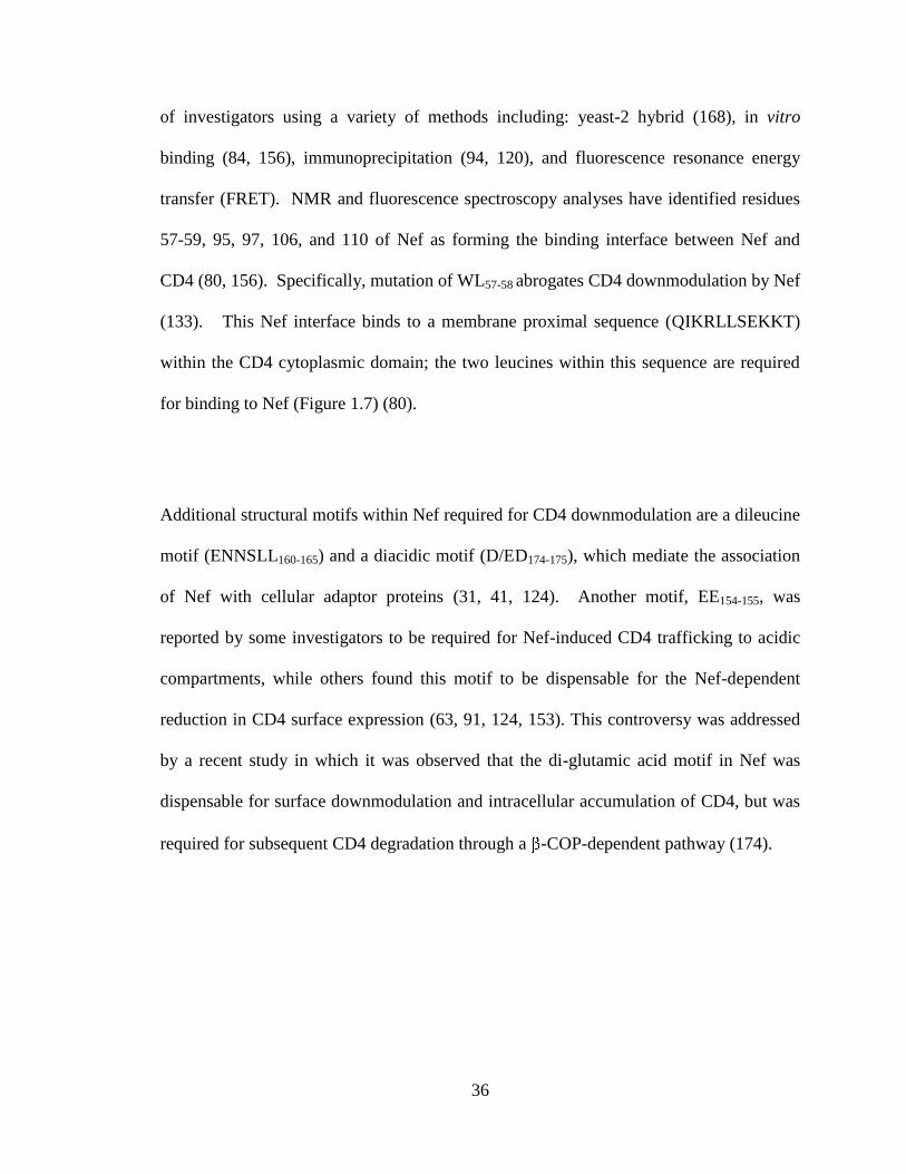
for binding to Nef (Figure 1.7) (80).
Additional structural motifs within Nef required for CD4 downmodulation are a dileucine
motif (ENNSLL160-165) and a diacidic motif (D/ED174-175), which mediate the association
of Nef with cellular adaptor proteins (31, 41, 124). Another motif, EE154-155, was
reported by some investigators to be required for Nef-induced CD4 trafficking to acidic
compartments, while others found this motif to be dispensable for the Nef-dependent
reduction in CD4 surface expression (63, 91, 124, 153). This controversy was addressed
by a recent study in which it was observed that the di-glutamic acid motif in Nef was
dispensable for surface downmodulation and intracellular accumulation of CD4, but was
required for subsequent CD4 degradation through a -COP-dependent pathway (174).

37
Figure 1.7. Cellular proteins targeted by Nef participate in immune activation and
signaling. (A) An interaction between an antigen presenting cell (APC) and a T
lymphocyte. The T cell receptor (TCR) and either CD4 or CD8 bind to MHC-II or
MHC-I, respectively. Activation of the T cell requires a second signal which is provided
through ligation of CD28 by CD80 or CD86. (B) The cytoplasmic tail domains of Nef
target proteins do not share significant homology. Sequence comparison of the
cytoplasmic regions of cellular proteins targeted by Nef. Regions reported to be involved
in Nef-binding or Nef-induced downmodulation are underlined, while critical residues
are highlighted in red.
38
Figure 1.7. Cellular proteins targeted by Nef participate in immune activation and
signaling.

39
1.7.3 Nef downmodulates CD4 by an endocytic mechanism
The mechanism by which Nef affects CD4 surface expression is quite distinct from the
mechanism of MHC-I downmodulation by Nef (Figure 1.6). Furthermore, the
understanding of Nef-induced CD4 downmodulation within the field is less controversial.
It has long been known that Nef associates with CD4 at the plasma membrane,
connecting CD4 to cellular endocytic machinery and inducing an increased rate of CD4
endocytosis (1, 66, 72, 78, 101, 124, 132, 138, 152, 153, 183). Consistent with an
endocytic model, Nef-induced downmodulation of CD4 was found to be inhibited by
dominant negative dynamin expression (93, 116), as well as by chemical inhibitors of
clathrin-mediated endocytosis (129). Additionally, microscopic analysis revealed that
Nef increases the number of CD4-containing clathrin pits (66), and RNAi knockdown of
the clathrin heavy and light chains inhibited Nef-induced downmodulation of CD4 (30).
Thus, downmodulation of CD4 by Nef depends on clathrin-associated endocytic
machinery.
1.7.4 Host factors that partner with Nef to induce CD4 downmodulation and
degradation
1.7.4.1. AP-2 mediates CD4 downmodulation by Nef
Nef links CD4 to cellular endocytic machinery through clathrin associated adaptor
protein 2 (AP-2) (Figure 1.6). An early study used fluorescence microscopy to observe
colocalization between Nef and the subunit of AP-2 at the plasma membrane. It was
soon noted that Nef contains a canonical dileucine trafficking signal, (ENNSLL160-165),

40
and that this domain was not only required for CD4 downmodulation but was also
capable of interacting with both AP-1 and AP-2 (21, 41, 76). AP-2 is known to mediate
clathrin-mediated endocytosis at the plasma membrane and this protein seemed the
likeliest candidate for the Nef-induced increase in CD4 turnover. RNAi knockdown of
the 2 subunit of AP-2 provided evidence for AP-2 involvement in CD4
downmodulation. While 2 knockdown alone did not impair Nef-induced
downregulation of CD4, co-expression of the siRNA with a dominant negative mutant of
an AP-2 accessory protein involved in clathrin-mediated endocytosis, Eps15 (16, 24),
reduced Nef-specific CD4 downmodulation (93). Subsequent reports of AP-2
knockdown have been conflicting, with some investigators observing a partial
requirement for AP-2 in CD4 downmodulation by Nef (30, 183), while others have not
(120, 166). This may depend in part on which subunit of AP-2 is targeted, as in one study
AP-2 knockdown inhibited downmodulation by approximately 50% in HeLa and S2
insect cells, but AP-2 knockdown did not (30).
Though AP-2 knockdown has been controversial, studies taking a different experimental
approach have provided ample evidence of an interaction between Nef and AP-2 in vitro.
Yeast 3-hybrid analyses identified an interaction between Nef and AP-2
hemicomplexes that required both the dileucine motif and D/ED174-175 diacidic motif
within Nef, and this interaction was confirmed with GST pulldown experiments (30,
124). This observation is consistent with a recent study which mapped the dileucine
binding site within AP-2 to an interface between the and subunits, as discussed

41
above (104). Interestingly, the requirement for Nef’s D/ED174-175 diacidic motif in
addition to the dileucine motif appears to represent an extended AP-2 binding site as
compared to other AP-2-cargo interactions. Yeast 3-hybrid experiments and mutational
analysis of AP-2 recently identified a patch of arginine and lysine residues which are
responsible for the interaction with the diacidic motif of Nef. Furthermore, Yeast 4-
hybrid analysis provided direct evidence for a tripartite complex between CD4, Nef, and
the hemicomplex (31). Taken together, this evidence supports the conclusion that
Nef utilizes AP-2 to connect CD4 to the endocytic machinery
1.7.4.2. -COP is required for Nef-induced CD4 degradation
In addition to decreasing CD4 surface expression through increased internalization, Nef
also induces degradation of CD4 (1, 63, 107, 124, 129, 153, 160, 171). Inhibition of
lysosomal acidification prevents Nef-induced CD4 degradation but does not restore CD4
surface expression, indicating that CD4 internalization and degradation are separable
functions of Nef (129, 159, 171). CD4 targeting to late endosomal and lysosomal
compartments requires an association between EE154-155 in Nef and the cellular adaptor
protein -COP, as discussed above (Figure 1.6) (63, 153, 174).
1.8 Nef is reported to disrupt trafficking of a variety of cellular proteins
In addition to the effects of Nef on MHC-I and CD4 trafficking, there are a number of
recent reports that Nef downmodulates a variety of other cell surface signaling proteins,
42
including CD28, CD8, CD80, CD86, and CD1d (Figure 1.7). Relative to MHC-I and
CD4 downmodulation, these functions of Nef are not well understood. These proteins
are all immunologically important molecules, but their cytoplasmic tail domains share no
obvious sequence homology (Figure 1.7). Therefore, it is surprising that Nef would be
able to affect the trafficking of so many different targets.
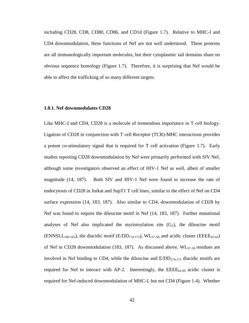
1.8.1. Nef downmodulates CD28
Like MHC-I and CD4, CD28 is a molecule of tremendous importance in T cell biology.
Ligation of CD28 in conjunction with T cell Receptor (TCR)-MHC interactions provides
a potent co-stimulatory signal that is required for T cell activation (Figure 1.7). Early
studies reporting CD28 downmodulation by Nef were primarily performed with SIV Nef,
although some investigators observed an effect of HIV-1 Nef as well, albeit of smaller
magnitude (14, 187). Both SIV and HIV-1 Nef were found to increase the rate of
endocytosis of CD28 in Jurkat and SupT1 T cell lines, similar to the effect of Nef on CD4
surface expression (14, 183, 187). Also similar to CD4, downmodulation of CD28 by
Nef was found to require the dileucine motif in Nef (14, 183, 187). Further mutational
analyses of Nef also implicated the myristoylation site (G2), the dileucine motif
(ENNSLL160-165), the diacidic motif (E/DD174-175), WL57-58, and acidic cluster (EEEE62-65)
of Nef in CD28 downmodulation (183, 187). As discussed above, WL57-58 residues are
involved in Nef binding to CD4, while the dileucine and E/DD174-175 diacidic motifs are
required for Nef to interact with AP-2. Interestingly, the EEEE62-65 acidic cluster is
required for Nef-induced downmodulation of MHC-I, but not CD4 (Figure 1.4). Whether

43
any these residues contribute to Nef-CD28 binding is unclear, as attempts to detect an
interaction between SIV Nef and CD28 by yeast two-hybrid analysis or co-
immunoprecipitation were not successful (14). However, fluorescence microscopy
experiments in IMR90 fibroblasts did reveal colocalization of Nef and CD28, as well as
colocalization of CD28 and AP-2 in cells expressing Nef (187). Therefore, it has been
proposed that Nef induces rapid CD28 endocytosis in an AP-2 dependent mechanism,
similar to the manner by which Nef induces CD4 downmodulation. However, neither an
association of Nef with CD28 nor adaptor protein involvement in Nef-induced CD28
downmodulation has been directly demonstrated.
1.8.2 Evidence for downmodulation of CD8 by Nef
CD8, like CD4, is a T cell co-receptor, but CD8 is expressed on CTLs while CD4 is
expressed on helper T lymphocytes. CD8 ligation to MHC I is required for efficient
signaling through the TCR, resulting in antigen recognition and increased avidity of
TCR-antigen interactions (Figure 1.7). Therefore, a lack of CD8 surface expression
would likely impair activation of anti-HIV CTLs. However, as CD4 is the viral receptor,
HIV infects CD4-expressing cells, not CTLs. Thus it is not clear under what
circumstances HIV-1 Nef might encounter CD8.
CD8 is a dimer classically formed by one and one subunit, although
homodimers do occur in some cell types, such as NK cells. HIV-1 Nef was reported to
downmodulate CD8 in the SupT1 T cell line as well as in primary T lymphocytes.
Specifically, Nef downmodulated CD8 / and CD8 / homodimers, but not CD8
44
homodimers. This indicated that Nef downmodulates CD8 by acting on the subunit
(183).
Similar to several other Nef targets, CD8 downmodulation by Nef was determined to be
due to an increased rate of internalization. In order to assess the involvement of
endocytic machinery, investigators used RNAi to knock down expression of the clathrin
heavy chain, dynamin, and AP-2 in the Daudi B cell lymphoma cell line. Dynamin and
AP-2 depletion each inhibited Nef-induced downmodulation of CD8, as well as CD4, by
approximately half. Though clathrin heavy chain knockdown had no effect on CD8 or
CD4 downmodulation, treatment with ikarugamycin, a chemical inhibitor of clathrin-
mediated endocytosis, inhibited Nef-induced internalization of CD8, indicating that Nef-
dependent endocytosis of CD8 is likely mediated by clathrin (183). Mutational analysis
of Nef revealed that downmodulation of CD8 requires the myristoylation site (G2), the
CD4 binding site (WL57-58) the dileucine motif (LL154-155) and diacidic motif (E/DD174-
175) of Nef (183). Each of these Nef domains has also been implicated in CD4 and CD28
downmodulation, suggesting that common molecular interactions mediate these
mechanisms (Figure 1.4). All together, this evidence is consistent with the conclusion
that HIV-1 Nef uses a common mechanism by which it downmodulates CD4, CD28, and
CD8.
The CD8 cytoplasmic tail is short, only 15 amino acids, and, unlike the CD4
cytoplasmic tail, it contains no known protein-protein interaction motifs. Furthermore,
45
CD8 shares no obvious sequence homology with the CD4 and CD28 cytoplasmic
domains (Figure 1.7). Mutational analysis of the CD8 sequence revealed that amino
acids FMK204-206 of the cytoplasmic domain were required for Nef-dependent
downmodulation (183). This motif does not resemble the dileucine signal that forms the
Nef-binding site within CD4, nor does it correspond to any motif within CD28.
Therefore, it is unclear how Nef can affect these targets by similar mechanisms, mediated
through dissimilar protein domains. Therefore, further studies are needed to clarify the
mechanism by which Nef affects CD8 and how CD8 is a physiologically relevant target
for Nef.
1.8.3. Evidence for CD1d downmodulation by HIV-1
Antigen-presenting molecule CD1d is structurally similar to MHC-I and presents lipid, as
opposed to peptide, antigens to Natural Killer T Cells (NKT cells). CD1d has long been
known to be capable of presenting the model antigen galactosylceramide (aGalCer), a
marine sponge-derived lipid (103, 143). Recently, CD1d has also been found to present
lipid antigens from microbial pathogens such as Sphingomonas, Borrelia burgdorferi,
Leishmania, and Mycobacteria (4, 65, 108, 109, 136, 182). It is not clear whether an
HIV-associated lipid antigen exists. It is also unknown whether CD1d-restricted NKT
cells have any antiviral activity against HIV and whether CD1d expression influences
HIV pathogenesis. The literature regarding CD1d downmodulation by HIV-1 is
conflicting. Two groups have reported downmodulation of CD1d by HIV-1 Nef but
report conflicting mechanisms (32, 33), while another group attributes this function to

46
Vpu (141). Other groups reported controversial CD80/86 or CD1a downmodulation, but
failed to observe CD1d downmodulation by HIV in primary human APCs (27, 180).
1.8.3.1 Reports of CD1d downmodulation by Nef
CD1d was first observed to be downmodulated in human Jurkat T cells infected with
NL4-3 HIV-1. Downmodulation of transiently expressed CD1d was observed when
NL4-3 Nef was expressed in human T2 cells. The authors state (as data not shown) that
HIV accessory protein Vpu had no effect on CD1d expression. In this study, the
magnitude of Nef-induced CD1d downmodulation always exceeded that of MHC-I or
CD4. Co-immunprecipitation experiments detected Nef in complex with CD1d, although
the interaction was described as weak or transient due to the necessity of using a
chemical crosslinker to observe co-precipitation. This interaction was dependent upon
the CD1d cytoplasmic domain, but was independent of a tyrosine trafficking motif in
CD1d (33).
Another group also demonstrated reduced CD1d expression on Jurkat cells infected with
NL4-3 HIV as compared to those infected with NL4-3 HIV nef. In contrast to the
previous study, these investigators reported moderate effects of Nef on CD1d expression
(approximately 30%) as compared to MHC-I (50%) and CD4 (70%). Transient
transfection of Nef alone resulted in decreased CD1d expression, although not to the
same extent as whole HIV, suggesting the involvement of other viral proteins. This study
attributed CD1d downmodulation to an increased rate of endocytosis in the presence of
47
Nef, though the rate increase, measured as the percent of labeled-CD1d unaffected by an
acid wash, was not observed at time points earlier than 45 minutes, at which time
recycling defects confound endocytosis measurements. Microscopic analysis of HeLa
cells revealed that CD1d colocalized with a Nef-GFP fusion protein and accumulated in
the TGN. The investigators observed a requirement for the tyrosine motif within the
CD1d cytoplasmic domain for Nef-induced downmodulation (Figure 1.7). Mutational
analysis of Nef provided evidence for a requirement for the myristoylation site (G2) as
well as a partial dependence upon CD4-downmodulation domains LL164-165 and DD174-175,
and MHC-I downmodulation domains PxxP72/75 and EEEE62-65, but not M20 (Figure 1.4)
(32). Therefore it is not apparent that CD1d downmodulation by Nef occurs by a
mechanism shared with MHC-I or CD4 downmodulation.
Interestingly, although differing binding domain requirements were reported, both studies
reported that Nef-expressing APCs demonstrated reduced capacity to activate CD1d-
restricted NKT cells through GalCer presentation as compared to cells expressing CD1d
in the absence of Nef (32, 33). This observation supports the potential functional
significance of HIV-dependent CD1d downmodulation.
1.8.3.2. Evidence for CD1d downmodulation by HIV-1 Vpu
Though in agreement with previous studies that HIV-1 induces downmodulation of
CD1d, a recent report has identified Vpu as the HIV accessory protein responsible for
this activity. As in previous reports, functional assays demonstrated impaired activation
48
of CD1d-restricted NKT cells by HIV-1-infected primary human monocyte-derived
dendritic cells (MDDC). However, in this system HIV-1 Vpu expression was sufficient
to induce CD1d downmodulation (141). Vpu did not affect CD1d internalization
between 0 and 30 minutes, but recycling of CD1d was impaired in cells expressing Vpu.
Fluorescence microscopy revealed colocalization of CD1d and a Vpu-GFP fusion protein
within EEA1-positive early endosomal compartments. CD1d, but not Vpu, could also be
found in LAMP-1-positive late endosomes/lysosomes in Vpu-expressing cells (141).
Interestingly, HIV-1 vpu or nef each demonstrated a reduced ability to affect CD1d
surface expression in human MDDCs, while inactivation of both Nef and Vpu
synergistically abrogated CD1d downmodulation by HIV (141). This observation
indicates that HIV-1 Nef and Vpu may both impair CD1d surface expression by distinct
pathways, similar to the redundant abilities of Nef and Vpu to induce CD4 degradation
(69, 200). Additionally, HIV-1 Vpu downmodulates viral restriction factor BST-
2/tetherin, a function that is performed by Nef in primate lentiviruses which lack Vpu,
such as SIVsmm, SIVmac, SIVsyk, and SIVagm (173). Therefore, perhaps Nef and Vpu
functions frequently overlap, and both may acquire the ability to downregulate CD1d,
depending upon the evolution of each virus strain.
1.8.4. Evidence for downmodulation of CD80 and CD86 by HIV-1 Nef
Like CD1d, CD80 and CD86 are expressed on APCs, though the latter two are not
antigen-presenting molecules. Indeed, CD80 and CD86, previously known jointly as B7,

49
are ligands for CD28, providing the critical co-stimulatory signal for T cell activation
(125). It is likely that a Nef-induced reduction in surface expression of these molecules
would prevent the activation of anti-HIV T lymphocytes. However, the literature on
CD80/86 downmodulation by Nef is conflicting; several groups report no change in
expression of these molecules in the presence of Nef (130, 157, 180), while other reports
describe a detailed mechanism by which Nef affects these molecules (27, 134).
CD80 and CD86 downmodulation by Nef was first reported in a mouse dendritic cell
(DC) line in a study characterizing the effects of Nef on immature DC function (134).
However, it is notable that every marker stained for, including CD40 and inter-cellular
adhesion molecule-1 (ICAM-I), was similarly downmodulated and the isotype
background was also highly reduced in the Nef-expressing cells. It is also important to
consider the ramifications of utilizing cell lines that stably express Nef, a cytotoxic
protein. Indeed, the authors observed changes in cell morphology such as smaller size
and loss of dendrites, as well as reduced antigen presentation and an inability to respond
to stimulation (134). An additional caveat is the use of a murine cell line. Nef has
reduced ability to downmodulate murine MHC-I allotypes, and Nef-induced
downmodulation of human MHC-I is impaired in murine cells, indicating that Nef may
not be able to bind to cellular cofactors in murine cells (33).
Other investigators did not observe downmodulation of CD86 or ICAM-I by Nef, but did
observe CD4 downmodulation, indicating that Nef was active in this experiment (130).
50
Additional studies detected Nef-induced downmodulation of MHC-I by Nef in primary
human DCs, but failed to detect CD86 or CD40 downmodulation (157, 180). Still other
experiments performed in primary monocytes did report Nef-dependent downmodulation
of CD80 and CD86 (27). Hence, differences in experimental systems must influence the
observed effects of Nef on CD80/86.
1.8.4.1 Proposed mechanism of CD80/86 downmodulation
Despite the controversy, one group has extensively studied CD80/86 downmodulation by
HIV-1 Nef in human APCs, thoroughly detailing a mechanism for this proposed Nef
function. They report robust downmodulation of endogenous CD80 and CD86, but not
CD1d, in U937 monocytic cells infected with NL43 HIV, as well as in primary human
monocytes infected with Ada HIV (27). Transfection of Indian subtype F2 Nef into a
mouse myeloid cell line or primary murine DCs and macrophages produced a similar
result. Surprisingly, the investigators did not observe degradation of CD80, CD86, or
MHC-I in the presence of Nef. Also unexpected was the observation that mutation of a
number of functional domains within Nef (WL56,57, E62-65, R77, P72/75/78, D86, R106, I109,
F121, P130, EE154-155, and LL164-165) did not inhibit CD80/86 downmodulation. Only
elimination of the myristoylation site in Nef (G2A) or truncation of Nef at amino acid 100
reduced downmodulation of CD80/86 (27). Despite a failure to identify responsible Nef
domains, the authors were able to provide evidence for a direct association of Nef with
CD80 and CD86 using microscopy and co-immunoprecipitation assays, as well as yeast-
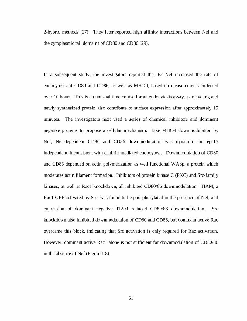
51
2-hybrid methods (27). They later reported high affinity interactions between Nef and
the cytoplasmic tail domains of CD80 and CD86 (29).
In a subsequent study, the investigators reported that F2 Nef increased the rate of
endocytosis of CD80 and CD86, as well as MHC-I, based on measurements collected
over 10 hours. This is an unusual time course for an endocytosis assay, as recycling and
newly synthesized protein also contribute to surface expression after approximately 15
minutes. The investigators next used a series of chemical inhibitors and dominant
negative proteins to propose a cellular mechanism. Like MHC-I downmodulation by
Nef, Nef-dependent CD80 and CD86 downmodulation was dynamin and eps15
independent, inconsistent with clathrin-mediated endocytosis. Downmodulation of CD80
and CD86 depended on actin polymerization as well functional WASp, a protein which
moderates actin filament formation. Inhibitors of protein kinase C (PKC) and Src-family
kinases, as well as Rac1 knockdown, all inhibited CD80/86 downmodulation. TIAM, a
Rac1 GEF activated by Src, was found to be phosphorylated in the presence of Nef, and
expression of dominant negative TIAM reduced CD80/86 downmodulation. Src
knockdown also inhibited downmodulation of CD80 and CD86, but dominant active Rac
overcame this block, indicating that Src activation is only required for Rac activation.
However, dominant active Rac1 alone is not sufficient for downmodulation of CD80/86
in the absence of Nef (Figure 1.8).

52
Figure 1.8. An endocytic model for CD80/86 downmodulation by Nef. Nef binds to
and activates an SFK, which requires Nef-induced PKC activity. The active SFK then
phosphorylates TIAM in an actin-remodeling-dependent manner. Active TIAM in turn
activates Rac1, which again induces actin remodeling. Nef binds tightly to the
cytoplasmic tails of CD80/86 and increases the rate of internalization from the plasma
membrane (PM). Nef then routes CD80/86 to Rab11+ recycling endosomes. Finally,
Nef activates PI3-kinase, which results in transport of CD80/86 to the TGN.
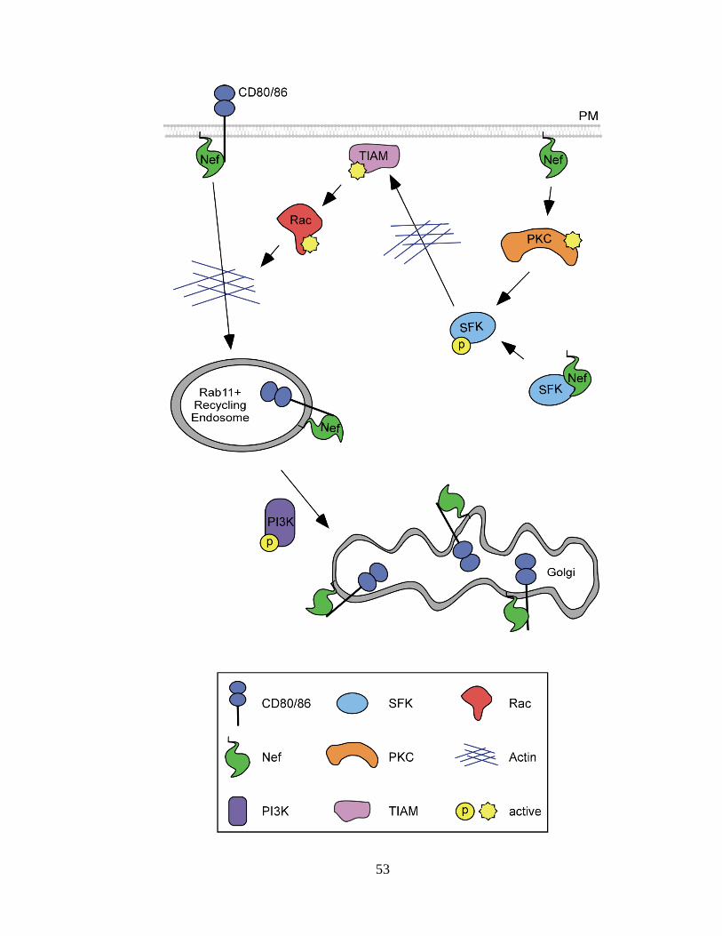
53
54
Finally, microscopic analysis was used to map CD80/86 trafficking in the presence of
Nef. CD80 or CD86 were labeled at the plasma membrane, then examined over time for
colocalization with markers of various cellular compartments. CD80 and CD86 initially
colocalized with Rab11, a key regulator of recycling endosomes. CD80/86 were
subsequently found to colocalize with Golgi markers Rab6 and GM130, but were never
observed in complex with ARF-6 or late endosome marker Rab7 (Figure 1.8) (28).
Therefore, it has been proposed that HIV-1 Nef downmodulates CD80/86 by a unique
signaling mechanism that does not rely on known Nef functional domains.
1.8.4.2. Functional importance of CD80/86 downmodulation
In an experimental system where Nef-dependent CD80 and CD86 downmodulation were
observed, Nef also reduced the ability of murine macrophages to activate OT-1 CTLs.
OT-1 is a transgenic mouse system in which T cells are engineered to express a TCR that
recognizes an ovalbumin epitope in the context of the mouse H-2kb MHC-I molecule. It
is expected that MHC-I downmodulation by Nef would abrogate CTL activation. To
differentiate between the effects of MHC-I and CD80/86 downmodulation, the
investigators expressed a Nef mutant capable of downmodulating CD80 and CD86, but
not MHC-I, and found it to behave similarly to wild type Nef in the OT-1 assay,
providing evidence that MHC-I antigen presentation is not sufficient for CTL activation,
and that Nef inhibits an additional required signal. A G2A myristoylation mutant of Nef,
which is incapable of affecting intracellular trafficking, had no effect on CTL activation.
Together, this evidence supports the conclusion that HIV-1 Nef induces downmodulation
55
of CD80/86 and thereby disrupts CTL activation by macrophages. However, numerous
conflicting reports are not in agreement that CD80 and CD86 are downmodulated by Nef,
indicating that further investigation is warranted.
1.9. Summary
In conclusion, data from a number of laboratories have contributed to our current
understanding of the mechanisms by which HIV-1 Nef affects the trafficking of cellular
proteins to disrupt immune cell activation and anti-HIV activity. We currently have a
good understanding of the mechanism by which Nef affects CD4 expression, although
the importance of this Nef function in vivo is not entirely clear. The field is beginning to
come to a consensus about the mechanism by which Nef downmodulates MHC-I and an
understanding of its role in immune evasion in vitro and in vivo. However, the extent to
which Nef affects other proteins remains ambiguous, with conflicting data originating
from various research groups. A greater understanding of which targets are most
important for Nef biology is needed. This dissertation describes the development of tools
to address this question, insight into the mechanisms involved in Nef-dependent
trafficking pathways, and the development of pharmacologic inhibitors of Nef function
for future therapeutic use.
56
References
1. Aiken, C., J. Konner, N. R. Landau, M. E. Lenburg, and D. Trono. 1994. Nef
induces CD4 endocytosis: requirement for a critical dileucine motif in the
membrane-proximal CD4 cytoplasmic domain. Cell 76:853-64.
2. Akari, H., S. Arold, T. Fukumori, T. Okazaki, K. Strebel, and A. Adachi.
2000. Nef-induced major histocompatibility complex class I down-regulation is
functionally dissociated from its virion incorporation, enhancement of viral
infectivity, and CD4 down-regulation. J Virol 74:2907-2912.
3. Alkhatib, G., C. Combadiere, C. C. Broder, Y. Feng, P. E. Kennedy, P. M.
Murphy, and E. A. Berger. 1996. CC CKR5: a RANTES, MIP-1alpha, MIP-
1beta receptor as a fusion cofactor for macrophage-tropic HIV-1. Science
272:1955-8.
4. Amprey, J. L., J. S. Im, S. J. Turco, H. W. Murray, P. A. Illarionov, G. S.
Besra, S. A. Porcelli, and G. F. Spath. 2004. A subset of liver NK T cells is
activated during Leishmania donovani infection by CD1d-bound
lipophosphoglycan. J Exp Med 200:895-904.
5. Aniento, F., F. Gu, R. G. Parton, and J. Gruenberg. 1996. An endosomal beta
COP is involved in the pH-dependent formation of transport vesicles destined for
late endosomes. J Cell Biol 133:29-41.
6. Arganaraz, E. R., M. Schindler, F. Kirchhoff, M. J. Cortes, and J. Lama.
2003. Enhanced CD4 down-modulation by late stage HIV-1 nef alleles is
associated with increased Env incorporation and viral replication. J Biol Chem
278:33912-9.
7. Arold, S., P. Franken, M. P. Strub, F. Hoh, S. Benichou, R. Benarous, and C.
Dumas. 1997. The crystal structure of HIV-1 Nef protein bound to the Fyn kinase
SH3 domain suggests a role for this complex in altered T cell receptor signaling.
Structure 5:1361-72.
8. Atkins, K. M., L. Thomas, R. T. Youker, M. J. Harriff, F. Pissani, H. You,
and G. Thomas. 2008. HIV-1 Nef binds PACS-2 to assemble a multikinase
cascade that triggers major histocompatibility complex class I (MHC-I) down-
regulation: analysis using short interfering RNA and knock-out mice. J Biol Chem
283:11772-84.
9. Austin, C., I. Hinners, and S. A. Tooze. 2000. Direct and GTP-dependent
interaction of ADP-ribosylation factor 1 with clathrin adaptor protein AP-1 on
immature secretory granules. J Biol Chem 275:21862-9.
10. Balch, W. E., R. A. Kahn, and R. Schwaninger. 1992. ADP-ribosylation factor
is required for vesicular trafficking between the endoplasmic reticulum and the
cis-Golgi compartment. J Biol Chem 267:13053-61.
11. Baltimore, D. 1970. RNA-dependent DNA polymerase in virions of RNA tumour
viruses. Nature 226:1209-11.
12. Bartz, S. R., M. E. Rogel, and M. Emerman. 1996. Human immunodeficiency
virus type 1 cell cycle control: Vpr is cytostatic and mediates G2 accumulation by
a mechanism which differs from DNA damage checkpoint control. J Virol
70:2324-31.
57
13. Baugh, L. L., J. V. Garcia, and J. L. Foster. 2008. Functional characterization
of the human immunodeficiency virus type 1 Nef acidic domain. J Virol 82:9657-
67.
14. Bell, I., T. M. Schaefer, R. P. Trible, A. Amedee, and T. A. Reinhart. 2001.
Down-modulation of the costimulatory molecule, CD28, is a conserved activity of
multiple SIV Nefs and is dependent on histidine 196 of Nef. Virology 283:148-
58.
15. Benichou, S., M. Bomsel, M. Bodeus, H. Durand, M. doute, F. Letourneur, J.
Camonis, and R. Benarous. 1994. Physical interaction of the HIV-1 Nef protein
wih b-cop, a component of non-clathrin coated vesicles essential for membrane
traffic. J Biol Chem 269:30073-30076.
16. Benmerah, A., C. Lamaze, B. Begue, S. L. Schmid, A. Dautry-Varsat, and N.
Cerf-Bensussan. 1998. AP-2/Eps15 interaction is required for receptor-mediated
endocytosis. J Cell Biol 140:1055-62.
17. Benson, R. E., A. Sanfridson, J. S. Ottinger, C. Doyle, and B. R. Cullen. 1993.
Downregulation of cell-surface CD4 expression by simian immunodeficiency
virus Nef prevents viral super infection. J Exp Med 177:1561-6.
18. Berke, G. 1995. The CTL's kiss of death. Cell 81:9-12.
19. Birch, M. R., J. C. Learmont, W. B. Dyer, N. J. Deacon, J. J. Zaunders, N.
Saksena, A. L. Cunningham, J. Mills, and J. S. Sullivan. 2001. An
examination of signs of disease progression in survivors of the Sydney Blood
Bank Cohort (SBBC). J Clin Virol 22:263-70.
20. Blagoveshchenskaya, A. D., L. Thomas, S. F. Feliciangeli, C. H. Hung, and G.
Thomas. 2002. HIV-1 Nef downregulates MHC-I by a PACS-1- and PI3K-
regulated ARF6 endocytic pathway. Cell 111:853-66.
21. Bresnahan, P. A., W. Yonemoto, S. Ferrell, D. Williams-Herman, R.
Geleziunas, and W. C. Greene. 1998. A dileucine motif in HIV-1 Nef acts as an
internalization signal for CD4 downregulation and binds the AP-1 clathrin
adaptor. Curr Biol 8:1235-8.
22. Bukrinsky, M. I., N. Sharova, M. P. Dempsey, T. L. Stanwick, A. G.
Bukrinskaya, S. Haggerty, and M. Stevenson. 1992. Active nuclear import of
human immunodeficiency virus type 1 preintegration complexes. Proc Natl Acad
Sci U S A 89:6580-4.
23. Bukrinsky, M. I., N. Sharova, T. L. McDonald, T. Pushkarskaya, W. G.
Tarpley, and M. Stevenson. 1993. Association of integrase, matrix, and reverse
transcriptase antigens of human immunodeficiency virus type 1 with viral nucleic
acids following acute infection. Proc Natl Acad Sci U S A 90:6125-9.
24. Carbone, R., S. Fre, G. Iannolo, F. Belleudi, P. Mancini, P. G. Pelicci, M. R.
Torrisi, and P. P. Di Fiore. 1997. eps15 and eps15R are essential components of
the endocytic pathway. Cancer Res 57:5498-504.
25. Carl, S., R. Daniels, A. J. Iafrate, P. Easterbrook, T. C. Greenough, J.
Skowronski, and F. Kirchhoff. 2000. Partial "repair" of defective NEF genes in
a long-term nonprogressor with human immunodeficiency virus type 1 infection.
J Infect Dis 181:132-40.
26. Carl, S., T. C. Greenough, M. Krumbiegel, M. Greenberg, J. Skowronski, J.
L. Sullivan, and F. Kirchhoff. 2001. Modulation of different human
58
immunodeficiency virus type 1 Nef functions during progression to AIDS. J Virol
75:3657-65.
27. Chaudhry, A., S. R. Das, A. Hussain, S. Mayor, A. George, V. Bal, S. Jameel,
and S. Rath. 2005. The Nef protein of HIV-1 induces loss of cell surface
costimulatory molecules CD80 and CD86 in APCs. J Immunol 175:4566-74.
28. Chaudhry, A., S. R. Das, S. Jameel, A. George, V. Bal, S. Mayor, and S.
Rath. 2008. HIV-1 Nef induces a Rab11-dependent routing of endocytosed
immune costimulatory proteins CD80 and CD86 to the Golgi. Traffic 9:1925-35.
29. Chaudhry, A., S. R. Das, S. Jameel, A. George, V. Bal, S. Mayor, and S.
Rath. 2007. A two-pronged mechanism for HIV-1 Nef-mediated endocytosis of
immune costimulatory molecules CD80 and CD86. Cell Host Microbe 1:37-49.
30. Chaudhuri, R., O. W. Lindwasser, W. J. Smith, J. H. Hurley, and J. S.
Bonifacino. 2007. Downregulation of CD4 by human immunodeficiency virus
type 1 Nef is dependent on clathrin and involves direct interaction of Nef with the
AP2 clathrin adaptor. J Virol 81:3877-90.
31. Chaudhuri, R., R. Mattera, O. W. Lindwasser, M. S. Robinson, and J. S.
Bonifacino. 2009. A basic patch on alpha-adaptin is required for binding of
human immunodeficiency virus type 1 Nef and cooperative assembly of a CD4-
Nef-AP-2 complex. J Virol 83:2518-30.
32. Chen, N., C. McCarthy, H. Drakesmith, D. Li, V. Cerundolo, A. J.
McMichael, G. R. Screaton, and X. N. Xu. 2006. HIV-1 down-regulates the
expression of CD1d via Nef. Eur J Immunol 36:278-86.
33. Cho, S., K. S. Knox, L. M. Kohli, J. J. He, M. A. Exley, S. B. Wilson, and R.
R. Brutkiewicz. 2005. Impaired cell surface expression of human CD1d by the
formation of an HIV-1 Nef/CD1d complex. Virology 337:242-52.
34. Clever, J., C. Sassetti, and T. G. Parslow. 1995. RNA secondary structure and
binding sites for gag gene products in the 5' packaging signal of human
immunodeficiency virus type 1. J Virol 69:2101-9.
35. Cohen, G. B., R. T. Gandhi, D. M. Davis, O. Mandelboim, B. K. Chen, J. L.
Strominger, and D. Baltimore. 1999. The selective downregulation of class I
major histocompatibility complex proteins by HIV-1 protects HIV-infected cells
from NK cells. Immunity 10:661-671.
36. Coleman, S. H., D. Hitchin, C. M. Noviello, and J. C. Guatelli. 2006. HIV-1
Nef stabilizes AP-1 on membranes without inducing ARF1-independent de novo
attachment. Virology 345:148-55.
37. Collins, K., B. Chen, S. Kalams, B. Walker, and D. Baltimore. 1998. HIV-1
Nef protein protects infected primary human cells from killing by cytotoxic T
lymphocytes. Nature 391:397-401.
38. Collins, K. L. 2004. Resistance of HIV-infected cells to cytotoxic T lymphocytes.
Microbes Infect 6:494-500.
39. Collins, K. L., B. K. Chen, S. A. Kalams, B. D. Walker, and D. Baltimore.
1998. HIV-1 Nef protein protects infected primary cells against killing by
cytotoxic T lymphocytes. Nature 391:397-401.
40. Craig, H., M. Pandori, and J. Guatelli. 1998. Interaction of HIV-1 Nef with the
cellular dileucine-based sorting pathway is required for CD4 down-regulation and
optimal viral infectivity. Proc Natl Acad Sci 95:11229-11234.

59
41. Craig, H. M., M. W. Pandori, and J. C. Guatelli. 1998. Interaction of HIV-1
Nef with the cellular dileucine-based sorting pathway is required for CD4 down-
regulation and optimal viral infectivity. Proc Natl Acad Sci U S A 95:11229-34.
42. Craig, H. M., T. R. Reddy, N. L. Riggs, P. P. Dao, and J. C. Guatelli. 2000.
Interactions of HIV-1 nef with the mu subunits of adaptor protein complexes 1, 2,
and 3: role of the dileucine-based sorting motif. Virology 271:9-17.
43. Crise, B., L. Buonocore, and J. K. Rose. 1990. CD4 is retained in the
endoplasmic reticulum by the human immunodeficiency virus type 1 glycoprotein
precursor. J Virol 64:5585-93.
44. Dalgleish, A. G., P. C. Beverley, P. R. Clapham, D. H. Crawford, M. F.
Greaves, and R. A. Weiss. 1984. The CD4 (T4) antigen is an essential
component of the receptor for the AIDS retrovirus. Nature 312:763-7.
45. Daro, E., D. Sheff, M. Gomez, T. Kreis, and I. Mellman. 1997. Inhibition of
endosome function in CHO cells bearing a temperature-sensitive defect in the
coatomer (COPI) component epsilon-COP. J Cell Biol 139:1747-59.
46. Deacon, N. J., A. Tsykin, A. Solomon, K. Smith, M. Ludford-Menting, D. J.
Hooker, D. A. McPhee, A. L. Greenway, A. Ellett, C. Chatfield, and et al. 1995. Genomic structure of an attenuated quasi species of HIV-1 from a blood
transfusion donor and recipients. Science 270:988-991.
47. Deacon, N. J., A. Tsykin, A. Solomon, K. Smith, M. Ludford-Menting, D. J.
Hooker, D. A. McPhee, A. L. Greenway, A. Ellett, C. Chatfield, V. A.
Lawson, S. Crowe, A. Maerz, S. Sonza, J. Learmont, J. S. Sullivan, A.
Cunningham, D. Dwyer, D. Dowton, and J. Mills. 1995. Genomic structure of
an attenuated quasi species of HIV-1 from a blood transfusion donor and
recipients. Science 270:988-91.
48. Deng, H., R. Liu, W. Ellmeier, S. Choe, D. Unutmaz, M. Burkhart, P. Di
Marzio, S. Marmon, R. E. Sutton, C. M. Hill, C. B. Davis, S. C. Peiper, T. J.
Schall, D. R. Littman, and N. R. Landau. 1996. Identification of a major co-
receptor for primary isolates of HIV-1. Nature 381:661-6.
49. Dikeakos, J. D., K. M. Atkins, L. Thomas, L. Emert-Sedlak, I. J. Byeon, J.
Jung, J. Ahn, M. D. Wortman, B. Kukull, M. Saito, H. Koizumi, D. M.
Williamson, M. Hiyoshi, E. Barklis, M. Takiguchi, S. Suzu, A. M.
Gronenborn, T. E. Smithgall, and G. Thomas. 2010. Small Molecule Inhibition
of HIV-1-Induced MHC-I Downregulation Identifies a Temporally Regulated
Switch in Nef Action. Mol Biol Cell.
50. Donaldson, J. G., D. Cassel, R. A. Kahn, and R. D. Klausner. 1992. ADP-
ribosylation factor, a small GTP-binding protein, is required for binding of the
coatomer protein beta-COP to Golgi membranes. Proc Natl Acad Sci U S A
89:6408-12.
51. Donaldson, J. G., and R. D. Klausner. 1994. ARF: a key regulatory switch in
membrane traffic and organelle structure. Curr Opin Cell Biol 6:527-32.
52. Doray, B., P. Ghosh, J. Griffith, H. J. Geuze, and S. Kornfeld. 2002.
Cooperation of GGAs and AP-1 in packaging MPRs at the trans-Golgi network.
Science 297:1700-3.
60
53. Doray, B., I. Lee, J. Knisely, G. Bu, and S. Kornfeld. 2007. The gamma/sigma1
and alpha/sigma2 hemicomplexes of clathrin adaptors AP-1 and AP-2 harbor the
dileucine recognition site. Mol Biol Cell 18:1887-96.
54. Douek, D. C., J. M. Brenchley, M. R. Betts, D. R. Ambrozak, B. J. Hill, Y.
Okamoto, J. P. Casazza, J. Kuruppu, K. Kunstman, S. Wolinsky, Z.
Grossman, M. Dybul, A. Oxenius, D. A. Price, M. Connors, and R. A. Koup. 2002. HIV preferentially infects HIV-specific CD4+ T cells. Nature 417:95-98.
55. Douglas, J. L., J. K. Gustin, K. Viswanathan, M. Mansouri, A. V. Moses, and
K. Fruh. 2010. The great escape: viral strategies to counter BST-2/tetherin. PLoS
Pathog 6:e1000913.
56. Dube, M., M. G. Bego, C. Paquay, and E. A. Cohen. 2010. Modulation of HIV-
1-host interaction: role of the Vpu accessory protein. Retrovirology 7:114.
57. Eichholtz, T., P. Vossebeld, M. van Overveld, and H. Ploegh. 1992. Activation
of protein kinase C accelerates internalization of transferrin receptor but not of
major histocompatibility complex class I, independent of their phosphorylation
status. J Biol Chem 267:22490-22495.
58. Ellison, V., and P. O. Brown. 1994. A stable complex between integrase and
viral DNA ends mediates human immunodeficiency virus integration in vitro.
Proc Natl Acad Sci U S A 91:7316-20.
59. Erdtmann, L., K. Janvier, G. Raposo, H. M. Craig, P. Benaroch, C. Berlioz-
Torrent, J. C. Guatelli, R. Benarous, and S. Benichou. 2000. Two independent
regions of HIV-1 Nef are required for connection with the endocytic pathway
through binding to the mu 1 chain of AP1 complex. Traffic 1:871-83.
60. Fackler, O. T., W. Luo, M. Geyer, A. S. Alberts, and B. M. Peterlin. 1999.
Activation of Vav by Nef induces cytoskeletal rearrangements and downstream
effector functions. Mol Cell 3:729-39.
61. Farnet, C. M., and F. D. Bushman. 1997. HIV-1 cDNA integration: requirement
of HMG I(Y) protein for function of preintegration complexes in vitro. Cell
88:483-92.
62. Farnet, C. M., and W. A. Haseltine. 1991. Determination of viral proteins
present in the human immunodeficiency virus type 1 preintegration complex. J
Virol 65:1910-5.
63. Faure, J., R. Stalder, C. Borel, K. Sobo, V. Piguet, N. Demaurex, J.
Gruenberg, and D. Trono. 2004. ARF1 regulates Nef-induced CD4 degradation.
Curr Biol 14:1056-64.
64. Feng, Y., C. C. Broder, P. E. Kennedy, and E. A. Berger. 1996. HIV-1 entry
cofactor: functional cDNA cloning of a seven-transmembrane, G protein-coupled
receptor. Science 272:872-7.
65. Fischer, K., E. Scotet, M. Niemeyer, H. Koebernick, J. Zerrahn, S. Maillet, R.
Hurwitz, M. Kursar, M. Bonneville, S. H. Kaufmann, and U. E. Schaible. 2004. Mycobacterial phosphatidylinositol mannoside is a natural antigen for
CD1d-restricted T cells. Proc Natl Acad Sci U S A 101:10685-90.
66. Foti, M., A. Mangasarian, V. Piguet, D. P. Lew, K. H. Krause, D. Trono, and
J. L. Carpentier. 1997. Nef-mediated clathrin-coated pit formation. J Cell Biol
139:37-47.

61
67. Franken, P., S. Arold, A. Padilla, M. Bodeus, F. Hoh, M. P. Strub, M. Boyer,
M. Jullien, R. Benarous, and C. Dumas. 1997. HIV-1 Nef protein: purification,
crystallizations, and preliminary X-ray diffraction studies. Protein Sci 6:2681-3.
68. Ganser-Pornillos, B. K., M. Yeager, and W. I. Sundquist. 2008. The structural
biology of HIV assembly. Curr Opin Struct Biol 18:203-17.
69. Garcia, J. V., and A. D. Miller. 1991. Serine phosphorylation-independent
downregulation of cell-surface CD4 by nef. Nature 350:508-11.
70. Geldmacher, C., J. R. Currier, E. Herrmann, A. Haule, E. Kuta, F.
McCutchan, L. Njovu, S. Geis, O. Hoffmann, L. Maboko, C. Williamson, D.
Birx, A. Meyerhans, J. Cox, and M. Hoelscher. 2007. CD8 T-cell recognition
of multiple epitopes within specific Gag regions is associated with maintenance of
a low steady-state viremia in human immunodeficiency virus type 1-seropositive
patients. J Virol 81:2440-8.
71. Gheysen, D., E. Jacobs, F. de Foresta, C. Thiriart, M. Francotte, D. Thines,
and M. De Wilde. 1989. Assembly and release of HIV-1 precursor Pr55gag
virus-like particles from recombinant baculovirus-infected insect cells. Cell
59:103-12.
72. Giolo, G., F. Neri, N. Casartelli, M. Potesta, F. Belleudi, M. R. Torrisi, and
M. Doria. 2007. Internalization and intracellular retention of CD4 are two
separate functions of the human immunodeficiency virus type 1 Nef protein. J
Gen Virol 88:3133-8.
73. Goh, W. C., M. E. Rogel, C. M. Kinsey, S. F. Michael, P. N. Fultz, M. A.
Nowak, B. H. Hahn, and M. Emerman. 1998. HIV-1 Vpr increases viral
expression by manipulation of the cell cycle: a mechanism for selection of Vpr in
vivo. Nat Med 4:65-71.
74. Gorry, P. R., M. Churchill, J. Learmont, C. Cherry, W. B. Dyer, S. L.
Wesselingh, and J. S. Sullivan. 2007. Replication-dependent pathogenicity of
attenuated nef-deleted HIV-1 in vivo. J Acquir Immune Defic Syndr 46:390-4.
75. Gorry, P. R., D. A. McPhee, E. Verity, W. B. Dyer, S. L. Wesselingh, J.
Learmont, J. S. Sullivan, M. Roche, J. J. Zaunders, D. Gabuzda, S. M.
Crowe, J. Mills, S. R. Lewin, B. J. Brew, A. L. Cunningham, and M. J.
Churchill. 2007. Pathogenicity and immunogenicity of attenuated, nef-deleted
HIV-1 strains in vivo. Retrovirology 4:66.
76. Greenberg, M., L. DeTulleo, I. Rapoport, J. Skowronski, and T.
Kirchhausen. 1998. A dileucine motif in HIV-1 Nef is essential for sorting into
clathrin-coated pits and for downregulation of CD4. Curr Biol 8:1239-42.
77. Greenberg, M., A. Iafrate, and J. Skowronski. 1998. The SH3 domain-binding
surface and an acidic motif in HIV-1 Nef regulate trafficking of class I MHC
complexes. EMBO J 17:2777-2789.
78. Greenberg, M. E., S. Bronson, M. Lock, M. Neumann, G. N. Pavlakis, and J.
Skowronski. 1997. Co-localization of HIV-1 Nef with the AP-2 adaptor protein
complex correlates with Nef-induced CD4 down-regulation. EMBO J 16:6964-
76.
79. Grzesiek, S., A. Bax, G. M. Clore, A. M. Gronenborn, J. S. Hu, J. Kaufman,
I. Palmer, S. J. Stahl, and P. T. Wingfield. 1996. The solution structure of HIV-

62
1 Nef reveals an unexpected fold and permits delineation of the binding surface
for the SH3 domain of Hck tyrosine protein kinase. Nat Struct Biol 3:340-5.
80. Grzesiek, S., S. J. Stahl, P. T. Wingfield, and A. Bax. 1996. The CD4
determinant for downregulation by HIV-1 Nef directly binds to Nef. Mapping of
the Nef binding surface by NMR. Biochemistry 35:10256-61.
81. Gu, F., F. Aniento, R. G. Parton, and J. Gruenberg. 1997. Functional
dissection of COP-I subunits in the biogenesis of multivesicular endosomes. J
Cell Biol 139:1183-95.
82. Gu, F., and J. Gruenberg. 2000. ARF1 regulates pH-dependent COP functions
in the early endocytic pathway. J Biol Chem 275:8154-60.
83. Hansen, T. H., and M. Bouvier. 2009. MHC class I antigen presentation:
learning from viral evasion strategies. Nat Rev Immunol 9:503-13.
84. Harris, M. P., and J. C. Neil. 1994. Myristoylation-dependent binding of HIV-1
Nef to CD4. J Mol Biol 241:136-42.
85. Helms, J. B., and J. E. Rothman. 1992. Inhibition by brefeldin A of a Golgi
membrane enzyme that catalyses exchange of guanine nucleotide bound to ARF.
Nature 360:352-4.
86. Hill, M., G. Tachedjian, and J. Mak. 2005. The packaging and maturation of
the HIV-1 Pol proteins. Curr HIV Res 3:73-85.
87. Hung, C. H., L. Thomas, C. E. Ruby, K. M. Atkins, N. P. Morris, Z. A.
Knight, I. Scholz, E. Barklis, A. D. Weinberg, K. M. Shokat, and G. Thomas. 2007. HIV-1 Nef assembles a Src family kinase-ZAP-70/Syk-PI3K cascade to
downregulate cell-surface MHC-I. Cell Host Microbe 1:121-33.
88. Jabbar, M. A., and D. P. Nayak. 1990. Intracellular interaction of human
immunodeficiency virus type 1 (ARV-2) envelope glycoprotein gp160 with CD4
blocks the movement and maturation of CD4 to the plasma membrane. J Virol
64:6297-304.
89. Jackson, C. L., and J. E. Casanova. 2000. Turning on ARF: the Sec7 family of
guanine-nucleotide-exchange factors. Trends Cell Biol 10:60-7.
90. Janvier, K., H. Craig, D. Hitchin, R. Madrid, N. Sol-Foulon, L. Renault, J.
Cherfils, D. Cassel, S. Benichou, and J. Guatelli. 2003. HIV-1 Nef stabilizes
the association of adaptor protein complexes with membranes. J Biol Chem
278:8725-32.
91. Janvier, K., H. Craig, S. Le Gall, R. Benarous, J. Guatelli, O. Schwartz, and
S. Benichou. 2001. Nef-induced CD4 downregulation: a diacidic sequence in
human immunodeficiency virus type 1 Nef does not function as a protein sorting
motif through direct binding to beta-COP. J Virol 75:3971-6.
92. Janvier, K., Y. Kato, M. Boehm, J. R. Rose, J. A. Martina, B. Y. Kim, S.
Venkatesan, and J. S. Bonifacino. 2003. Recognition of dileucine-based sorting
signals from HIV-1 Nef and LIMP-II by the AP-1 gamma-sigma1 and AP-3 delta-
sigma3 hemicomplexes. J Cell Biol 163:1281-90.
93. Jin, Y. J., C. Y. Cai, X. Zhang, H. T. Zhang, J. A. Hirst, and S. J. Burakoff.
2005. HIV Nef-mediated CD4 down-regulation is adaptor protein complex 2
dependent. J Immunol 175:3157-64.
63
94. Jin, Y. J., X. Zhang, J. G. Boursiquot, and S. J. Burakoff. 2004. CD4
phosphorylation partially reverses Nef down-regulation of CD4. J Immunol
173:5495-500.
95. Jouvenet, N., S. J. Neil, C. Bess, M. C. Johnson, C. A. Virgen, S. M. Simon,
and P. D. Bieniasz. 2006. Plasma membrane is the site of productive HIV-1
particle assembly. PLoS Biol 4:e435.
96. Jovanovic, O. A., F. D. Brown, and J. G. Donaldson. 2006. An effector domain
mutant of Arf6 implicates phospholipase D in endosomal membrane recycling.
Mol Biol Cell 17:327-35.
97. Kahn, R. A., P. Randazzo, T. Serafini, O. Weiss, C. Rulka, J. Clark, M.
Amherdt, P. Roller, L. Orci, and J. E. Rothman. 1992. The amino terminus of
ADP-ribosylation factor (ARF) is a critical determinant of ARF activities and is a
potent and specific inhibitor of protein transport. J Biol Chem 267:13039-46.
98. Kalpana, G. V., S. Marmon, W. Wang, G. R. Crabtree, and S. P. Goff. 1994.
Binding and stimulation of HIV-1 integrase by a human homolog of yeast
transcription factor SNF5. Science 266:2002-6.
99. Kao, S. Y., A. F. Calman, P. A. Luciw, and B. M. Peterlin. 1987. Anti-
termination of transcription within the long terminal repeat of HIV-1 by tat gene
product. Nature 330:489-93.
100. Karageorgos, L., P. Li, and C. Burrell. 1993. Characterization of HIV
replication complexes early after cell-to-cell infection. AIDS Res Hum
Retroviruses 9:817-23.
101. Kasper, M. R., and K. L. Collins. 2003. Nef-mediated disruption of HLA-A2
transport to the cell surface in T cells. J Virol 77:3041-9.
102. Kasper, M. R., J. F. Roeth, M. Williams, T. M. Filzen, R. I. Fleis, and K. L.
Collins. 2005. HIV-1 Nef disrupts antigen presentation early in the secretory
pathway. J Biol Chem 280:12840-8.
103. Kawano, T., J. Cui, Y. Koezuka, I. Toura, Y. Kaneko, K. Motoki, H. Ueno,
R. Nakagawa, H. Sato, E. Kondo, H. Koseki, and M. Taniguchi. 1997. CD1d-
restricted and TCR-mediated activation of valpha14 NKT cells by
glycosylceramides. Science 278:1626-9.
104. Kelly, B. T., A. J. McCoy, K. Spate, S. E. Miller, P. R. Evans, S. Honing, and
D. J. Owen. 2008. A structural explanation for the binding of endocytic dileucine
motifs by the AP2 complex. Nature 456:976-979.
105. Kestler, H. W., 3rd, D. J. Ringler, K. Mori, D. L. Panicali, P. K. Sehgal, M. D.
Daniel, and R. C. Desrosiers. 1991. Importance of the nef gene for maintenance
of high virus loads and for development of AIDS. Cell 65:651-62.
106. Kiepiela, P., K. Ngumbela, C. Thobakgale, D. Ramduth, I. Honeyborne, E.
Moodley, S. Reddy, C. de Pierres, Z. Mncube, N. Mkhwanazi, K. Bishop, M.
van der Stok, K. Nair, N. Khan, H. Crawford, R. Payne, A. Leslie, J. Prado,
A. Prendergast, J. Frater, N. McCarthy, C. Brander, G. H. Learn, D. Nickle,
C. Rousseau, H. Coovadia, J. I. Mullins, D. Heckerman, B. D. Walker, and P.
Goulder. 2007. CD8+ T-cell responses to different HIV proteins have discordant
associations with viral load. Nat Med 13:46-53.
107. Kim, Y. H., S. H. Chang, J. H. Kwon, and S. S. Rhee. 1999. HIV-1 Nef plays
an essential role in two independent processes in CD4 down-regulation:
64
dissociation of the CD4-p56(lck) complex and targeting of CD4 to lysosomes.
Virology 257:208-19.
108. Kinjo, Y., E. Tupin, D. Wu, M. Fujio, R. Garcia-Navarro, M. R. Benhnia, D.
M. Zajonc, G. Ben-Menachem, G. D. Ainge, G. F. Painter, A. Khurana, K.
Hoebe, S. M. Behar, B. Beutler, I. A. Wilson, M. Tsuji, T. J. Sellati, C. H.
Wong, and M. Kronenberg. 2006. Natural killer T cells recognize diacylglycerol
antigens from pathogenic bacteria. Nat Immunol 7:978-86.
109. Kinjo, Y., D. Wu, G. Kim, G. W. Xing, M. A. Poles, D. D. Ho, M. Tsuji, K.
Kawahara, C. H. Wong, and M. Kronenberg. 2005. Recognition of bacterial
glycosphingolipids by natural killer T cells. Nature 434:520-5.
110. Kirchhoff, F. 2010. Immune evasion and counteraction of restriction factors by
HIV-1 and other primate lentiviruses. Cell Host Microbe 8:55-67.
111. Kirchoff, F., T. Greenough, D. Brettler, J. Sullivan, and R. Desrosiers. 1995.
Absence of intact nef sequences in a long-term survivor with nonprogressive
HIV-1 infection. N Engl J Med 332:228-232.
112. Klumperman, J., A. Hille, T. Veenendaal, V. Oorschot, W. Stoorvogel, K.
von Figura, and H. J. Geuze. 1993. Differences in the endosomal distributions
of the two mannose 6-phosphate receptors. J Cell Biol 121:997-1010.
113. Lama, J., A. Mangasarian, and D. Trono. 1999. Cell-surface expression of
CD4 reduces HIV-1 infectivity by blocking Env incorporation in a Nef- and Vpu-
inhibitable manner. Curr Biol 9:622-31.
114. Larsen, J. E., R. H. Massol, T. J. Nieland, and T. Kirchhausen. 2004. HIV
Nef-mediated major histocompatibility complex class I down-modulation is
independent of Arf6 activity. Mol Biol Cell 15:323-31.
115. Lasky, L. A., G. Nakamura, D. H. Smith, C. Fennie, C. Shimasaki, E. Patzer,
P. Berman, T. Gregory, and D. J. Capon. 1987. Delineation of a region of the
human immunodeficiency virus type 1 gp120 glycoprotein critical for interaction
with the CD4 receptor. Cell 50:975-85.
116. Le Gall, S., F. Buseyne, A. Trocha, B. D. Walker, J. M. Heard, and O.
Schwartz. 2000. Distinct trafficking pathways mediate Nef-induced and clathrin-
dependent major histocompatibility complex class I down-regulation. J Virol
74:9256-66.
117. Le Gall, S., L. Erdtmann, S. Benichou, C. Berlloz-Torrent, L. Liu, R.
Benarous, J. Heard, and O. Schwartz. 1998. Nef interacts with mu subunit of
clathrin adaptor complexes and reveals a cryptic sorting signal in MHC I
molecules. Immunity 8:483-495.
118. Learmont, J., B. Tindall, L. Evans, A. Cunningham, P. Cunningham, J.
Wells, R. Penny, J. Kaldor, and D. A. Cooper. 1992. Long-term symptomless
HIV-1 infection in recipients of blood products from a single donor. Lancet
340:863-7.
119. Lee, C. H., K. Saksela, U. A. Mirza, B. T. Chait, and J. Kuriyan. 1996. Crystal
structure of the conserved core of HIV-1 Nef complexed with a Src family SH3
domain. Cell 85:931-42.
120. Leonard, J. A., T. Filzen, C. C. Carter, M. Schaefer, and K. L. Collins. 2011.
HIV-1 Nef Disrupts Intracellular Trafficking of Major Histocompatibility

65
Complex Class I, CD4, CD8, and CD28 by Distinct Pathways That Share
Common Elements. J Virol 85:6867-81.
121. Lever, A., H. Gottlinger, W. Haseltine, and J. Sodroski. 1989. Identification of
a sequence required for efficient packaging of human immunodeficiency virus
type 1 RNA into virions. J Virol 63:4085-7.
122. Lewinsohn, D. A., R. Lines, D. M. Lewinsohn, S. R. Riddell, P. D. Greenberg,
M. Emerman, and S. R. Bartz. 2002. HIV-1 Vpr does not inhibit CTL-mediated
apoptosis of HIV-1 infected cells. Virology 294:13-21.
123. Lewis, M. J., A. Balamurugan, A. Ohno, S. Kilpatrick, H. L. Ng, and O. O.
Yang. 2008. Functional adaptation of Nef to the immune milieu of HIV-1
infection in vivo. J Immunol 180:4075-81.
124. Lindwasser, O. W., W. J. Smith, R. Chaudhuri, P. Yang, J. H. Hurley, and J.
S. Bonifacino. 2008. A diacidic motif in human immunodeficiency virus type 1
Nef is a novel determinant of binding to AP-2. J Virol 82:1166-74.
125. Linsley, P. S., W. Brady, L. Grosmaire, A. Aruffo, N. K. Damle, and J. A.
Ledbetter. 1991. Binding of the B cell activation antigen B7 to CD28
costimulates T cell proliferation and interleukin 2 mRNA accumulation. J Exp
Med 173:721-30.
126. Liu, L. X., N. Heveker, O. T. Fackler, S. Arold, S. Le Gall, K. Janvier, B. M.
Peterlin, C. Dumas, O. Schwartz, S. Benichou, and R. Benarous. 2000.
Mutation of a conserved residue (D123) required for oligomerization of human
immunodeficiency virus type 1 Nef protein abolishes interaction with human
thioesterase and results in impairment of Nef biological functions. J Virol
74:5310-5319.
127. Lubben, N. B., D. A. Sahlender, A. M. Motley, P. J. Lehner, P. Benaroch, and
M. S. Robinson. 2007. HIV-1 Nef-induced Down-Regulation of MHC Class I
Requires AP-1 and Clathrin but Not PACS-1 and Is Impeded by AP-2. Mol Biol
Cell 18:3351-65.
128. Lundquist, C. A., M. Tobiume, J. Zhou, D. Unutmaz, and C. Aiken. 2002.
Nef-mediated downregulation of CD4 enhances human immunodeficiency virus
type 1 replication in primary T lymphocytes. J Virol 76:4625-33.
129. Luo, T., S. J. Anderson, and J. V. Garcia. 1996. Inhibition of Nef- and phorbol
ester-induced CD4 degradation by macrolide antibiotics. J Virol 70:1527-34.
130. Maccormac, L. P., J. M. Jacque, and B. Chain. 2004. The functional
consequences of delivery of HIV-1 Nef to dendritic cells using an adenoviral
vector. Vaccine 22:528-35.
131. Machy, P., A. Truneh, D. Gennaro, and S. Hoffstein. 1987. Major
histocompatibility complex class I molecules internalized via coated pits in T
lymphocytes. Nature 328:724-726.
132. Mangasarian, A., M. Foti, C. Aiken, D. Chin, J. L. Carpentier, and D. Trono.
1997. The HIV-1 Nef protein acts as a connector with sorting pathways in the
Golgi and at the plasma membrane. Immunity 6:67-77.
133. Mangasarian, A., V. Piguet, J. K. Wang, Y. L. Chen, and D. Trono. 1999.
Nef-induced CD4 and major histocompatibility complex class I (MHC-I) down-
regulation are governed by distinct determinants: N-terminal alpha helix and
proline repeat of Nef selectively regulate MHC-I trafficking. J Virol 73:1964-73.
66
134. Mann, J., C. N. Patrick, M. S. Cragg, J. Honeychurch, D. A. Mann, and M.
Harris. 2005. Functional analysis of HIV type 1 Nef reveals a role for PAK2 as a
regulator of cell phenotype and function in the murine dendritic cell line, DC2.4. J
Immunol 175:6560-9.
135. Mariani, R., and J. Skowronski. 1993. CD4 down-regulation by nef alleles
isolated from human immunodeficiency virus type 1-infected individuals. Proc
Natl Acad Sci U S A 90:5549-53.
136. Mattner, J., K. L. Debord, N. Ismail, R. D. Goff, C. Cantu, 3rd, D. Zhou, P.
Saint-Mezard, V. Wang, Y. Gao, N. Yin, K. Hoebe, O. Schneewind, D.
Walker, B. Beutler, L. Teyton, P. B. Savage, and A. Bendelac. 2005.
Exogenous and endogenous glycolipid antigens activate NKT cells during
microbial infections. Nature 434:525-9.
137. Mellors, J. W., C. Rinaldo, P. Gupta, R. M. White, J. A. Todd, and L. A.
Kingsley. 1996. Prognosis in HIV-1 infection predicted by the quantity of virus in
plasma. Science 272:1167-1170.
138. Michel, N., I. Allespach, S. Venzke, O. T. Fackler, and O. T. Keppler. 2005.
The Nef protein of human immunodeficiency virus establishes superinfection
immunity by a dual strategy to downregulate cell-surface CCR5 and CD4. Curr
Biol 15:714-23.
139. Miller, M. D., M. B. Feinberg, and W. C. Greene. 1994. The HIV-1 nef gene
acts as a positive viral infectivity factor. Trends Microbiol 2:294-8.
140. Miller, M. D., M. T. Warmerdam, I. Gaston, W. C. Greene, and M. B.
Feinberg. 1994. The human immunodeficiency virus-1 nef gene product: a
positive factor for viral infection and replication in primary lymphocytes and
macrophages. J Exp Med 179:101-13.
141. Moll, M., S. K. Andersson, A. Smed-Sorensen, and J. K. Sandberg. 2010.
Inhibition of lipid antigen presentation in dendritic cells by HIV-1 Vpu
interference with CD1d recycling from endosomal compartments. Blood
116:1876-84.
142. Munch, J., N. Stolte, D. Fuchs, C. Stahl-Hennig, and F. Kirchhoff. 2001.
Efficient class I major histocompatibility complex down-regulation by simian
immunodeficiency virus Nef is associated with a strong selective advantage in
infected rhesus macaques. J Virol 75:10532-6.
143. Naidenko, O. V., J. K. Maher, W. A. Ernst, T. Sakai, R. L. Modlin, and M.
Kronenberg. 1999. Binding and antigen presentation of ceramide-containing
glycolipids by soluble mouse and human CD1d molecules. J Exp Med 190:1069-
80.
144. Natarajan, K., N. Dimasi, J. Wang, R. A. Mariuzza, and D. H. Margulies.
2002. Structure and function of natural killer cell receptors: multiple molecular
solutions to self, nonself discrimination. Annu Rev Immunol 20:853-85.
145. Neil, S. J., T. Zang, and P. D. Bieniasz. 2008. Tetherin inhibits retrovirus release
and is antagonized by HIV-1 Vpu. Nature 451:425-30.
146. Nekhai, S., and K. T. Jeang. 2006. Transcriptional and post-transcriptional
regulation of HIV-1 gene expression: role of cellular factors for Tat and Rev.
Future Microbiol 1:417-26.

67
147. Noviello, C. M., S. Benichou, and J. C. Guatelli. 2008. Cooperative binding of
the class I major histocompatibility complex cytoplasmic domain and human
immunodeficiency virus type 1 Nef to the endosomal AP-1 complex via its mu
subunit. J Virol 82:1249-58.
148. Orcl, L., D. J. Palmer, M. Amherdt, and J. E. Rothman. 1993. Coated vesicle
assembly in the Golgi requires only coatomer and ARF proteins from the cytosol.
Nature 364:732-4.
149. Owen, D. J., and P. R. Evans. 1998. A structural explanation for the recognition
of tyrosine-based endocytotic signals. Science 282:1327-32.
150. Palmer, D. J., J. B. Helms, C. J. Beckers, L. Orci, and J. E. Rothman. 1993.
Binding of coatomer to Golgi membranes requires ADP-ribosylation factor. J Biol
Chem 268:12083-9.
151. Peden, A. A., V. Oorschot, B. A. Hesser, C. D. Austin, R. H. Scheller, and J.
Klumperman. 2004. Localization of the AP-3 adaptor complex defines a novel
endosomal exit site for lysosomal membrane proteins. J Cell Biol 164:1065-76.
152. Piguet, V., Y. L. Chen, A. Mangasarian, M. Foti, J. L. Carpentier, and D.
Trono. 1998. Mechanism of Nef-induced CD4 endocytosis: Nef connects CD4
with the mu chain of adaptor complexes. EMBO J 17:2472-81.
153. Piguet, V., F. Gu, M. Foti, N. Demaurex, J. Gruenberg, J. L. Carpentier, and
D. Trono. 1999. Nef-induced CD4 degradation: a diacidic-based motif in Nef
functions as a lysosomal targeting signal through the binding of beta-COP in
endosomes. Cell 97:63-73.
154. Piguet, V., L. Wan, C. Borel, A. Mangasarian, N. Demaurex, G. Thomas, and
D. Trono. 2000. HIV-1 Nef protein binds to the cellular protein PACS-1 to
downregulate class I major histocompatibility complexes. Nat Cell Biol 2:163-
167.
155. Pollard, V. W., and M. H. Malim. 1998. The HIV-1 Rev protein. Annu Rev
Microbiol 52:491-532.
156. Preusser, A., L. Briese, A. S. Baur, and D. Willbold. 2001. Direct in vitro
binding of full-length human immunodeficiency virus type 1 Nef protein to CD4
cytoplasmic domain. J Virol 75:3960-4.
157. Quaranta, M. G., E. Tritarelli, L. Giordani, and M. Viora. 2002. HIV-1 Nef
induces dendritic cell differentiation: a possible mechanism of uninfected CD4(+)
T cell activation. Exp Cell Res 275:243-54.
158. Radhakrishna, H., and J. G. Donaldson. 1997. ADP-ribosylation factor 6
regulates a novel plasma membrane recycling pathway. J Cell Biol 139:49-61.
159. Rhee, S. S., and J. W. Marsh. 1994. HIV-1 Nef activity in murine T cells. CD4
modulation and positive enhancement. J Immunol 152:5128-34.
160. Rhee, S. S., and J. W. Marsh. 1994. Human immunodeficiency virus type 1 Nef-
induced down-modulation of CD4 is due to rapid internalization and degradation
of surface CD4. J Virol 68:5156-63.
161. Robinson, M. S. 2004. Adaptable adaptors for coated vesicles. Trends Cell Biol
14:167-74.
162. Robinson, M. S., and J. S. Bonifacino. 2001. Adaptor-related proteins. Curr
Opin Cell Biol 13:444-53.
68
163. Robinson, M. S., and T. E. Kreis. 1992. Recruitment of coat proteins onto Golgi
membranes in intact and permeabilized cells: effects of brefeldin A and G protein
activators. Cell 69:129-38.
164. Roeth, J. F., and K. L. Collins. 2006. Human immunodeficiency virus type 1
Nef: adapting to intracellular trafficking pathways. Microbiol Mol Biol Rev
70:548-63.
165. Roeth, J. F., M. R. Kasper, M. Williams, T. M. Filzen, and K. L. Collins.
2004. HIV-1 Nef disrupts MHC-I trafficking by recruiting AP-1 to the MHC-I
cytoplasmic tail. J. Cell Biol. 167:903-913.
166. Rose, J. J., K. Janvier, S. Chandrasekhar, R. P. Sekaly, J. S. Bonifacino, and
S. Venkatesan. 2005. CD4 down-regulation by HIV-1 and simian
immunodeficiency virus (SIV) Nef proteins involves both internalization and
intracellular retention mechanisms. J Biol Chem 280:7413-26.
167. Ross, T. M., A. E. Oran, and B. R. Cullen. 1999. Inhibition of HIV-1 progeny
virion release by cell-surface CD4 is relieved by expression of the viral Nef
protein. Curr Biol 9:613-21.
168. Rossi, F., A. Gallina, and G. Milanesi. 1996. Nef-CD4 physical interaction
sensed with the yeast two-hybrid system. Virology 217:397-403.
169. Rothman, J. E. 1994. Mechanisms of intracellular protein transport. Nature
372:55-63.
170. Saad, J. S., J. Miller, J. Tai, A. Kim, R. H. Ghanam, and M. F. Summers.
2006. Structural basis for targeting HIV-1 Gag proteins to the plasma membrane
for virus assembly. Proc Natl Acad Sci U S A 103:11364-9.
171. Sanfridson, A., B. R. Cullen, and C. Doyle. 1994. The simian
immunodeficiency virus Nef protein promotes degradation of CD4 in human T
cells. J Biol Chem 269:3917-20.
172. Sattentau, Q. J., and J. P. Moore. 1991. Conformational changes induced in the
human immunodeficiency virus envelope glycoprotein by soluble CD4 binding. J
Exp Med 174:407-15.
173. Sauter, D., A. Specht, and F. Kirchhoff. 2010. Tetherin: holding on and letting
go. Cell 141:392-8.
174. Schaefer, M. R., E. R. Wonderlich, J. F. Roeth, J. A. Leonard, and K. L.
Collins. 2008. HIV-1 Nef targets MHC-I and CD4 for degradation via a final
common beta-COP-dependent pathway in T cells. PLoS Pathog 4:e1000131.
175. Schaefer, M. S., Williams, M., Gonzalez, P.K., Collins, K.L. 2007. The HLA-C
cytoplasmic tail contains trafficking signals that allow regulated expression with
differentiation of macrophages. submitted.
176. Schmid, S. L. 1997. Clathrin-coated vesicle formation and protein sorting: an
integrated process. Annu Rev Biochem 66:511-48.
177. Schwartz, O., V. Marechal, S. Le Gall, F. Lemonnier, and J. Heard. 1996.
Endocytosis of major histocompatibility complex class I molecules is induced by
the HIV-1 Nef protein. Nature Medicine 2:338-342.
178. Schwartz, O., V. Marechal, S. Le Gall, F. Lemonnier, and J. M. Heard. 1996.
Endocytosis of major histocompatibility complex class I molecules is induced by
the HIV-1 Nef protein. Nat Med 2:338-42.
69
179. Sheehy, A. M., N. C. Gaddis, J. D. Choi, and M. H. Malim. 2002. Isolation of a
human gene that inhibits HIV-1 infection and is suppressed by the viral Vif
protein. Nature 418:646-50.
180. Shinya, E., A. Owaki, M. Shimizu, J. Takeuchi, T. Kawashima, C. Hidaka,
M. Satomi, E. Watari, M. Sugita, and H. Takahashi. 2004. Endogenously
expressed HIV-1 nef down-regulates antigen-presenting molecules, not only class
I MHC but also CD1a, in immature dendritic cells. Virology 326:79-89.
181. Singh, R. K., D. Lau, C. M. Noviello, P. Ghosh, and J. C. Guatelli. 2009. An
MHC-I cytoplasmic domain/HIV-1 Nef fusion protein binds directly to the mu
subunit of the AP-1 endosomal coat complex. PLoS One 4:e8364.
182. Sriram, V., W. Du, J. Gervay-Hague, and R. R. Brutkiewicz. 2005. Cell wall
glycosphingolipids of Sphingomonas paucimobilis are CD1d-specific ligands for
NKT cells. Eur J Immunol 35:1692-701.
183. Stove, V., I. Van de Walle, E. Naessens, E. Coene, C. Stove, J. Plum, and B.
Verhasselt. 2005. Human immunodeficiency virus Nef induces rapid
internalization of the T-cell coreceptor CD8alphabeta. J Virol 79:11422-33.
184. Stumptner-Cuvelette, P., S. Morchoisne, M. Dugast, S. Le Gall, G. Raposo,
O. Schwartz, and P. Benaroch. 2001. HIV-1 Nef impairs MHC class II antigen
presentation and surface expression. Proc Natl Acad Sci U S A 98:12144-9.
185. Swann, S. A., M. Williams, C. M. Story, K. R. Bobbitt, R. Fleis, and K. L.
Collins. 2001. HIV-1 Nef blocks transport of MHC class I molecules to the cell
surface via a PI 3-kinase-dependent pathway. Virology 282:267-277.
186. Swigut, T., L. Alexander, J. Morgan, J. Lifson, K. G. Mansfield, S. Lang, R.
P. Johnson, J. Skowronski, and R. Desrosiers. 2004. Impact of Nef-mediated
downregulation of major histocompatibility complex class I on immune response
to simian immunodeficiency virus. J Virol 78:13335-44.
187. Swigut, T., N. Shohdy, and J. Skowronski. 2001. Mechanism for down-
regulation of CD28 by Nef. Embo J 20:1593-1604.
188. Taylor, T. C., R. A. Kahn, and P. Melancon. 1992. Two distinct members of
the ADP-ribosylation factor family of GTP-binding proteins regulate cell-free
intra-Golgi transport. Cell 70:69-79.
189. Tomiyama, H., H. Akari, A. Adachi, and M. Takiguchi. 2002. Different effects
of Nef-mediated HLA class I down-regulation on human immunodeficiency virus
type 1-specific CD8(+) T-cell cytolytic activity and cytokine production. J Virol
76:7535-43.
190. Traub, L. M. 2003. Sorting it out: AP-2 and alternate clathrin adaptors in
endocytic cargo selection. J Cell Biol 163:203-8.
191. Tritel, M., and M. D. Resh. 2000. Kinetic analysis of human immunodeficiency
virus type 1 assembly reveals the presence of sequential intermediates. J Virol
74:5845-55.
192. Tse, D. B., and B. Pernis. 1984. Spontaneous internalization of Class I major
histocompatibility complex molecules in T lymphoid cells. J Exp Med 159:193-
207.
193. Turner, C. E., K. A. West, and M. C. Brown. 2001. Paxillin-ARF GAP
signaling and the cytoskeleton. Curr Opin Cell Biol 13:593-9.

70
194. UNAIDS. 2009. AIDS epidemic update.
http://www.unaids.org/en/KnowledgeCentre/HIVData/EpiUpdate/EpiUpdArchive
/2009/default.asp.
195. Venkateswarlu, K., and P. J. Cullen. 2000. Signalling via ADP-ribosylation
factor 6 lies downstream of phosphatidylinositide 3-kinase. Biochem J 345 Pt
3:719-24.
196. Vogt, V. M. 1996. Proteolytic processing and particle maturation. Curr Top
Microbiol Immunol 214:95-131.
197. Waguri, S., F. Dewitte, R. Le Borgne, Y. Rouille, Y. Uchiyama, J. F.
Dubremetz, and B. Hoflack. 2003. Visualization of TGN to endosome
trafficking through fluorescently labeled MPR and AP-1 in living cells. Mol Biol
Cell 14:142-55.
198. Waters, M. G., T. Serafini, and J. E. Rothman. 1991. 'Coatomer': a cytosolic
protein complex containing subunits of non-clathrin-coated Golgi transport
vesicles. Nature 349:248-51.
199. Wildum, S., M. Schindler, J. Munch, and F. Kirchhoff. 2006. Contribution of
Vpu, Env, and Nef to CD4 down-modulation and resistance of human
immunodeficiency virus type 1-infected T cells to superinfection. J Virol
80:8047-59.
200. Willey, R. L., F. Maldarelli, M. A. Martin, and K. Strebel. 1992. Human
immunodeficiency virus type 1 Vpu protein induces rapid degradation of CD4. J
Virol 66:7193-200.
201. Willey, R. L., F. Maldarelli, M. A. Martin, and K. Strebel. 1992. Human
immunodeficiency virus type 1 Vpu protein regulates the formation of
intracellular gp160-CD4 complexes. J Virol 66:226-34.
202. Williams, M., J. F. Roeth, M. R. Kasper, T. Filzen, and K. L. Collins. 2005.
Human Immunodeficiency Virus Type 1 Nef Domains Required for Disruption of
Major Histocompatibility Complex Class I Trafficking Are Also Necessary for
Coprecipitation of Nef with HLA-A2. Journal of Virology 79:632-636.
203. Williams, M., J. F. Roeth, M. R. Kasper, R. I. Fleis, C. G. Przybycin, and K.
L. Collins. 2002. Direct binding of human immunodeficiency virus type 1 Nef to
the major histocompatibility complex class I (MHC-I) cytoplasmic tail disrupts
MHC-I trafficking. J Virol 76:12173-12184.
204. Wonderlich, E. R., M. Williams, and K. L. Collins. 2008. The tyrosine binding
pocket in the adaptor protein 1 (AP-1) mu1 subunit is necessary for Nef to recruit
AP-1 to the major histocompatibility complex class I cytoplasmic tail. J Biol
Chem 283:3011-22.
205. Wonderlich, E. R. L., J.A.; Collins, K.L. 2011. HIV Immune Evasion:
Disruption of antigen presentation by the HIV Nef protein. Advances in Virus Research
80.
206. Wong, J. K., M. C. Strain, R. Porrata, E. Reay, S. Sankaran-Walters, C. C.
Ignacio, T. Russell, S. K. Pillai, D. J. Looney, and S. Dandekar. 2010. In vivo
CD8+ T-cell suppression of siv viremia is not mediated by CTL clearance of
productively infected cells. PLoS Pathog 6:e1000748.
207. Yang, O. O., P. T. Nguyen, S. A. Kalams, T. Dorfman, H. G. Gottlinger, S.
Stewart, I. S. Chen, S. Threlkeld, and B. D. Walker. 2002. Nef-mediated

71
resistance of human immunodeficiency virus type 1 to antiviral cytotoxic T
lymphocytes. J Virol 76:1626-31.
208. Yi, L., T. Rosales, J. J. Rose, B. Chaudhury, J. R. Knutson, and S.
Venkatesan. 2010. HIV-1 Nef binds a subpopulation of MHC-I throughout its
trafficking itinerary and down-regulates MHC-I by perturbing both anterograde
and retrograde trafficking. J Biol Chem 285:30884-905.

72
Chapter 2
HIV-1 Nef disrupts intracellular trafficking of MHC-I, CD4,
CD8, and CD28 by distinct pathways that share common
elements
2.1 Abstract
The Nef protein is an important HIV virulence factor that promotes the degradation of
host proteins to augment virus production and facilitate immune evasion. The best
characterized targets of Nef are MHC-I and CD4, but Nef has also been reported to target
several other proteins, including CD8 , CD28, CD80, CD86, and CD1d. To compare and
contrast the effects of Nef on each protein we constructed a panel of chimeric proteins in
which the extracellular and transmembrane regions of MHC-I allele HLA-A2 were fused
to the cytoplasmic tails of CD4, CD28, CD8β, CD80, CD86, and CD1d. We found that
Nef co-precipitated with and disrupted the expression of molecules with cytoplasmic tails
from MHC-I HLA-A2, CD4, CD8β, and CD28, but Nef did not bind to or alter the
expression of molecules with cytoplasmic tails from CD80, CD86, and CD1d. In
addition, we used siRNA knockdown and co-precipitation experiments to implicate AP-1
73
as a cellular co-factor for Nef in the downmodulation of both CD28 and CD8 . The
interaction with AP-1 required for CD28 and CD8 differed from the AP-1 interaction
required for MHC-I downmodulation in that it was mediated through the dileucine motif
within Nef (LL164,165AA) and did not require the tyrosine binding pocket of the AP-1
subunit. In addition, we demonstrate a requirement for -COP as a cellular co-factor for
Nef that was necessary for the degradation of targeted molecules HLA-A2, CD4, and
CD8. These studies provide important new information on the similarities and differences
with which Nef affects intracellular trafficking and help focus future research on the best
potential pharmaceutical targets.

74
2.2 Introduction
Nef is an important virulence factor for human immunodeficiency virus (HIV)-1
pathogenesis which functions to increase viral spread and promote disease progression.
The significance of Nef expression on disease progression is highlighted by the delayed
progression to AIDS observed in a cohort infected with an HIV with a deletion the nef
open reading frame and LTR (8, 26, 28, 56, 57). Similarly, rhesus macaques infected
with SIV Nef demonstrated low viral loads and delayed disease progression (50). Nef is
a 25-34kDa myristoylated early viral gene product that acts as a multifunctional adaptor
protein, containing a number of protein-protein interaction domains. Additionally, Nef
has several disordered regions that confer flexibility to facilitate exposure of different
interaction domains in response to the local environment ((34) and reviewed in (3)).
Nef has been implicated in signal transduction, intracellular trafficking, and viral
infectivity. One of its best studied activities is its role in promoting immune evasion
from anti-HIV cytotoxic T lymphocytes (CTL) (22, 58, 80, 87, 95). Nef also
downmodulates CD4, the HIV receptor, from the plasma membrane (33) to prevent
superinfection (5) and allow more efficient release of budding virus (53, 76). In
addition, Nef has been reported to downmodulate a number of other surface molecules,
including CD8 (mediated through its subunit), CD28, CD80, CD86, and CD1d (12-14,
18, 19, 82, 86). Relative to MHC-I and CD4, little is known about the mechanisms and
the functional significance of downmodulation of these molecules by Nef.
75
We have previously demonstrated that Nef disrupts MHC-I trafficking by binding to the
cytoplasmic tail of MHC-I in the endoplasmic reticulum or early Golgi, and this complex
subsequently recruits adaptor protein (AP)-1 in the trans-Golgi network (64, 74, 81, 91-
93). AP-1 then directs MHC-I into an endolysosomal pathway as opposed to the cell
surface (79, 91-93). In contrast, CD4 anterograde traffic to the plasma membrane is not
inhibited in the presence of Nef. Instead, Nef promotes CD4 internalization at the plasma
membrane (11, 23, 61, 70, 79). CD4 downmodulation has been shown to require AP-2
and it has been proposed that Nef-dependent AP-2 recruitment to the cytoplasmic domain
of CD4 at the plasma membrane promotes clathrin-dependent endocytosis (16, 45, 82).
Our lab and others have also demonstrated that Nef utilizes a -COP-dependent pathway
to ultimately target MHC-I and internalized CD4 to lysosomes for degradation (4, 30, 70,
79).
The other putative targets of Nef, CD28, CD8 , CD80, CD86, and CD1d, are expressed
in a variety of immune cells, including helper T lymphocytes and CTLs, as well as
antigen presenting cells (APCs), such as macrophage and dendritic cells (DC). Though
Nef is multifunctional, it is surprising that Nef would affect so many different molecular
targets. Therefore, we sought to directly compare the relative effect of Nef on CD28,
CD8 , CD80, CD86, and CD1d to assess the relative importance of each target and to
provide mechanistic insights. Here we show that HIV-1 Nef dramatically reduces the cell
surface expression of MHC-I, CD4, CD8 and, to a lesser degree, CD28. We were
76
unable to detect significant downmodulation of CD80, CD86, or CD1d by HIV-1 Nef in a
variety of cell lines or primary antigen presenting cells (APCs). Downmodulation of
CD8 and CD28 was partially dependant on AP-1 expression and we detected a Nef-
dependant interaction of AP-1 with the CD8 and CD28 cytoplasmic tails that relied on
the dileucine sorting signal within Nef. Additionally, we found that Nef promoted the
degradation of CD8 and that this function of Nef required -COP expression. These
new data help define the most relevant cellular targets of HIV-1 Nef and provide insights
into the mechanism by which HIV-1 promotes disease.
2.3 Results
2.3.1 Nef targets HLA-A2, CD4, CD8 , and CD28 through their cytoplasmic tail
domains
Nef has been reported to downmodulate a large number of host proteins including MHC-
I, CD4, CD28, CD8 , CD80, CD86, and CD1d. To assess the relative effects of Nef on
these targets, we created a panel of chimeric proteins in which the extracellular and
transmembrane regions of hemagglutinin epitope (HA) tagged HLA-A2 were fused to the
cytoplasmic tail domain of each protein of interest (Figure 2.1). This allowed us to use
the same antibodies and established assays to directly compare the effect of Nef on each
cytoplasmic tail domain. We then measured the impact of HIV-1 and SIV Nef on the cell
surface expression of each chimeric molecule. We found that HLA-A2, the HLA-A2
chimera with the CD4 cytoplasmic tail (A2/CD4), and the HLA-A2 chimera with the

77
Figure 2.1. Diagrammatic representation of chimeric molecules HLA-A2, A2/CD4,
A2/CD8 , and A2/CD28 in CEM T cells.

78
Figure 2.2. Nef downmodulates HLA-A2, A2/CD4, A2/CD8 , and A2/CD28 in CEM
T cells. (A) Flow cytometric analysis of CEM SS T cells expressing the indicated
chimeric proteins and transduced with the indicated adenoviral vector. Cells were
analyzed at 3 days post infection (dpi). (B) Quantitation of flow cytometry experiments
from part B. The mean fold downmodulation +/- standard deviation is shown. n=3
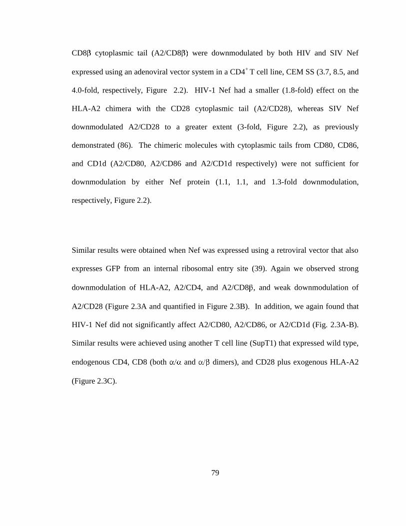
79
CD8 cytoplasmic tail (A2/CD8 were downmodulated by both HIV and SIV Nef
expressed using an adenoviral vector system in a CD4+
T cell line, CEM SS (3.7, 8.5, and
4.0-fold, respectively, Figure 2.2). HIV-1 Nef had a smaller (1.8-fold) effect on the
HLA-A2 chimera with the CD28 cytoplasmic tail (A2/CD28), whereas SIV Nef
downmodulated A2/CD28 to a greater extent (3-fold, Figure 2.2), as previously
demonstrated (86). The chimeric molecules with cytoplasmic tails from CD80, CD86,
and CD1d (A2/CD80, A2/CD86 and A2/CD1d respectively) were not sufficient for
downmodulation by either Nef protein (1.1, 1.1, and 1.3-fold downmodulation,
respectively, Figure 2.2).
Similar results were obtained when Nef was expressed using a retroviral vector that also
expresses GFP from an internal ribosomal entry site (39). Again we observed strong
downmodulation of HLA-A2, A2/CD4, and A2/CD8 , and weak downmodulation of
A2/CD28 (Figure 2.3A and quantified in Figure 2.3B). In addition, we again found that
HIV-1 Nef did not significantly affect A2/CD80, A2/CD86, or A2/CD1d (Fig. 2.3A-B).
Similar results were achieved using another T cell line (SupT1) that expressed wild type,
endogenous CD4, CD8 (both and dimers), and CD28 plus exogenous HLA-A2
(Figure 2.3C).

80
Figure 2.3. Nef downmodulates HLA-A2, A2/CD4, A2/CD8 , and A2/CD28 in T cell
lines. (A) Flow cytometric analysis of CEM SS T cells transduced with a retroviral vector
expressing GFP alone, or NL43 Nef and GFP reporter. (B) Quantitation of flow
cytometry experiments from part D. The mean fold downmodulation of GFP+ cells +/-
standard deviation is shown. n=3. (C) Flow cytometric analysis of SupT1 cells
transduced with the indicated adenoviral vector. Cells were analyzed at 3 dpi. Sup T1
cells express endogenous CD4, CD28 and CD8.

81
2.3.2 Endogenous CD80 and CD86 are not downmodulated by Nef in Primary APCs
Because CD80, CD86, and CD1d are normally expressed in antigen presenting cells
(APCs), we employed the human monocytic cell lines THP-1 and U937 for further
experiments with these molecules. We were unable to detect significant cell surface
expression of endogenous CD80 or CD86 in either cell line (data not shown). Therefore
we created stable U937 and THP-1 lines expressing the chimeric molecules. Nef was
introduced into undifferentiated cells using a retroviral vector and then the cells were
stimulated with LPS and PMA to induce macrophage differentiation. Overall, we
observed that Nef was less active in the APC lines than in T cells, downmodulating HLA-
A2 two to three-fold in APC lines as compared to 17-fold downmodulation in CEM cells
(Figure 2.4B). Even so, Nef had minimal effect on the CD80 and CD86 chimeras in all
three cell lines (Figure 2.4A-B, 0.8-2.0-fold), indicating that the cytoplasmic tails of
CD80 and CD86 were not sufficient for downmodulation by HIV-1 Nef even in
macrophage cell lines. In contrast we observed a decrease in MHC-I HLA-A2 surface
expression under all conditions. Because PMA also reduces CD4 surface expression (7),
we were unable to assess the effect of Nef on CD4 in the PMA-treated cells.
We used the same experimental approach to examine the ability of HIV-1 Nef to
downmodulate full length endogenous CD1d in the THP-1 cell line, which also expresses
endogenous HLA-A2. We observed minimal downmodulation of CD1d by Nef in either
undifferentiated or differentiated THP-1 cells (Figure 2.4C-D).
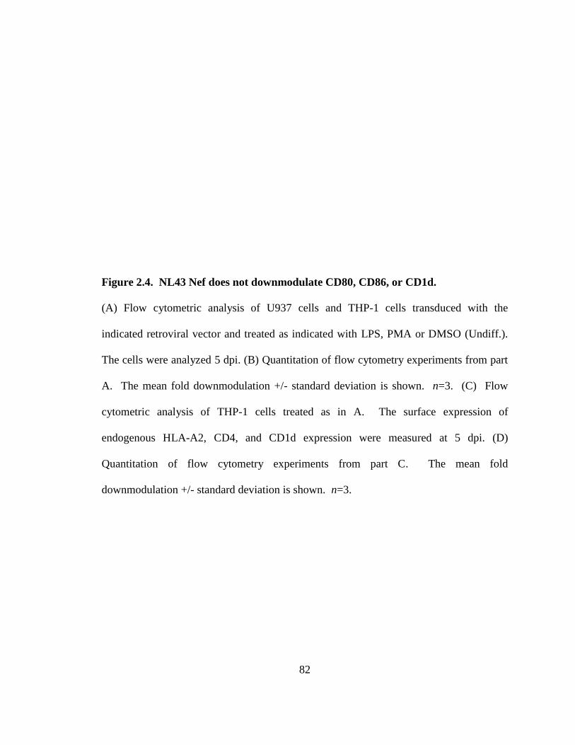
82
Figure 2.4. NL43 Nef does not downmodulate CD80, CD86, or CD1d.
(A) Flow cytometric analysis of U937 cells and THP-1 cells transduced with the
indicated retroviral vector and treated as indicated with LPS, PMA or DMSO (Undiff.).
The cells were analyzed 5 dpi. (B) Quantitation of flow cytometry experiments from part
A. The mean fold downmodulation +/- standard deviation is shown. n=3. (C) Flow
cytometric analysis of THP-1 cells treated as in A. The surface expression of
endogenous HLA-A2, CD4, and CD1d expression were measured at 5 dpi. (D)
Quantitation of flow cytometry experiments from part C. The mean fold
downmodulation +/- standard deviation is shown. n=3.
83
Figure 2.4. NL43 Nef does not downmodulate CD80, CD86, or CD1d.

84
Figure 2.5. NL43 Nef does not downmodulate endogenous CD80 or CD86 in
primary antigen presenting cells. (A) Flow cytometric analysis of primary antigen
presenting cells that were transduced with the indicated adenoviral vector. Endogenous
HLA-A2, CD4, CD80, and CD86 expression were assessed at 3 dpi. Primary
macrophages; GM-CSF treated prior to transduction, dendritic cells; GM-CSF, IL-4, and
TNF- treated prior to transduction. (B) Quantitation of flow cytometry experiments
from part E. The mean fold downmodulation +/- standard deviation is shown. n=3
85
To examine the ability of Nef to downmodulate full length CD80 and CD86, we obtained
primary antigen presenting cells from human peripheral blood mononuclear cells. These
cells were stimulated with GM-CSF alone or GM-CSF, IL-4, and TNF- to induce
macrophage or monocyte-derived dendritic cell phenotypes, respectively (78, 96). We
observed no significant effect of Nef on the surface expression of endogenous CD80 and
CD86 in either cell type (Figure 2.5A, quantified in 2.5B). As controls we demonstrated
downmodulation of endogenous MHC-I HLA-A2 and CD4 by Nef, albeit to a lesser
extent than in T cells (Figure 2.5A-B).
2.3.3 Multiple HIV-1 Nef variants downmodulate HLA-A2, CD4, CD8 , and CD28
All experiments thus far utilized Nef from the NL4-3 molecular clone of HIV (1). While
many of the known functional domains of Nef are conserved, significant sequence
variation does occur. Therefore, we examined whether the activity of Nef varied
amongst HIV isolates. Although we observed variation in the magnitude of MHC-I
HLA-A2 downmodulation, all Nef variants tested (except YU-2) reduced HLA-A2
surface expression dramatically (6-35-fold, Figure 2.6A-B). Similarly, all the Nef
proteins downmodulated endogenous CD4, as well as A2/CD8 , and A2/CD28. CD4
was downmodulated to the greatest extent, followed by HLA-A2, then A2/CD8 .
A2/CD28 was only moderately affected by all of the Nef variants tested. In contrast,
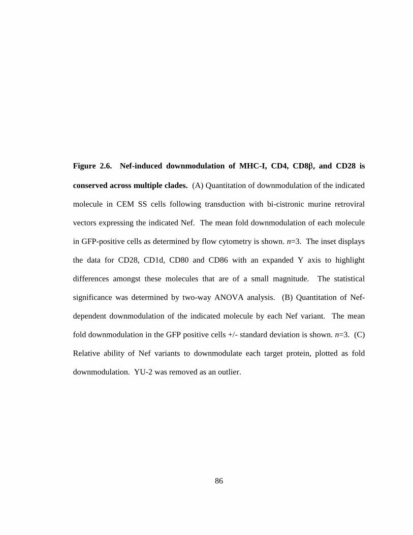
86
Figure 2.6. Nef-induced downmodulation of MHC-I, CD4, CD8 , and CD28 is
conserved across multiple clades. (A) Quantitation of downmodulation of the indicated
molecule in CEM SS cells following transduction with bi-cistronic murine retroviral
vectors expressing the indicated Nef. The mean fold downmodulation of each molecule
in GFP-positive cells as determined by flow cytometry is shown. n=3. The inset displays
the data for CD28, CD1d, CD80 and CD86 with an expanded Y axis to highlight
differences amongst these molecules that are of a small magnitude. The statistical
significance was determined by two-way ANOVA analysis. (B) Quantitation of Nef-
dependent downmodulation of the indicated molecule by each Nef variant. The mean
fold downmodulation in the GFP positive cells +/- standard deviation is shown. n=3. (C)
Relative ability of Nef variants to downmodulate each target protein, plotted as fold
downmodulation. YU-2 was removed as an outlier.
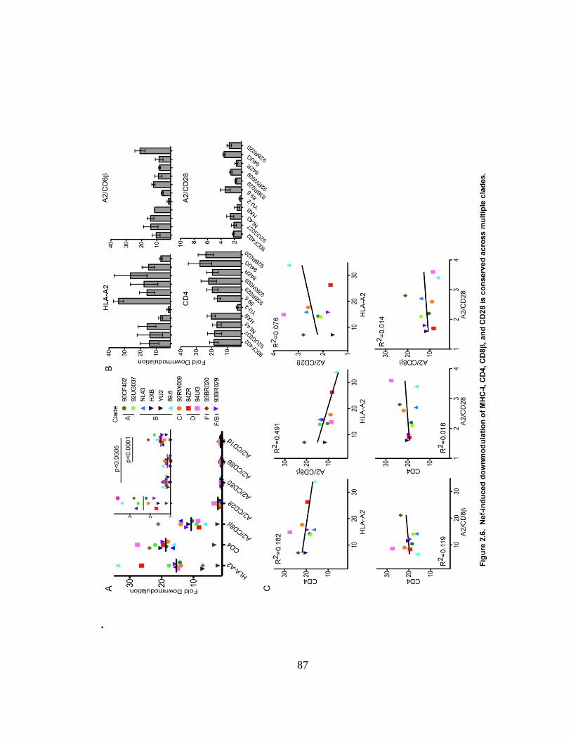
87
.
88
none of the Nef proteins reduced A2/CD80 or A2/CD86 surface expression more than
1.5-fold (Figure 2.6A and inset).
Interestingly, some of the Nef variants downmodulated A2/CD1d more than 1.5-fold
(Figure 2.6A, inset). 93BR020 Nef, a clade F isolate (32), downmodulated A2/CD1d to
the greatest extent. Thus, certain HIV subtypes may evolve the ability to significantly
affect CD1d trafficking, although the effect was comparatively small.
Notably, Nef proteins which demonstrated higher downmodulation activity on one target
protein did not necessarily demonstrate similarly high activity for all other targets
examined (Fig. 2.6B). When relative Nef activities were plotted for each target molecule,
a number of correlative observations were made (Figure 2.6C). HLA-A2 and A2/CD8
were negatively correlated, indicating that the ability of Nef to downmodulate these
proteins did not seem to co-evolve. There is a weak positive correlation between HLA-
A2 and A2/CD28, indicating that Nef sequences that contribute to the downmodulation
of these targets may be partially shared. Because CD4 and CD8 are analogous
molecules, one might hypothesize that CD8 downmodulation is an evolutionary
byproduct of CD4 downmodulation. Significantly, however, there was no correlation
between CD4 and A2/CD8 downmodulation (Figure 2.6C).
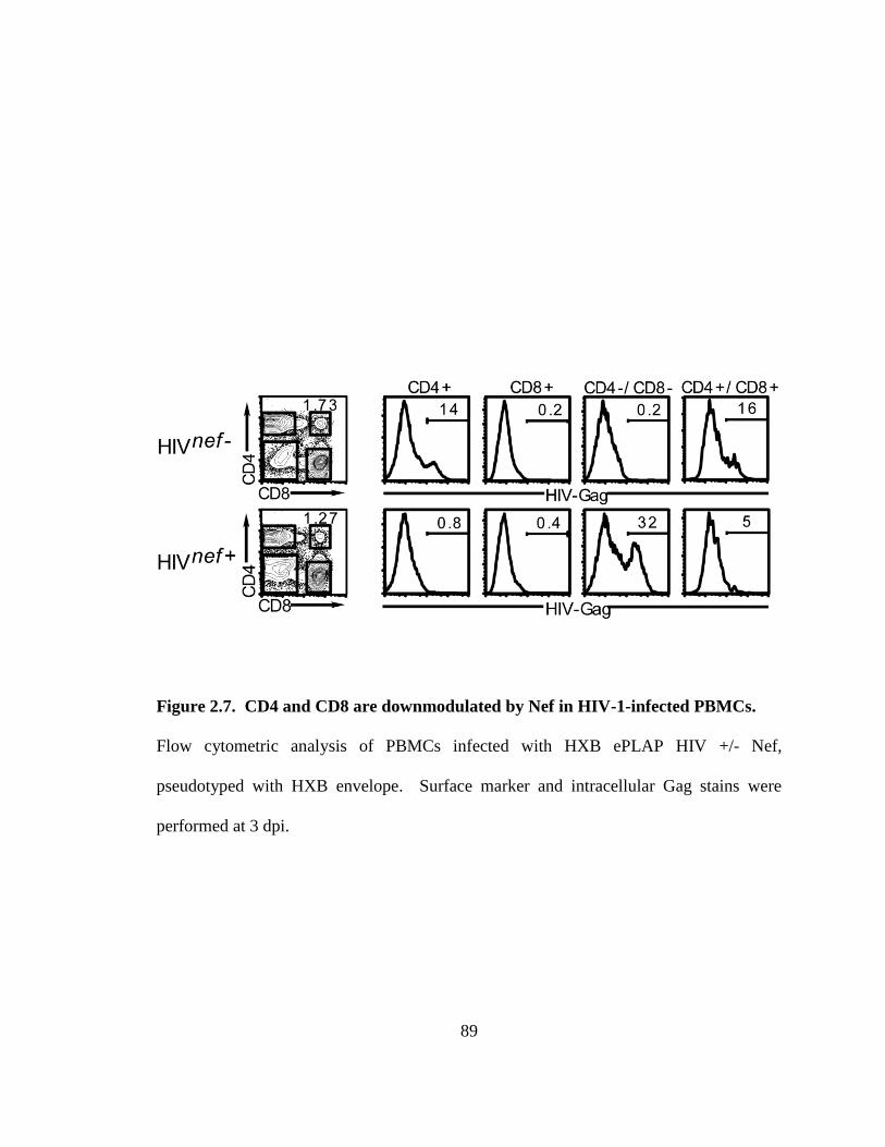
89
Figure 2.7. CD4 and CD8 are downmodulated by Nef in HIV-1-infected PBMCs.
Flow cytometric analysis of PBMCs infected with HXB ePLAP HIV +/- Nef,
pseudotyped with HXB envelope. Surface marker and intracellular Gag stains were
performed at 3 dpi.
90
2.3.4 HIV-1 Nef downmodulates CD4 and CD8 in primary T lymphocytes
CD8 can be expressed as a CD8 / homodimer or as a CD8 heterodimer. Because
Nef targets CD8 only the CD8 heterodimer can be affected by Nef (82). T
lymphocytes, DCs, and NK cells are all CD8+, but the CD8 heterodimer is expressed
exclusively in T cells. Typically, T lymphocytes are either CD4+ or CD8
+ and only the
CD4+ subset can be infected by HIV. Thus, it is unclear how Nef might come in contact
with the CD8 molecule. Significantly, a small population of CD4+CD8
+ T lymphocytes
circulates in peripheral blood (9, 31, 66, 67, 99) and the frequency of these double
positive cells increases in response to infection with a number of viruses, including HIV
(42, 54, 60). Additionally, these cells have been reported to be susceptible to infection by
HIV (6, 20, 41, 42, 52, 94, 98). While the role of CD4+CD8
+ double positive cells is not
entirely clear, they are known to exhibit cytolytic activity as well as antigen-dependant
secretion of IFN- and IL-2. Moreover, they are enriched for HIV-specific responses in
chronically infected patients (40, 66, 84, 97). Importantly, these cells are reported to
express the CD8 heterodimer (66, 83). Thus, this population represents a potential
physiologic target of HIV in which Nef would encounter CD8 .
To investigate the ability of HIV to infect CD4+CD8
+ cells, we infected PBMCs with
HIV HXB ePLAP (17) pseudotyped with an HIV envelope (HXB) that lacked Nef
expression (HIVnef -) so that the expression of cell surface markers would be maintained.
As expected, we observed similar rates of infection in CD4+CD8
+ and CD4
+CD8
- cells
(16 and 14% respectively, Figure 2.7, upper panel). In contrast, when these cells were

91
infected with an HIV HXB ePLAP that expressed Nef, most of the infected cells became
negative for both CD4 and CD8 (Figure 2.7, lower panel). Therefore, CD4+CD8
+ cells
are a physiologically relevant target of HIV-1 and Nef actively downmodulates both CD4
and CD8 in these cells.
2.3.5 Nef recruits AP-1 to the cytoplasmic tail of CD8 and CD28
Prior research has shown that Nef disrupts MHC-I and CD4 host protein trafficking by
binding to sequences in their cytoplasmic tail domains (79, 91). Here we confirmed that
Nef co-precipitated with HLA-A2 and A2/CD4. Moreover, we observed that Nef co-
precipitated with A2/CD8 , and A2/CD28, but not A2/Cw4, A2/CD80, A2/CD86, or
A2/CD1d (Figure 2.8A). A2/Cw4 is a negative control in these experiments as Nef does
not bind to or downmodulate HLA-C (or E or F) MHC-I allotypes (21, 36, 92). Thus,
NL43 Nef physically associates with each of the proteins which it potently
downmodulates in T cells.
In Nef expressing cells, HLA-A2 also coprecipitates with the clathrin adaptor protein AP-
1 (when degradation is inhibited by ammonium chloride), and this interaction is required
for Nef to disrupt MHC-I trafficking (74, 93). Similarly, we observed AP-1
coprecipitation with A2/CD28 and A2/CD8 (Figure 2.8B), although the interaction with
A2/CD8 seemed weaker than that observed with HLA-A2 and A2/CD28. In contrast,

92
Figure 2.8. Nef physically associates with the cytoplasmic domains of HLA-A2,
CD4, CD8 , and CD28 and recruits AP-1 to HLA-A2, CD8 , and CD28.
Immunoprecipitation and western blot analysis of the indicated chimeric molecule
expressed in CEM T cells transduced with the indicated adenoviral vector. The cells were
harvested at 3 dpi after an overnight incubation in ammonium chloride to inhibit
degradation. Lysates were immunoprecipitated with an antibody directed against HLA-
A2 (BB7.2) and the presence of (HA)-HLA-A2, Nef (A) or AP-1 (B) was detected by
western blot analysis.

93

94
we did not observe AP-1 coprecipitating with A2/CD4, A2/Cw4, A2/CD80, A2/CD86, or
A2/CD1d.
We also asked whether the other adaptor proteins AP-2 and AP-3 coprecipitated with the
chimeras. In two of five experiments we observed AP-2 recruitment to A2/CD28
(Figure 2.9C), which would be consistent with previous reports of AP-2 involvement in
CD28 downmodulation (82, 86). However, we were unable to detect coprecipitation of
any other chimeras with AP-2 or AP-3 (data not shown).
2.3.6 Nef recruits AP-1 to CD8 and CD28 through its dileucine motif
The AP proteins can utilize either Yxx or (E/D)xxxLL motifs in cargo molecules (x
stands for any amino acid, and indicates a hydrophobic residue). While Nef recruits
AP-1 to the tyrosine residue in the MHC-I cytoplasmic tail, such a sequence is not
present in the cytoplasmic tail domains of CD28 or CD8 Interestingly, Nef also
contains a conserved canonical dileucine motif (ExxxLL165), which has been reported to
mediate binding to AP-1, AP-2 and AP-3 (11, 25, 29, 35, 43, 44, 55, 69). Furthermore,
Nef LL164,165AA is defective at downmodulating CD8 or CD28, yet retains the ability
to downmodulate HLA-A2 (Figure 2.9A and (82)). Thus we hypothesized that
recruitment of AP-1 to CD8 or CD28 might occur through this dileucine motif.
Consistent with our hypothesis, Nef LL164,165AA did not support coprecipitation of AP-1

95
Figure 2.9. Nef recruits AP-1 for the downmodulation of CD8 and CD28 in a
dileucine dependant manner. (A) Flow cytometric analysis of CEM SS T cells
expressing the indicated chimeric protein and transduced with the indicated adenoviral
vector. Nef LL164,165AA is indicated as xLL. (B) Immunoprecipitation and western blot
analysis of the indicated chimeric molecule expressed in CEM T cells transduced with
the indicated adenoviral vector. The cells were harvested at 3 dpi. Lysates were
immunoprecipitated with an antibody directed against HLA-A2 (BB7.2) and the presence
of Nef, AP-1 subunits and MHC-I (HA) were detected by western blot analysis.
Intervening lanes were removed, as indicated by white gap. The data are representative of
four independent experiments. (C) Immunoprecipitation and western blot analysis of the
indicated chimeric molecule expressed in CEM T cells transduced with the indicated
adenoviral vector and treated as described for part B. AP-2 co-precipitation is
representative of two of four experiments.
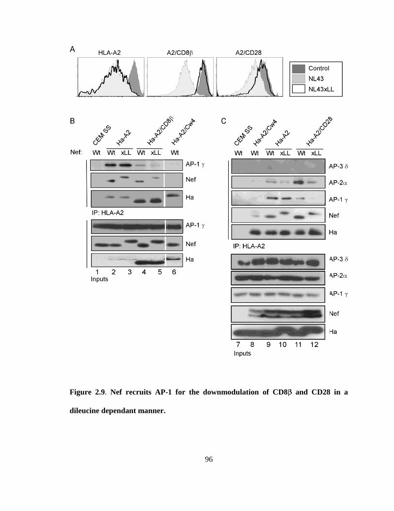
96
Figure 2.9. Nef recruits AP-1 for the downmodulation of CD8 and CD28 in a
dileucine dependant manner.
97
with A2/CD8 and A2/CD28 (Figure 2.9B lanes 4-5 and C lanes 11-12) but did promote
co-precipitation of AP-1 with HLA-A2 (Figure 2.9B-C). Therefore, Nef can recruit AP-1
to cytoplasmic tail domains using two different signals, depending upon which
cytoplasmic tail domain Nef is bound to.
In experiments where we observed AP-2 coprecipitation with the CD28 cytoplasmic tail,
we were able to determine that the AP-2 recruitment also depended upon the dileucine
motif in Nef (Figure 2.9C).
The subunit of heterotetrameric adaptor protein complexes recognizes tyrosine based
motifs (10, 65) whereas an interface between the and large subunit ( or
recognizes dileucine motifs (15, 27, 48). For MHC-I downmodulation, Nef promotes an
interaction between the tyrosine binding motif in the 1 subunit of AP-1 and a conserved
tyrosine in the cytoplasmic tail of MHC-I. An AP-1 mutant with an inactive tyrosine
binding pocket acts as a dominant negative inhibitor of MHC-I downmodulation (74, 93).
To confirm that downmodulation of CD8 and CD28 did not utilize the AP-1 tyrosine
binding pocket, we tested the impact of the tyrosine binding pocket mutant (TBPM) on
these pathways. In contrast to its effect on MHC-I, TBPM did not significantly inhibit
CD8 or CD28 downmodulation by Nef (Figure 2.10A-B). Similarly, knockdown of the
AP-1 1 subunit had no significant effect on CD8 or CD28 downmodulation by Nef
(Figure 2.11A-B). Importantly, depletion of the AP-1 subunit did not significantly
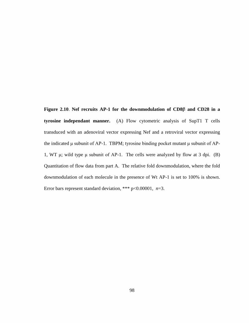
98
Figure 2.10. Nef recruits AP-1 for the downmodulation of CD8 and CD28 in a
tyrosine independant manner. (A) Flow cytometric analysis of SupT1 T cells
transduced with an adenoviral vector expressing Nef and a retroviral vector expressing
the indicated μ subunit of AP-1. TBPM; tyrosine binding pocket mutant μ subunit of AP-
1, WT μ; wild type μ subunit of AP-1. The cells were analyzed by flow at 3 dpi. (B)
Quantitation of flow data from part A. The relative fold downmodulation, where the fold
downmodulation of each molecule in the presence of Wt AP-1 is set to 100% is shown.
Error bars represent standard deviation, *** p<0.00001, n=3.
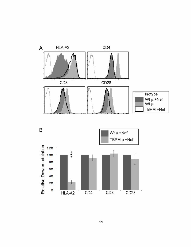
99

100
reduce AP1 expression, and AP-1 depletion had a partial destabilizing effect on AP-
1 (Figure 2.11C). Therefore, AP-1 involvement in CD8 and CD28 downmodulation
by Nef is independent of the AP-1 tyrosine-binding activity but could still depend on AP-
1 dileucine motif binding.
Based on the requirement for Nef’s dileucine motif for CD8 and CD28 downmodulation
as well as the lack of an effect of knocking down the AP-1 subunit, we next examined
the effect of knocking down the large subunits of the clathrin associated adaptor proteins,
which contain in part the dileucine motif recognition site (15, 27, 48). Knockdown was
achieved using lentiviral vectors expressing shRNAs directed against the large subunits
of AP-1( ), AP-2( ), AP-3( ), and -COP. Western blot analysis confirmed that we
efficiently and specifically reduced expression of each target (Figure 2.11D). We found
that knocking down AP-1 and -COP significantly reduced the downmodulation of both
CD8 and CD28 by Nef but the effect was only partial (Figure 2.11E). The partial
effects we observed may be due to incomplete knockdown or Nef utilization of multiple
adaptor proteins in redundant pathways to achieve downmodulation of CD8 and CD28.
Additionally, AP-2 knockdown had a small but significant effect on CD28
downmodulation, consistent with previous reports and the interaction we observed
between CD28 and AP-2 (Figure 2.11E) (82, 86). Surprisingly, knockdown of AP-2 had
no significant impact on CD4 downmodulation by Nef, despite a body of evidence tying
CD4 downmodulation to this adaptor in other cell systems (16, 45, 82). It is possible

101
Figure 2.11. Nef requires AP-1 and -COP for downmodulation of CD8 and CD28.
(A) Flow cytometric analysis of CEM SS cells expressing the indicated chimeric protein
and transduced with lentivirus expressing shRNA and GFP and subsequently transduced
with the indicated adenoviral vector at 3 days post lentivirus. Flow cytometric analysis
was performed at 3 days post adenoviral transduction. Histograms represent GFP-
expressing cells. (B) Quantitation of the flow data from part A. Fold downmodulation of
the indicated molecule in the presence of shRNA. (C) Western blot analysis confirming
specific knockdown of AP-1 subunits in CEM SS cells. (D) Western blot analysis
confirming adaptor protein knockdown in SupT1 cells. (E) Relative downmodulation of
the indicated molecule in SupT1 T cells transduced with the indicated lentivirus
expressing shRNA and GFP plus a bicistronic retrovirus expressing Nef and a PLAP
marker gene. shNC; negative control. The cells were analyzed by flow at 3 days post
retroviral transduction. The fold downmodulation normalized to the negative control in
cells positive for both GFP and PLAP is shown. * p<0.05, ** p< 0.01, ***p<0.001.

102
Figure 2.11. Nef requires AP-1 and -COP for downmodulation of CD8 and CD28.

103
CD4 downmodulation by Nef can utilize multiple adaptor proteins and the degree to
which it does so might vary in different cell systems. However, this possibility remains
ambiguous because when we attempted to knockdown more than one adaptor protein
large subunits the toxicity was too great to assess Nef function (data not shown).
2.3.7 Nef increases CD28 recycling
We were surprised to observe robust recruitment of adaptor proteins to A2/CD28 in the
presence of Nef, because the effect of Nef on steady-state surface expression of A2/CD28
is relatively small. Interestingly, CD28 is internalized at a rate similar to, or even greater
than, that of CD4 in the presence of Nef ((82) and data not shown). To address this
discrepancy, we performed a recycling assay in which we observed a significant increase
in CD28 recycling in the presence of Nef (Figure 2.12). Thus, Nef may initially induce a
potent downmodulation of CD28 through recruitment of AP-1 and/or AP-2 in parallel or
redundant pathways, and then a subset of CD28 is rapidly recycled to the plasma
membrane. Thus, the effect of Nef on CD28 may vary depending on the recycling rates
in different cell types.
\
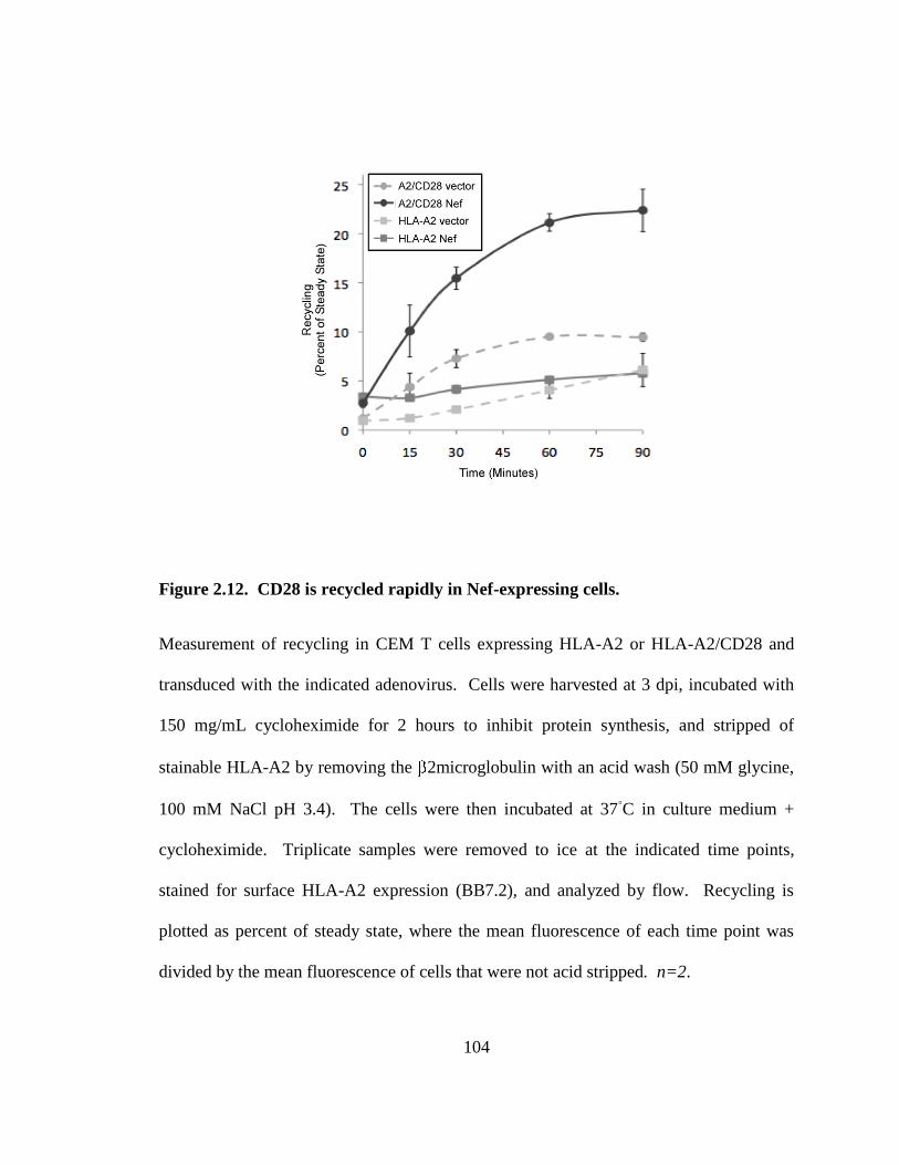
104
Figure 2.12. CD28 is recycled rapidly in Nef-expressing cells.
Measurement of recycling in CEM T cells expressing HLA-A2 or HLA-A2/CD28 and
transduced with the indicated adenovirus. Cells were harvested at 3 dpi, incubated with
150 mg/mL cycloheximide for 2 hours to inhibit protein synthesis, and stripped of
stainable HLA-A2 by removing the 2microglobulin with an acid wash (50 mM glycine,
100 mM NaCl pH 3.4). The cells were then incubated at 37◦C in culture medium +
cycloheximide. Triplicate samples were removed to ice at the indicated time points,
stained for surface HLA-A2 expression (BB7.2), and analyzed by flow. Recycling is
plotted as percent of steady state, where the mean fluorescence of each time point was
divided by the mean fluorescence of cells that were not acid stripped. n=2.
105
2.3.8 Nef utilizes -COP in CD8 and CD28 downmodulation
We also observed that knockdown of -COP significantly reduced the effect of Nef on
CD8 and CD28 surface expression (Figure 2.11E (SupT1 cells) and 2.13A (CEM SS
cells)). This is interesting because COP is required for Nef-dependant trafficking of
HLA-A2 and CD4 to lysosomes for degradation (79). COP knockdown has different
effects on cell surface expression of MHC-I and CD4 in Nef expressing cells. Whereas
COP knockdown increases cell surface levels of MHC-I in Nef-expressing cells,
COP knockdown does not always alter CD4 surface expression in Nef expressing cells
(Figure 2.11E and (79)). Instead, CD4 accumulates in intracellular vesicles and is unable
to recycle to the cell surface in some cell types (70, 79).
Nef contains two -COP binding sites, RR17,19 and EE155,156 (70, 79). Nef RR17,19AA is
defective at targeting MHC-I for degradation, while Nef EE155,156AA is defective at
targeting CD4 for degradation, and both have a partially reduced ability to co-precipitate
with -COP (79). When both domains are mutated (R/E), Nef is unable to efficiently
bind -COP and is defective at promoting the degradation of either MHC-1 or CD4 (79).
We assessed the relative contributions of these Nef domains in CD8 and CD28
downmodulation, and found that the R/E double-mutant was defective at
downmodulating CD8 and CD28, while each of the single mutant Nef proteins
demonstrated intermediate defects (Figure 2.13B-C). Similar results were observed
whether we assayed the chimeric panel (Figure 13B) or the full length target molecules
(Figure 2.13C).

106
Figure 2.13. -COP is required for Nef-mediated downmodulation of HLA-
A2/CD8 . (A) Quantitation of relative downmodulation (normalized to control) of the
indicated molecule expressed in CEM SS cells transduced with the indicated lentivirus
expressing shRNA and a retroviral vector expressing Nef. The cells were harvested at 3
days post retroviral transduction. n=3. (B and C) Quantitation of fold downmodulation
of the indicated molecule expressed in (B) CEM SS cells or (C) Sup T1 cells transduced
with a retroviral vector expressing Nef or the indicated Nef mutants. Cells with equal
GFP expression to control for Nef levels were analyzed at 3dpi. The mean fold
downmodulation +/- standard deviation is shown. n=3. * p<0.05, **p<0.01, ***p<0.001.
107
2.3.9 Combined effects of AP-1 and -COP
To further investigate the contribution of AP-1 and -COP to A2/CD8 degradation, we
again utilized the shRNA lentivirus system to knockdown AP-1 and -COP, both singly
and in combination. We observed that knockdown of either AP-1 or -COP reduced the
ability of Nef to downmodulate HLA-A2 and A2/CD8 . However, knocking down both
adaptors in combination did not have a significant additional inhibitory effect on
downmodulation (Figure 2.13A). Thus, the effect of Nef on CD8 results from effects of
at least two cellular trafficking factors and possibly others (82).
To examine whether HIV-1 Nef promoted degradation of CD8 , we performed western
blot analysis focusing on the mature, endoglycosidase H (endo H) resistant form of each
molecule. As reported previously, Nef expression reduces the amount of endo H resistant
HLA-A2 and this degradation is reversed by knocking down AP-1 or -COP (Figure
2.14A, left panel and quantified in Figure 2.14B). As previously observed, knocking
down -COP also affected the migration of MHC-I, probably by disrupting the
trafficking of enzymes required for proper glycosylation. -COP knockdown does not
inhibit trafficking of HLA-A2 through the Golgi, nor does it disrupt gross Golgi structure
(79).
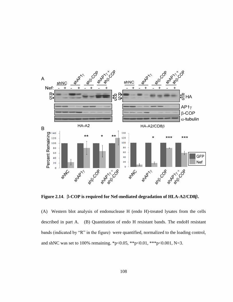
108
Figure 2.14. -COP is required for Nef-mediated degradation of HLA-A2/CD8 .
(A) Western blot analysis of endonuclease H (endo H)-treated lysates from the cells
described in part A. (B) Quantitation of endo H resistant bands. The endoH resistant
bands (indicated by “R” in the figure) were quantified, normalized to the loading control,
and shNC was set to 100% remaining. *p<0.05, **p<0.01, ***p<0.001, N=3.

109
In addition, we found that knockdown of COP dramatically reduced degradation of
A2/CD8 (Figure 2.14A, right panel and quantified in Figure 2.14B). A small but
statistically significant effect of knocking down AP-1 on A2/CD8 degradation was also
noted (Figure 2.14B).
2.4 Discussion
In summary, we have found that HIV-1 Nef has three major cellular targets: MHC-I,
CD4, and CD8. The ability of Nef to downmodulate endogenous CD8 was confirmed in
HIV infected primary CD4+CD8
+ T cells. We also report here that the effect of Nef on
MHC-I and CD8 depended at least in part on expression of AP-1 and -COP. In
addition, we found that the AP-1 binding motif utilized by Nef to recruit AP-1 to
cytoplasmic tails varied depending on which cytoplasmic tail domain Nef was bound to.
Finally, we found that mutation of both -COP binding sites in Nef reduced the activity
of Nef against CD8. Thus, one explanation for the capacity of Nef to affect multiple
targets is that it has evolved redundant mechanisms to recruit the same adaptor proteins.
Downmodulation of CD8, which participates in the recognition of antigen in association
with MHC-1 provides a second Nef-dependent mechanism by which HIV-infected cells
could avoid CTL lysis. While CD8+ T cells are not generally thought to be targeted by
HIV, there is mounting evidence that a subset of CD8+ T cells upregulate CD4 in
response to activation stimuli, and that these cells demonstrate antiviral activity, thus
110
creating an infectable target cell, the inhibition of which would be beneficial to HIV (31,
51, 52, 83, 94). Nef might also access CD8+ T cells through cell-cell contacts, as was
described to occur between infected and bystander T cells (63). Cell-cell conduits have
also been described between Nef expressing APCs and bystander B cells. Moreover
these nanotubular conduits have been shown to transmit Nef (72). If Nef were
transferred when CTLs contacted an infected target, the transmitted Nef could
downmodulate CD8 and reduce the efficacy of the anti-HIV-specific CTL attack.
Finally, there are reports of CD8-tropic HIV strains providing another potential scenario
in which Nef could encounter CD8 (37, 38, 59, 71, 77, 89).
This study is the first to assess the contribution of AP-1 and -COP to the
downmodulation of CD28 and CD8 . Another group has reported a partial requirement
for AP-2 in the downmodulation of CD4 and CD8 that we did not observe here (82).
However, the literature is conflicting on this point; some groups report a defect in CD4
downmodulation when AP-2 is knocked down, while other groups observe no effect (15,
75, 82). Additionally, co-depletion of Eps15 and AP-2 may be required to observe an
inhibition of Nef activity against CD4 (45). Knockdown of adaptor protein subunits is
technically difficult, as highly efficient depletion of these proteins can be lethal to cells.
For these reasons we may have underestimated the contribution of AP-2 in CD28 and
CD8 downmodulation by Nef in our system. AP-1- and AP-2-dependant mechanisms
may represent parallel or redundant pathways, which Nef can utilize depending upon
cellular localization and availability of cellular adaptors.

111
Here we have presented direct evidence that Nef can recruit AP-1 by utilizing either the
tyrosine motif recognition site or the dileucine recognition site of AP-1, and that this is
determined by which cytoplasmic tail domain Nef is bound to (Figure 2.15A-B).
Previous studies found that the dileucine motif in Nef is required for glutathione-S-
transferase (GST)-Nef pull-down of AP-1 complexes from mammalian cell lysates (11,
44) and for interactions detected in yeast two and three hybrid analyses (11, 24, 25, 29,
35, 43, 44, 55, 69) but the dileucine motif is dispensable for MHC-I downmodulation
even though AP-1 is required (62). CD4 downmodulation by Nef requires the dileucine
motif, but does not involve AP-1 (35). Thus, the interaction between AP-1 and the
dileucine motif has remained unexplained. Here we provide evidence that this interaction
is utilized to target CD8 (and CD28).
We have also identified -COP as a common factor in the downregulation and
degradation of all three major targets of Nef: MHC-I, CD4, and CD8 . The evidence
presented here is consistent with a model in which Nef utilizes AP-1 or AP-2 to direct
cellular targets into endosomal compartments, where Nef subsequently recruits -COP to
route targets to the lysosomes for degradation as previously proposed for MHC-I and
CD4 (Figure 2.15F). However, the fact that the degradation of CD8 was far more
dependent on -COP expression than on AP-1 expression indicates that -COP may also
directly target intracellular CD8 for degradation in Nef expressing cells without the need
112
for additional adaptor proteins (Figure 2.15F). The requirement for -COP in multiple
Nef pathways may represent a good target for therapeutic inhibition of Nef.
CD28 appears to be a unique target of Nef, in that it is downmodulated consistently by a
panel of Nef variants, but the effect is small relative to the other targets of Nef. The
small effect of Nef on steady-state surface CD28 seems at odds with the observation that
CD28 is internalized at a rate similar to that of CD4 in the presence of Nef ((82) and data
not shown). This discrepancy may be explained by the observation that CD28 recycling
is significantly increased in Nef-expressing cells. SIV Nef downmodulates CD28 more
potently than HIV-1 Nef; therefore CD28 downmodulation may be more important for
SIV pathogenesis. It is also possible that CD28 recycling may be regulated in some cell
systems, and that a physiologically relevant set of conditions exists which would result in
less recycling of CD28 and more downmodulation of CD28 by HIV-1 Nef.
We were unable to reproduce data reported by another group that CD80 and CD86 were
downmodulated by Nef. There were some differences in the cell lines used in our study
compared with the previous report. The previous studies used endogenously expressed
CD80 and CD86 in the U937 monocytic cell line. However, even when replicating the
published culture conditions, we were able to observe only low levels of CD80 on the cell
surface, and no CD86 at all, leading us to believe that our clone of the U937 line is
inherently different from that used to report downmodulation of CD80 and CD86. This

113
may be the cause for the discrepancy between our results and others, and is the reason
that we utilized primary macrophages and DCs as a highly relevant system for
investigating Nef activity against these potential targets. We were unable to detect an
effect of Nef on CD80 or CD86 under established stimulation and culture conditions for
primary macrophages and DCs, indicating that Nef targeting of these proteins must only
occur under specialized circumstance that we were unable to replicate. CD1d also
appears to be a special case, in that all Nef isolates do not retain the ability to reduce
surface expression of this molecule, yet one or more Nef variants may be capable of
doing so to a limited extent. This may be rare, evolutionarily driven by some
infrequently occurring host factor, or may be a feature of certain HIV-1 clades.
In summary, we have identified three major targets of HIV-1 Nef: MHC-I, CD4 and CD8
and these results were generalizable to multiple Nef variants. We also demonstrated that
downmodulation of CD8 and CD28 required AP-1 recruitment and was dependent upon
Nef’s dileucine motif. This observation indicates that Nef can interact with AP-1 through
either the tyrosine binding pocket or dileucine recognition domain of AP-1, depending
upon which cytoplasmic tail Nef is associated with. Finally, we discovered a role for -
COP in the downregulation and degradation of CD8, as well as MHC-I and CD4, which
represents a convergence of the all the trafficking mechanisms utilized by Nef.

114
Figure 2.15. Schematic representation of the two AP-1 binding sites that Nef utilizes
when bound to the MHC-I cytoplasmic domain (A), Nef recruits AP-1 through the
tyrosine recognition site in the AP-1 subunit (93). In this model the three-way complex
requires amino acid residues in both MHC-I, especially Y320, as well as in Nef, M20 in
particular (although assays using purified proteins and a Nef-MHC-I cytoplasmic tail
fusion protein did not demonstrate a requirement for M20) (64, 74, 81, 93). When bound
to the CD8 or CD28 cytoplasmic tails, Nef recruits AP-1 through the dileucine
recognition site at the interface between the and subunits of AP-1 (49). C-E)
Schematic representation of the Nef domains involved in -COP recruitment. Nef
residues R17,19 are required for -COP involvement in MHC-I degradation (C) (79). A
diacidic motif in Nef, EE155,156, is required for -COP involvement in CD4 degradation
(D) (30, 70, 79). Either or both Nef domains likely participate in -COP recruitment for
the degradation of CD8 by Nef (E). F) Model of the trafficking pathways Nef uses to
achieve downmodulation and degradation of target proteins. Nef blocks anterograde
transport in an AP-1-dependant pathway (47, 74, 85). Nef also induces internalization of
target molecules at the plasma membrane through an association with AP-1 or AP-2 (11,
16, 24, 45, 61, 79, 82, 86). These pathways likely converge at a -COP-dependant step
(79). Because AP-1 knockdown did not block -COP-dependent degradation of
A2/CD8, Nef may be able to direct some targets into endolysosomal compartments by a
pathway in which only -COP recruitment is necessary, and clathrin-associated adaptor
proteins are dispensable. PM stands for plasma membrane; LY indicates lysosome; LE/
MVB indicates late endosome or multivesicular body compartments.
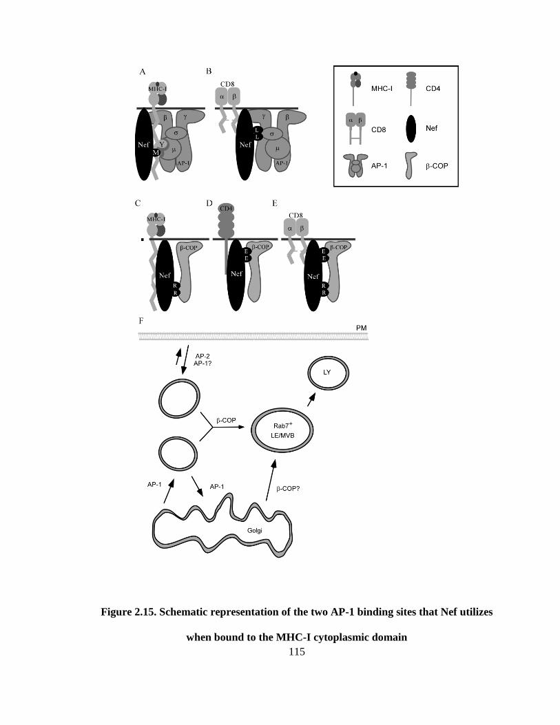
115
Figure 2.15. Schematic representation of the two AP-1 binding sites that Nef utilizes
when bound to the MHC-I cytoplasmic domain

116
2.5 Experimental methods
2.5.1 Cell culture
Bosc and 293T viral packaging cells were cultured in high glucose DMEM supplemented
with 10% fetal bovine serum (FBS) and 2 mM penicillin, streptomycin, and glutamine
(PSG). CEM-SS and SupT1 cells lines were maintained in RPMI 1640 supplemented
with 10% FBS and 2 mM PSG.
2.5.2 Preparation of T cell lines expressing HLA-A2-chimeric molecules
Stable cell lines were made as previously described (3). Briefly, each construct was
introduced into CEM-SS or SupT1 cells using the Murine Stem Cell Virus (MSCV)
retroviral vector pseudotyped with VSV-G. A uniform population was selected by
culturing the cells in neomycin.
2.5.3 Primary T cell preparation
Leukopaks were obtained from the New York Blood Center. Peripheral blood
mononuclear cells (PBMCs) were purified using ficoll gradients. Non-adherent cells
were stimulated with 10μg/mL phytohemaglutinin (PHA, Sigma-Aldrich) overnight and
the next day 50 U/mL interleukin(IL)-2 was added.
117
2.5.4 Preparation of macrophages and dendritic cells
Primary monocytes were obtained from whole PBMCs by positive selection using a
magnetic sorting kit (EasySep Human CD14 selection kit, StemCell Technologies). The
CD14+ cells were induced to mature into macrophages by culturing in RPMI + 10 ng/mL
GM-CSF (R&D Systems) for 5 days. The CD14+ cells were induced to mature into
dendritic cells (DC) by culturing in RPMI + 800 U/mL GM-CSF and 500 U/mL IL-
4,with the addition of 100 U/mL TNF on day 3 (all from R&D Systems). DCs were
stimulated 9 days in total before infection.
2.5.5 DNA Constructs
The CD80, CD1d, and CD8 cytoplasmic tails were fused to the extracellular and
transmembrane regions of HA-tagged HLA-A2 using PCR cloning methods. HA-HLA-
A2 (1), was used as the PCR template. The following oligonucleotide primers were used:
HA-HLA-A2/CD8 forward primer 5’-CGGGATCCACCATGCGGGTCACGGCG-3’
and reverse primer 5’-
GTGGTCGCTGCTGTGATGTGGTGCTGCCGGCGGAGGAGAGCCCGGCTTCGTT
TCATGAAACAATTTTACAAATAACTCGAGCGG-3’. HA-HLA-A2/CD80 forward
primer 5’-CGGGATCCACCATGCGGGTCACGGCG-3’ and reverse primer
5’AGCTGTGGTCGCTGCTGTGATGTGGAGATGCAGAGAGAGAAGGAGGAATG
AGAGATTGAGAAGGGAAAGTGTACGCCCTGTATAACTCGAGCGG3’. HA-
HLA-A2/CD1d forward primer 5’-CGGGATCCACCATGCGGGTCACGGCG-3’ and
118
reverse primer 5’-
GCTGCTGTGATGTGGTCCCGGTTTAAGAGGCAAACTTCCTATCAGGGCGT
CCTGTGACCGCTCGAGCGG-3’. The CD28 tail was fused to HA-HLA-A2 in a two-
step cloning method. First a 3’ fragment was created using two overlapping oligos that
span the entire CD28 cytoplasmic tail sequence, along with a forward primer which
appended a 15 base pair sequence which overlapped with the transmembrane region of
HLA-A2, and a reverse primer to introduce a 3’ XhoI site. The oligo sequences are as
follows: 5’ oligo 5’-
AGGAGTAAGAGGAGCAGGCTCCTGCACAGTGACTACATGAACATGACTCC
CCGCCGCCCCGGGCCCACCCGCAAG-3’, 5’-
CGCCGCCCCGGGCCCACCCGCAAGCATTACCAGCCCTATGCCCCACCACG
CGACTTCGCAGCCTATCGCTCCTGA-3’, 5’-
GGTCGCTGCTGTGATGTGGAGGAGTAAGAGGAGCAGGCTC-3’ AND 5’-
TCGCAGCCTATCGCTCCTGACTCGAGGG-3’. A second round of PCR then
generated the HA-HLA-A2/CD28 using HA-HLA-A2 and the CD28 fragment generated
in the first PCR reaction as templates. The following primers were used: 5’-
CGGGATCCACCATGCGGGTCACGGCG-3’ and
5’-TCGCAGCCTATCGCTCCTGACTCGAGGG-3’. The full length CD86 gene was
cloned from the Megaman human cDNA library (Stratagene) into pLITMUS29 (NEB).
The CD86 cytoplasmic tail was fused to HA-HLA-A2 in a manner similar to CD28
above. A first round of PCR generated a CD86 cytoplasmic tail fragment with 15 base
119
pairs 5’ to the CD86 tail that overlap with the transmembrane region of HLA-A2 using
full length CD86 as the template and the forward primer 5’-
gtggtcgctgctgtgatgtggaaatggaagaagaagaagc-3’ and reverse primer 5’-
aagtgatacatgtttttaactcgagcgg-3’. A second PCR reaction was performed to create HA-
HLA-A2 with a 15 base pair 3’ region that overlapped with the 5’sequence of the CD86
tail. HA-HLA-A2 was the template and the primer sequences were forward primer 5’-
CGGGATCCACCATGCGGGTCACGGCG-3’ and reverse primer 5’-
GTGGTCGCTGCTGTGATGTGGAAATGGAAGAAGAAGAAGC-3’. A third round
of PCR, using the products of the first and second PCR reactions, resulted in the full
length HA-HLA-A2/CD86 chimera. The following primers were used: forward primer
5’-CGGGATCCACCATGCGGGTCACGGCG-3’ and reverse primer 5’-
AAGTGATACATGTTTTTAACTCGAGCGG-3’. The PCR products were digested
BamHI to XhoI and then ligated into the BglII and XhoI sites of MSCV2.2 (2). The
resultant constructs were sequenced to ensure that no mutations were introduced during
cloning.
2.5.6 Cloning of nef alleles into pMIG
Nef alleles were cloned into the Bgl II and Eco RI sites of pMIG vector obtained from
Dr. Luk Van Parijs (MIT) (28). Nef alleles containing a 5’ Bam HI and 3’ Eco RI site
were PCR-amplified from cloned HIV genomes obtained through the AIDS Research
Reference Reagent Program, Division of AIDS, NIAID, NIH (7, 8, 12). The following
oligonucleotide primers were used: 5’ oligonucleotides: 5’ Nef-1 5’-
120
CGGGATCCACCATGGGTGGCAAGTGGTCAAAA-3’ (All except 92UG037,
93BR029 and 92RW009)
5’Nef-2 5’-CGGGATCCACCATGGGTAACAAGTGGTCAAAG-3’ (92UG037)
5’Nef-4 5’-CGGGATCCACCATGGGTAGCAAGTGGTCAAAA-3’ (93BR029 and
92RW009)
3’ oligonucleotides:
89.6 5’-CGGAATTCTCAGTTCTTGAAGTACTCCGGATG-3’
92UG037 5’-CGGAATTCTCAGCAGTCTTTGTAAAACTCCGG-3’
93BR020 5’-CGGAATTCTCAGTCTTGGTAGTACTCCGGATG-3’
NL4-3/84ZR 5’-CGGAATTCTCAGCAGTTCTTGAAGTACTCCGG-3’
PEYYKDC 5’-CGGAATTCTCAGCAGTCTTTGTAGTACTCCGG-3’
(93BR029, 92RW009, 94UG114)
YU-2 5’-CGGAATTCTCAGTTCTTGTAGTACTCCGGATG-3’
All alleles were sequenced to ensure that no mutations were introduced during cloning.
2.5.7 shRNA constructs
FG12 shRNA lentiviral vectors were constructed as previously described (73, 79). ShNC
and construct targeting -COP were previously described (79). The target sequence for
shAP1 began at position 1201-
GATCCCCGCAGACTGTGCATCTGGAATTCAAGAGATTCCAGATGCACAGTCT
GCTTTTTGGAAA. The AP2 target sequence was
GATCCCCGTCAGAGTTCCGACAGAACTTCAAGAGAGTTCTGTCGGAACTCTG

121
ACTTTTTGGAAA. The target sequence for shAP3 began at position 4180-
GATCCCCGAGAGGTTGCAGTCAGAACTTCAAGAGAGTTCTGACTGCAACCTC
TCTTTTTGGAAA
2.5.8 Viral transductions
Adenovirus was prepared by University of Michigan Gene Vector Core, and infection of
CEM-SS cells were performed as previously described (74, 91). Murine retroviral vector
preparation and transduction were performed as described (68, 90, 93). shRNA-
containing lentivirus preparation and transduction have also been described (73, 79).
Infection of PBMCs with HIV constructs was performed as in (22). Primary APCs were
infected with adenovirus at MOIs titrated to generate similar Nef expression levels
compared to what we typically obtain in CEM cells. In these experiments, CEM cells
were infected with an MOI of 50, primary macrophages were infected with an MOI of
250, and primary DCs were infected with an MOI of 500.
2.5.9 Western blot analyses and immunoprecipitations
Cells were lysed in PBS 0.3% CHAPS, 0.1% SDS pH 8, 1 mM PMSF, normalized for
total protein and separated by SDS-polyacrylamide gel electrophoresis. Endo H (NEB)
digestion was performed according to the manufacturer's protocol. Immunoprecipitations
were performed as previously described (93). Briefly, cells were lysed in 1% digitonin
(Wako) and normalized for total protein. Lysates were precleared overnight with A/G
122
beads (Pierce) and isotype control antibody (BD Biosciences). BB7.2-crosslinked A/G
beads were used for immunoprecipitation, and were washed five times with 0.1%
digitonin buffer prior to elution with 150 mM DTT.
Samples were separated by SDS-PAGE. HA-HLA-A2 and the chimeric proteins were
detected using the anti-HA antibody HA.11 (1:5000; Covance). Nef was detected with
AG11 (1:5000), and -COP with M3A5 (2) which were purified as described (46).
Adaptor protein subunits were detected with anti-AP-2 , anti-AP1 and anti-AP3 (all
1:500; BD Bioscience) or Ry/1 anti-AP1 (1:5000) (88). The secondary antibody for
anti-Nef, β-COP, HA, AP-2 , and anti-AP3 was HRP-rat anti-mouse IgG1 (Zymed), for
anti-AP1 was HRP-rat anti- -μ1 was HRP-goat
anti-rabbit (Zymed).
2.5.10 Flow cytometry
Intact cells were stained in FACS Buffer (2% FBS, 1% human serum, 1% Hepes, and
0.025% NaN3 in PBS). Primary cells were pre-incubated with 10% Fc-Receptor Blocker
(Accurate Chemical and Scientific Corps) prior to primary antibody incubation.
Chimeric molecules were detected with HA.11 (1:50; Covance) or BB7.2 (1:500).
Endogenous proteins were detected with BB7.2 (1:500), OKT4 (1:500), OKT8 (1:500),
all prepared by the University of Michigan Hybridoma Core and purified as previously
described (46), or anti-CD8 5F2 (1:100; Santa Cruz), CD28.2 (1:100; Biolegend), CD80

123
(1:100; BD Pharmingen), CD86 (1:100; BD Pharmingen), CD1d (1:100; eBioscience).
Isotype-specific secondary antibodies conjugated to PE, alexafluor 488, or alexafluor 647
were used at 1:250 (Invitrogen).
Acknowledgements
We thank members of the Collins lab for helpful discussions; the University of Michigan Gene
Vector Core facility for producing adenoviruses; and the University of Michigan Hybridoma Core
for producing ascites. This work was funded by NIH RO1A1046998. Jolie Leonard and Chris
Carter were supported by the University of Michigan Cellular and Molecular Biology Training
Program and the University of Michigan Molecular Mechanisms of Microbial Pathogenesis
Training Program. We would also like to thank Linton Traub (University of Pittsburgh) for his
gift of antibody to AP-1; Alice Telesnitsky (University of Michigan) for her gift of SupT1 cells;
and Beatrice Hahn (University of Pennsylvania) for her gift of HIV-1 Molecular clones (32).
Notes
This work has been published as “HIV-1 Nef Disrupts Intracellular Trafficking of Major
Histocompatibility Complex Class 1, CD4, CD8, and CD28 by Distinct Pathways That
Share Common Elements” Journal of Virology, 2011 Jul;85(14):6867-81. The work
presented here was the primary effort of Jolie Leonard. Contributions from coauthors are
as follows: Tracey Filzen cloned the nef alleles from primary HIV-1 isolates and into the
pMIG vector. Chris Carter performed the HIV infection and flow cytometric analysis for
Figure 2.7. Malinda Schaefer cloned the A2/CD1d chimeric construct into MSCV 2.2
vector. Jolie Leonard designed and performed all other experiments and analyses,
compiled the data into figures, and wrote the text of the manuscript.

124
2.6 References
1. Adachi, A., H. E. Gendelman, S. Koenig, T. Folks, R. Willey, A. Rabson, and
M. A. Martin. 1986. Production of acquired immunodeficiency syndrome-
associated retrovirus in human and nonhuman cells transfected with an infectious
molecular clone. J Virol 59:284-291.
2. Allan, V. J., and T. E. Kreis. 1986. A microtubule-binding protein associated
with membranes of the Golgi apparatus. J Cell Biol 103:2229-2239.
3. Arold, S. T., and A. S. Baur. 2001. Dynamic Nef and Nef dynamics: how
structure could explain the complex activities of this small HIV protein. Trends
Biochem Sci 26:356-363.
4. Benichou, S., M. Bomsel, M. Bodeus, H. Durand, M. doute, F. Letourneur, J.
Camonis, and R. Benarous. 1994. Physical interaction of the HIV-1 Nef protein
wih b-cop, a component of non-clathrin coated vesicles essential for membrane
traffic. J Biol Chem 269:30073-30076.
5. Benson, R. E., A. Sanfridson, J. S. Ottinger, C. Doyle, and B. R. Cullen. 1993.
Downregulation of cell-surface CD4 expression by simian immunodeficiency
virus Nef prevents viral super infection. J Exp Med 177:1561-1566.
6. Betts, M. R., D. A. Price, J. M. Brenchley, K. Lore, F. J. Guenaga, A. Smed-
Sorensen, D. R. Ambrozak, S. A. Migueles, M. Connors, M. Roederer, D. C.
Douek, and R. A. Koup. 2004. The functional profile of primary human antiviral
CD8+ T cell effector activity is dictated by cognate peptide concentration. J
Immunol 172:6407-6417.
7. Bigby, M., P. Wang, J. F. Fierro, and M. S. Sy. 1990. Phorbol myristate
acetate-induced down-modulation of CD4 is dependent on calmodulin and
intracellular calcium. J Immunol 144:3111-3116.
8. Birch, M. R., J. C. Learmont, W. B. Dyer, N. J. Deacon, J. J. Zaunders, N.
Saksena, A. L. Cunningham, J. Mills, and J. S. Sullivan. 2001. An
examination of signs of disease progression in survivors of the Sydney Blood
Bank Cohort (SBBC). J Clin Virol 22:263-270.
9. Blue, M. L., J. F. Daley, H. Levine, and S. F. Schlossman. 1985. Coexpression
of T4 and T8 on peripheral blood T cells demonstrated by two-color fluorescence
flow cytometry. J Immunol 134:2281-2286.
10. Boll, W., H. Ohno, Z. Songyang, I. Rapoport, L. C. Cantley, J. S. Bonifacino,
and T. Kirchhausen. 1996. Sequence requirements for the recognition of
tyrosine-based endocytic signals by clathrin AP-2 complexes. Embo J 15:5789-
5795.
11. Bresnahan, P. A., W. Yonemoto, S. Ferrell, D. Williams-Herman, R.
Geleziunas, and W. C. Greene. 1998. A dileucine motif in HIV-1 Nef acts as an
internalization signal for CD4 downregulation and binds the AP-1 clathrin
adaptor. Curr Biol 8:1235-1238.
12. Chaudhry, A., S. R. Das, A. Hussain, S. Mayor, A. George, V. Bal, S. Jameel,
and S. Rath. 2005. The Nef protein of HIV-1 induces loss of cell surface
costimulatory molecules CD80 and CD86 in APCs. J Immunol 175:4566-4574.
125
13. Chaudhry, A., S. R. Das, S. Jameel, A. George, V. Bal, S. Mayor, and S.
Rath. 2008. HIV-1 Nef induces a Rab11-dependent routing of endocytosed
immune costimulatory proteins CD80 and CD86 to the Golgi. Traffic 9:1925-
1935.
14. Chaudhry, A., S. R. Das, S. Jameel, A. George, V. Bal, S. Mayor, and S.
Rath. 2007. A two-pronged mechanism for HIV-1 Nef-mediated endocytosis of
immune costimulatory molecules CD80 and CD86. Cell Host Microbe 1:37-49.
15. Chaudhuri, R., O. W. Lindwasser, W. J. Smith, J. H. Hurley, and J. S.
Bonifacino. 2007. Downregulation of CD4 by human immunodeficiency virus
type 1 Nef is dependent on clathrin and involves direct interaction of Nef with the
AP2 clathrin adaptor. J Virol 81:3877-3890.
16. Chaudhuri, R., R. Mattera, O. W. Lindwasser, M. S. Robinson, and J. S.
Bonifacino. 2009. A basic patch on alpha-adaptin is required for binding of
human immunodeficiency virus type 1 Nef and cooperative assembly of a CD4-
Nef-AP-2 complex. J Virol 83:2518-2530.
17. Chen, B. K., R. T. Gandhi, and D. Baltimore. 1996. CD4 down-modulation
during infection of human T cells with human immunodeficiency virus type 1
involves independent activities of vpu, env, and nef. J Virol 70:6044-6053.
18. Chen, N., C. McCarthy, H. Drakesmith, D. Li, V. Cerundolo, A. J.
McMichael, G. R. Screaton, and X. N. Xu. 2006. HIV-1 down-regulates the
expression of CD1d via Nef. Eur J Immunol 36:278-286.
19. Cho, S., K. S. Knox, L. M. Kohli, J. J. He, M. A. Exley, S. B. Wilson, and R.
R. Brutkiewicz. 2005. Impaired cell surface expression of human CD1d by the
formation of an HIV-1 Nef/CD1d complex. Virology 337:242-252.
20. Cochrane, A., S. Imlach, C. Leen, G. Scott, D. Kennedy, and P. Simmonds.
2004. High levels of human immunodeficiency virus infection of CD8
lymphocytes expressing CD4 in vivo. J Virol 78:9862-9871.
21. Cohen, G. B., R. T. Gandhi, D. M. Davis, O. Mandelboim, B. K. Chen, J. L.
Strominger, and D. Baltimore. 1999. The selective downregulation of class I
major histocompatibility complex proteins by HIV-1 protects HIV-infected cells
from NK cells. Immunity 10:661-671.
22. Collins, K., B. Chen, S. Kalams, B. Walker, and D. Baltimore. 1998. HIV-1
Nef protein protects infected primary human cells from killing by cytotoxic T
lymphocytes. Nature 391:397-401.
23. Craig, H., M. Pandori, and J. Guatelli. 1998. Interaction of HIV-1 Nef with the
cellular dileucine-based sorting pathway is required for CD4 down-regulation and
optimal viral infectivity. Proc Natl Acad Sci 95:11229-11234.
24. Craig, H. M., M. W. Pandori, and J. C. Guatelli. 1998. Interaction of HIV-1
Nef with the cellular dileucine-based sorting pathway is required for CD4 down-
regulation and optimal viral infectivity. Proc Natl Acad Sci U S A 95:11229-
11234.
25. Craig, H. M., T. R. Reddy, N. L. Riggs, P. P. Dao, and J. C. Guatelli. 2000.
Interactions of HIV-1 nef with the mu subunits of adaptor protein complexes 1, 2,
and 3: role of the dileucine-based sorting motif. Virology 271:9-17.

126
26. Deacon, N. J., A. Tsykin, A. Solomon, K. Smith, M. Ludford-Menting, D. J.
Hooker, D. A. McPhee, A. L. Greenway, A. Ellett, C. Chatfield, and et al. 1995. Genomic structure of an attenuated quasi species of HIV-1 from a blood
transfusion donor and recipients. Science 270:988-991.
27. Doray, B., I. Lee, J. Knisely, G. Bu, and S. Kornfeld. 2007. The gamma/sigma1
and alpha/sigma2 hemicomplexes of clathrin adaptors AP-1 and AP-2 harbor the
dileucine recognition site. Mol Biol Cell 18:1887-1896.
28. Dyer, W. B., A. F. Geczy, S. J. Kent, L. B. McIntyre, S. A. Blasdall, J. C.
Learmont, and J. S. Sullivan. 1997. Lymphoproliferative immune function in
the Sydney Blood Bank Cohort, infected with natural nef/long terminal repeat
mutants, and in other long-term survivors of transfusion-acquired HIV-1
infection. AIDS 11:1565-1574.
29. Erdtmann, L., K. Janvier, G. Raposo, H. M. Craig, P. Benaroch, C. Berlioz-
Torrent, J. C. Guatelli, R. Benarous, and S. Benichou. 2000. Two independent
regions of HIV-1 Nef are required for connection with the endocytic pathway
through binding to the mu 1 chain of AP1 complex. Traffic 1:871-883.
30. Faure, J., R. Stalder, C. Borel, K. Sobo, V. Piguet, N. Demaurex, J.
Gruenberg, and D. Trono. 2004. ARF1 regulates Nef-induced CD4 degradation.
Curr Biol 14:1056-1064.
31. Flamand, L., R. W. Crowley, P. Lusso, S. Colombini-Hatch, D. M. Margolis,
and R. C. Gallo. 1998. Activation of CD8+ T lymphocytes through the T cell
receptor turns on CD4 gene expression: implications for HIV pathogenesis. Proc
Natl Acad Sci U S A 95:3111-3116.
32. Gao, F., D. Robertson, C. Carruthers, S. Morrison, B. Jian, Y. Chen, F.
Barre-Sinoussi, M. Girard, A. Srinivasan, A. Abimiku, G. Shaw, P. Sharp,
and B. Hahn. 1998. A comprehensive panel of near-full-length clones and
reference sequences of non-subtype B isolates of human immunodeficiency virus
type 1. J Virol 72:5680-5698.
33. Garcia, J. V., and A. D. Miller. 1991. Serine phosphorylation-independent
downregulation of cell-surface CD4 by nef. Nature 350:508-511.
34. Geyer, M., H. Yu, R. Mandic, T. Linnemann, Y. H. Zheng, O. T. Fackler,
and B. M. Peterlin. 2002. Subunit H of the V-ATPase binds to the medium chain
of adaptor protein complex 2 and connects Nef to the endocytic machinery. J Biol
Chem 277:28521-28529.
35. Greenberg, M., L. DeTulleo, I. Rapoport, J. Skowronski, and T.
Kirchhausen. 1998. A dileucine motif in HIV-1 Nef is essential for sorting into
clathrin-coated pits and for downregulation of CD4. Curr Biol 8:1239-1242.
36. Greenberg, M. E., S. Bronson, M. Lock, M. Neumann, G. N. Pavlakis, and J.
Skowronski. 1997. Co-localization of HIV-1 Nef with the AP-2 adaptor protein
complex correlates with Nef-induced CD4 down-regulation. Embo J 16:6964-
6976.
37. Gulzar, N., S. Balasubramanian, G. Harris, J. Sanchez-Dardon, and K. F.
Copeland. 2008. Infection of CD8+CD45RO+ memory T-cells by HIV-1 and
their proliferative response. Open AIDS J 2:43-57.
127
38. Gulzar, N., B. Diker, J. Mihowich, J. Deschatelets, M. E. Arsenault, N.
Lamoureux, D. W. Cameron, J. E. Kim, and K. F. Copeland. 2008. Proportion
of HIV-1 infected CD8+CD4- T lymphocytes in vivo. Curr HIV Res 6:585-596.
39. Hahn, W. C., S. K. Dessain, M. W. Brooks, J. E. King, B. Elenbaas, D. M.
Sabatini, J. A. DeCaprio, and R. A. Weinberg. 2002. Enumeration of the
simian virus 40 early region elements necessary for human cell transformation.
Mol Cell Biol 22:2111-2123.
40. Howe, R., S. Dillon, L. Rogers, B. Palmer, S. MaWhinney, N. Blyveis, R.
Schlichtemeier, M. D'Souza, L. Ingoldby, J. E. Harwood, C. Rietmeijer, G.
Ray, E. Connick, and C. C. Wilson. 2009. Phenotypic and functional
characterization of HIV-1-specific CD4+CD8+ double-positive T cells in early
and chronic HIV-1 infection. J Acquir Immune Defic Syndr 50:444-456.
41. Hughes, G. J., A. Cochrane, C. Leen, S. Morris, J. E. Bell, and P. Simmonds.
2008. HIV-1-infected CD8+CD4+ T cells decay in vivo at a similar rate to
infected CD4 T cells during HAART. AIDS 22:57-65.
42. Imlach, S., S. McBreen, T. Shirafuji, C. Leen, J. E. Bell, and P. Simmonds.
2001. Activated peripheral CD8 lymphocytes express CD4 in vivo and are targets
for infection by human immunodeficiency virus type 1. J Virol 75:11555-11564.
43. Janvier, K., H. Craig, D. Hitchin, R. Madrid, N. Sol-Foulon, L. Renault, J.
Cherfils, D. Cassel, S. Benichou, and J. Guatelli. 2003. HIV-1 Nef stabilizes
the association of adaptor protein complexes with membranes. J Biol Chem
278:8725-8732.
44. Janvier, K., Y. Kato, M. Boehm, J. R. Rose, J. A. Martina, B. Y. Kim, S.
Venkatesan, and J. S. Bonifacino. 2003. Recognition of dileucine-based sorting
signals from HIV-1 Nef and LIMP-II by the AP-1 gamma-sigma1 and AP-3 delta-
sigma3 hemicomplexes. J Cell Biol 163:1281-1290.
45. Jin, Y. J., C. Y. Cai, X. Zhang, H. T. Zhang, J. A. Hirst, and S. J. Burakoff.
2005. HIV Nef-mediated CD4 down-regulation is adaptor protein complex 2
dependent. J Immunol 175:3157-3164.
46. Kasper, M. R., and K. L. Collins. 2003. Nef-mediated disruption of HLA-A2
transport to the cell surface in T cells. J Virol 77:3041-3049.
47. Kasper, M. R., J. F. Roeth, M. Williams, T. M. Filzen, R. I. Fleis, and K. L.
Collins. 2005. HIV-1 Nef disrupts antigen presentation early in the secretory
pathway. J Biol Chem 280:12840-12848.
48. Kelly, B. T., A. J. McCoy, K. Spate, S. E. Miller, P. R. Evans, S. Honing, and
D. J. Owen. 2008. A structural explanation for the binding of endocytic dileucine
motifs by the AP2 complex. Nature 456:976-979.
49. Kelly, B. T., A. J. McCoy, K. Spate, S. E. Miller, P. R. Evans, S. Honing, and
D. J. Owen. 2008. A structural explanation for the binding of endocytic dileucine
motifs by the AP2 complex. Nature 456:976-979.
50. Kestler, H. W., 3rd, D. J. Ringler, K. Mori, D. L. Panicali, P. K. Sehgal, M. D.
Daniel, and R. C. Desrosiers. 1991. Importance of the nef gene for maintenance
of high virus loads and for development of AIDS. Cell 65:651-662.
51. Kitchen, S. G., N. R. Jones, S. LaForge, J. K. Whitmire, B. A. Vu, Z. Galic,
D. G. Brooks, S. J. Brown, C. M. Kitchen, and J. A. Zack. 2004. CD4 on

128
CD8(+) T cells directly enhances effector function and is a target for HIV
infection. Proc Natl Acad Sci U S A 101:8727-8732.
52. Kitchen, S. G., Y. D. Korin, M. D. Roth, A. Landay, and J. A. Zack. 1998.
Costimulation of naive CD8(+) lymphocytes induces CD4 expression and allows
human immunodeficiency virus type 1 infection. J Virol 72:9054-9060.
53. Lama, J., A. Mangasarian, and D. Trono. 1999. Cell-surface expression of
CD4 reduces HIV-1 infectivity by blocking Env incorporation in a Nef- and Vpu-
inhibitable manner. Curr Biol 9:622-631.
54. Laux, I., A. Khoshnan, C. Tindell, D. Bae, X. Zhu, C. H. June, R. B. Effros,
and A. Nel. 2000. Response differences between human CD4(+) and CD8(+) T-
cells during CD28 costimulation: implications for immune cell-based therapies
and studies related to the expansion of double-positive T-cells during aging. Clin
Immunol 96:187-197.
55. Le Gall, S., L. Erdtmann, S. Benichou, C. Berlloz-Torrent, L. Liu, R.
Benarous, J. Heard, and O. Schwartz. 1998. Nef interacts with mu subunit of
clathrin adaptor complexes and reveals a cryptic sorting signal in MHC I
molecules. Immunity 8:483-495.
56. Learmont, J., B. Tindall, L. Evans, A. Cunningham, P. Cunningham, J.
Wells, R. Penny, J. Kaldor, and D. A. Cooper. 1992. Long-term symptomless
HIV-1 infection in recipients of blood products from a single donor [see
comments]. Lancet 340:863-867.
57. Learmont, J. C., A. F. Geczy, J. Mills, L. J. Ashton, C. H. Raynes-Greenow,
R. J. Garsia, W. B. Dyer, L. McIntyre, R. B. Oelrichs, D. I. Rhodes, N. J.
Deacon, and J. S. Sullivan. 1999. Immunologic and virologic status after 14 to
18 years of infection with an attenuated strain of HIV-1. A report from the
Sydney Blood Bank Cohort. N Engl J Med 340:1715-1722.
58. Lewinsohn, D. A., R. Lines, D. M. Lewinsohn, S. R. Riddell, P. D. Greenberg,
M. Emerman, and S. R. Bartz. 2002. HIV-1 Vpr Does Not Inhibit CTL-
Mediated Apoptosis of HIV-1 Infected Cells. Virology 294:13-21.
59. Livingstone, W. J., M. Moore, D. Innes, J. E. Bell, and P. Simmonds. 1996.
Frequent infection of peripheral blood CD8-positive T-lymphocytes with HIV-1.
Edinburgh Heterosexual Transmission Study Group. Lancet 348:649-654.
60. Macchi, B., G. Graziani, J. Zhang, and A. Mastino. 1993. Emergence of
double-positive CD4/CD8 cells from adult peripheral blood mononuclear cells
infected with human T cell leukemia virus type I (HTLV-I). Cell Immunol
149:376-389.
61. Mangasarian, A., M. Foti, C. Aiken, D. Chin, J. L. Carpentier, and D. Trono.
1997. The HIV-1 Nef protein acts as a connector with sorting pathways in the
Golgi and at the plasma membrane. Immunity 6:67-77.
62. Mangasarian, A., V. Piguet, J. K. Wang, Y. L. Chen, and D. Trono. 1999.
Nef-induced CD4 and major histocompatibility complex class I (MHC-I) down-
regulation are governed by distinct determinants: N-terminal alpha helix and
proline repeat of Nef selectively regulate MHC-I trafficking. J Virol 73:1964-
1973.

129
63. Muratori, C., L. E. Cavallin, K. Kratzel, A. Tinari, A. De Milito, S. Fais, P.
D'Aloja, M. Federico, V. Vullo, A. Fomina, E. A. Mesri, F. Superti, and A. S.
Baur. 2009. Massive secretion by T cells is caused by HIV Nef in infected cells
and by Nef transfer to bystander cells. Cell Host Microbe 6:218-230.
64. Noviello, C. M., S. Benichou, and J. C. Guatelli. 2008. Cooperative binding of
the class I major histocompatibility complex cytoplasmic domain and human
immunodeficiency virus type 1 Nef to the endosomal AP-1 complex via its mu
subunit. J Virol 82:1249-1258.
65. Ohno, H., J. Stewart, M. C. Fournier, H. Bosshart, I. Rhee, S. Miyatake, T.
Saito, A. Gallusser, T. Kirchhausen, and J. S. Bonifacino. 1995. Interaction of
tyrosine-based sorting signals with clathrin-associated proteins. Science
269:1872-1875.
66. Ortolani, C., E. Forti, E. Radin, R. Cibin, and A. Cossarizza. 1993.
Cytofluorimetric identification of two populations of double positive
(CD4+,CD8+) T lymphocytes in human peripheral blood. Biochem Biophys Res
Commun 191:601-609.
67. Parel, Y., and C. Chizzolini. 2004. CD4+ CD8+ double positive (DP) T cells in
health and disease. Autoimmun Rev 3:215-220.
68. Pear, W. S., G. P. Nolan, M. L. Scott, and D. Baltimore. 1993. Production of
high-titer helper-free retroviruses by transient transfection. Proc Natl Acad Sci U
S A 90:8392-8396.
69. Piguet, V., Y. L. Chen, A. Mangasarian, M. Foti, J. L. Carpentier, and D.
Trono. 1998. Mechanism of Nef-induced CD4 endocytosis: Nef connects CD4
with the mu chain of adaptor complexes. Embo J 17:2472-2481.
70. Piguet, V., F. Gu, M. Foti, N. Demaurex, J. Gruenberg, J. L. Carpentier, and
D. Trono. 1999. Nef-induced CD4 degradation: a diacidic-based motif in Nef
functions as a lysosomal targeting signal through the binding of beta-COP in
endosomes. Cell 97:63-73.
71. Potter, S. J., D. E. Dwyer, and N. K. Saksena. 2003. Differential cellular
distribution of HIV-1 drug resistance in vivo: evidence for infection of CD8+ T
cells during HAART. Virology 305:339-352.
72. Qiao, X., B. He, A. Chiu, D. M. Knowles, A. Chadburn, and A. Cerutti. 2006.
Human immunodeficiency virus 1 Nef suppresses CD40-dependent
immunoglobulin class switching in bystander B cells. Nat Immunol 7:302-310.
73. Qin, X. F., D. S. An, I. S. Chen, and D. Baltimore. 2003. Inhibiting HIV-1
infection in human T cells by lentiviral-mediated delivery of small interfering
RNA against CCR5. Proc Natl Acad Sci U S A 100:183-188.
74. Roeth, J. F., M. R. Kasper, M. Williams, T. M. Filzen, and K. L. Collins.
2004. HIV-1 Nef disrupts MHC-I trafficking by recruiting AP-1 to the MHC-I
cytoplasmic tail. J. Cell Biol. 167:903-913.
75. Rose, J. J., K. Janvier, S. Chandrasekhar, R. P. Sekaly, J. S. Bonifacino, and
S. Venkatesan. 2005. CD4 down-regulation by HIV-1 and simian
immunodeficiency virus (SIV) Nef proteins involves both internalization and
intracellular retention mechanisms. J Biol Chem 280:7413-7426.

130
76. Ross, T. M., A. E. Oran, and B. R. Cullen. 1999. Inhibition of HIV-1 progeny
virion release by cell-surface CD4 is relieved by expression of the viral Nef
protein. Curr Biol 9:613-621.
77. Saha, K., J. Zhang, A. Gupta, R. Dave, M. Yimen, and B. Zerhouni. 2001.
Isolation of primary HIV-1 that target CD8+ T lymphocytes using CD8 as a
receptor. Nat Med 7:65-72.
78. Schaefer, M. R., M. Williams, D. A. Kulpa, P. K. Blakely, A. Q. Yaffee, and
K. L. Collins. 2008. A novel trafficking signal within the HLA-C cytoplasmic tail
allows regulated expression upon differentiation of macrophages. J Immunol
180:7804-7817.
79. Schaefer, M. R., E. R. Wonderlich, J. F. Roeth, J. A. Leonard, and K. L.
Collins. 2008. HIV-1 Nef targets MHC-I and CD4 for degradation via a final
common beta-COP-dependent pathway in T cells. PLoS Pathog 4:e1000131.
80. Schwartz, O., V. Marechal, S. Le Gall, F. Lemonnier, and J. Heard. 1996.
Endocytosis of major histocompatibility complex class I molecules is induced by
the HIV-1 Nef protein. Nature Medicine 2:338-342.
81. Singh, R. K., D. Lau, C. M. Noviello, P. Ghosh, and J. C. Guatelli. 2009. An
MHC-I cytoplasmic domain/HIV-1 Nef fusion protein binds directly to the mu
subunit of the AP-1 endosomal coat complex. PLoS One 4:e8364.
82. Stove, V., I. Van de Walle, E. Naessens, E. Coene, C. Stove, J. Plum, and B.
Verhasselt. 2005. Human immunodeficiency virus Nef induces rapid
internalization of the T-cell coreceptor CD8alphabeta. J Virol 79:11422-11433.
83. Sullivan, Y. B., A. L. Landay, J. A. Zack, S. G. Kitchen, and L. Al-Harthi.
2001. Upregulation of CD4 on CD8+ T cells: CD4dimCD8bright T cells
constitute an activated phenotype of CD8+ T cells. Immunology 103:270-280.
84. Suni, M. A., S. A. Ghanekar, D. W. Houck, H. T. Maecker, S. B. Wormsley,
L. J. Picker, R. B. Moss, and V. C. Maino. 2001. CD4(+)CD8(dim) T
lymphocytes exhibit enhanced cytokine expression, proliferation and cytotoxic
activity in response to HCMV and HIV-1 antigens. Eur J Immunol 31:2512-2520.
85. Swann, S. A., M. Williams, C. M. Story, K. R. Bobbitt, R. Fleis, and K. L.
Collins. 2001. HIV-1 Nef blocks transport of MHC class I molecules to the cell
surface via a PI 3-kinase-dependent pathway. Virology 282:267-277.
86. Swigut, T., N. Shohdy, and J. Skowronski. 2001. Mechanism for down-
regulation of CD28 by Nef. Embo J 20:1593-1604.
87. Tomiyama, H., H. Akari, A. Adachi, and M. Takiguchi. 2002. Different effects
of Nef-mediated HLA class I down-regulation on human immunodeficiency virus
type 1-specific CD8(+) T-cell cytolytic activity and cytokine production. J Virol
76:7535-7543.
88. Traub, L. M., S. Kornfeld, and E. Ungewickell. 1995. Different domains of the
AP-1 adaptor complex are required for Golgi membrane binding and clathrin
recruitment. J Biol Chem 270:4933-4942.
89. Tsubota, H., D. J. Ringler, M. Kannagi, N. W. King, K. R. Solomon, J. J.
MacKey, D. G. Walsh, and N. L. Letvin. 1989. CD8+CD4- lymphocyte lines
can harbor the AIDS virus in vitro. J Immunol 143:858-863.
131
90. Van Parijs, L., Y. Refaeli, J. D. Lord, B. H. Nelson, A. K. Abbas, and D.
Baltimore. 1999. Uncoupling IL-2 signals that regulate T cell proliferation,
survival, and Fas-mediated activation-induced cell death. Immunity 11:281-288.
91. Williams, M., J. F. Roeth, M. R. Kasper, T. Filzen, and K. L. Collins. 2005.
Human Immunodeficiency Virus Type 1 Nef Domains Required for Disruption of
Major Histocompatibility Complex Class I Trafficking Are Also Necessary for
Coprecipitation of Nef with HLA-A2. Journal of Virology 79:632-636.
92. Williams, M., J. F. Roeth, M. R. Kasper, R. I. Fleis, C. G. Przybycin, and K.
L. Collins. 2002. Direct binding of human immunodeficiency virus type 1 Nef to
the major histocompatibility complex class I (MHC-I) cytoplasmic tail disrupts
MHC-I trafficking. J Virol 76:12173-12184.
93. Wonderlich, E. R., M. Williams, and K. L. Collins. 2008. The tyrosine binding
pocket in the adaptor protein 1 (AP-1) mu1 subunit is necessary for Nef to recruit
AP-1 to the major histocompatibility complex class I cytoplasmic tail. J Biol
Chem 283:3011-3022.
94. Yang, L. P., J. L. Riley, R. G. Carroll, C. H. June, J. Hoxie, B. K. Patterson,
Y. Ohshima, R. J. Hodes, and G. Delespesse. 1998. Productive infection of
neonatal CD8+ T lymphocytes by HIV-1. J Exp Med 187:1139-1144.
95. Yang, O. O., P. T. Nguyen, S. A. Kalams, T. Dorfman, H. G. Gottlinger, S.
Stewart, I. S. Chen, S. Threlkeld, and B. D. Walker. 2002. Nef-Mediated
Resistance of Human Immunodeficiency Virus Type 1 to Antiviral Cytotoxic T
Lymphocytes. J Virol 76:1626-1631.
96. Zhou, L. J., and T. F. Tedder. 1996. CD14+ blood monocytes can differentiate
into functionally mature CD83+ dendritic cells. Proc Natl Acad Sci U S A
93:2588-2592.
97. Zloza, A., J. M. Schenkel, A. R. Tenorio, J. A. Martinson, P. M. Jeziorczak,
and L. Al-Harthi. 2009. Potent HIV-specific responses are enriched in a unique
subset of CD8+ T cells that coexpresses CD4 on its surface. Blood 114:3841-
3853.
98. Zloza, A., Y. B. Sullivan, E. Connick, A. L. Landay, and L. Al-Harthi. 2003.
CD8+ T cells that express CD4 on their surface (CD4dimCD8bright T cells)
recognize an antigen-specific target, are detected in vivo, and can be productively
infected by T-tropic HIV. Blood 102:2156-2164.
99. Zuckermann, F. A. 1999. Extrathymic CD4/CD8 double positive T cells. Vet
Immunol Immunopathol 72:55-66.
132
Chapter 3
ARF-1 activity is required to recruit AP-1 to the MHC-I
cytoplasmic tail and disrupt MHC-I trafficking in HIV-1
infected primary T cells
3.1 Abstract
HIV-1 infected cells are partially resistant to anti-HIV cytotoxic T lymphocytes due to
the effects of the HIV Nef protein on antigen presentation by MHC-I and evidence has
been accumulating that this function of Nef is important in vivo. HIV Nef disrupts
normal expression of MHC-I by stabilizing a protein-protein interaction between the
clathrin adaptor protein AP-1 and the MHC-I cytoplasmic tail. There is also evidence
that Nef activates a PI3-kinase-dependent GTPase (ARF-6) to stimulate MHC-I
internalization. However, the relative importance of these two pathways is unclear. Here
we report that a GTPase required for AP-1 activity (ARF-1) was needed for Nef to
disrupt MHC-I surface levels whereas no significant requirement for ARF-6 was
observed in Nef expressing T cell lines and in HIV-infected primary T cells. An ARF-1
inhibitor blocked the ability of Nef to recruit AP-1 to the MHC-I cytoplasmic tail and a
dominant active ARF-1 mutant stabilized the Nef-MHC-I-AP-1 complex. These data
support a model in which Nef and ARF-1 stabilize an interaction between MHC-I and
AP-1 to disrupt presentation of HIV-1 epitopes to CTLs.
133
3.2 Introduction
CD8+ cytotoxic T lymphocytes (CTLs) are important for the control of chronic viral
infections. In a virally infected cell, MHC-I molecules present peptides derived from
viral proteins. Once the T cell receptor (TCR) on CD8+ CTLs recognizes a “non-self”
signal presented by MHC-I, the CTL releases perforins and granzymes which kill the
virally infected cell preventing further spread of the virus (reviewed in (3)). CTLs play
an important role in the control of HIV infection (for review see (9)) and recent evidence
indicates that individuals mounting a Gag-specific CTL response have improved
parameters with regard to controlling disease (16, 24). Despite the efficacy with which
CTLs control viral load early in infection, anti-HIV CTLs ultimately fail to prevent
progression of disease in most infected people.
Studies performed in vitro have revealed that the HIV Nef protein protects infected cells
from CTL-mediated lysis (8, 26, 52, 61). Nef has been shown to protect HIV-infected
primary T cells from CTL lysis using flow cytometric killing assays (8, 26), CTL co-
culture assays (61) and chromium release assays (52). Although Nef limits the ability of
CTLs to recognize and kill infected cells, it does not appear to abrogate the capacity of
CTLs to produce inhibitory cytokines in response to infected cells (52). Recent in vivo
evidence supports the hypothesis that CTLs may control HIV infection in vivo primarily
by the elaboration of inhibitory cytokines, but fail to eradicate the infection because the
CTLs cannot efficiently lyse the infected cell source of new virions (60).
134
Nef binds directly to the cytoplasmic tail of MHC-I allotypes (58) and recruits the
clathrin adaptor protein AP-1 to redirect MHC-I into an endolysosomal pathway from the
trans-Golgi network (TGN) and to prevent its expression at the cell surface (41). There
is evidence that MHC-I delivered to endosomes via the activity of AP-1 is subsequently
targeted for degradation via Nef binding to the COP-I coatomer, -COP (44).
Nef has also been implicated in promoting internalization of MHC-I from the cell surface
(45). The relative contribution of internalization versus disruption of forward transport to
overall MHC-I downmodulation varies depending on the cell type (23) and has been
attributed to both ARF-6 (4) and AP-1 (23, 28) dependent pathways. The relative
importance of ARF-6 versus AP-1 has not yet been determined in studies that directly
compared their roles.
The adaptor protein AP-1 is a heterotetrameric complex that recognizes trafficking
signals in cargo and recruits clathrin-sorting machinery to the trans-Golgi network. AP-1
is made up of 1, 1, , and 1 subunits, which collectively sort cargo containing Yxx
or [D/E]xxxLL trafficking signals into the endo-lysosomal network (for review see (40)).
MHC-I downmodulation can be inhibited by knocking down expression of the AP-1 1
subunit (41, 44) or by overexpressing a dominant negative mutant of AP-1 1 which
lacks a functional tyrosine-binding pocket (47, 59). Mutation of a tyrosine residue in the
MHC-I cytoplasmic tail disrupts binding of AP-1 to the Nef-MHC-I complex in cells (41,
59) and in experiments using purified proteins (32, 47). Based on these data, the AP-1
135
tyrosine-binding pocket has been proposed to interact with the MHC-I cytoplasmic tail
tyrosine and this interaction is stabilized by Nef.
Binding of adaptor proteins to trafficking signals is normally regulated by the activity of
ADP-ribosylation factors (ARFs), which are small GTPases that control assembly and
disassembly of intracellular trafficking complexes. ARF activation and recruitment to
cellular membranes is cyclical and regulated by its GTP binding state. Guanine
nucleotide exchange factors (GEFs) promote the exchange of GTP for GDP. GTPase-
activating proteins (GAPs) support ARF catalysis of GTP and thus are important to
inactivate ARF ((20) and reviewed in (13)). There are a number of different ARF
proteins expressed by cells. ARF-1 is a clathrin regulatory protein that, upon binding
GTP, undergoes a conformational change exposing a myristoyl group that inserts into
membranes and subsequently stabilizes AP-1 (1, 48, 54) or COP-I coatomer (46, 54)
binding to trafficking signals. The ARF-1 GEF inhibitor brefeldin A (BFA) stabilizes an
abortive ARF-GDP-bound complex (35) thus preventing ARF-1 cycling.
ARF-6 is a related GTPase that regulates the recycling of cargo that has been internalized
by clathrin-independent endocytosis (10, 38). There is evidence that ARF-6 is required
for Nef-dependent MHC-I downmodulation based on experiments with ARF-6 mutants
that are “locked” in a GDP-bound, inactive state (ARF-6-T27N) or a GTP-bound,
constitutively active state (ARF-6-Q67L) (4, 10, 38).

136
Here, we demonstrate that ARF-1 activity was required for Nef-dependent MHC-I
trafficking via AP-1. Co-precipitation of AP-1 with the MHC-I-Nef complex was
specifically inhibited by BFA and stabilized by constitutively active ARF-1 Q71L (51). In
contrast, we were unable to detect a clear requirement for ARF-6. These data help to
clarify the relative contributions of the two major pathways implicated in HIV immune
evasion and highlight the AP-1/ARF-1 pathway as an important potential target for drug
development.
3.3 Results
3.3.1 Functional ARF-1 is required for Nef to disrupt the trafficking of MHC-I
Nef binds directly to the cytoplasmic tail of MHC-I allotypes (58) and recruits the
clathrin adaptor protein AP-1 to redirect MHC-I into an endolysosomal pathway from the
trans-Golgi network (TGN) and to prevent its expression at the cell surface (41).
However, it is not known whether Nef-dependent AP-1 binding and clathrin recruitment
bypass the normal AP-1 regulatory steps that include the GTPase, ARF-1. To determine
if ARF-1 activity was necessary for Nef to disrupt the transport of MHC-I to the cell
surface, we utilized two ARF-1 mutants, ARF-1 T31N and ARF-1 Q71L. ARF-1 T31N is
defective because it does not exchange GDP for GTP (11). ARF-1 Q71L is a dominant
active form of the molecule that stably binds GTP (51). We also tested BFA, which
inhibits the activity of ARF-1 GTP exchange factors (GEFs).

137
Figure 3.1. ARF-1 activity is required for Nef-induced downmodulation of HLA-
A2. (A) Flow cytometric analysis of HLA-A2, HLA-A2 A323V, and CD4 surface
expression plus or minus Nef in cells expressing ARF-1 constructs as indicated. Cells
expressing ARF-1 vector control were GFP+ and these cells were gated on for this
analysis. Dark gray shaded curve, vector alone; Light gray shaded curve, adeno-Nef;
black curve, adeno-Nef plus ARF-1 as indicated. BFA, brefeldin A. (B) Western blot
analysis of Myc-ARF-1 expression levels in transduced CEM T cells (C and D)
Quantitation of part (A). The median fluorescence normalized to wild type ARF-1 is
shown sd, n ≥ 3. * denotes a p-value of <0.05, ** denotes a p-value of <0.01, and ***
denotes a p-value of <0.001.
138
We found that overexpression of wild type and T31N ARF-1 did not significantly affect
Nef-dependent MHC-I downmodulation (Figure 3.1A and 3.2, quantified in Figure 3.1C-
D). However, the ARF-1 inhibitor, BFA, significantly increased MHC-I surface
expression in the presence of Nef (Figure 3.1A and quantified in Figure 3.1C-D). The
lack of a phenotype with overexpression of MHC-I T31N was not due to poor expression
(Figure 3.1B) but likely resulted from the presence of endogenous wild type ARF-1 that
was sufficient for Nef activity. CD4 downmodulation by Nef was unaffected by these
inhibitors consistent with previously published data showing that internalization and
degradation of CD4 are independent of the GTP-bound state of ARF-1 (Figure 3.1A and
C) (15).
In addition, we observed a striking inhibitory effect of dominant active ARF-1 (Q71L) on
Nef-dependent MHC-I downmodulation (Figure 3.1A, quantified in Figure 3.1C). Based
on this result, we postulated that this mutant might inhibit MHC-I downmodulation by
blocking the cycling of ARF-1 and thus sequestering essential trafficking machinery
required to maintain low MHC-I surface levels.
We also observed an inhibitory effect of BFA and ARF-1 (Q71L) on MHC-I surface
expression in the absence of Nef (quantified in Figure 3.1C-D). This is expected due to
the known effects of these factors on protein export form the endoplasmic reticulum (27).
Despite the inhibitory effect of these factors on MHC-I surface expression in the absence
of Nef, both BFA and ARF-1 (Q71L) caused a significant increase in MHC-I surface
expression in the presence of Nef (Figure 3.1). The opposing effects observed in the
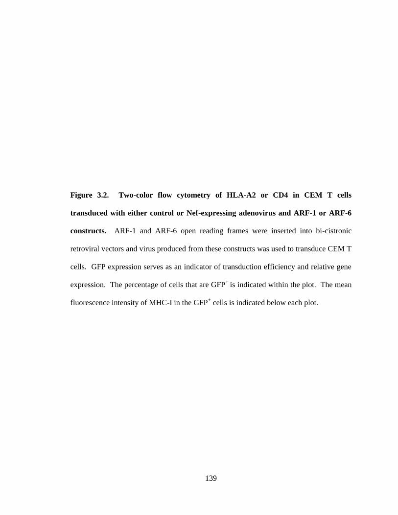
139
Figure 3.2. Two-color flow cytometry of HLA-A2 or CD4 in CEM T cells
transduced with either control or Nef-expressing adenovirus and ARF-1 or ARF-6
constructs. ARF-1 and ARF-6 open reading frames were inserted into bi-cistronic
retroviral vectors and virus produced from these constructs was used to transduce CEM T
cells. GFP expression serves as an indicator of transduction efficiency and relative gene
expression. The percentage of cells that are GFP+
is indicated within the plot. The mean
fluorescence intensity of MHC-I in the GFP+ cells is indicated below each plot.
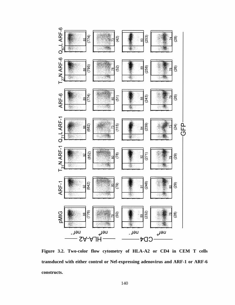
140
Figure 3.2. Two-color flow cytometry of HLA-A2 or CD4 in CEM T cells
transduced with either control or Nef-expressing adenovirus and ARF-1 or ARF-6
constructs.
141
absence versus the presence of Nef supports the conclusion that these inhibitors are
specifically affecting Nef-dependent pathways.
For comparison, we also included a molecule that contained a more natural AP-1
trafficking signal, which was previously shown to bind AP-1 in the absence of Nef. This
signal was made by substituting a valine for an alanine in the cytoplasmic tail of MHC-I
HLA-A2 to create a Yxx sorting signal [Y320SQV323 (HLA-A2 A323V)] (59). Compared
to wild type MHC-I HLA-A2, this molecule has relatively low surface expression due to
its interaction with AP-1 (59). Interestingly, the effect of ARF-1 mutants and BFA on
the surface expression of HLA-A2 A323V in the absence of Nef was somewhat different
than what occurred in the presence of Nef. The main difference was that, in the absence
of Nef, there was a relatively small but statistically significant further reduction of HLA-
A2 A323V expression with the dominant active mutant ARF-1 Q71L (Figure 3.1C).
Whereas in the presence of Nef, ARF-1 Q71L inhibited the Nef-dependent reduction in
surface expression of HLA-A2 molecules (Figure 3.1A and C). Given that HLA-A2
A323V is a model for trafficking due to AP-1, these differences provide suggestive
evidence that the effects of Nef on MHC-I HLA-A2 are not due to AP-1 alone and
probably involve additional steps that are disrupted by ARF-1 Q71L.
3.3.2 Functional ARF-6 is dispensable for Nef-dependent MHC-I downmodulation
ARF-6 may also be important for Nef-dependent internalization of MHC-I based on
evidence that dominant active ARF-6 (Q67L) inhibited internalization of MHC-I by Nef
in HeLa cells (4). To examine the relative effect of ARF-1 and ARF-6 mutants on Nef

142
Figure 3.3. ARF-1 but not ARF-6 activity is required for Nef-dependent HLA-A2
and CD4 downmodulation in T cell lines. (A) Flow cytometric analysis of HLA-A2 or
CD4 surface expression in CEM-SS cells transduced with control or Nef-expressing
adenovirus plus the indicated ARF-6 construct. ARF-6 proteins were expressed via bi-
cistronic murine retroviral vectors that also expressed GFP. Similar GFP+ populations
based on FL-1 mean were analyzed for HLA-A2 or CD4. Gray shaded curve, control
vector; gray outlined curve, adeno-Nef; black curve, adeno-Nef plus the indicated ARF-6
protein. (B) Quantitation of part (A). The median fluorescence normalized to wild type
ARF-6 is shown sd, n=3. (C) Western blot of ARF-6 and Nef expression.

143
activity in our system, we generated ARF-6 constructs analogous to the ARF-1 constructs
tested in Figure 3.1. We found that, in contrast to the effect of ARF-1 mutants, there was
no significant effect of ARF-6 mutants on Nef activity (Figure 3.3A-B). As controls, we
demonstrated that the ARF-6 mutants were expressed in the cells (Figure 3.3C).
Additionally, we verified that the ARF-1 and ARF-6 constructs transduced the cells with
similar efficiency (Figure 3.2). For these experiments the ARF-1 and ARF-6 constructs
were made identically as bi-cistronic elements that co-expressed GFP from an internal
ribosome entry site (Figure 3.2) allowing GFP to serve both as a measure of transduction
efficiency and as a measure of relative expression levels.
ARF-6 Q67L was active in our experimental system based on its effect on MHC-I surface
expression in the absence of Nef. Microscopic analysis revealed intracellular
accumulation of HLA-A2 in CEM-SS cells expressing ARF-6 Q67L, but not in cells
expressing wild type ARF-6 or ARF-6 T27N (Figure 3.4). This phenotype is consistent
with previous reports that proteins that normally traffic through ARF-6 compartments are
trapped in the presence of ARF-6 Q67L (5, 30).
Previous studies have suggested that Nef activates ARF-6 via a PI3-kinase-dependent
step. Thus, levels of the PI3-kinase inhibitor, phosphatase and tensin homolog (PTEN),
could influence the relative amount of ARF-6 dependent activity we observed. Indeed, it
has been suggested that lower expression level of PTEN in the CEM-SS cell line used in
some of our studies may influence PI3K-induced ARF-6-dependent effects on MHC-I
and limit our ability to detect this pathway (19). To address this possibility, we used

144
Figure 3.4 ARF-6 Q67L disrupts HLA-A2 localization in a Nef-independent manner
in T cell lines. Visualization of HLA-A2 expression in CEM-SS cells transduced with
the indicated ARF-6 construct. ARF-6 proteins were expressed via bi-cistronic murine
retroviral vectors that also expressed GFP. GFP and HLA-A2 were detected by confocal
microscopy.
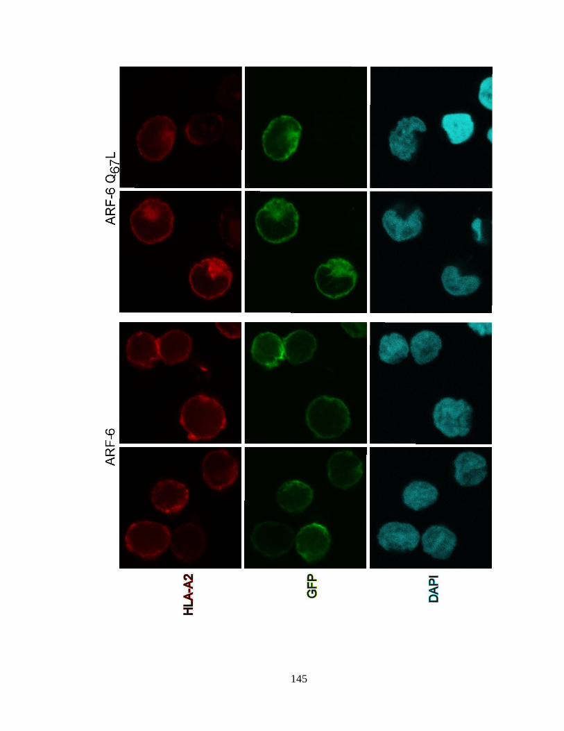
145

146
Figure 3.5. ARF-1 but not ARF-6 activity is required for Nef-dependent HLA-A2
and CD4 downmodulation in PTEN-expressing T cell lines. (A) Western blot of
PTEN or control (GAPDH) levels in the indicated cell type. (B) Flow cytometric
analysis of HLA-A2 surface expression in SupT1 cells transduced with control or Nef-
expressing adenovirus plus the indicated ARF-6 construct and analyzed as described in
part A. Dark gray shaded curve, control adenovirus plus ARF as indicated;
Light gray shaded curve, adeno-Nef plus vector; black curve, adeno-Nef
plus ARF as indicated. (C) Quantitation of part (B). The median fluorescence normalized
to empty vector treatment is shown sd n=3. Values obtained with wild type ARF-1
were significantly different than those with ARF-1 Q71L (p< 0.01). Values obtained with
wild type ARF-6 were not significantly different than those with ARF-6 Q67L (p= 0.46).

147
western blot analysis to examine the levels of PTEN expression in several cell lines, as
well as in primary CD4+ T lymphocytes. We confirmed that PTEN expression varied
dramatically amongst cell lines and that CEM-SS cells did not express much, if any,
PTEN (Figure 3.5A). We also performed experiments in a cell line (the SupT1
lymphoblastoid line) with very high PTEN expression to determine whether PTEN levels
might be affecting our ability to detect a requirement for ARF-6 activity. Similar to our
observations in CEM-SS cells, we found that expression of ARF-1 Q71L significantly
reduced HLA-A2 downmodulation by Nef, while expression of ARF-6 Q67L did not
(Figure 3.5B-C). As a control we again demonstrated that the ARF-1 and ARF-6
constructs transduced the cells and expressed the bi-cistronic message to similar degrees
(Figure 3.6).
Finally, we compared the effects of ARF-1 and ARF-6 mutants in HIV-infected primary
T lymphocytes expressing endogenous HLA-A2. For these experiments, wild type ARF
or its dominant active mutant was inserted directly into the envelope open reading frame
of a GFP-expressing HIV molecular clone (Figure 3.7A, NL-GIarf). Primary T cells
were infected with the virus and then assayed 36 hours post-infection. We reproducibly
observed a two-to-three fold inhibition of Nef-dependent MHC-I downmodulation when
the infected cells expressed the ARF-1 mutant Q71L, but not when the infected cells
expressed the corresponding ARF-6 mutant Q67L (Figure 3.7B). These data support a
model in which an ARF-1-dependent pathway rather than an ARF-6-dependent pathway
is needed for Nef-dependent MHC-I downmodulation in HIV-infected primary T cells.

148
Figure 3.6. Two color flow cytometry of GFP and HLA-A2 or CD4 staining in cells
transduced with control or adeno-Nef. The ARF-1 and ARF-6 constructs used in these
experiments were made identically by inserting the open reading frames into bi-cistronic
retroviral vectors. Virus produced from these constructs was used to transduce Sup T1
cells. GFP expression serves as an indicator of transduction efficiency and relative gene
expression.

149
Further evidence that ARF-6 Q67L was active in this experimental system is its effect on
MHC-I surface expression in the absence of Nef. For HLA-A2, Bw4 and Bw6 MHC-I
allotypes we observed an 18% (Figure 3.7B), 31% and 29% (data not shown) reduction in
MHC-I expression respectively. Over three independent experiments in which HLA-A2
expression was measured in primary T cells expressing ARF-6 Q67L the average
reduction in surface expression was 13% (p< 0.002). These data are consistent with the
inhibitory effect of ARF-6 Q67L on MHC-I recycling (5, 30).
To further examine the contribution of ARF-6 to Nef-dependent MHC-I
downmodulation, we used a lentiviral system expressing shRNA to knock down ARF-6
expression in CEM-SS cells. Knockdown of ARF-6 has previously been demonstrated to
inhibit clathrin-independent endocytosis of G-protein coupled receptors (18). We found
that depletion of ARF-6, as confirmed by western blot, had no significant impact on
HLA-A2 or endogenous CD4 downmodulation by Nef, as compared to negative control
shRNA (Figure 3.8). This observation lends additional support to the conclusion that
ARF-6 is dispensable for Nef-induced MHC-I downmodulation in T cells. Similar
analysis was not possible for ARF-1, as knockdown of single Golgi ARFs does not affect
cellular trafficking (56).
3.3.3 Dominant active ARF-1 increases AP-1 recruitment to HLA-A2 A323V and the
HLA-A2-Nef complex
To determine the mechanism by which ARF-1 mutants affect MHC-I surface expression,
we used co-immunoprecipitation assays to measure AP-1 binding to cargo plus or minus
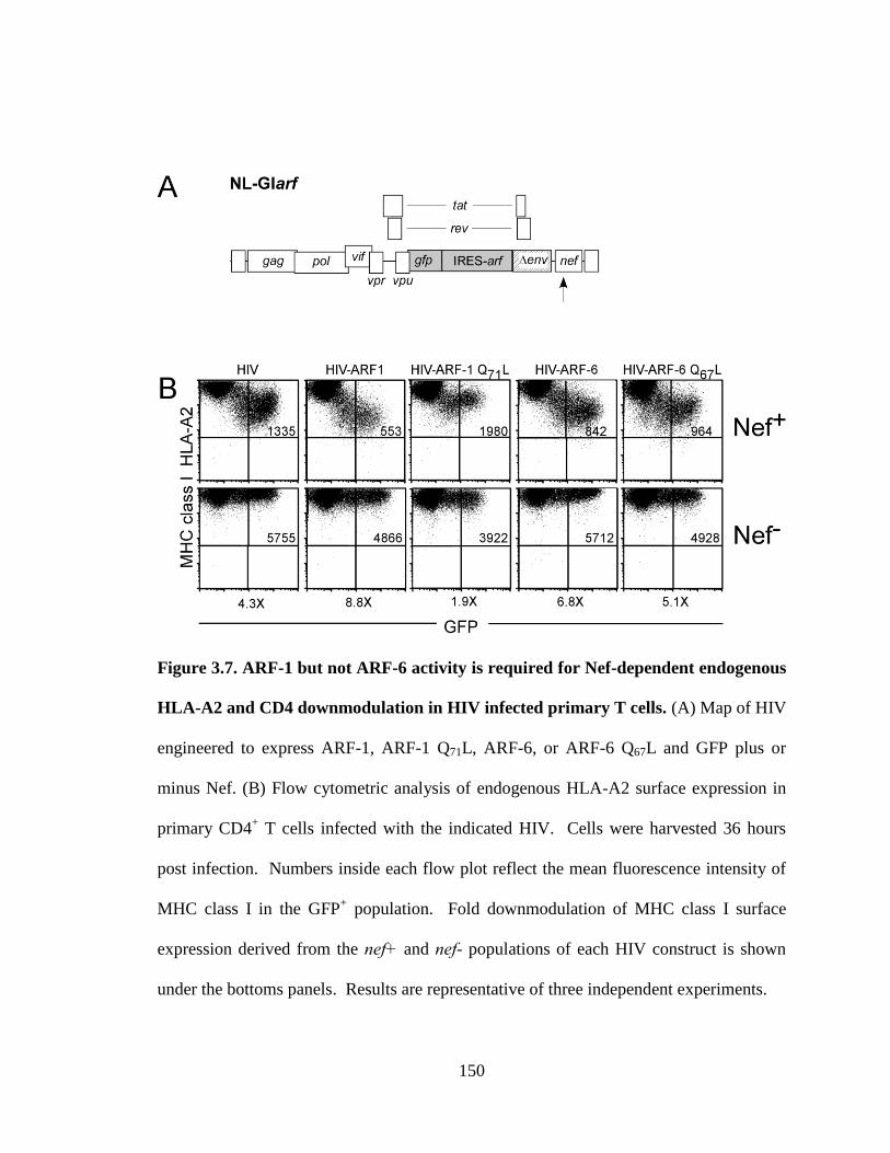
150
Figure 3.7. ARF-1 but not ARF-6 activity is required for Nef-dependent endogenous
HLA-A2 and CD4 downmodulation in HIV infected primary T cells. (A) Map of HIV
engineered to express ARF-1, ARF-1 Q71L, ARF-6, or ARF-6 Q67L and GFP plus or
minus Nef. (B) Flow cytometric analysis of endogenous HLA-A2 surface expression in
primary CD4+ T cells infected with the indicated HIV. Cells were harvested 36 hours
post infection. Numbers inside each flow plot reflect the mean fluorescence intensity of
MHC class I in the GFP+ population. Fold downmodulation of MHC class I surface
expression derived from the nef+ and nef- populations of each HIV construct is shown
under the bottoms panels. Results are representative of three independent experiments.

151
expression of ARF-1 mutants. As a positive control, we first examined AP-1 binding to a
natural Yxx sorting signal using HLA-A2 A323V in the absence of Nef. As seen in
Figure 3.9A lanes 2, 3, 4, and 5, and as previously reported (59), AP-1 co-precipitated
with HLA-A2 A323V in the absence of Nef. Expression of wild type (lane 3) or T31N
ARF-1 (lane 5) did not affect AP-1 recruitment by this assay. However expression of
dominant active ARF-1 Q71L (lane 4) dramatically increased co-precipitation of AP-1
with HLA-A2 A323V.
Next, we used the same assay system to determine whether ARF-1 activity affected
formation of the Nef-MHC-I-AP-1 complex. Similar to what was observed with HLA-
A2 A323V, we found that dominant active ARF-1 Q71L dramatically increased co-
precipitation of AP-1 with HLA-A2 in Nef expressing cells. Conversely, we found that
treatment with BFA disrupted complex formation (Figure 3.9B lanes 10 and 11). The
effect of BFA was specific because it could be rescued by overexpression of ARF-1 Q71L
as previously reported (Figure 3.9B lane 12) (62). Similar to what we observed with
HLA-A2 A323V, expression of ARF-1 T31N had little effect on AP-1 co-precipitation
(Figure 3.9, lane 9), presumably because endogenous ARF-1 was sufficient for this
purpose.
We were also able to detect the presence of ARF-1 in the Nef-MHC-I-AP-1 complex
(Figure 3.9B). ARF-1 Q71L co-precipitated readily, but both wild type and ARF-1 T31N
could also be detected, albeit at lower efficiency. Of note, expression of ARF-1 mutants
and BFA treatment had no significant effect on Nef co-precipitation with HLA-A2,

152
Figure 3.8. ARF-6 knockdown does not inhibit Nef-induced MHC-I
downmodulation. A) Flow cytometric analysis of CEM SS cells transduced with
lentivirus expressing shRNA and GFP and subsequently transduced with the indicated
adenoviral vector at 3 days post lentivirus. Flow cytometric analysis was performed at 3
days post adenoviral transduction. B) Western blot analysis confirming specific
knockdown of ARF-6. C) Quantitation of the flow data from part A. Relative
downmodulation of the indicated molecule. Errors bars represent standard deviation.

153
Figure 3.9. Dominant active ARF-1 stabilizes AP-1 binding to the Nef-MHC-I
complex. (A) Immunoprecipitation of HLA-A2 A323V with western blot analysis of
associated AP-1. HLA-A2 A323V was immunoprecipitated from CEM-SS cells
transduced with a bicistronic murine retroviral vector expressing GFP and ARF-1
proteins as indicated. Based on GFP expression, 50-70% of the cells were transduced.
(n=4) (B) Immunoprecipitation of HLA-A2 with western blot analysis of associated
proteins. HLA-A2 was immunoprecipitated from CEM-SS cells transduced with control
or adeno-Nef and a bi-cistronic murine retroviral vector expressing GFP and ARF-1
proteins as indicated. Based on GFP expression 50-70% of the cells were transduced
n=4)

154
Figure 3.9. Dominant active ARF-1 stabilizes AP-1 binding to the Nef-MHC-I
complex.

155
confirming previous data (58, 59) that Nef can bind to the cytoplasmic tail of HLA-A2 in
the absence of AP-1.
We also performed the reverse experiment in which we asked whether we could observe
co-precipitation of AP-1, Nef and HLA-A2 when we immunoprecipitated ARF-1. For
this study we examined complexes formed both in the absence and presence of Nef.
Indeed, we were able to detect AP-1 co-precipitating with ARF-1 Q71L (Figure 3.10). In
Nef-expressing cells we were also able to detect Nef co-precipitating with ARF-1
independent of the GTP-bound state of the ARF-1 molecule (Figure 3.10 lanes 4, 5, and
6), as previously reported (36). Additionally, in Nef expressing cells, there was an
enhancement in the amount of AP-1 that co-precipitated with ARF-1 Q71L (Figure 3.10,
lane 6). Finally, we also detected HLA-A2 co-precipitating with ARF-1 Q71L in Nef
expressing cells. These data suggest that ARF-1 Q71L potently stabilizes interactions
amongst AP-1, Nef, and MHC-I HLA-A2 and that its mode of inhibition of MHC-I
downmodulation is not disruption of complex formation but rather the formation of a
static complex that sequesters necessary trafficking components. Therefore we propose a
model in which ARF-1 is necessary to form the trafficking vesicles at the trans-Golgi
network that contain MHC-I, Nef, and AP-1 (Figure 3.11).
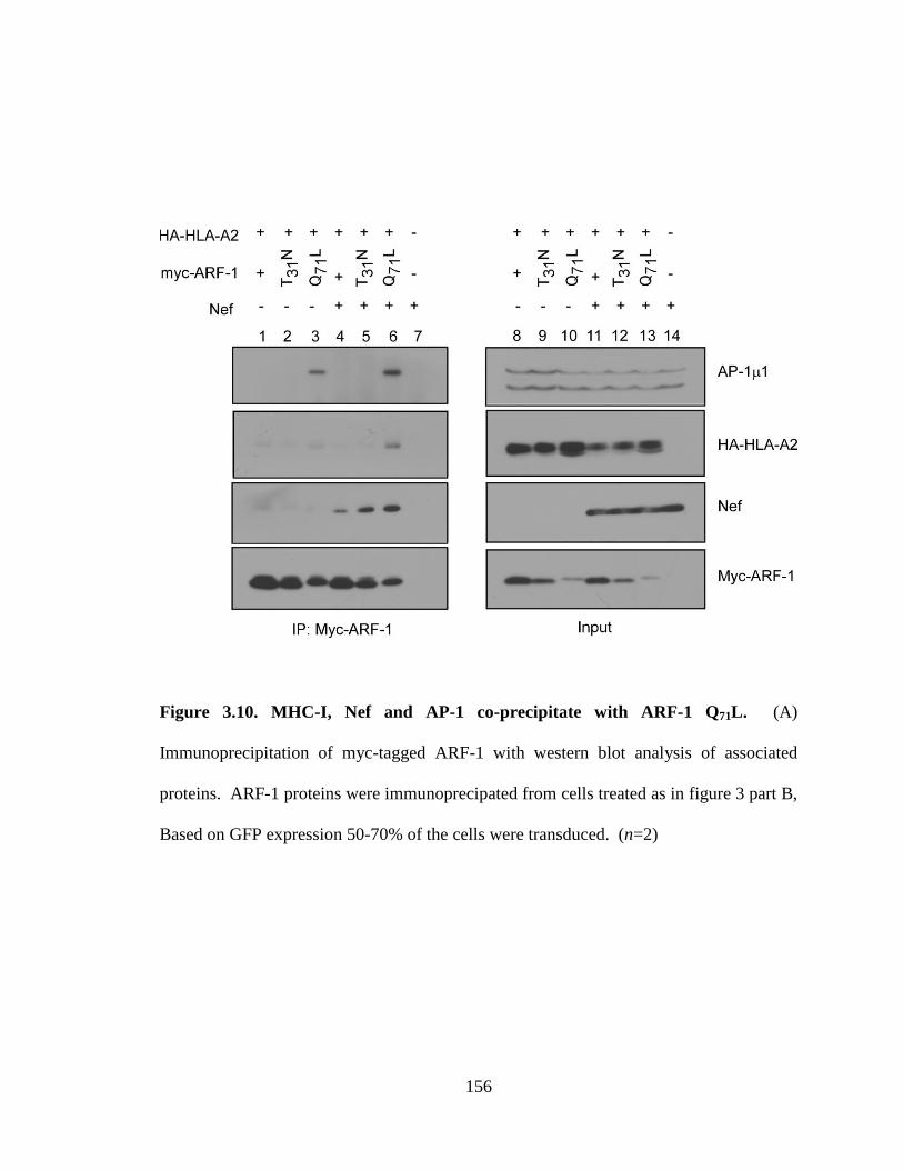
156
Figure 3.10. MHC-I, Nef and AP-1 co-precipitate with ARF-1 Q71L. (A)
Immunoprecipitation of myc-tagged ARF-1 with western blot analysis of associated
proteins. ARF-1 proteins were immunoprecipated from cells treated as in figure 3 part B,
Based on GFP expression 50-70% of the cells were transduced. (n=2)
157
3.4 Discussion
HIV causes a persistent infection that evades eradication by anti-HIV CTLs. Viral
persistence is mediated in part by the activity of the HIV Nef protein, which disrupts
antigen presentation by MHC-I to CTLs. A number of cellular factors have been
implicated in this pathway and thus it is important to clarify the relative contribution of
each to help focus the development of inhibitors.
Here we confirm a requirement for AP-1 and we provide new evidence that the small
GTPase ARF-1 is also needed for Nef„s effects on MHC-I trafficking. The ARF-1
dominant active mutant, ARF-1-Q71L and the ARF-1 inhibitor, BFA both had dramatic
effects on the formation of Nef-MHC-I–AP-1 complex and on surface MHC-I expression
in Nef-expressing cells. Our new data, that ARF-1 is needed for Nef to recruit AP-1 to
the MHC-I cytoplasmic tail, provide insights into what is required for complex formation
and further elucidates how this process occurs (Figure 3.11).
The effect of dominant active ARF-1 Q71L on MHC-I downmodulation may be explained
by other studies examining COP-1 coats, which have demonstrated that when ARF-1
Q71L is expressed, ARF-1-GTP cannot be hydrolyzed into ARF-1-GDP, resulting in a
static coat (43, 50). This effect compromises the cycles of recruitment and dissociation
from membranes.
Prior studies have shown that AP-1 is required for Nef-dependent trafficking and can be
found in complexes with MHC-I and Nef in transformed T cell lines and in HIV-infected

158
Figure 3.11. Model of Nef-dependent CD4 and MHC-I trafficking. LY, lysosome;
LE, late endosome; MVB, multi-vesicular body; ER, endoplasmic reticulum.
159
primary T lymphocytes (41). Work with purified proteins has revealed that Nef directly
contacts the MHC-I cytoplasmic tail (58) and that a Nef-MHC-I fusion protein directly
interacts with the AP-1 subunit (32, 47). A tyrosine in the MHC-I cytoplasmic tail and
the tyrosine binding pocket of AP-1 1 are both required for AP-1 to coprecipitate with
HLA-A2 in Nef-expressing cells (59) and with purified proteins (32, 47). These data
have led to a model in which Nef disrupts MHC-I trafficking from the TGN through an
interaction with AP-1 that re-directs MHC-I into an endolysosomal pathway instead of
the cell surface (Figure 3.11). In cells that demonstrate a greater degree of Nef-
dependent MHC-I internalization (e.g. HeLa), AP-1 is also required for Nef-dependent
internalization of MHC-I (23).
Nef also downmodulates the HIV receptor, CD4 to prevent viral superinfection (2) and to
promote viral assembly and release (25, 42). Downmodulation of CD4 and MHC-I by
Nef occurs by distinct mechanisms and requires different Nef domains (29, 44). Work
from a number of laboratories has supported a model in which the separate Nef-
dependent trafficking pathways of MHC-I and CD4 ultimately converge into a common
-COP-dependent pathway necessary for lysosomal targeting (15, 36, 44). In this model
(Figure 3.11), Nef promotes accelerated internalization of CD4 in an AP-2 dependent
manner (7, 17, 21, 49) and internalized CD4 is targeted into acidic compartments (36)
and MVBs (12, 44) for accelerated degradation. Previous research has shown that ARF-1
is involved in COP-I coatomer recruitment to internalized Nef-CD4 complexes and is
necessary for localization of these complexes to acidic compartments (15). However,
GTP binding and hydrolysis is not needed for this process (15).
160
In contrast to our results with ARF-1, we did not detect a significant requirement for
ARF-6 activity for Nef to reduce MHC-I surface expression. The difference in our
results compared with prior publications from another laboratory may be related to a
number of technical factors. The effect of ARF-6-Q67L was reported in HeLa cells (4),
which have been shown to traffic MHC-I differently than T cells (23). In addition, the
prior study (4), did not examine the effects of these mutants on steady state surface
expression as was done here.
In sum, we have further defined the mechanism by which Nef allows HIV-1 infected
cells to evade the host immune response. Implicating ARF-1 in downmodulation of
MHC-I by Nef provides further support for the role of the AP-1 dependent pathway
shown in Figure 3.11. These data help to elucidate the mechanism by which Nef
downmodulates MHC-I and reveal further targets for pharmaceuticals that may inhibit
immune evasion.
3.5 Materials and methods
3.5.1 Cell lines and primary cell isolation
CEM T cells and SupT1 cells expressing HA-tagged HLA-A2 were created and
maintained as previously published (59). Primary T cells were purified from buffy coats
obtained from the New York Blood Center. Mononuclear cells were purified by ficoll-
hypaque centrifugation, and primary T lymphocytes were isolated through adherence,
CD8 and CD56 depletion of mononuclear cells. Following isolation, T cells were
161
stimulated with PHA at 10 g/mL (Sigma-Aldrich). After 24 hours, 50U/mL interleukin-
2 [IL-2] was added to the culture medium. 48 hours post-IL-2 stimulation, the T
lymphocytes were used for HIV transduction. Donor MHC-I allotype (HLA-A2, HLA-
BW4, or HLA-BW6) was determined at the time of CD4+ T cell isolation using flow
cytometric techniques.
3.5.2 DNA constructs
3.5.2.1 Construction of pMSCV IRES GFP vectors expressing ARF-1 and ARF-6
pCB6 expressing Myc-tagged wild type or T31N ARF-1 were obtained from Didier Trono
(Ecole Polytechnique Fédérale de Lausanne). The arf-1 ORF was amplified by PCR
using either wild type or T31N pCB6 Myc-ARF-1 as a template with the primers listed in
Supplementary Table 1. MSCV Myc-ARF-1 Q71L IRES GFP was created through
standard two-step PCR mutagenesis using wild type MSCV ARF-1 IRES GFP as a
template with the primers listed in Supplementary Table 1. The PCR products were
cloned into the BamHI site of MSCV IRES GFP (pMIG) (55).
pXS expressing HA-tagged wild type, T27N, or Q67L ARF-6 was obtained from Julie
Donaldson (National Institutes of Health). ARF-6 constructs were amplified using pXS
HA-ARF-6 as a template with the primers listed in Supplementary Table 1. The PCR
product was cloned into Bgl II and EcoRI sites of pMIG.

162
3.5.2.2 Construction of HIV vectors expressing ARF-1 and ARF-6
To construct HIVs that also contained both GFP and ARF ORFs, we first made a version
of HIV (pNL-GI) in which a portion of the env ORF was replaced by a GFP IRES
multiple cloning site cassette. PCR was used to amplify the IRES from pNL-PI (8) and
to add additional restriction enzyme sites downstream of the IRES. The PCR product was
ligated into the NheI and BglII sites in the env ORF of pNL4-3-deltaE-EGFP (63) just
downstream of GFP. pNL-GInef – was generated by creating a frame shift within the nef
ORF of pNL-GI by digesting with XhoI, filling-in and re-ligating the ends.
pNL-GI was then used to create HIV constructs expressing ARF-1 and ARF-6. To
create pNL-GIarf constructs, linker primers were designed to create a XbaI site in the 5‟
end and a MluI site in the 3‟ end of the amplicon during PCR amplification of the ARF-1
and ARF-1 Q71L from MSCV ARF-1 IRES GFP and MSCV Myc-ARF-1 Q71L IRES
GFP respectively. Primers for this step are listed in Supplementary Table 1. Digested
PCR products were ligated into the XbaI/MluI sites downstream of the IRES element in
pNL-GI.
Due to an internal XbaI site present in ARF-6, pNL-GI ARF-6 +/- nef and pNL-GI ARF-
6 Q67L +/- nef were engineered by designing linker primers to create a SpeI site in the 5‟
end, which is compatible with XbaI overhang ligation, and a MluI site in the 3‟ end of the
amplicon during PCR amplification of the ARF-6 and ARF-6 Q67L from MSCV ARF-6
IRES GFP and MSCV ARF-6 Q67L IRES GFP respectively. PCR primers used are listed
163
in Supplementary Table 1. Digested PCR products were ligated into the XbaI/MluI
digested parental vector. All constructs were confirmed by sequencing.
3.5.2.3 shRNA constructs
FG12 shRNA lentiviral vectors were constructed as previously described (37, 44). The
negative control shNC construct was previously described (44). The target sequence for
shARF-6 began at position 247:
GATCCCCGGTCTCATCTTCGTAGTGGTTCAAGAGACCACTACGAAGATGAGA
CCTTTTTGGAAA.
3.5.3 Virus preparation and transductions.
3.5.3.1 Retrovirus
Retroviral supernatants were prepared as described previously (34, 55). Bosc cells (34)
were transfected with MSCV constructs described above, the retrovirus packaging vector
pCL-Eco (31), and pHCMV-G (34). Briefly, 5x10⁵ CEM or SupT1 cells were spin-
transduced with 1 ml of retroviral supernatants plus 8 g/ml polybrene at 2500 RPM for
two hours in a tabletop centrifuge at room temperature.
3.5.3.2 Adenovirus
Replication defective adenovirus was produced by the University of Michigan Gene
Vector Core facility. Adenoviral transductions were performed as previously described
(57). Transductions were performed using 1x10⁶ cells in 1 mL of RPMI 1640 containing
2% fetal bovine serum; 10mM HEPES; and 2mM penicillin, streptomycin, and
glutamine. Multiplicity of infection was 200 for CEM and 100 for SupT1 (based on 293
164
cell infectivity, which is greater than CEM infectivity). Low serum transductions ranged
from 4-6 hours.
3.5.3.3 HIV
HIV constructs were produced by transfecting 293 cells with each construct and
harvesting the supernatant. 5x10⁵ PHA and IL-2 stimulated CD8-CD56-depleted T
lymphocytes were transduced using 1 mL of supernatant with 8 g/mL polybrene using a
spin-transduction protocol. Primary T lymphocytes were harvested for analysis of MHC-
I downmodulation at 36 hours post transduction.
3.5.4 Flow cytometry and antibodies
Cells were stained for 20 minutes on ice in FACS buffer (PBS, 1% Human Serum,
1%FBS, 1% HEPES, and 1% NaN3), washed, and then stained for 20 additional minutes
in secondary antibody. Mouse antibody to HLA-A2 antibody, BB7.2 (33) and mouse
antibody to CD4, OKT4 (39) were purified from ascites (22) provided by the University
of Michigan Hybridoma core facility. Mouse antibody to PLAP antibody was obtained
from Serotec. The secondary antibodies used were goat anti-mouse IgG2B-phycoerythrin
(Invitrogen, 1:250) and goat anti-mouse IgG1-Alexa Fluor 647 (Invitrogen, 1:250).
Isotype controls were obtained from BD Biosciences. Stained cells were analyzed on a
Becton Dickinson FACSCanto cytometer. Analysis was performed using FlowJo
software (Tree Star Inc.).

165
3.5.5 Immunofluorescence microscopy and antibodies
ARF-6-transduced CEM cells were adhered to glass slides with Celltak (BD
Biosciences), fixed, permeabilized, and stained for indirect immunofluorescence as
previously described (Roeth JBC 2004). Images were collected using a Zeiss LSM 510
confocal microscope and processed using Deneba Canvas software. The following
antibodies were utilized to localize proteins via microscopy: anti-HLA-A2 (BB7.2) and
anti-GFP (3E6, Invitrogen). Secondary antibodies were obtained from Molecular Probes
and were used at a dilution of 1:250: goat anti-mouse IgG2B-Alexa Fluor 546 and goat
anti-mouse IgG2A-Alexa Fluor 488.
3.5.6 Immunoprecipitations and western blotting
Immunoprecipitations were performed as previously described (59). 15x106 CEM cells
were transduced with either control or Nef-expressing Adenovirus. At 48 hours post-
adenoviral transduction, the cells were spin-transduced with either DMEM (mock) or
MSCV ARF-1 IRES GFP viral supernatants. At 72 hours post-adenoviral transduction,
all cells were incubated in 20mM NH4Cl for 16 hours. Where indicated, samples were
also incubated in 50 M BFA for 16 hours. Cells were then harvested and lysed in 1%
digitonin (Wako) lysis buffer described previously (44, 59). Lysates were normalized for
total protein and GFP transduction rates, when appropriate, prior to immunoprecipitation.
Input controls were 1% of the immunoprecipitated protein. After an overnight pre-clear at
4oC, lysates were immunoprecipitated for either HLA-A2 (BB7.2 crosslinked beads) (59)
or Myc-tagged ARF-1 (9e10 (14) crosslinked beads). Immunoprecipitates were eluted
166
and analyzed by western blot as previously described (57). Western blot antibodies used
were anti-Nef (AG11 (6)) and anti-Myc (9e10 (14)), which were produced by the
University of Michigan Hybridoma Core Facility and purified as previously described
(22). Antibodies also used for western blotting were anti-AP-1 (BD Biosciences), HA
(HA.11, Covance), anti-PTEN (Cell Signaling Technology), anti-GAPDH (Santa Cruz
Biotechnology), and anti-AP-1 1 (RY/1 (53)). The secondary antibody for anti-Nef,
anti-HA, and anti-Myc was goat anti-mouse IgG1-HRP (Zymed Laboratories Inc). The
secondary antibody for anti-AP-1 1 and anti-PTEN was goat anti-rabbit-HRP (Zymed
Laboratories Inc). The secondary antibody for AP-1 was rabbit anti-mouse-HRP
(Zymed Laboratories Inc).
Acknowledgements
This work was supported by NIH grant AI046998. We thank members of the Collins lab
for helpful discussions; the University of Michigan Gene Vector Core Facility for
producing adenoviruses; and the University of Michigan Hybridoma Core for producing
ascites. Jolie Leonard and Elizabeth Wonderlich were supported by the Cellular and
Molecular Biology Training Grant from NIH. Jolie Leonard was supported by the
Molecular Mechanisms in Microbial Pathogenesis Training Grant from NIH. Elizabeth
Wonderlich was also supported by a University of Michigan Rackham Predoctoral
Fellowship. Deanna Kulpa was supported by a the Irvington Institute Fellowship
Program of the Cancer Research Institute. pNL4-3deltaE-GFP (Cat# 1100) was obtained
through the AIDS Research and Reference Reagent Program, Division of AIDS, National
Institute of Allergy and Infectious Diseases, US National Institutes of Health from Drs.
167
Haili Zhang, Yan Zhou, and Robert Siliciano (Johns Hopkins University). We would also
like to thank Linton Traub (University of Pittsburgh) for his gift of antibody to AP-1;
Didier Trono (Ecole Polytechnique Fédérale de Lausanne) for his gift of Myc-tagged
ARF-1 DNA; and Julie Donaldson (National Institutes of Health) for her gift of HA-
tagged ARF-6 DNA.
Notes
This work is the result of a collaborative effort. This project was the primary effort of
Elizabeth Wonderlich, who performed the experiments shown in Figures 3.1, 3.2, 3.3,
3.9, and 3.10. Jolie Leonard contributed the work depicted in Figures 3.3, 3.5, 3.6, and
3.8. Deanna Kulpa performed the experiments in Figure 3.7, and cloned NL-GIarf from
constructs previously created by Jason Normam.
3.6 References
1. Austin, C., I. Hinners, and S. A. Tooze. 2000. Direct and GTP-dependent
interaction of ADP-ribosylation factor 1 with clathrin adaptor protein AP-1 on
immature secretory granules. J Biol Chem 275:21862-9.
2. Benson, R. E., A. Sanfridson, J. S. Ottinger, C. Doyle, and B. R. Cullen. 1993.
Downregulation of cell-surface CD4 expression by simian immunodeficiency
virus Nef prevents viral super infection. J Exp Med 177:1561-6.
3. Berke, G. 1995. The CTL's kiss of death. Cell 81:9-12.
4. Blagoveshchenskaya, A. D., L. Thomas, S. F. Feliciangeli, C. H. Hung, and G.
Thomas. 2002. HIV-1 Nef downregulates MHC-I by a PACS-1- and PI3K-
regulated ARF6 endocytic pathway. Cell 111:853-66.
5. Brown, F. D., A. L. Rozelle, H. L. Yin, T. Balla, and J. G. Donaldson. 2001.
Phosphatidylinositol 4,5-bisphosphate and Arf6-regulated membrane traffic. J
Cell Biol 154:1007-17.
6. Chang, A. H., J. A. Hoxie, S. Cassol, M. O'Shaughnessy, and F. Jirik. 1998.
Construction of single-chain antibodies that bind an overlapping epitope of HIV-1
Nef. FEBS Lett 441:307-12.
7. Chaudhuri, R., O. W. Lindwasser, W. J. Smith, J. H. Hurley, and J. S.
Bonifacino. 2007. Downregulation of CD4 by human immunodeficiency virus

168
type 1 Nef is dependent on clathrin and involves direct interaction of Nef with the
AP2 clathrin adaptor. J Virol 81:3877-90.
8. Collins, K., B. Chen, S. Kalams, B. Walker, and D. Baltimore. 1998. HIV-1
Nef protein protects infected primary human cells from killing by cytotoxic T
lymphocytes. Nature 391:397-401.
9. Collins, K. L. 2004. Resistance of HIV-infected cells to cytotoxic T lymphocytes.
Microbes Infect 6:494-500.
10. D'Souza-Schorey, C., G. Li, M. I. Colombo, and P. D. Stahl. 1995. A
regulatory role for ARF6 in receptor-mediated endocytosis. Science 267:1175-8.
11. Dascher, C., and W. E. Balch. 1994. Dominant inhibitory mutants of ARF1
block endoplasmic reticulum to Golgi transport and trigger disassembly of the
Golgi apparatus. J Biol Chem 269:1437-48.
12. daSilva, L. L., R. Sougrat, P. V. Burgos, K. Janvier, R. Mattera, and J. S.
Bonifacino. 2009. Human immunodeficiency virus type 1 Nef protein targets
CD4 to the multivesicular body pathway. J Virol 83:6578-90.
13. Donaldson, J. G., and R. D. Klausner. 1994. ARF: a key regulatory switch in
membrane traffic and organelle structure. Curr Opin Cell Biol 6:527-32.
14. Evan, G. I., G. K. Lewis, G. Ramsay, and J. M. Bishop. 1985. Isolation of
monoclonal antibodies specific for human c-myc proto-oncogene product. Mol
Cell Biol 5:3610-6.
15. Faure, J., R. Stalder, C. Borel, K. Sobo, V. Piguet, N. Demaurex, J.
Gruenberg, and D. Trono. 2004. ARF1 regulates Nef-induced CD4 degradation.
Curr Biol 14:1056-64.
16. Geldmacher, C., J. R. Currier, E. Herrmann, A. Haule, E. Kuta, F.
McCutchan, L. Njovu, S. Geis, O. Hoffmann, L. Maboko, C. Williamson, D.
Birx, A. Meyerhans, J. Cox, and M. Hoelscher. 2007. CD8 T-cell recognition
of multiple epitopes within specific Gag regions is associated with maintenance of
a low steady-state viremia in human immunodeficiency virus type 1-seropositive
patients. J Virol 81:2440-8.
17. Greenberg, M. E., S. Bronson, M. Lock, M. Neumann, G. N. Pavlakis, and J.
Skowronski. 1997. Co-localization of HIV-1 Nef with the AP-2 adaptor protein
complex correlates with Nef-induced CD4 down-regulation. EMBO J 16:6964-
76.
18. Houndolo, T., P. L. Boulay, and A. Claing. 2005. G protein-coupled receptor
endocytosis in ADP-ribosylation factor 6-depleted cells. J Biol Chem 280:5598-
604.
19. Hung, C. H., L. Thomas, C. E. Ruby, K. M. Atkins, N. P. Morris, Z. A.
Knight, I. Scholz, E. Barklis, A. D. Weinberg, K. M. Shokat, and G. Thomas. 2007. HIV-1 Nef assembles a Src family kinase-ZAP-70/Syk-PI3K cascade to
downregulate cell-surface MHC-I. Cell Host Microbe 1:121-33.
20. Jackson, C. L., and J. E. Casanova. 2000. Turning on ARF: the Sec7 family of
guanine-nucleotide-exchange factors. Trends Cell Biol 10:60-7.
21. Jin, Y. J., C. Y. Cai, X. Zhang, H. T. Zhang, J. A. Hirst, and S. J. Burakoff.
2005. HIV Nef-mediated CD4 down-regulation is adaptor protein complex 2
dependent. J Immunol 175:3157-64.
169
22. Kasper, M. R., and K. L. Collins. 2003. Nef-mediated disruption of HLA-A2
transport to the cell surface in T cells. J Virol 77:3041-9.
23. Kasper, M. R., J. F. Roeth, M. Williams, T. M. Filzen, R. I. Fleis, and K. L.
Collins. 2005. HIV-1 Nef disrupts antigen presentation early in the secretory
pathway. J Biol Chem 280:12840-8.
24. Kiepiela, P., K. Ngumbela, C. Thobakgale, D. Ramduth, I. Honeyborne, E.
Moodley, S. Reddy, C. de Pierres, Z. Mncube, N. Mkhwanazi, K. Bishop, M.
van der Stok, K. Nair, N. Khan, H. Crawford, R. Payne, A. Leslie, J. Prado,
A. Prendergast, J. Frater, N. McCarthy, C. Brander, G. H. Learn, D. Nickle,
C. Rousseau, H. Coovadia, J. I. Mullins, D. Heckerman, B. D. Walker, and P.
Goulder. 2007. CD8+ T-cell responses to different HIV proteins have discordant
associations with viral load. Nat Med 13:46-53.
25. Lama, J., A. Mangasarian, and D. Trono. 1999. Cell-surface expression of
CD4 reduces HIV-1 infectivity by blocking Env incorporation in a Nef- and Vpu-
inhibitable manner. Curr Biol 9:622-31.
26. Lewinsohn, D. A., R. Lines, D. M. Lewinsohn, S. R. Riddell, P. D. Greenberg,
M. Emerman, and S. R. Bartz. 2002. HIV-1 Vpr does not inhibit CTL-mediated
apoptosis of HIV-1 infected cells. Virology 294:13-21.
27. Lippincott-Schwartz, J., L. C. Yuan, J. S. Bonifacino, and R. D. Klausner.
1989. Rapid redistribution of Golgi proteins into the ER in cells treated with
brefeldin A: evidence for membrane cycling from Golgi to ER. Cell 56:801-13.
28. Lubben, N. B., D. A. Sahlender, A. M. Motley, P. J. Lehner, P. Benaroch, and
M. S. Robinson. 2007. HIV-1 Nef-induced Down-Regulation of MHC Class I
Requires AP-1 and Clathrin but Not PACS-1 and Is Impeded by AP-2. Mol Biol
Cell 18:3351-65.
29. Mangasarian, A., V. Piguet, J. K. Wang, Y. L. Chen, and D. Trono. 1999.
Nef-induced CD4 and major histocompatibility complex class I (MHC-I) down-
regulation are governed by distinct determinants: N-terminal alpha helix and
proline repeat of Nef selectively regulate MHC-I trafficking. J Virol 73:1964-73.
30. Naslavsky, N., R. Weigert, and J. G. Donaldson. 2004. Characterization of a
nonclathrin endocytic pathway: membrane cargo and lipid requirements. Mol Biol
Cell 15:3542-52.
31. Naviaux, R. K., E. Costanzi, M. Haas, and I. M. Verma. 1996. The pCL vector
system: rapid production of helper-free, high-titer, recombinant retroviruses. J
Virol 70:5701-5.
32. Noviello, C. M., S. Benichou, and J. C. Guatelli. 2008. Cooperative binding of
the class I major histocompatibility complex cytoplasmic domain and human
immunodeficiency virus type 1 Nef to the endosomal AP-1 complex via its mu
subunit. J Virol 82:1249-58.
33. Parham, P., and F. M. Brodsky. 1981. Partial purification and some properties
of BB7.2. A cytotoxic monoclonal antibody with specificity for HLA-A2 and a
variant of HLA-A28. Hum Immunol 3:277-299.
34. Pear, W. S., G. P. Nolan, M. L. Scott, and D. Baltimore. 1993. Production of
high-titer helper-free retroviruses by transient transfection. Proc Natl Acad Sci U
S A 90:8392-6.

170
35. Peyroche, A., B. Antonny, S. Robineau, J. Acker, J. Cherfils, and C. L.
Jackson. 1999. Brefeldin A acts to stabilize an abortive ARF-GDP-Sec7 domain
protein complex: involvement of specific residues of the Sec7 domain. Mol Cell
3:275-85.
36. Piguet, V., F. Gu, M. Foti, N. Demaurex, J. Gruenberg, J. L. Carpentier, and
D. Trono. 1999. Nef-induced CD4 degradation: a diacidic-based motif in Nef
functions as a lysosomal targeting signal through the binding of beta-COP in
endosomes. Cell 97:63-73.
37. Qin, X. F., D. S. An, I. S. Chen, and D. Baltimore. 2003. Inhibiting HIV-1
infection in human T cells by lentiviral-mediated delivery of small interfering
RNA against CCR5. Proc Natl Acad Sci U S A 100:183-8.
38. Radhakrishna, H., and J. G. Donaldson. 1997. ADP-ribosylation factor 6
regulates a novel plasma membrane recycling pathway. J Cell Biol 139:49-61.
39. Reinherz, E. L., P. C. Kung, G. Goldstein, and S. F. Schlossman. 1979.
Separation of functional subsets of human T cells by a monoclonal antibody. Proc
Natl Acad Sci U S A 76:4061-5.
40. Robinson, M. S. 2004. Adaptable adaptors for coated vesicles. Trends Cell Biol
14:167-74.
41. Roeth, J. F., M. R. Kasper, M. Williams, T. M. Filzen, and K. L. Collins.
2004. HIV-1 Nef disrupts MHC-I trafficking by recruiting AP-1 to the MHC-I
cytoplasmic tail. J. Cell Biol. 167:903-913.
42. Ross, T. M., A. E. Oran, and B. R. Cullen. 1999. Inhibition of HIV-1 progeny
virion release by cell-surface CD4 is relieved by expression of the viral Nef
protein. Curr Biol 9:613-21.
43. Rothman, J. E., and F. T. Wieland. 1996. Protein sorting by transport vesicles.
Science 272:227-34.
44. Schaefer, M. R., E. R. Wonderlich, J. F. Roeth, J. A. Leonard, and K. L.
Collins. 2008. HIV-1 Nef targets MHC-I and CD4 for degradation via a final
common beta-COP-dependent pathway in T cells. PLoS Pathog 4:e1000131.
45. Schwartz, O., V. Marechal, S. Le Gall, F. Lemonnier, and J. M. Heard. 1996.
Endocytosis of major histocompatibility complex class I molecules is induced by
the HIV-1 Nef protein. Nat Med 2:338-42.
46. Serafini, T., L. Orci, M. Amherdt, M. Brunner, R. A. Kahn, and J. E.
Rothman. 1991. ADP-ribosylation factor is a subunit of the coat of Golgi-derived
COP-coated vesicles: a novel role for a GTP-binding protein. Cell 67:239-53.
47. Singh, R. K., D. Lau, C. M. Noviello, P. Ghosh, and J. C. Guatelli. 2009. An
MHC-I cytoplasmic domain/HIV-1 Nef fusion protein binds directly to the micro
subunit of the AP-1 endosomal coat complex. PLoS One 4:e8364.
48. Stamnes, M. A., and J. E. Rothman. 1993. The binding of AP-1 clathrin adaptor
particles to Golgi membranes requires ADP-ribosylation factor, a small GTP-
binding protein. Cell 73:999-1005.
49. Stove, V., I. Van de Walle, E. Naessens, E. Coene, C. Stove, J. Plum, and B.
Verhasselt. 2005. Human immunodeficiency virus Nef induces rapid
internalization of the T-cell coreceptor CD8alphabeta. J Virol 79:11422-33.

171
50. Tanigawa, G., L. Orci, M. Amherdt, M. Ravazzola, J. B. Helms, and J. E.
Rothman. 1993. Hydrolysis of bound GTP by ARF protein triggers uncoating of
Golgi-derived COP-coated vesicles. J Cell Biol 123:1365-71.
51. Teal, S. B., V. W. Hsu, P. J. Peters, R. D. Klausner, and J. G. Donaldson.
1994. An activating mutation in ARF1 stabilizes coatomer binding to Golgi
membranes. J Biol Chem 269:3135-8.
52. Tomiyama, H., H. Akari, A. Adachi, and M. Takiguchi. 2002. Different effects
of Nef-mediated HLA class I down-regulation on human immunodeficiency virus
type 1-specific CD8(+) T-cell cytolytic activity and cytokine production. J Virol
76:7535-43.
53. Traub, L. M., S. Kornfeld, and E. Ungewickell. 1995. Different domains of the
AP-1 adaptor complex are required for Golgi membrane binding and clathrin
recruitment. J Biol Chem 270:4933-42.
54. Traub, L. M., J. A. Ostrom, and S. Kornfeld. 1993. Biochemical dissection of
AP-1 recruitment onto Golgi membranes. J Cell Biol 123:561-73.
55. Van Parijs, L., Y. Refaeli, J. D. Lord, B. H. Nelson, A. K. Abbas, and D.
Baltimore. 1999. Uncoupling IL-2 signals that regulate T cell proliferation,
survival, and Fas-mediated activation-induced cell death. Immunity 11:281-288.
56. Volpicelli-Daley, L. A., Y. Li, C. J. Zhang, and R. A. Kahn. 2005. Isoform-
selective effects of the depletion of ADP-ribosylation factors 1-5 on membrane
traffic. Mol Biol Cell 16:4495-508.
57. Williams, M., J. F. Roeth, M. R. Kasper, T. Filzen, and K. L. Collins. 2005.
Human Immunodeficiency Virus Type 1 Nef Domains Required for Disruption of
Major Histocompatibility Complex Class I Trafficking Are Also Necessary for
Coprecipitation of Nef with HLA-A2. Journal of Virology 79:632-636.
58. Williams, M., J. F. Roeth, M. R. Kasper, R. I. Fleis, C. G. Przybycin, and K.
L. Collins. 2002. Direct binding of human immunodeficiency virus type 1 Nef to
the major histocompatibility complex class I (MHC-I) cytoplasmic tail disrupts
MHC-I trafficking. J Virol 76:12173-12184.
59. Wonderlich, E. R., M. Williams, and K. L. Collins. 2008. The tyrosine binding
pocket in the adaptor protein 1 (AP-1) mu1 subunit is necessary for Nef to recruit
AP-1 to the major histocompatibility complex class I cytoplasmic tail. J Biol
Chem 283:3011-22.
60. Wong, J. K., M. C. Strain, R. Porrata, E. Reay, S. Sankaran-Walters, C. C.
Ignacio, T. Russell, S. K. Pillai, D. J. Looney, and S. Dandekar. 2010. In vivo
CD8+ T-cell suppression of siv viremia is not mediated by CTL clearance of
productively infected cells. PLoS Pathog 6:e1000748.
61. Yang, O. O., P. T. Nguyen, S. A. Kalams, T. Dorfman, H. G. Gottlinger, S.
Stewart, I. S. Chen, S. Threlkeld, and B. D. Walker. 2002. Nef-mediated
resistance of human immunodeficiency virus type 1 to antiviral cytotoxic T
lymphocytes. J Virol 76:1626-31.
62. Zhang, C. J., A. G. Rosenwald, M. C. Willingham, S. Skuntz, J. Clark, and
R. A. Kahn. 1994. Expression of a dominant allele of human ARF1 inhibits
membrane traffic in vivo. J Cell Biol 124:289-300.
63. Zhang, H., Y. Zhou, C. Alcock, T. Kiefer, D. Monie, J. Siliciano, Q. Li, P.
Pham, J. Cofrancesco, D. Persaud, and R. F. Siliciano. 2004. Novel single-
172
cell-level phenotypic assay for residual drug susceptibility and reduced replication
capacity of drug-resistant human immunodeficiency virus type 1. J Virol
78:1718-29.
173
Chapter 4
Cell-based high throughput screening methodology to identify
small molecule inhibitors of HIV-1 Nef
4.1 Introduction
HIV is a worldwide epidemic, with approximately 2.6 million new infections occurring
annually (UNAIDS, 2009). Moreover, while treatments do exist, there is no cure for
HIV. Instead, HIV is a chronic infection, persisting over the lifetime of an infected
individual. Therefore, treatment must be also delivered over the lifetime of the patient.
Many of the drug therapies currently available have significant toxic side effects, making
long term adherence difficult. Furthermore, drug resistance can arise, making additional
viral targets important.
The existing therapies for HIV target viral enzymes, including reverse transcriptase,
protease, and integrase. Another class of inhibitors prevents viral entry by blocking
envelope fusion or attachment to the CCR5 coreceptor. These drugs all target viral
particles and inhibit viral replication. None of these inhibitors target HIV-infected cells
174
or enable the host immune system to better recognize and respond to HIV. There are
currently no drugs on the market or in clinical studies which target the HIV-1 Nef
protein, despite ample evidence that Nef is critically important for viral pathogenesis and
disease progression in both animal models and human patients. Macaques infected with a
Nef-deleted strain of SIV do not progress to AIDS (22). Furthermore, a cohort of blood
transfusion recipients who received an HIV strain with a deletion in Nef and the LTR
demonstrate delayed disease progression or elite control of viral loads (10, 19, 24).
These observations highlight the potential efficacy of a Nef inhibitor in HIV treatment.
Traditional drug development strategies often rely on enzyme substrate modification and
active site targeting. These methods are not suitable for identifying a Nef inhibitor, as
Nef has no enzymatic activity and therefore requires a different approach. Nef is a
structurally flexible, multifunctional adaptor protein with multiple cellular binding
partners (13, 15-17, 21, 25). Nef downmodulates MHC-I, allowing infected cells to
evade detection by cytotoxic T lymphocytes (CTLs) (9, 27, 38, 41, 44), as well as the
viral receptor CD4, preventing superinfection and increasing virion release (14, 23, 37).
Nef also modulates the activity of kinases, such as Pak2 and Hck, and promotes T cell
activation, which may in turn promote viral replication (1, 4, 5, 18, 31, 39, 45).
Additionally, Nef enhances viral infectivity in vitro by a poorly understood mechanism
that is independent of CD4 downmodulation (6, 29, 34-36). It is likely that all of these
Nef functions make contributions to HIV pathogenesis in vivo. However, the ability of
Nef to downmodulate MHC-I and evade CTL detection is a particularly tempting target
for therapeutic intervention. Several studies have used SIV systems to demonstrate that

175
the capacity to downmodulate MHC-I is selected for in vivo (7, 32, 40) . In addition, it
has recently been demonstrated that the ability of in vivo-derived Nef to down-regulate
MHC-I predicted the resistance of HIV-1 to suppression by CTL in vitro (28) . Taken
together, these data demonstrate that the ability of Nef to down-regulate MHC-I in vivo is
maintained by the need of HIV-1 to cope with the antiviral CTL response. Therefore,
inhibition of this Nef function early in infection may enable the host immune system to
better counteract, or even clear, HIV-1. While recent efforts have been applied to
identifying Nef inhibitors, these studies have not specifically targeted the ability of Nef to
downmodulate MHC-I.
The first report to identify chemical inhibitors of Nef function used a competition ELISA
assay to identify guanidine alkaloid analogs capable of disrupting Nef’s interactions with
p56lck
, p53, and actin (33). These proteins all bind to the N-terminus of Nef, although the
functional significance of these interactions is not clear. Furthermore, all of the putative
Nef inhibitors were highly cytotoxic even at submicromolar concentrations and therefore
could not be tested for their impact on viral replication or Nef function within cells.
These compounds have limited therapeutic value.
A few studies have focused on inhibiting interactions with the polyproline, or SH3-
binding, motif in Nef. Mutation of this domain reduces Nef’s ability to downmodulate
MHC-I and also inhibits association of Nef with cellular Src family kinases such as Hck
(5, 20, 21, 30, 31, 42, 45). The importance of Hck activation by Nef in HIV pathogenesis
176
is unclear, as mutation of the SH3-binding domain of Nef results in a Nef protein which
is defective for both Hck activation and MHC-I downmodulation. An in silico screen
resulted in the identification of ten candidate molecules (2). Follow-up in vitro screening
of those compounds, as well as structural analogs, narrowed the candidates to two
molecules capable of inhibiting Nef-SH3 interactions in COS7 African green monkey
kidney cells. Neither compound demonstrated significant cytotoxicity in the COS7 cell
line. Inhibitor treatment did not affect CD4 downmodulation by Nef, which is
independent of the SH3-binding domain of Nef, indicating that the inhibition was specific
to this Nef domain. Disappointingly, treatment of resting peripheral blood mononuclear
cells (PBMC) with up to 25 M inhibitor, the highest concentration tested, inhibited Nef-
induced downmodulation of MHC-I A,B, and C by only 25%.
Subsequent studies also focused on the SH3-binding domain of Nef, specifically looking
for inhibitors of Nef-interactions with Src family kinases (SFK) such as Hck. Screening
of a 10,000 compound library enriched for kinase and protease inhibitors identified a
compound capable of inhibiting Hck in the presence of Nef (12). The compound, termed
DFP-4AP, was found to inhibit Hck activation even in the absence of Nef, and therefore
is likely not specific enough for therapeutic use. However, analysis of DFP-4AP analogs
lead to the discovery of DFP-AB, a molecule capable of specifically inhibited Hck
activity in a Nef-dependant manner. Interestingly, both DFP-4AP and DFP-AB inhibited
HIV replication in U87MG astrocytoma cells with an IC50 in the low M range.
Another study also identified a chemical inhibitor of Nef-induced activation of Hck
which also partially reduced MHC-I HLA-A, B, and C downmodulation by Nef in

177
primary T lymphocytes at 8-days post HIV infection, a late time point (11). While
mutation of multiple prolines within the SH3-binding domain of Nef inhibit both SFK
interactions with Nef and downmodulation of MHC-I, point mutations disrupt kinase
activation but do not affect MHC-I downmodulation (8, 43). It is therefore perhaps
unsurprising that compounds which target this region of Nef have relatively small impact
on MHC-I surface expression in the presence of Nef. While inhibition of kinase activity
is a tractable readout for high throughput methodology, the correlation between this Nef
function and HIV disease progression is unclear.
While targeting the polyproline motif of Nef has had limited success, another recent
study took a different approach by simultaneously targeting multiple Nef functional
domains in conjunction with the SH3-binding activity. The investigators designed a
series of soluble peptides that wrap around Nef, interacting with several domains (3).
The SH3 domain of Hck containing six amino acid modifications that enhance affinity
for Nef was fused to the CD4 cytoplasmic tail with a GGGG linker in the presence of an
N-terminal lipidation signal. This molecule was found to bind to purified Nef with a
dissociation constant of 26 nM, the strongest interaction of a molecule with Nef ever
reported. Additionally, this inhibitor (termed NI3-9) inhibited CD4 and MHC-I
downmodulation by Nef, and blocked Nef-dependant enhancement of virion infectivity in
HeLa and Chinese hamster ovary cells, which may not recapitulate Nef activity in
primary T lymphocytes. The therapeutic potential of an inhibitory protein, however, is
complicated by technical challenges in delivery, stability, and immunogenicity.
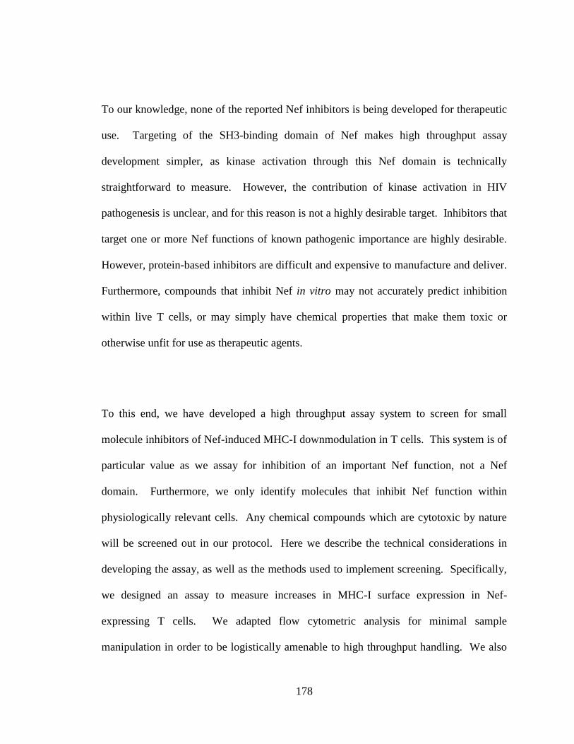
178
To our knowledge, none of the reported Nef inhibitors is being developed for therapeutic
use. Targeting of the SH3-binding domain of Nef makes high throughput assay
development simpler, as kinase activation through this Nef domain is technically
straightforward to measure. However, the contribution of kinase activation in HIV
pathogenesis is unclear, and for this reason is not a highly desirable target. Inhibitors that
target one or more Nef functions of known pathogenic importance are highly desirable.
However, protein-based inhibitors are difficult and expensive to manufacture and deliver.
Furthermore, compounds that inhibit Nef in vitro may not accurately predict inhibition
within live T cells, or may simply have chemical properties that make them toxic or
otherwise unfit for use as therapeutic agents.
To this end, we have developed a high throughput assay system to screen for small
molecule inhibitors of Nef-induced MHC-I downmodulation in T cells. This system is of
particular value as we assay for inhibition of an important Nef function, not a Nef
domain. Furthermore, we only identify molecules that inhibit Nef function within
physiologically relevant cells. Any chemical compounds which are cytotoxic by nature
will be screened out in our protocol. Here we describe the technical considerations in
developing the assay, as well as the methods used to implement screening. Specifically,
we designed an assay to measure increases in MHC-I surface expression in Nef-
expressing T cells. We adapted flow cytometric analysis for minimal sample
manipulation in order to be logistically amenable to high throughput handling. We also
179
optimized the assay for maximal statistical power. Preliminary findings suggest that
molecules identified under the stringent conditions of this screen will be valid and
effective Nef inhibitors.
4.2 Methods and Results:
4.2.1 Assay design
In order to screen for molecules which inhibit downmodulation of MHC-I by Nef in T
lymphocytes, we first wanted to develop a cell-based flow cytometric screening protocol
to measure MHC-I surface expression. A typical FACS staining protocol requires
multiple antibody incubations and a number of rigorous washes. These steps are not
amenable to high throughput methodology, as minimal manipulation of sample is
technically desirable in these systems. Therefore, we needed to establish a FACS
staining protocol in which we could reliably differentiate between high and low MHC-I
surface expression, in as few steps as possible. We tested two strategies for reducing
sample manipulation: condensing antibody incubations, and using dilution of the staining
reaction to reduce background.
We examined three conditions for condensing antibody incubations. First, we tested a
primary antibody against HLA-A2 (BB7.2) directly conjugated to Alexa Fluor-488 to
eliminate separate primary and secondary antibody incubation steps. As two-step
antibody stains can amplify signal, we also tried co-incubating primary and secondary
180
antibodies, as well as sequential primary and secondary antibody incubations in the
absence of intervening wash steps. We used these staining protocols to compare CEM
SS cells stably expressing exogenous HLA-A2, a common MHC-I allele, with the same
cell line (CEM A2) transduced with an adenoviral vector expressing NL4-3 Nef. We
found that the directly-conjugated antibody (BB7.2-488) was most amenable to a staining
protocol without washes, resulting in the highest signal over background (Figure 4.1A-
B).
We then optimized dilution conditions in order to achieve thorough background reduction
while keeping sample volume low. Additionally, we further condensed the protocol by
combining the dilution and fixation steps. This was accomplished by including
paraformaldehyde (PFA) in the dilution buffer. To determine signal over background, we
compared CEM A2 cells to parental CEM SS, incubated with BB7.2-488 and then
subjected to a variety of dilution volumes. We found that dilution of two- to three-fold
resulted in high specific signal and low background, while greater dilution factors
reduced signal without further reducing background (Figure 4.1C-D). Thus we
determined optimal conditions for a high throughput FACS stain, condensing a multi-step
protocol into two simple steps which produce robust, specific signal.
181
Figure 4.1 FACS stain protocol development. (A-B) CEM A2 cells infected with the
indicated adenovirus were stained with the indicated antibodies. (A) Flow cytometric
analysis of HLA-A2 expression. (B) Mean fluorescence intensity (MFI) plus standard
deviation is shown, and the Z’-factor for each +/- Nef pair is displayed above the bars.
(C-D) CEM SS and CEM A2 cells were stained with fluorophore-conjugated antibody
and dilution with PFA was carried out at the indicated dilution factor. (C) Flow
cytometric analysis of background and specific staining. (D) The MFI is shown and the
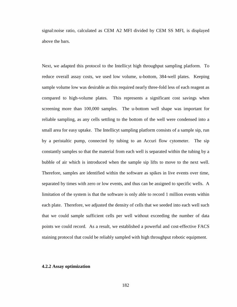
182
signal:noise ratio, calculated as CEM A2 MFI divided by CEM SS MFI, is displayed
above the bars.
Next, we adapted this protocol to the Intellicyt high throughput sampling platform. To
reduce overall assay costs, we used low volume, u-bottom, 384-well plates. Keeping
sample volume low was desirable as this required nearly three-fold less of each reagent as
compared to high-volume plates. This represents a significant cost savings when
screening more than 100,000 samples. The u-bottom well shape was important for
reliable sampling, as any cells settling to the bottom of the well were condensed into a
small area for easy uptake. The Intellicyt sampling platform consists of a sample sip, run
by a peristaltic pump, connected by tubing to an Accuri flow cytometer. The sip
constantly samples so that the material from each well is separated within the tubing by a
bubble of air which is introduced when the sample sip lifts to move to the next well.
Therefore, samples are identified within the software as spikes in live events over time,
separated by times with zero or low events, and thus can be assigned to specific wells. A
limitation of the system is that the software is only able to record 1 million events within
each plate. Therefore, we adjusted the density of cells that we seeded into each well such
that we could sample sufficient cells per well without exceeding the number of data
points we could record. As a result, we established a powerful and cost-effective FACS
staining protocol that could be reliably sampled with high throughput robotic equipment.
4.2.2 Assay optimization

183
Once we had established a FACS staining protocol that was amenable to high throughput
handling, it was important to assess the potential statistical power of this assay for
screening. An important measurement when piloting high throughput assays is the Z’-
factor. The Z’-factor is calculated as follows:
Where “s” is the standard deviation of the positive (p) and negative (n) controls, and “m”
is the mean value of the controls. Therefore this value relies on the magnitude of the
difference between positive and negative controls, as well as the variability within these
samples. This provides a measure of the reliability with which the assay may detect
samples with a difference of more than three standard deviations from the control
samples. Z’-factors fall between 0 and 1, with 1 being the ideal Z’. Assays with Z’-
factors greater than 0.5 are considered excellent assays, while lower Z’ values indicate
that additional considerations are warranted. In this screen, CEM A2 cells expressing
Nef, and therefore displaying low HLA-A2 surface expression, were the negative
controls. CEM A2 cells transduced with control adenovirus were the positive controls, as
a Nef inhibitor would restore high levels of HLA-A2 surface expression (Figure 4.2A-C).
Hence our technical goals were to minimize standard deviation and maximize the fold-
dowmodulation of HLA-A2 by Nef.
In order to improve downmodulation of HLA-A2 by Nef, we optimized our adenovirus
transductions to achieve efficient delivery of Nef. The most obvious strategy for
enhancing infection rate is to use a higher multiplicity of infection (MOI). However,
184
A B C
HLA-A2
Nef Inhibitor
BB7.2-488
Figure 4.2. Schematic representation of experimental controls and potential
inhibitor results. (A) Positive control. CEM A2 cells display high surface-expression
of MHC-I in the absence of Nef. (B) Negative control. Nef reduces MHC-I surface
expression, which in turn reduces antibody binding and fluorescent signal. (C) A
chemical inhibitor prevents Nef from disrupting MHC-I surface expression, resulting in
elevated fluorescent signal.

185
adenovirus preparations, which we obtained from the University of Michigan vector core,
are a costly reagent when required in large amounts over the course of a screen.
Therefore, it was important to balance optimal infection rates with expense. An
additional strategy was to improve infection efficiency without using more virus.
Adenovirus is inhibited by serum, so we typically perform these infections in low serum
media, RPMI 1640 supplemented with 2% fetal bovine serum (FBS). We tested whether
eliminating serum from the infection medium altogether would enhance transduction over
a range of MOIs. Indeed, we found that the use of serum-free infection media resulted in
a greater fold-reduction in HLA-A2 surface expression as compared to the 2% serum-
containing media (Figure 4.3A-B). We also determined that an MOI of 600 resulted in
uniform infection of the cells within the population and nearly twice as much Nef-
induced downmodulation of HLA-A2 as the lower MOIs (Figure 4.3A-B).
Once we had maximized the measurable difference between our positive and negative
controls, we investigated possible methods to reduce variability between samples. We
suspected that compound addition was a source of variation, as we observed high
standard deviation in plates to which we added the DMSO solvent control versus plates
that had no solvent or compound addition. DMSO is the solvent for the chemical
compounds within the test libraries and can affect HLA-A2 surface expression.
Compound addition was performed with a robotic arm that has 384 small metal pins that
dip into each well of plates containing the chemical libraries and rely on surface tension

186
Figure 4.3 High throughput assay optimization. (A) Flow cytometric analysis of
HLA-A2 expression in CEM A2 cells infected with a control adenovirus or an adenovirus
expressing Nef at a range of MOIs in the indicated infection medium. (B) Quantitation of
(A) showing fold downmodulation, calculated as the mean fluorescence intensity (MFI)
of vector transduced cells divided by the MFI of the Nef-expressing cells. (C)
Adenovirus-infected CEM A2 cells were either directly subjected to pintool addition of
DMSO, or dispensed into plates containing PBS to which DMSO had been added by
pintool. Graph summarizing the results of high throughput sampling of each plate is
represented by the median MFI for control vector treated or Nef-expressing cells. Error
bars indicate standard deviation. Z’ values are displayed above bars. (D) CEM A2 cells
were transduced with adenovirus or an adenovirus expressing Nef. At 2 dpi, the cells
were incubated overnight at high density in low serum assay media plus DMSO solvent
control. Cell viability, as determined by forward scatter and side scatter profile, is
shown. Error bars represent standard deviation.

187
Figure 4.3 High throughput assay optimization

188
to pick up and deliver the contents of the library plates. Therefore, the depth (ie volume)
within the wells of the experimental plate determines the amount of solvent or compound
delivered. We found that pintooling solvent directly into cells and media in the
experimental plates resulted in a high standard deviation in the concentration delivered to
each well. Alternatively, delivery into 4 L of PBS in each well of experimental plates
resulted in lower standard deviations and a desirable concentration of 15-25 M for the
compounds within the library. A 2x cell suspension could then be delivered into plates
already containing PBS and experimental compounds (Figure 4.3C). In this manner, we
were able to reduce the variation between identical samples.
An additional factor to consider was potential cytotoxicity. It was important both that the
cells survived the assay protocol and that we avoided registering toxic chemicals as
potential hits for extensive follow up. With regard to the first, we examined viability of
cells exposed to our assay protocol. A source of potential cytotoxicity was an overnight
incubation in low-serum media in the presence of chemical compounds or DMSO
solvent. The low-serum incubation was intended to avoid interference by undefined
factors within serum. Therefore cells were incubated with the compounds in RPMI 1640
supplemented with 0.1% FBS. The cells were exposed to the putative inhibitors
overnight, or approximately 16 hours, in hopes of achieving maximal delivery and
activity of each experimental molecule. Cells subjected to this treatment in the presence
of 1% DMSO tolerated this treatment well, as determined by cell viability (Figure 4.3D).
Additionally, inclusion of a live-cell exclusion reagent, 7-Aminoactinomysin-D (7AAD),
in the dilution buffer during the FACS stain allowed us to exclude dead or dying cells
189
from our analysis. Therefore, we were confident that we could identify compounds that
inhibit Nef while excluding compounds with significant toxicity in T cells.Despite
protocol optimization, pilot experiments of the assay yielded marginal Z’ values of 0.2-
0.4. The non-ideal Z’ values were a reflection of relatively high variation in the positive
controls, a variation that we found to be inherent to dipping the pintool into the wells.
The negative controls, or Nef-expressing cells, displayed very little variation. Therefore,
despite the low overall Z’ values, we could quite reliably identify compounds which
increased HLA-A2 expression greater than three standard deviations as compared to
negative controls (Figure 4.1B and 4.3C). The high variation within the positive controls
made elimination of false positive samples more difficult. Hence it was necessary to
develop a method for eliminating false positives.
False positives could be the result of compounds that have intrinsic fluorescence within
the detection channel (Figure 4.4B) or compounds that induce non-specific binding of the
fluorophore-conjugated antibody to the cells (Figure 4.4C). To factor out such
compounds, we developed a counter-screen. This involved testing the same compound
set against CEM SS parental cells, which do not express HLA-A2, using the same
screening protocol as that for the primary screen of CEM A2 cells. This counter-
screened allowed us to detect any increase in fluorescence intensity that was not specific
to increased HLA-A2 surface expression. Any compound which registered as a hit in the
counter-screen could be eliminated as non-specific. Therefore, despite the non-ideal Z’
value for the primary screen, the stringent counter-screen allowed us to confidently
eliminate false positives.
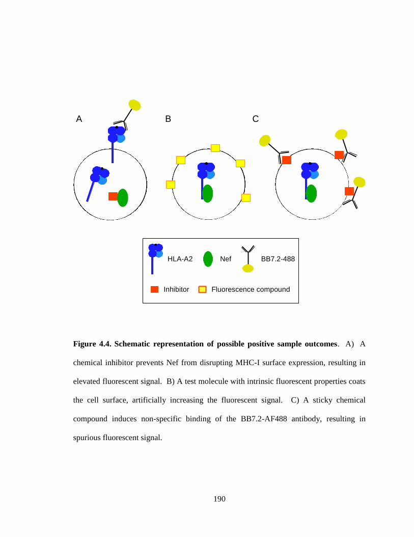
190
CBA
NefHLA-A2
Fluorescence compound
BB7.2-488
Inhibitor
Figure 4.4. Schematic representation of possible positive sample outcomes. A) A
chemical inhibitor prevents Nef from disrupting MHC-I surface expression, resulting in
elevated fluorescent signal. B) A test molecule with intrinsic fluorescent properties coats
the cell surface, artificially increasing the fluorescent signal. C) A sticky chemical
compound induces non-specific binding of the BB7.2-AF488 antibody, resulting in
spurious fluorescent signal.

191
4.2.3 Protocol
We have developed a high throughput screening system with sufficient statistical power
to reliably identify small molecules that display potent Nef inhibition in T lymphocytes.
The detailed protocol is as follows (Figure 4.5): First, CEM A2 cells were transduced
with an empty adenoviral vector control or Nef-expressing adenovirus at an MOI of 600
for six hours in serum-free RPMI 1640. At two days post infection, the cells were
counted and resuspended at a density of 2 million cells per mL in RPMI 1640
supplemented with 0.2% FBS. Experimental plates were then prepared by dispensing 4
L PBS into each well, delivering experimental compounds, and subsequently dispensing
4 L of cell suspension into each well. The cells were incubated with the compounds at
37°C for approximately 16 hours. On day three, 2 L of a 5x antibody solution of
BB7.2-488 (1:500) in 5x FACS buffer (10% FBS, 5% human A/B serum, 5% Hepes, and
0.02% sodium azide in PBS) was added directly to the cells and culture media within
each well and incubated for 30 minutes at 4°C. After antibody incubation, each sample
was diluted and fixed with 20 L 1.5% PFA in 1x FACS buffer (2% FBS, 1% human
A/B serum, 1% Hepes, and 0.02% sodium azide in PBS). After fixation, plates were
stable at 4°C for up to one week and thus were sampled within this time frame.
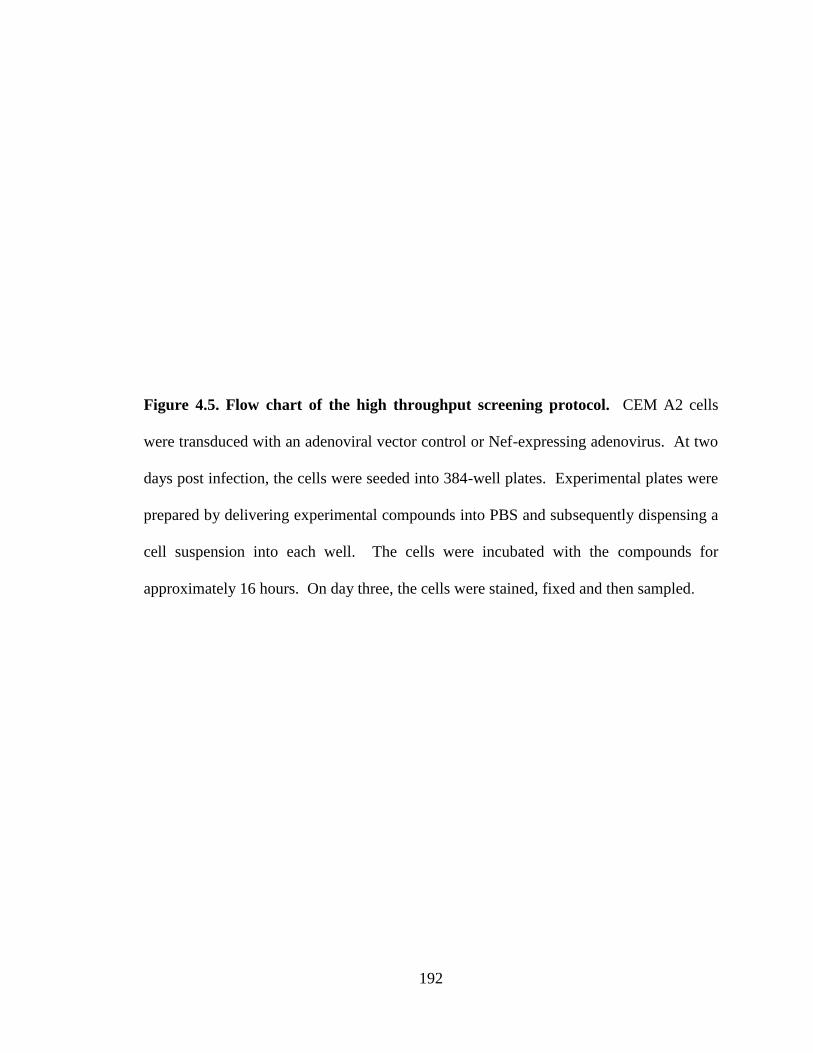
192
Figure 4.5. Flow chart of the high throughput screening protocol. CEM A2 cells
were transduced with an adenoviral vector control or Nef-expressing adenovirus. At two
days post infection, the cells were seeded into 384-well plates. Experimental plates were
prepared by delivering experimental compounds into PBS and subsequently dispensing a
cell suspension into each well. The cells were incubated with the compounds for
approximately 16 hours. On day three, the cells were stained, fixed and then sampled.
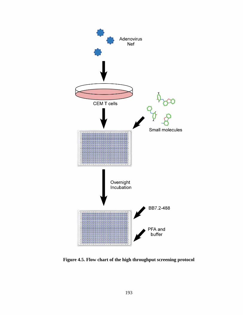
193
Figure 4.5. Flow chart of the high throughput screening protocol

194
4.2.4 Preliminary results
With an optimized protocol in place, we began screening experimental compounds. The
initial phase consisted of screening 38,351 small molecules, including 2000 compounds
within the Spectrum library of FDA-approved compounds, as well as Chembridge and
Maybridge collections of drug-like compounds. The Z-factor for this portion of the
screen averaged to 0.25, which is a marginal value. This screen identified 613 primary
hits, as defined by activity greater than three standard deviations above the negative
control samples within each plate, within the entire assay, or greater than 20% activity,
where the average HLA-A2 staining intensity in adenovirus-treated cells is set to 100%
activity, and that in Nef-expressing cells is set to 0%. This represents a hit rate of 1.3%
within the primary screen. 110 of these hits occurred in wells where fewer than 50 live
cell events were recorded. These compounds might be interpreted as cytotoxic, or,
because of potential sampling error, may be considered as unsampled and thus worth
including in a secondary screen. These 613 compounds were tested in triplicate in a
confirmation screen with a protocol identical to that of the primary screen. Only
compounds which tested as hits in at least 2 of 3 replicate wells were counted as
confirmed hits. To factor out false positive hits, we counter-screened the same
compound set against CEM SS parental cells. Any compound which registered as a hit in
the counter-screen was eliminated as a false positive. Of the 613 primary hits, only 1
compound demonstrated Nef inhibition in replicate wells of the confirmation screen
without non-specifically increasing fluorescence values in the parental cell counter-
screen. Therefore, despite the non-ideal Z’ value for the primary screen, we were able to
195
stringently eliminate false positive and identify a small number of functionally active
small molecule Nef inhibitors.
The next test for putative Nef inhibitors is a dose response curve. This assay also
consists of the same protocol as the primary screen, but tests each “hit” compound at a
range of known concentrations. Unfortunately, our initial hit demonstrated negligible
potency in the dose response assay. However, we felt there was value in continuing to
screen a broader library of chemical compounds.
A phase 2 screen of 64,316 compounds within the Chemdiv 100,000 library resulted in
1084 primary hits, defined by the same standards as phase 1. This represents a hit rate of
1.7% within the primary screen. The Z’ factor for this phase of the screen averaged to
0.45, an improvement that was likely the result of exclusion of DMSO from the positive
control wells. This change was necessary due to the setup of the chemical library source
plates, not a deliberate alteration in experimental design. In total, 113 of the hits occurred
in low event wells. These 1084 compounds were subjected to confirmatory and counter-
screening protocols. Of those, 506 compounds were confirmed as active in two of three
replicate wells, a confirmation rate of 47%, which is quite typical for high throughput
screens. However, all but 33 of the confirmed compounds also scored as active in the
counter-screen, eliminating 95% of the hits as false positives. Thus, we had a final hit
rate of 0.05% for phase 2 of the screen. The 33 compounds of interest will be subjected
to dose response analysis in order to identify the most potent Nef inhibitors.
196
4.3 Discussion
Here we have described and verified a high throughput assay system to identify chemical
compounds that potently inhibit a critical function of Nef in live T lymphocytes in the
absence of significant cytotoxicity. To date we have screened 102,667 drug-like small
molecules for their ability to inhibit downmodulation of MHC-I by HIV-1 Nef in T
lymphocytes. Preliminarily, we observed 1,697 primary hits, a hit rate of 1.7%. While
the Z’-factor measurement for the assay is non-ideal, this is a reflection of the relatively
high variation in the positive control samples. Conversely, the Nef-expressing negative
control samples have a very low standard deviation (Figure 4.1). Therefore, sample wells
with a significant increase in HLA-A2 expression as compared to the negative controls
are unambiguous. Hits are defined as compounds that have a signal greater than three
standard deviations, or 20%, over the negative controls. In addition, we have
implemented a stringent counter-screening protocol to eliminate false positive hits.
Therefore, we have developed a powerful system for the discovery of chemical
compounds that inhibit an important function of Nef in T cells.
We have not, as yet, identified a potent inhibitor of Nef. We have, however, identified a
small pool of candidate molecules for further testing. We will also continue screening
the chemical diversity libraries available to us through the University of Michigan Center
for Chemical Genomics. This includes, among others, the remainder of the Chemdiv
library, as well as a locally assembled natural compounds library, totaling approximately
50,000 additional chemical compounds. Should these libraries prove insufficient, NIH-

197
supported screening centers offer libraries of 300,000-500,000 small molecules for larger
scale screening. We fully expect to identify compounds of interest for further
characterization.
Once we have identified potent inhibitory molecules, we will perform cluster analysis in
order to detect structural similarities. Using this information, we will obtain structurally
related analogs and test these for improved activity. Our most promising inhibitory
compounds will be subjected to rigorous follow up testing. We will first confirm that the
inhibitor functions similarly in primary human T lymphocytes, as well as in other HIV
targets, such as macrophages. Importantly, we will assess the functional impact of a Nef
inhibitor in protecting HIV-infected PBMCs from anti-HIV CTLs, similar to previously
published assays (9). Further work will involve elucidating the molecular mechanism of
Nef inhibition, including mapping an interaction between the inhibitor and Nef through
crystallography or NMR analysis. The ability of Nef to physically associate with cellular
binding partners, such as MHC-I, SFKs, and clathrin-associated adaptor proteins, in the
presence of the inhibitor will also be an important line of investigation.
While we are primarily interested in compounds that inhibit downmodulation of MHC-I
by Nef, it is also interesting to determine whether other Nef functions are also inhibited.
A compound that interferes with the effects of Nef on intracellular trafficking may also
prevent downmodulation of other Nef targets such as CD4, CD28, and CD8. These
molecules are affected by Nef through distinct mechanisms, but some common cellular
198
factors are involved (26). Furthermore, an inhibitor that had profound effects on Nef
stability, subcellular localization, or conformation might also prevent Nef-induced kinase
activation or enhanced HIV infectivity. A compound which inhibits multiple Nef
functions might have the greatest impact on HIV pathogenesis.
While a Nef inhibitor would be a powerful laboratory tool for studying Nef function, our
primary long term goals would be to develop such compounds for therapeutic use. Early
steps would involve testing the efficacy of a Nef inhibitor in animal models such as
humanized mice and macaques. It would be important to assess the effects of a Nef
inhibitor on disease progression in animals with an established infection. It is also
interesting to hypothesize that treatment with a Nef inhibitor as a prophylactic measure
concurrent with exposure, or very early in HIV infection, might enhance the ability of
CTLs to recognize and target HIV-infected cells before the virus can fully establish long-
lived reservoirs. Therefore, it will be important to measure not only HIV replication and
disease progression in animal models, but immune cell function as well. Depending upon
the success of animal trials, we would then proceed to human clinical trials, with the end
goal of bringing a Nef-targeting drug to market.
With this assay, we strive to identify compounds that potently inhibit a critical function
of Nef in live T lymphocytes in belief that such a drug would have a significant impact
on HIV-1 pathogenesis. Inhibition of MHC-I downmodulation by Nef during acute
infection could fundamentally impact the ability of the human immune system to identify
and eliminate HIV-1, and perhaps increase the efficacy of vaccine-induced memory
199
responses. This screen specifically targets this important Nef function without bias for a
particular Nef domain and is performed in live T cells. Thus, the nature of the assay
excludes cytotoxic molecules and identifies compounds that are active within a
physiologically relevant environment. For these reasons, molecules identified in this
screen will have significant therapeutic potential against HIV.
Acknowledgements
We thank members of the Collins lab for helpful discussions, in particular Pennelope
Blakely for technical assistance; the University of Michigan Gene Vector Core facility
for producing adenoviruses; and the University of Michigan Hybridoma Core for
producing ascites. We are especially grateful to the University of Michigan Center for
Chemical Genomics for providing the robotic high throughput equipment and chemical
compound libraries, as well as helpful discussion and analytical support. This work was
funded by an NIH R21 grant. JL was supported by the University of Michigan Cellular
and Molecular Biology Training Program and the University of Michigan Molecular
Mechanisms of Microbial Pathogenesis Training Program.
Notes
The work presented here is the result of a long term collaborative effort. Jolie Leonard
designed and performed the experiments presented above, developing and optimizing the
primary and counter-screening assays, performed a pilot screen of 2,000 compounds, and
wrote the text of this manuscript. Jolie Leonard supervised Kay Leopold in screening the
102,000 compounds, the results of which are detailed as preliminary data.

200
4.4 References:
1. Arora, V. K., R. P. Molina, J. L. Foster, J. L. Blakemore, J. Chernoff, B. L.
Fredericksen, and J. V. Garcia. 2000. Lentivirus Nef specifically activates
Pak2. J Virol 74:11081-7.
2. Betzi, S., A. Restouin, S. Opi, S. T. Arold, I. Parrot, F. Guerlesquin, X.
Morelli, and Y. Collette. 2007. Protein protein interaction inhibition (2P2I)
combining high throughput and virtual screening: Application to the HIV-1 Nef
protein. Proc Natl Acad Sci U S A 104:19256-61.
3. Breuer, S., S. I. Schievink, A. Schulte, W. Blankenfeldt, O. T. Fackler, and
M. Geyer. 2011. Molecular Design, Functional Characterization and Structural
Basis of a Protein Inhibitor Against the HIV-1 Pathogenicity Factor Nef. PLoS
One 6:e20033.
4. Briggs, S. D., E. C. Lerner, and T. E. Smithgall. 2000. Affinity of Src family
kinase SH3 domains for HIV Nef in vitro does not predict kinase activation by
Nef in vivo. Biochemistry 39:489-95.
5. Briggs, S. D., M. Sharkey, M. Stevenson, and T. E. Smithgall. 1997. SH3-
mediated Hck tyrosine kinase activation and fibroblast transformation by the Nef
protein of HIV-1. J Biol Chem 272:17899-902.
6. Campbell, E. M., R. Nunez, and T. J. Hope. 2004. Disruption of the actin
cytoskeleton can complement the ability of Nef to enhance human
immunodeficiency virus type 1 infectivity. J Virol 78:5745-55.
7. Carl, S., T. C. Greenough, M. Krumbiegel, M. Greenberg, J. Skowronski, J.
L. Sullivan, and F. Kirchhoff. 2001. Modulation of different human
immunodeficiency virus type 1 Nef functions during progression to AIDS. J Virol
75:3657-3665.
8. Casartelli, N., G. Giolo, F. Neri, C. Haller, M. Potesta, P. Rossi, O. T.
Fackler, and M. Doria. 2006. The Pro78 residue regulates the capacity of the
human immunodeficiency virus type 1 Nef protein to inhibit recycling of major
histocompatibility complex class I molecules in an SH3-independent manner. J
Gen Virol 87:2291-6.
9. Collins, K. L., B. K. Chen, S. A. Kalams, B. D. Walker, and D. Baltimore.
1998. HIV-1 Nef protein protects infected primary cells against killing by
cytotoxic T lymphocytes. Nature 391:397-401.
10. Deacon, N. J., A. Tsykin, A. Solomon, K. Smith, M. Ludford-Menting, D. J.
Hooker, D. A. McPhee, A. L. Greenway, A. Ellett, C. Chatfield, V. A.
Lawson, S. Crowe, A. Maerz, S. Sonza, J. Learmont, J. S. Sullivan, A.
Cunningham, D. Dwyer, D. Dowton, and J. Mills. 1995. Genomic structure of
an attenuated quasi species of HIV-1 from a blood transfusion donor and
recipients. Science 270:988-91.
11. Dikeakos, J. D., K. M. Atkins, L. Thomas, L. Emert-Sedlak, I. J. Byeon, J.
Jung, J. Ahn, M. D. Wortman, B. Kukull, M. Saito, H. Koizumi, D. M.
Williamson, M. Hiyoshi, E. Barklis, M. Takiguchi, S. Suzu, A. M.
Gronenborn, T. E. Smithgall, and G. Thomas. 2010. Small molecule inhibition
of HIV-1-induced MHC-I down-regulation identifies a temporally regulated
switch in Nef action. Mol Biol Cell 21:3279-92.

201
12. Emert-Sedlak, L., T. Kodama, E. C. Lerner, W. Dai, C. Foster, B. W. Day, J.
S. Lazo, and T. E. Smithgall. 2009. Chemical library screens targeting an HIV-1
accessory factor/host cell kinase complex identify novel antiretroviral
compounds. ACS Chem Biol 4:939-47.
13. Franken, P., S. Arold, A. Padilla, M. Bodeus, F. Hoh, M. P. Strub, M. Boyer,
M. Jullien, R. Benarous, and C. Dumas. 1997. HIV-1 Nef protein: purification,
crystallizations, and preliminary X-ray diffraction studies. Protein Sci 6:2681-3.
14. Garcia, J. V., and A. D. Miller. 1991. Serine phosphorylation-independent
downregulation of cell-surface CD4 by nef. Nature 350:508-11.
15. Geyer, M., O. T. Fackler, and B. M. Peterlin. 2001. Structure--function
relationships in HIV-1 Nef. EMBO Rep 2:580-5.
16. Geyer, M., C. E. Munte, J. Schorr, R. Kellner, and H. R. Kalbitzer. 1999.
Structure of the anchor-domain of myristoylated and non-myristoylated HIV-1
Nef protein. J Mol Biol 289:123-38.
17. Geyer, M., and B. M. Peterlin. 2001. Domain assembly, surface accessibility
and sequence conservation in full length HIV-1 Nef. FEBS Lett 496:91-5.
18. Giorgi, J. V., L. E. Hultin, J. A. McKeating, T. D. Johnson, B. Owens, L. P.
Jacobson, R. Shih, J. Lewis, D. J. Wiley, J. P. Phair, S. M. Wolinsky, and R.
Detels. 1999. Shorter survival in advanced human immunodeficiency virus type 1
infection is more closely associated with T lymphocyte activation than with
plasma virus burden or virus chemokine coreceptor usage. J Infect Dis 179:859-
70.
19. Gorry, P. R., M. Churchill, J. Learmont, C. Cherry, W. B. Dyer, S. L.
Wesselingh, and J. S. Sullivan. 2007. Replication-dependent pathogenicity of
attenuated nef-deleted HIV-1 in vivo. J Acquir Immune Defic Syndr 46:390-4.
20. Greenberg, M. E., A. J. Iafrate, and J. Skowronski. 1998. The SH3 domain-
binding surface and an acidic motif in HIV-1 Nef regulate trafficking of class I
MHC complexes. EMBO J 17:2777-89.
21. Grzesiek, S., A. Bax, G. M. Clore, A. M. Gronenborn, J. S. Hu, J. Kaufman,
I. Palmer, S. J. Stahl, and P. T. Wingfield. 1996. The solution structure of HIV-
1 Nef reveals an unexpected fold and permits delineation of the binding surface
for the SH3 domain of Hck tyrosine protein kinase. Nat Struct Biol 3:340-5.
22. Kestler, H. W., 3rd, D. J. Ringler, K. Mori, D. L. Panicali, P. K. Sehgal, M. D.
Daniel, and R. C. Desrosiers. 1991. Importance of the nef gene for maintenance
of high virus loads and for development of AIDS. Cell 65:651-62.
23. Lama, J. 2003. The physiological relevance of CD4 receptor down-modulation
during HIV infection. Curr HIV Res 1:167-84.
24. Learmont, J., B. Tindall, L. Evans, A. Cunningham, P. Cunningham, J.
Wells, R. Penny, J. Kaldor, and D. A. Cooper. 1992. Long-term symptomless
HIV-1 infection in recipients of blood products from a single donor. Lancet
340:863-7.
25. Lee, C. H., K. Saksela, U. A. Mirza, B. T. Chait, and J. Kuriyan. 1996. Crystal
structure of the conserved core of HIV-1 Nef complexed with a Src family SH3
domain. Cell 85:931-42.
26. Leonard, J. A., T. Filzen, C. C. Carter, M. Schaefer, and K. L. Collins. 2011.
HIV-1 Nef Disrupts Intracellular Trafficking of Major Histocompatibility
202
Complex Class I, CD4, CD8, and CD28 by Distinct Pathways That Share
Common Elements. J Virol 85:6867-81.
27. Lewinsohn, D. A., R. Lines, D. M. Lewinsohn, S. R. Riddell, P. D. Greenberg,
M. Emerman, and S. R. Bartz. 2002. HIV-1 Vpr Does Not Inhibit CTL-
Mediated Apoptosis of HIV-1 Infected Cells. Virology 294:13-21.
28. Lewis, M. J., A. Balamurugan, A. Ohno, S. Kilpatrick, H. L. Ng, and O. O.
Yang. 2008. Functional adaptation of Nef to the immune milieu of HIV-1
infection in vivo. J Immunol 180:4075-81.
29. Madrid, R., K. Janvier, D. Hitchin, J. Day, S. Coleman, C. Noviello, J.
Bouchet, A. Benmerah, J. Guatelli, and S. Benichou. 2005. Nef-induced
alteration of the early/recycling endosomal compartment correlates with
enhancement of HIV-1 infectivity. J Biol Chem 280:5032-44.
30. Mangasarian, A., V. Piguet, J. K. Wang, Y. L. Chen, and D. Trono. 1999.
Nef-induced CD4 and major histocompatibility complex class I (MHC-I) down-
regulation are governed by distinct determinants: N-terminal alpha helix and
proline repeat of Nef selectively regulate MHC-I trafficking. J Virol 73:1964-
1973.
31. Moarefi, I., M. LaFevre-Bernt, F. Sicheri, M. Huse, C. H. Lee, J. Kuriyan,
and W. T. Miller. 1997. Activation of the Src-family tyrosine kinase Hck by SH3
domain displacement. Nature 385:650-3.
32. Munch, J., N. Stolte, D. Fuchs, C. Stahl-Hennig, and F. Kirchhoff. 2001.
Efficient class I major histocompatibility complex down-regulation by simian
immunodeficiency virus Nef is associated with a strong selective advantage in
infected rhesus macaques. J Virol 75:10532-10536.
33. Olszewski, A., K. Sato, Z. D. Aron, F. Cohen, A. Harris, B. R. McDougall, W.
E. Robinson, Jr., L. E. Overman, and G. A. Weiss. 2004. Guanidine alkaloid
analogs as inhibitors of HIV-1 Nef interactions with p53, actin, and p56lck. Proc
Natl Acad Sci U S A 101:14079-84.
34. Pizzato, M., A. Helander, E. Popova, A. Calistri, A. Zamborlini, G. Palu, and
H. G. Gottlinger. 2007. Dynamin 2 is required for the enhancement of HIV-1
infectivity by Nef. Proc Natl Acad Sci U S A 104:6812-7.
35. Qi, M., and C. Aiken. 2008. Nef enhances HIV-1 infectivity via association with
the virus assembly complex. Virology 373:287-97.
36. Qi, M., and C. Aiken. 2007. Selective restriction of Nef-defective human
immunodeficiency virus type 1 by a proteasome-dependent mechanism. J Virol
81:1534-6.
37. Ross, T. M., A. E. Oran, and B. R. Cullen. 1999. Inhibition of HIV-1 progeny
virion release by cell-surface CD4 is relieved by expression of the viral Nef
protein. Curr Biol 9:613-21.
38. Schwartz, O., V. Marechal, S. Le Gall, F. Lemonnier, and J. M. Heard. 1996.
Endocytosis of major histocompatibility complex class I molecules is induced by
the HIV-1 Nef protein. Nat Med 2:338-42.
39. Sousa, A. E., J. Carneiro, M. Meier-Schellersheim, Z. Grossman, and R. M.
Victorino. 2002. CD4 T cell depletion is linked directly to immune activation in
the pathogenesis of HIV-1 and HIV-2 but only indirectly to the viral load. J
Immunol 169:3400-6.
203
40. Swigut, T., L. Alexander, J. Morgan, J. Lifson, K. G. Mansfield, S. Lang, R.
P. Johnson, J. Skowronski, and R. Desrosiers. 2004. Impact of Nef-mediated
downregulation of major histocompatibility complex class I on immune response
to simian immunodeficiency virus. J Virol 78:13335-44.
41. Tomiyama, H., H. Akari, A. Adachi, and M. Takiguchi. 2002. Different effects
of Nef-mediated HLA class I down-regulation on human immunodeficiency virus
type 1-specific CD8(+) T-cell cytolytic activity and cytokine production. J Virol
76:7535-7543.
42. Williams, M., J. F. Roeth, M. R. Kasper, T. M. Filzen, and K. L. Collins.
2005. Human immunodeficiency virus type 1 Nef domains required for disruption
of major histocompatibility complex class I trafficking are also necessary for
coprecipitation of Nef with HLA-A2. J Virol 79:632-6.
43. Yamada, T., N. Kaji, T. Odawara, J. Chiba, A. Iwamoto, and Y. Kitamura.
2003. Proline 78 is crucial for human immunodeficiency virus type 1 Nef to
down-regulate class I human leukocyte antigen. J Virol 77:1589-1594.
44. Yang, O. O., P. T. Nguyen, S. A. Kalams, T. Dorfman, H. G. Gottlinger, S.
Stewart, I. S. Chen, S. Threlkeld, and B. D. Walker. 2002. Nef-Mediated
Resistance of Human Immunodeficiency Virus Type 1 to Antiviral Cytotoxic T
Lymphocytes. J Virol 76:1626-1631.
45. Ye, H., H. J. Choi, J. Poe, and T. E. Smithgall. 2004. Oligomerization is
required for HIV-1 Nef-induced activation of the Src family protein-tyrosine
kinase, Hck. Biochemistry 43:15775-84.
204
Chapter 5
Discussion of results and future directions
5.1 Overview
The data presented in the previous chapters advance our understanding of the primary
cellular factors targeted by the HIV-1 Nef protein. Though a variety of reports existed
previously, there was little agreement on which proteins were targeted by Nef, or the
mechanisms involved. We developed tools that allowed us to directly compare the
effects of Nef on putative target molecules. As a result, we found that HIV-1 Nef has
three major cellular targets: MHC-I, CD4, and CD8, and, to a lesser extent, CD28.
Moreover, we were able to demonstrate the ability of Nef to downmodulate endogenous
CD8 in HIV infected primary CD4+CD8
+ T cells. We also performed biochemical
analyses to determine that the effect of Nef on MHC-I and CD8 depended at least in part
on expression of cellular trafficking adaptors AP-1 and -COP. In addition, we found
that the AP-1 binding motif utilized by Nef to recruit AP-1 to cytoplasmic tails varied
depending on which target protein Nef was bound to. Thus, one explanation for the
capacity of Nef to affect multiple targets is that it has evolved redundant mechanisms to
recruit the same adaptor proteins. Additionally, we provided evidence that ARF-1, but
not ARF-6, is required for Nef-dependent MHC-I downmodulation, consistent with a
Nef-induced block in MHC-I transport to the plasma membrane. Finally, we described
our ongoing efforts to identify and develop a potent Nef inhibitor for clinical application.
205
5.2 Adaptor protein involvement in Nef-dependent trafficking
The data presented in chapter 2 is the first to assess the contribution of AP-1 and -COP
to the downmodulation of CD28 and CD8 . Another group has reported a partial
requirement for AP-2 in the downmodulation of CD28 and CD8 that we were unable to
reproduce (53). Knockdown of adaptor protein subunits is technically challenging, as
highly efficient depletion of these proteins can be lethal to cells. Additionally, co-
depletion of Eps15 and AP-2 may be required to observe an inhibition of Nef activity
against CD4 (25). For these reasons we may have underestimated the contribution of
AP-2 in CD28 and CD8 downmodulation by Nef in our system. AP-1- and AP-2-
dependant mechanisms may represent parallel or redundant pathways, which Nef can
utilize depending upon cellular localization and availability of cellular adaptors.
Knocking down multiple adaptor proteins in combination to look for synergistic
inhibitory effects on downmodulation of Nef targets would clarify whether multiple
parallel pathways exist, or whether the adaptors function within the same
downmodulation pathway. Our preliminary efforts to perform such experiments were
unsuccessful due to technical issues. As mentioned, knockdown of even one cellular
adaptor is cytotoxic, and knocking down two adaptors further reduces cell viability.
Additionally, this requires introducing shRNA-encoding constructs singly on successive
days, broadening the timeline during which we must maintain live cells. Furthermore, we
have observed that successive viral transductions decrease in efficiency, and knockdown
in our system requires highly efficient transduction and shRNA expression. Therefore, a
recently developed tool for expressing up to three shRNAs from the same plasmid might
allow us to target up to three adaptor proteins simultaneously, at equal efficiency, and
206
perhaps within a timeframe that would allow analysis of cells prior to massive death of
the culture (54).
In chapter 2 we present direct evidence that Nef can recruit AP-1 by utilizing either the
tyrosine motif recognition site or the dileucine recognition site of AP-1, and that this is
determined by which cytoplasmic tail domain Nef is bound to. Previous studies found
that the dileucine motif in Nef is required for glutathione-S-transferase (GST)-Nef pull-
down of AP-1 complexes from mammalian cell lysates (5, 24) and for interactions
detected in yeast two and three hybrid analyses (5, 9, 10, 12, 16, 23, 24, 32, 43) but the
dileucine motif is dispensable for MHC-I downmodulation even though AP-1 is required
(36). Conversely, CD4 downmodulation by Nef requires the dileucine motif, but does
not involve AP-1 (16). Thus, the interaction between AP-1 and the dileucine motif has
remained unexplained. Here we provide evidence that this interaction is utilized to target
CD8, as well as CD28. Previously, we found that mutation of residues within the
tyrosine binding pocket of the AP-1 subunit inhibited AP-1 binding to tyrosine-based
trafficking motifs in a dominant negative manner (60). It is possible that mutating
residues within the AP-1 and 2 subunits that mediate binding to dileucine motifs (A263
and L103 in 2) may also result in a dominant negative phenotype (26). This may
provide an alternative approach to assessing the involvement of AP-2 in CD8 and CD28
downmodulation by Nef. This approach may also be a less cytotoxic method of
comparing the relative contribution of multiple adaptor proteins to dileucine-dependent
downmodulation of proteins targeted by Nef.
207
5.3 -COP is a common element in multiple Nef-induced pathways
We have also identified -COP as a common factor required for maximal downregulation
and degradation of all major targets of Nef: MHC-I, CD4, and CD8 and CD28. The
evidence presented here is consistent with a model in which Nef utilizes AP-1 or AP-2 to
direct cellular targets into endosomal compartments, where Nef subsequently recruits -
COP to route targets to the lysosomes for degradation as previously proposed for MHC-I
and CD4. However, the fact that the degradation of CD8 was far more dependent on -
COP expression than on AP-1 expression indicates that -COP may also directly target
intracellular CD8 for degradation in Nef expressing cells without the need for additional
adaptor proteins.
The requirement for -COP in multiple Nef pathways may represent a good target for
therapeutic inhibition of Nef. As presented in chapter 2, RNAi knockout of -COP
partially inhibits Nef-induced downmodulation of MHC-I, CD8, and CD28. Whether
this level of inhibition is sufficient to restore antigen presentation by MHC-I could be
tested by assaying for restored CTL activity against HIV-infected T cells when -COP
expression is knocked down. A fluorescence-resonance energy transfer (FRET) system
could be used to screen for chemical compounds that disrupt Nef/ -COP interactions.
However, the fact that Nef may use either of two domains to interact with -COP may
complicate the experimental setup, as the two types of interaction may not both orient

208
protein-conjugated fluorophores in sufficient proximity to induce FRET signal. Another
system amenable to high throughput screening methodology would be a mammalian two-
hybrid system similar to that described by Betzi et al (3). This system involves co-
expressing a Nef-VP16 fusion protein and a -COP-GAL4 fusion protein, where an
interaction of Nef and -COP would result in transcriptional activation of a GAL4-
luciferase reporter construct (3, 47). However, this system may also be subject to the
technical issues associated with the multiple conformations of the Nef- -COP interaction.
Therefore, using the inhibition of Nef function as an experimental readout, as described
in chapter 4, may be a more reliable approach to identifying Nef inhibitors.
5.4 CD80 and CD86
We were unable to confirm an effect of Nef on CD80 and CD86 surface expression in
chapter 2. This may be a result of differences in experimental systems. Previous
mechanistic studies of CD80/86 downmodulation by Nef were largely conducted on
endogenously expressed molecules in the monocytic cell line U937. Even when
replicating the published culture conditions, we were able to observe only low levels of
CD80 on the cell surface, and no CD86 at all, leading us to believe that our clone of the
U937 line is inherently different from that used to report downmodulation of CD80 and
CD86. Additionally, the method of Nef delivery may influence the outcome. Chaudhury
et al. introduced Nef into U937 cells by transfection, while we used retroviral and
adenoviral expression systems designed to express Nef at levels similar to that observed
with HIV infection. Other investigators that used adenovirus to introduce Nef expression
209
also failed to observe CD80/86 downmodulation in primary dendritic cells (35). This
discrepancy is the reason that we utilized primary human macrophages and DCs as a
highly relevant system for investigating Nef activity against these potential targets. It is
also possible that the culture conditions and activation state of primary cells might
influence Nef activity. Studies in immature and mature DCs reported no effect of Nef on
CD80/86 expression (35, 46, 50), while those performed in mouse DCs or human
monocytes did observe Nef-induced CD80/86 downmodulation (6, 37). The differences
in experimental systems are also reflected in the magnitude of Nef-induced MHC-I
downmodulation observed. Chaudhury et al. report 5-10-fold reduction of MHC-I using
a pan-MHC-I antibody which recognizes HLA-A and –B allotypes, but also allotypes that
are not affected by Nef. In contrast, we consistently observed only a 2-3-fold Nef-
induced reduction in HLA-A2 expression in both monocytic cell lines and primary
antigen presenting cells. Because we were unable to detect an effect of Nef on CD80 or
CD86 under established stimulation and culture conditions for primary macrophages and
DCs in which Nef was active against other targets, we conclude that if Nef targets CD80
and CD86 it must do so under specialized circumstance that we were unable to replicate.
5.5 Nef has a small effect on CD1d
CD1d targeting by HIV-1 appears to be a special case. In chapter 2, we observed that not
all Nef isolates retain the ability to downmodulated CD1d, yet one or more nef alleles are
capable of doing so. Therefore, this function may be rare, evolutionarily driven by some
infrequently occurring host factor, or may be a feature of certain HIV-1 clades.

210
Furthermore, the magnitude of the affect of Nef on CD1d expression is quite small,
around 1.5-fold on average. This observation is consistent with published reports, yet
previous work found that this minor reduction in CD1d surface expression had a
functional impact on NKT cell activation in vitro (6, 7, 35). However, it is unclear what
impact CD1d downmodulation might have on HIV-1 pathogenesis in vivo. While CD1d
is able to present lipid antigens from bacterial and parasitic pathogens such as
Sphingomonas, Borrelia burgdorferi, Leishmania, and Mycobacteria (1, 14, 27, 28, 38,
52), there is no evidence that an HIV-associated lipid antigen exists. It is also unknown
whether CD1d-restricted NKT cells have any antiviral activity against HIV or whether
CD1d expression influences HIV pathogenesis. CD1d-/-
transgenic mice have been
widely used to elucidate the impact of CD1d on a variety of pathogens (13, 51). While
mice normally cannot be infected by HIV-1, the recent development of BLT (bone
marrow, liver, thymus) mice with humanized immune systems allows mice to be used as
a model organism for HIV studies (11, 39). Another useful technology is the use of
homing endonucleases to specifically knock out genes of interest in hematopoietic
progenitor cells, as recently developed for knock out of the HIV co-receptor, CCR5 (19).
Combining these two technologies to create BLT mice transplanted with fetal bone
marrow in which CD1d has been knocked out, followed by HIV infection, may shed light
on the role CD1d plays in HIV-1 pathogenesis.
Reduced CD1d expression in HIV-infected cells has also been attributed to Vpu.
Interestingly, it has been reported that deletion of either Nef or Vpu from HIV partially
reduced CD1d downmodulation, with the greatest inhibition occurring when both HIV-1
211
accessory proteins were deleted. This data supports the conclusion that both Nef and
Vpu remove CD1d from the plasma membrane through distinct mechanisms. There is
precedence for such functional redundancy in HIV-1 proteins, as both Nef and Vpu also
target CD4 for degradation. An alternative hypothesis is that either Nef or Vpu may
retain the ability to affect CD1d expression depending upon the virus strain. This is
supported by the observation in chapter 2 that some, but not all, HIV-1 Nef isolates have
the ability to downmodulate CD1d. This scenario resembles that of the restriction factor
BST-2/tetherin, which inhibits virus release from the plasma membrane (40, 58).
Tetherin is antagonized by HIV-1 Vpu, while this function is performed by Nef in
primate lentiviruses which lack Vpu, such as SIV strains that infect sooty mangabey,
macaque, and African green monkey species (49). A comparison of the ability of Nef
and Vpu proteins from a variety of human and primate lentiviruses, as well as additional
HIV-1 isolates, to remove CD1d from the cell surface would be informative.
5.6 CD28 recycling may be regulated by Nef
We did confirm that HIV-1 Nef has a moderate effect on CD28 surface expression.
CD28 appears to be a unique target of Nef, in that this function is conserved among a
panel of Nef variants, but the effect is small relative to the other targets of Nef. CD28 is
internalized at a rate similar to that of CD4 in the presence of Nef, yet displays a much
smaller change in steady state expression (53). This discrepancy may be explained by the
observation in chapter 2 that CD28 recycling is significantly increased in Nef-expressing
cells. SIV Nef downmodulates CD28 more potently than HIV-1 Nef; therefore CD28
212
downmodulation may be more important for SIV pathogenesis. It would be interesting to
assess the impact of SIV Nef on CD28 recycling; presumably CD28 would be recycled to
a lesser extent in the presence of SIV Nef than HIV-1 Nef. It is also possible that a
physiologically relevant set of conditions exists which would result in less recycling of
CD28 and more downmodulation of CD28 by HIV-1 Nef.
An important remaining question is the functional importance of Nef-induced CD28
downmodulation. CD28 is an important co-stimulatory molecule for T lymphocyte
activation; downmodulation of CD28 could conceivably regulate the activation state of
HIV-infected T cells. Since all Nef domains known to be important for CD28
downmodulation are also required for either MHC-I or CD4 downmodulation, it is not
possible to study the pathogenesis of HIV constructs in which Nef has been mutated in
such a way that only CD28 downmodulation is affected. Until a Nef mutation is
discovered which genetically separates CD28 downmodulation from other Nef functions,
mutational analysis of CD28 may be more informative. It is not currently known what
residues within CD28 participate in an association with Nef and/or cellular adaptor
proteins. Identification of mutations that render CD28 insensitive to Nef would further
illuminate the mechanisms by which Nef interacts with and affects cellular targets.
Furthermore, it might be possible to “knock-in” a Nef-insensitive CD28 into an animal
model in order to assess the importance of this Nef function in HIV replication and
disease progression.
213
5.7 The role of Nef-induced CD8 downmodulation
We consistently observed that multiple Nef variants induced a robust decrease in CD8
surface expression in the presence of multiple Nef variants, yet it is not clear in what
context Nef encounters CD8. Downmodulation of CD8, the T cell receptor co-receptor,
would prevent activation of CTLs, perhaps providing a second mechanism by which
HIV-infected cells avoid CTL lysis. However, CD8 is not expressed in cell types that are
classically infected by HIV. Reports of CD8-tropic HIV strains do exist, which provide a
potential scenario for Nef coming into contact with CD8 (17, 18, 33, 44, 48, 57). While
this phenomenon may in fact occur, the relatively small body of evidence in support of
this may suggest that this occurrence is rare, arguing against a strong contribution to HIV
evolution.
Another plausible scenario is the transfer of Nef through cell-cell contacts such as
virological synapses or nanotubules. This has already been reported to occur between
APCs and B cells, resulting in disregulation of the humoral response without direct
infection of B cells by HIV (45). It is conceivable that this could similarly occur when
CTLs contact an infected target. Nef would be transmitted through cellular contacts and
then downmodulate CD8 locally to prevent activation of an HIV-specific CTL.
Preliminary attempts to demonstrate transfer of a Nef-GFP fusion protein from THP-1
monocytic cells to SupT1 T cells were inconclusive, as the GFP protein alone was
observed to transfer to the bystander cells (data not shown). We also examined whether
Nef protected primary human macrophages from lysis by non-autologous CTLs. Again,
214
initial studies were not promising (data not shown). However, the experiments were
performed with an adenoviral system for expressing Nef and it is possible that other HIV
proteins may mediate Nef transfer.
There is yet another possible scenario in which HIV Nef could encounter CD8. Usually,
T lymphocytes are either CD4+ or CD8+, however a small population of CD4+CD8+ T
lymphocytes does exist in peripheral blood (4, 15, 41, 42, 64). There is evidence that
these cells may be a subset of CD8+ T cells that upregulate CD4 in response to activation
stimuli (15, 28, 29, 54, 58. Importantly, these cells express the CD8 heterodimer, and
thus a CD8 molecule which would be susceptible to Nef (41, 55). This subset of T cells
is small, generally less than 5% of total circulating lymphocyte population. The
frequency of these CD4+CD8+ double positive cells has been reported to increase in
response to infection with a number of viruses, including HIV (22, 31, 34). Additionally,
these cells are susceptible to infection by HIV (2, 7, 21, 22, 30, 61, 63). While the role of
CD4+CD8+ double positive cells is not entirely clear, they are known to exhibit antigen-
dependant IFN- and IL-2 secretion and cytolytic activity and are enriched for HIV-
specific responses in chronically infected patients (20, 41, 56, 62). Therefore,
CD4+CD8+ T lymphocytes are an infectable target cell, the inhibition of which would be
beneficial to HIV (15, 29, 30, 55, 61). Indeed, when we infected peripheral blood
mononuclear cells with HIV +/- Nef, we observed a marked reduction in the CD4+CD8-
single positive cells and CD4+CD8+ double positive cells which expressed HIV-Gag,
and a substantial population of CD4-CD8- cells which stained positive for Gag,
indicating that both CD4 and CD8 were downmodulated by Nef.
215
Once again, CD8 downmodulation by Nef is not genetically separable from other
functions of Nef. This means that we cannot create a mutated HIV construct lacking only
the ability to downmodulate CD8 and assess this virus for pathogenic defects. However,
studying the impact of Nef on CD4+CD8+ T lymphocyte function in vitro may shed light
on the potential benefit of CD8 downmodulation to the virus. Such experiments are
complicated by the fact that once these cells express Nef, they lose their identity as CD4+
or CD8+, as these molecules are removed from the cell surface. Therefore, sorting
strategies will be important. Positive selection of CD8+ lymphocytes would result in a
mixed population of CD8 single-positive and CD4, CD8 double-positive cells. We
would then infect with population with HIV constructs, +/- Nef and pseudotyped with a
natural HIV envelope, such that only the CD4-expressing cells could be infected. Thus,
the CD4-CD8- HIV Gag-expressing cells would have originated from the double-positive
T cells, and we could test these cells for functional responses. For example, if we
obtained these cells from a blood donor with previous exposure to human
cytomegalovirus (hCMV), we could examine whether Nef inhibits the ability of these
cells to bind CMV tetramers, or four fluorophore-labeled peptide-loaded MHC-I
complexes. We could also co-incubate the primary cells with target cells loaded with
CMV peptide. We could then use multi-parameter flow cytometry assess the ability of
the primary T cells to upregulate cytokine expression or display cytotoxic potential, as
apparent by CD107a/Lamp1 expression at the plasma membrane. A cleaner way to do
this would be to positively select for CD8+ cells, followed by CD4 positive selection in
order to isolate only the CD4+CD8+ T lymphocytes. This may be fraught with technical
216
problems, as this population is quite small. If we were to succeed in sorting this
population, we would perform the experiments described for the CD8+ mixed population.
These experiments would help elucidate the impact of Nef on CD4+CD8+ T cell activity,
but would not provide evidence that Nef gains access to these cells in vivo.
In order to explore the ability of Nef to downmodulate CD8 in vivo, we could obtain
lymphocytes from SIV-infected primates. Again, CD4+CD8+ T lymphocytes lose their
identity as such if they express Nef. Therefore, we could sort lymphocytes into CD4+,
CD8+, and double-negative populations and use qPCR methods to detect Nef and CD8
mRNA in the absence of CD8 protein expression. These same methods could also be
applied to lymphocytes acquired from HIV-infected human donors. This would provide
direct evidence that Nef removes CD8 from the surface of lymphocytes in vivo.
5.8 Nef inhibits antigen presentation in an ARF-1-dependant manner
We have also provided additional detail of the mechanism by which Nef affects MHC-I
expression and addressed a controversy within the field. In chapter 3, we observed a
requirement for ARF-1 in Nef-induced MHC-I downmodulation, while we found ARF-6
to be dispensable for this function. We observed that a constitutively GTP-bound mutant
of ARF-1 (ARF Q71L) reduced the effects of Nef on MHC-I in human T cell lines and in
primary human T lymphocytes. The analogous ARF-6 mutant (ARF-6 Q67L) did not
inhibit downmodulation of MHC-I HLA-A and –B allotypes by Nef in human T cell
lines, regardless of PTEN expression levels, as well as in primary human T lymphocytes.

217
RNAi knockdown of ARF-6 expression also had no effect on MHC-I downmodulation by
Nef. An ARF-1 inhibitor prevented Nef from recruiting AP-1 to the cytoplasmic tail of
HLA-A2, while the dominant active ARF-1 mutant stabilized the Nef-AP-1-MHC-I
complex. This data supports a model in which Nef recruits AP-1 to the MHC-I
cytoplasmic domain in an ARF-1-dependent manner to prevent forward transport of
MHC-I. Experiments using shRNA to knock down ARF-1 expression in T cells are
ongoing in order to confirm a requirement for this molecule. However, Golgi-associated
ARFs may be capable of substituting for one another, and ARF-1 knockdown has been
shown to have no effect on membrane trafficking unless co-depleted with other ARF
isoforms (59). Therefore, it would be unsurprising if we were unable to demonstrate a
requirement for ARF-1 in Nef-induced MHC-I downmodulation with this experimental
strategy.
5.9 Methods for the identification of a pharmacologic inhibitor of Nef
Understanding the specific mechanisms through which Nef impairs the host immune
response may help direct future efforts to design anti-HIV drugs. An alternative
approach to developing pharmacologic agents against Nef is to screen libraries of drug-
like small molecules for Nef inhibitors. In chapter 4, we have presented preliminary
results from a screen of over 100,000 chemical compounds from which we have
identified a pool of 33 potential Nef inhibitors. The next step is to test the efficacy of
each of these active compounds over a range of concentrations. Once we have identified
potent inhibitory molecules, we will perform cluster analysis in order to detect structural
218
similarities. This structure-activity relationship analysis will allow us to determine key
chemical moieties that likely participate in Nef inhibition. Using this information, we will
obtain structurally related analogs and test these for improved activity.
Our most promising inhibitory compounds will be subjected to rigorous follow up
testing. We will first confirm that the inhibitor similarly disrupts Nef-induced MHC-I
downmodulation in primary human T lymphocytes, as well as in other HIV targets, such
as macrophages and DCs, without significant cellular toxicity. Importantly, we will
assess the functional impact of a Nef inhibitor in protecting HIV-infected PBMCs from
anti-HIV CTLs in co-culture assays (8).
The functional impact of a Nef inhibitor on HIV pathogenesis is of primary importance.
However, it will also be important to elucidate the molecular mechanism of Nef
inhibition. First, we will use crystallography or NMR analysis to determine whether the
inhibitor directly binds to Nef, and where, as well as how this affects the conformation of
Nef. We will also assess the effect of the inhibitor on Nef protein stability and
subcellular localization. The ability of Nef to physically associate with cellular binding
partners, such as MHC-I and clathrin-associated adaptor proteins, in the presence of the
inhibitor will also be an important line of investigation. These questions can be answered
with immunoprecipitation experiments like those conducted in chapters 2 and 3.

219
While we are primarily interested in compounds that inhibit downmodulation of MHC-I
by Nef, it is also interesting to determine whether other Nef functions are also inhibited.
A compound that interferes with the effects of Nef on intracellular trafficking may also
prevent downmodulation of other Nef targets such as CD4, CD28, and CD8.
Furthermore, an inhibitor that had profound effects on Nef stability, subcellular
localization, or conformation might also prevent Nef-induced kinase activation or
enhanced HIV infectivity. Kinase activation can be detected with phospho-specific
antibodies. Effects on viral infectivity can be measured by producing HIV +/- Nef in T
cell lines or primary T lymphocytes, and determining the relative efficiency with which
equal titers of the viruses, as determined by p24 ELISA, infect a second round of target
cells. If a Nef inhibitor does impact this poorly understood Nef function, it would be
interesting to determine whether the inhibitor is effective when delivered to producer
cells or target cells. A compound which inhibits multiple Nef functions might have the
greatest impact on HIV pathogenesis.
While a Nef inhibitor would be a powerful laboratory tool for studying Nef function, our
long term goals are be to develop such compounds for therapeutic use. Early steps will
involve testing the efficacy of a Nef inhibitor in animal models such as humanized mice
and non-human primates. Important parameters to measure will include the time to AIDS
progression, as well as virus titers and CD4+ T cell counts. It will be important to assess
the effects of a Nef inhibitor on disease progression in animals with an established
infection, as this would resemble the most common clinical scenario. It is also interesting
to hypothesize that treatment with a Nef inhibitor as a prophylactic measure concurrent
220
with exposure, or very early in HIV infection, might enhance the ability of the immune
system to recognize and target HIV-infected cells before the virus can fully establish
long-lived reservoirs. Therefore, it will be essential to measure not only HIV replication
and disease progression in inhibitor-treated animals, but immune cell function as well.
Adaptive immune responses can be measured by HIV-tetramer staining, IFN- ELISpot,
and intracellular cytokine staining. Contingent upon success in animal trials, we would
then proceed to human clinical trials with the goal of bringing a Nef-targeting drug to
market.
5.10 Concluding remarks
The work presented in this dissertation advances our understanding of Nef, a protein
required for maximal HIV-1 pathogenesis. The identification of which cellular proteins
are targeted by Nef will help direct future study of how Nef influences host immune
responses to HIV. Additional work is needed to determine how Nef’s functions
contribute to HIV-1 persistence and disease progression. In addition to a greater
mechanistic understanding of Nef, this work represents preliminary efforts in developing
chemical inhibitors of Nef for pharmaceutical intervention in HIV-1 pathogenesis.
Furthermore, understanding how modulation of host proteins by Nef affects HIV-specific
immune responses will expand the current understanding of host immune responses to
intracellular pathogens.

221
5.11 References
1. Amprey, J. L., J. S. Im, S. J. Turco, H. W. Murray, P. A. Illarionov, G. S. Besra, S.
A. Porcelli, and G. F. Spath. 2004. A subset of liver NK T cells is activated during
Leishmania donovani infection by CD1d-bound lipophosphoglycan. J Exp Med 200:895-
904.
2. Betts, M. R., D. A. Price, J. M. Brenchley, K. Lore, F. J. Guenaga, A. Smed-
Sorensen, D. R. Ambrozak, S. A. Migueles, M. Connors, M. Roederer, D. C. Douek,
and R. A. Koup. 2004. The functional profile of primary human antiviral CD8+ T cell
effector activity is dictated by cognate peptide concentration. J Immunol 172:6407-17.
3. Betzi, S., A. Restouin, S. Opi, S. T. Arold, I. Parrot, F. Guerlesquin, X. Morelli, and
Y. Collette. 2007. Protein protein interaction inhibition (2P2I) combining high
throughput and virtual screening: Application to the HIV-1 Nef protein. Proc Natl Acad
Sci U S A 104:19256-61.
4. Blue, M. L., J. F. Daley, H. Levine, and S. F. Schlossman. 1985. Coexpression of T4
and T8 on peripheral blood T cells demonstrated by two-color fluorescence flow
cytometry. J Immunol 134:2281-6.
5. Bresnahan, P. A., W. Yonemoto, S. Ferrell, D. Williams-Herman, R. Geleziunas,
and W. C. Greene. 1998. A dileucine motif in HIV-1 Nef acts as an internalization
signal for CD4 downregulation and binds the AP-1 clathrin adaptor. Curr Biol 8:1235-8.
6. Chaudhry, A., S. R. Das, A. Hussain, S. Mayor, A. George, V. Bal, S. Jameel, and S.
Rath. 2005. The Nef protein of HIV-1 induces loss of cell surface costimulatory
molecules CD80 and CD86 in APCs. J Immunol 175:4566-74.
7. Cochrane, A., S. Imlach, C. Leen, G. Scott, D. Kennedy, and P. Simmonds. 2004.
High levels of human immunodeficiency virus infection of CD8 lymphocytes expressing
CD4 in vivo. J Virol 78:9862-71.
8. Collins, K. L., B. K. Chen, S. A. Kalams, B. D. Walker, and D. Baltimore. 1998.
HIV-1 Nef protein protects infected primary cells against killing by cytotoxic T
lymphocytes. Nature 391:397-401.
9. Craig, H. M., M. W. Pandori, and J. C. Guatelli. 1998. Interaction of HIV-1 Nef with
the cellular dileucine-based sorting pathway is required for CD4 down-regulation and
optimal viral infectivity. Proc Natl Acad Sci U S A 95:11229-34.
10. Craig, H. M., T. R. Reddy, N. L. Riggs, P. P. Dao, and J. C. Guatelli. 2000.
Interactions of HIV-1 nef with the mu subunits of adaptor protein complexes 1, 2, and 3:
role of the dileucine-based sorting motif. Virology 271:9-17.
11. Denton, P. W., J. D. Estes, Z. Sun, F. A. Othieno, B. L. Wei, A. K. Wege, D. A.
Powell, D. Payne, A. T. Haase, and J. V. Garcia. 2008. Antiretroviral pre-exposure
prophylaxis prevents vaginal transmission of HIV-1 in humanized BLT mice. PLoS Med
5:e16.
12. Erdtmann, L., K. Janvier, G. Raposo, H. M. Craig, P. Benaroch, C. Berlioz-Torrent,
J. C. Guatelli, R. Benarous, and S. Benichou. 2000. Two independent regions of HIV-1
Nef are required for connection with the endocytic pathway through binding to the mu 1
chain of AP1 complex. Traffic 1:871-83.
13. Exley, M. A., N. J. Bigley, O. Cheng, A. Shaulov, S. M. Tahir, Q. L. Carter, J.
Garcia, C. Wang, K. Patten, H. F. Stills, F. W. Alt, S. B. Snapper, and S. P. Balk. 2003. Innate immune response to encephalomyocarditis virus infection mediated by
CD1d. Immunology 110:519-26.
14. Fischer, K., E. Scotet, M. Niemeyer, H. Koebernick, J. Zerrahn, S. Maillet, R.
Hurwitz, M. Kursar, M. Bonneville, S. H. Kaufmann, and U. E. Schaible. 2004.

222
Mycobacterial phosphatidylinositol mannoside is a natural antigen for CD1d-restricted T
cells. Proc Natl Acad Sci U S A 101:10685-90.
15. Flamand, L., R. W. Crowley, P. Lusso, S. Colombini-Hatch, D. M. Margolis, and R.
C. Gallo. 1998. Activation of CD8+ T lymphocytes through the T cell receptor turns on
CD4 gene expression: implications for HIV pathogenesis. Proc Natl Acad Sci U S A
95:3111-6.
16. Greenberg, M., L. DeTulleo, I. Rapoport, J. Skowronski, and T. Kirchhausen. 1998.
A dileucine motif in HIV-1 Nef is essential for sorting into clathrin-coated pits and for
downregulation of CD4. Curr Biol 8:1239-42.
17. Gulzar, N., S. Balasubramanian, G. Harris, J. Sanchez-Dardon, and K. F. Copeland.
2008. Infection of CD8+CD45RO+ memory T-cells by HIV-1 and their proliferative
response. Open AIDS J 2:43-57.
18. Gulzar, N., B. Diker, J. Mihowich, J. Deschatelets, M. E. Arsenault, N. Lamoureux,
D. W. Cameron, J. E. Kim, and K. F. Copeland. 2008. Proportion of HIV-1 infected
CD8+CD4- T lymphocytes in vivo. Curr HIV Res 6:585-96.
19. Holt, N., J. Wang, K. Kim, G. Friedman, X. Wang, V. Taupin, G. M. Crooks, D. B.
Kohn, P. D. Gregory, M. C. Holmes, and P. M. Cannon. 2010. Human hematopoietic
stem/progenitor cells modified by zinc-finger nucleases targeted to CCR5 control HIV-1
in vivo. Nat Biotechnol 28:839-47.
20. Howe, R., S. Dillon, L. Rogers, B. Palmer, S. MaWhinney, N. Blyveis, R.
Schlichtemeier, M. D'Souza, L. Ingoldby, J. E. Harwood, C. Rietmeijer, G. Ray, E.
Connick, and C. C. Wilson. 2009. Phenotypic and functional characterization of HIV-1-
specific CD4+CD8+ double-positive T cells in early and chronic HIV-1 infection. J
Acquir Immune Defic Syndr 50:444-56.
21. Hughes, G. J., A. Cochrane, C. Leen, S. Morris, J. E. Bell, and P. Simmonds. 2008.
HIV-1-infected CD8+CD4+ T cells decay in vivo at a similar rate to infected CD4 T cells
during HAART. AIDS 22:57-65.
22. Imlach, S., S. McBreen, T. Shirafuji, C. Leen, J. E. Bell, and P. Simmonds. 2001.
Activated peripheral CD8 lymphocytes express CD4 in vivo and are targets for infection
by human immunodeficiency virus type 1. J Virol 75:11555-64.
23. Janvier, K., H. Craig, D. Hitchin, R. Madrid, N. Sol-Foulon, L. Renault, J. Cherfils,
D. Cassel, S. Benichou, and J. Guatelli. 2003. HIV-1 Nef stabilizes the association of
adaptor protein complexes with membranes. J Biol Chem 278:8725-32.
24. Janvier, K., Y. Kato, M. Boehm, J. R. Rose, J. A. Martina, B. Y. Kim, S.
Venkatesan, and J. S. Bonifacino. 2003. Recognition of dileucine-based sorting signals
from HIV-1 Nef and LIMP-II by the AP-1 gamma-sigma1 and AP-3 delta-sigma3
hemicomplexes. J Cell Biol 163:1281-90.
25. Jin, Y. J., C. Y. Cai, X. Zhang, H. T. Zhang, J. A. Hirst, and S. J. Burakoff. 2005.
HIV Nef-mediated CD4 down-regulation is adaptor protein complex 2 dependent. J
Immunol 175:3157-64.
26. Kelly, B. T., A. J. McCoy, K. Spate, S. E. Miller, P. R. Evans, S. Honing, and D. J.
Owen. 2008. A structural explanation for the binding of endocytic dileucine motifs by
the AP2 complex. Nature 456:976-79.
27. Kinjo, Y., E. Tupin, D. Wu, M. Fujio, R. Garcia-Navarro, M. R. Benhnia, D. M.
Zajonc, G. Ben-Menachem, G. D. Ainge, G. F. Painter, A. Khurana, K. Hoebe, S. M.
Behar, B. Beutler, I. A. Wilson, M. Tsuji, T. J. Sellati, C. H. Wong, and M.
Kronenberg. 2006. Natural killer T cells recognize diacylglycerol antigens from
pathogenic bacteria. Nat Immunol 7:978-86.
28. Kinjo, Y., D. Wu, G. Kim, G. W. Xing, M. A. Poles, D. D. Ho, M. Tsuji, K.
Kawahara, C. H. Wong, and M. Kronenberg. 2005. Recognition of bacterial
glycosphingolipids by natural killer T cells. Nature 434:520-5.

223
29. Kitchen, S. G., N. R. Jones, S. LaForge, J. K. Whitmire, B. A. Vu, Z. Galic, D. G.
Brooks, S. J. Brown, C. M. Kitchen, and J. A. Zack. 2004. CD4 on CD8(+) T cells
directly enhances effector function and is a target for HIV infection. Proc Natl Acad Sci
U S A 101:8727-32.
30. Kitchen, S. G., Y. D. Korin, M. D. Roth, A. Landay, and J. A. Zack. 1998.
Costimulation of naive CD8(+) lymphocytes induces CD4 expression and allows human
immunodeficiency virus type 1 infection. J Virol 72:9054-60.
31. Laux, I., A. Khoshnan, C. Tindell, D. Bae, X. Zhu, C. H. June, R. B. Effros, and A.
Nel. 2000. Response differences between human CD4(+) and CD8(+) T-cells during
CD28 costimulation: implications for immune cell-based therapies and studies related to
the expansion of double-positive T-cells during aging. Clin Immunol 96:187-97.
32. Le Gall, S., L. Erdtmann, S. Benichou, C. Berlloz-Torrent, L. Liu, R. Benarous, J.
Heard, and O. Schwartz. 1998. Nef interacts with mu subunit of clathrin adaptor
complexes and reveals a cryptic sorting signal in MHC I molecules. Immunity 8:483-495.
33. Livingstone, W. J., M. Moore, D. Innes, J. E. Bell, and P. Simmonds. 1996. Frequent
infection of peripheral blood CD8-positive T-lymphocytes with HIV-1. Edinburgh
Heterosexual Transmission Study Group. Lancet 348:649-54.
34. Macchi, B., G. Graziani, J. Zhang, and A. Mastino. 1993. Emergence of double-
positive CD4/CD8 cells from adult peripheral blood mononuclear cells infected with
human T cell leukemia virus type I (HTLV-I). Cell Immunol 149:376-89.
35. Maccormac, L. P., J. M. Jacque, and B. Chain. 2004. The functional consequences of
delivery of HIV-1 Nef to dendritic cells using an adenoviral vector. Vaccine 22:528-35.
36. Mangasarian, A., V. Piguet, J. K. Wang, Y. L. Chen, and D. Trono. 1999. Nef-
induced CD4 and major histocompatibility complex class I (MHC-I) down-regulation are
governed by distinct determinants: N-terminal alpha helix and proline repeat of Nef
selectively regulate MHC-I trafficking. J Virol 73:1964-73.
37. Mann, J., C. N. Patrick, M. S. Cragg, J. Honeychurch, D. A. Mann, and M. Harris.
2005. Functional analysis of HIV type 1 Nef reveals a role for PAK2 as a regulator of cell
phenotype and function in the murine dendritic cell line, DC2.4. J Immunol 175:6560-9.
38. Mattner, J., K. L. Debord, N. Ismail, R. D. Goff, C. Cantu, 3rd, D. Zhou, P. Saint-
Mezard, V. Wang, Y. Gao, N. Yin, K. Hoebe, O. Schneewind, D. Walker, B. Beutler,
L. Teyton, P. B. Savage, and A. Bendelac. 2005. Exogenous and endogenous glycolipid
antigens activate NKT cells during microbial infections. Nature 434:525-9.
39. Melkus, M. W., J. D. Estes, A. Padgett-Thomas, J. Gatlin, P. W. Denton, F. A.
Othieno, A. K. Wege, A. T. Haase, and J. V. Garcia. 2006. Humanized mice mount
specific adaptive and innate immune responses to EBV and TSST-1. Nat Med 12:1316-
22.
40. Neil, S. J., T. Zang, and P. D. Bieniasz. 2008. Tetherin inhibits retrovirus release and is
antagonized by HIV-1 Vpu. Nature 451:425-30.
41. Ortolani, C., E. Forti, E. Radin, R. Cibin, and A. Cossarizza. 1993. Cytofluorimetric
identification of two populations of double positive (CD4+,CD8+) T lymphocytes in
human peripheral blood. Biochem Biophys Res Commun 191:601-9.
42. Parel, Y., and C. Chizzolini. 2004. CD4+ CD8+ double positive (DP) T cells in health
and disease. Autoimmun Rev 3:215-20.
43. Piguet, V., Y. L. Chen, A. Mangasarian, M. Foti, J. L. Carpentier, and D. Trono.
1998. Mechanism of Nef-induced CD4 endocytosis: Nef connects CD4 with the mu chain
of adaptor complexes. EMBO J 17:2472-81.
44. Potter, S. J., D. E. Dwyer, and N. K. Saksena. 2003. Differential cellular distribution of
HIV-1 drug resistance in vivo: evidence for infection of CD8+ T cells during HAART.
Virology 305:339-52.

224
45. Qiao, X., B. He, A. Chiu, D. M. Knowles, A. Chadburn, and A. Cerutti. 2006. Human
immunodeficiency virus 1 Nef suppresses CD40-dependent immunoglobulin class
switching in bystander B cells. Nat Immunol 7:302-10.
46. Quaranta, M. G., E. Tritarelli, L. Giordani, and M. Viora. 2002. HIV-1 Nef induces
dendritic cell differentiation: a possible mechanism of uninfected CD4(+) T cell
activation. Exp Cell Res 275:243-54.
47. Sadowski, I., J. Ma, S. Triezenberg, and M. Ptashne. 1988. GAL4-VP16 is an
unusually potent transcriptional activator. Nature 335:563-4.
48. Saha, K., J. Zhang, A. Gupta, R. Dave, M. Yimen, and B. Zerhouni. 2001. Isolation
of primary HIV-1 that target CD8+ T lymphocytes using CD8 as a receptor. Nat Med
7:65-72.
49. Sauter, D., A. Specht, and F. Kirchhoff. 2010. Tetherin: holding on and letting go. Cell
141:392-8.
50. Shinya, E., A. Owaki, M. Shimizu, J. Takeuchi, T. Kawashima, C. Hidaka, M.
Satomi, E. Watari, M. Sugita, and H. Takahashi. 2004. Endogenously expressed HIV-
1 nef down-regulates antigen-presenting molecules, not only class I MHC but also CD1a,
in immature dendritic cells. Virology 326:79-89.
51. Sonoda, K. H., M. Exley, S. Snapper, S. P. Balk, and J. Stein-Streilein. 1999. CD1-
reactive natural killer T cells are required for development of systemic tolerance through
an immune-privileged site. J Exp Med 190:1215-26.
52. Sriram, V., W. Du, J. Gervay-Hague, and R. R. Brutkiewicz. 2005. Cell wall
glycosphingolipids of Sphingomonas paucimobilis are CD1d-specific ligands for NKT
cells. Eur J Immunol 35:1692-701.
53. Stove, V., I. Van de Walle, E. Naessens, E. Coene, C. Stove, J. Plum, and B.
Verhasselt. 2005. Human immunodeficiency virus Nef induces rapid internalization of
the T-cell coreceptor CD8alphabeta. J Virol 79:11422-33.
54. Stove, V. S., K.; Naessens, E.; Plum, J.; Verhasselt, B. 2006. Multiple gene knock-
down by a single lentiviral vector expressing an array of short hairpin RNAs Electronic
Journal of Bioctechnology 9:13.
55. Sullivan, Y. B., A. L. Landay, J. A. Zack, S. G. Kitchen, and L. Al-Harthi. 2001.
Upregulation of CD4 on CD8+ T cells: CD4dimCD8bright T cells constitute an activated
phenotype of CD8+ T cells. Immunology 103:270-80.
56. Suni, M. A., S. A. Ghanekar, D. W. Houck, H. T. Maecker, S. B. Wormsley, L. J.
Picker, R. B. Moss, and V. C. Maino. 2001. CD4(+)CD8(dim) T lymphocytes exhibit
enhanced cytokine expression, proliferation and cytotoxic activity in response to HCMV
and HIV-1 antigens. Eur J Immunol 31:2512-20.
57. Tsubota, H., D. J. Ringler, M. Kannagi, N. W. King, K. R. Solomon, J. J. MacKey,
D. G. Walsh, and N. L. Letvin. 1989. CD8+CD4- lymphocyte lines can harbor the
AIDS virus in vitro. J Immunol 143:858-63.
58. Van Damme, N., D. Goff, C. Katsura, R. L. Jorgenson, R. Mitchell, M. C. Johnson,
E. B. Stephens, and J. Guatelli. 2008. The interferon-induced protein BST-2 restricts
HIV-1 release and is downregulated from the cell surface by the viral Vpu protein. Cell
Host Microbe 3:245-52.
59. Volpicelli-Daley, L. A., Y. Li, C. J. Zhang, and R. A. Kahn. 2005. Isoform-selective
effects of the depletion of ADP-ribosylation factors 1-5 on membrane traffic. Mol Biol
Cell 16:4495-508.
60. Wonderlich, E. R., M. Williams, and K. L. Collins. 2008. The tyrosine binding pocket
in the adaptor protein 1 (AP-1) mu1 subunit is necessary for Nef to recruit AP-1 to the
major histocompatibility complex class I cytoplasmic tail. J Biol Chem 283:3011-22.
225
61. Yang, L. P., J. L. Riley, R. G. Carroll, C. H. June, J. Hoxie, B. K. Patterson, Y.
Ohshima, R. J. Hodes, and G. Delespesse. 1998. Productive infection of neonatal CD8+
T lymphocytes by HIV-1. J Exp Med 187:1139-44.
62. Zloza, A., M. C. Jagoda, G. E. Lyons, M. C. Graves, F. J. Kohlhapp, J. A.
O'Sullivan, A. T. Lacek, M. I. Nishimura, and J. A. Guevara-Patino. 2010. CD8 Co-
receptor promotes susceptibility of CD8(+) T cells to transforming growth factor-beta
(TGF-beta)-mediated suppression. Cancer Immunol Immunother.
63. Zloza, A., Y. B. Sullivan, E. Connick, A. L. Landay, and L. Al-Harthi. 2003. CD8+ T
cells that express CD4 on their surface (CD4dimCD8bright T cells) recognize an antigen-
specific target, are detected in vivo, and can be productively infected by T-tropic HIV.
Blood 102:2156-64.
64. Zuckermann, F. A. 1999. Extrathymic CD4/CD8 double positive T cells. Vet Immunol
Immunopathol 72:55-66.