mechanisms for spontaneous termination of monomorphic ... · experimental studies mechanisms for...
TRANSCRIPT

EXPERIMENTAL STUDIES
Mechanisms for Spontaneous Termination of Monomorphic,Sustained Ventricular Tachycardia
Results of Activation Mapping of Reentrant Circuits in the Epicardial BorderZone of Subacute Canine Infarcts
HEIKO SCHMITT, MD, ANDREW L. WIT, PHD, FACC, JAMES COROMILAS, MD, FACC,BERND WALDECKER, MD*
New York, New York and Giessen, Germany
Objectives. The objective of this study was to determine whysustained ventricular tachycardias (VT) sometimes stop withoutoutside intervention.
Background. Sustained, monomorphic VT in patients withischemic heart disease is often caused by reentrant excitation.These tachycardias can degenerate into rapid polymorphicrhythms or occasionally terminate spontaneously.
Methods. Sustained VT was induced by programmed stimula-tion in dog hearts 4 to 5 days after ligation of the left anteriordescending coronary artery. Activation in reentrant circuits in theepicardial border zone of the infarct was mapped using 192 to 312bipolar electrodes.
Results. Spontaneous termination of sustained VT always oc-curred when the reentrant wave front blocked in the centralcommon pathway in reentrant circuits with a figure-of-eightconfiguration. Two major patterns of termination were identifiedfrom activation maps of the circuits that were not distinguishable
from each other on the surface electrocardiogram: 1) Abrupttermination was not preceded by any change in the pattern ofactivation or cycle length. It could occur at different locationswithin the central common pathway, was not related to thedirections of the muscle fiber orientation and was not caused by ashort excitable gap. 2) Termination caused by premature activa-tion (after a short cycle) either resulted from shortening of thefunctional lines of block around which the reentrant impulsecirculated or was caused by wave fronts originating outside thereentrant circuit. In only one episode were oscillations of cyclelength associated with termination.
Conclusions. The mechanisms for termination of reentry infunctional circuits causing VT are different from those in ana-tomic circuits where oscillatory behavior precedes termination.
(J Am Coll Cardiol 1998;31:460–72)©1998 by the American College of Cardiology
Sustained ventricular tachycardia (VT) can occur in patientswith ischemic heart disease (1–3), where the major mechanismfor the arrhythmia is reentrant excitation (4). Although oftenlasting for many minutes or occasionally changing into morerapid polymorphic rhythms, sustained tachycardias may alsoterminate suddenly and for no apparent reason after they haveattained a relatively stable rate and QRS configuration formany cycles (5). The reason for spontaneous termination is notwell understood.
The mechanisms for termination of sustained reentry havebeen studied in detail using experimental models of reentryaround an anatomic obstacle (6). Activation mapping hasrevealed “oscillations” of local cycle length before termination(6). The refractory period lengthens after a long cycle length,followed by conduction block in the refractory myocardiumduring the subsequent short cycle length. However, othermechanisms might be involved in the termination of sustainedreentry caused by functional reentrant mechanisms, becausefunctional circuits are able to undergo changes that cannotoccur in anatomic circuits, such as alterations in size or shape,which might lead to termination of reentry (7).
Sustained VT in a canine model of healing myocardialinfarction has many similar characteristics to tachycardias inhumans (8–11), including spontaneous termination. Thesetachycardias are often caused by functional (anisotropic) reen-try (9). In this study we investigated the mechanisms respon-sible for spontaneous termination by mapping impulse propa-gation in the reentrant circuits. A preliminary report of ourresults has been presented in abstract form (12).
From the Departments of Pharmacology and Medicine, College of Physi-cians and Surgeons of Columbia University, New York, New York; and*Department of Medicine, University of Giessen, Giessen, Germany. This studywas supported by Grant R37-HL31393 and by Program Project Grant HL 30557from the National Heart, Lung, and Blood Institute, National Institutes ofHealth, Bethesda, Maryland. Dr. Schmitt was supported by a grant from theDeutsche Forschungsgemeinschaft, Bonn, Germany.
Manuscript received July 9, 1997; revised manuscript received October 9,1997, accepted October 30, 1997.
Address for correspondence: Dr. Andrew L. Wit, Department of Pharma-cology, Columbia University College of Physicians and Surgeons, 630 West 168thStreet, New York, New York 10032. E-mail: [email protected].
JACC Vol. 31, No. 2February 1998:460–72
460
©1998 by the American College of Cardiology 0735-1097/98/$19.00Published by Elsevier Science Inc. PII S0735-1097(97)00513-5
MethodsCanine model of myocardial infarction. Myocardial in-
farcts were produced in adult mongrel dogs weighing 30 to40 kg by ligation of the left anterior descending coronary artery(LAD) near its origin (9,13). Four to 5 days after the opera-tion, the dogs were reanesthetized (pentobarbital sodium, 20to 30 mg/kg) and ventilated with a positive-pressure respiratorfor electrophysiologic study. At this time VT can often beinduced and reentrant circuits identified by mapping theepicardial border zone (8,9). Blood pressure was recordedthrough a cannulated left femoral artery and displayed alongwith leads II and III of the electrocardiogram (ECG) on anElectronics for Medicine DR12 oscillographic recorder. Theleft femoral vein was cannulated for administration of fluids.The chest was opened through a median sternotomy, and amapping electrode array was sutured on the infarcted antero-lateral left ventricular surface.
Electrophysiologic study. Electrode array and recording in-strumentation. In some of the experiments, the epicardialelectrode array consisted of 196 bipolar electrodes arranged ina 9 3 13-cm flexible polymer sheet that covered the entire leftventricle, with the exception of part of the posterior wall. Theelectrodes were made from silver disks 1 mm in diameter. Thedistance between poles in the bipolar pairs was 2 mm, and thedistance between bipolar pairs was 5 to 10 mm (14). In otherexperiments, the electrode array consisted of 312 bipolarelectrodes covering an area similar in size to the first electrodearray. The electrodes were also made from silver disks with adiameter of 1 mm. The distance between poles in the bipolarpairs was 3.2 mm, and the distance between bipolar pairs was4.8 to 6.4 mm.
The electrode leads for the studies with the 196-electrodearray were led into preamplifiers with automatic gain con-trolled by a microprocessor. The electrograms were filtered(bandpass of 10 to 1,000 Hz), multiplexed, sampled (2,000samples/s) and digitized by an 8-bit analog-to-digital converter(9,10,14). For experiments with the 320-electrode array, theelectrograms were filtered with a bandpass of 15 to 500 Hz,sampled at 1,000 samples/s and digitized by a 16-bit analog-to-digital converter. All digitized signals were pulse code modu-lated and stored on a wide-band tape recorder (Ampex).
Experimental protocol. For induction of VT, standard pro-grammed stimulation protocols with single, double or tripleextrastimuli (2 ms in duration, two to three times the diastolicpacing threshold) delivered during basic drive of the ventricleswere used. Tachycardia induction was attempted from stimu-lating electrodes located on the right ventricle adjacent to theLAD, from electrodes along the basal or lateral margins of the
electrode array or from electrodes in the center of the elec-trode array. Activation patterns during pacing from this centralsite were also used to determine the orientation of the long axisof the myocardial fibers (9,10). In this study we included onlyVT episodes that had 1) a monomorphic QRS pattern on theECG; 2) a duration of at least 30 s, a standard criterion forsustained tachycardia (1–3); 3) spontaneous termination unre-lated to pacing or pharmacologic intervention; and 4) acomplete reentrant circuit identified by epicardial mapping.
Data analysis. The magnetic tapes from each experimentwere replayed afterward for off-line data analysis. A detaileddescription of our methods for determining activation timesand constructing activation maps has been reported (9,10,14).
ResultsCharacteristics of sustained, monomorphic VT. Seventeen
episodes of sustained, monomorphic VT in 11 hearts in whichthe complete reentrant circuit could be mapped in the epicar-dial border zone, stopped spontaneously after 30 to 215 s(mean duration 81 6 49 s) and did not require electricalstimulation for termination (10) (Fig. 1). In all experiments,tachycardia termination resulted from conduction block of the
Abbreviations and Acronyms
ECG 5 electrocardiogram, electrocardiographicLAD 5 left anterior descending coronary arteryVT 5 ventricular tachycardia
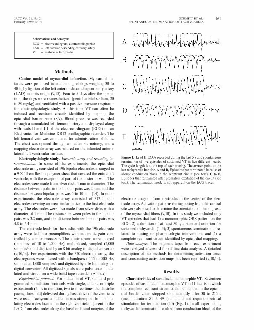
Figure 1. Lead II ECGs recorded during the last 5 s and spontaneoustermination of five episodes of sustained VT in five different hearts.The cycle length is at the top of each tracing. The arrows point to thelast tachycardia impulse. A and B, Episodes that terminated because ofabrupt conduction block in the reentrant circuit (see text). C to E,Episodes that terminated after premature excitation of the circuit (seetext). The termination mode is not apparent on the ECG traces.
461JACC Vol. 31, No. 2 SCHMITT ET AL.February 1998:460–72 SPONTANEOUS TERMINATION OF TACHYCARDIA
reentrant wave front in the circuit. Two major patterns ofspontaneous tachycardia termination were identified fromanalysis of the activation maps. The first was abrupt termina-tion not preceded by any change in the pattern of activationaround the reentrant circuit, nor by a change in the circuitcycle length .5 ms (,2%, which is within the error of ourability to accurately mark activation times) during the last fivetachycardia cycles (Fig. 1, A and B). This pattern occurred insix tachycardia episodes with a mean cycle length of 248 625 ms and a mean duration of 108 6 73 s. The second patternof termination was termination that followed a short cyclelength (premature activation) in the reentrant circuit, resultingfrom changes in the route of impulse propagation in the circuit.This pattern occurred in 11 episodes with a mean cycle lengthof 209 6 15 ms and a mean duration of 67 6 22 s. This type oftermination is not evident from the surface ECG because eachtachycardia is characterized by abrupt termination on the ECGwith little or no change in the cycle length (Fig. 1, C and D),with the exception of the tachycardia shown in Figure 1E,which had oscillating cycle lengths from the beginning. Anadditional five episodes of sustained tachycardia, during whichreentrant circuits could not be mapped, also terminated spon-taneously without previous changes in cycle length on theECG.
Abrupt termination without changes in activation patternor cycle length. An example of one of the episodes of VTclassified as abrupt termination and that lasted 57 s is shown in
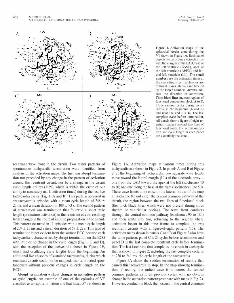
Figure 1A. Activation maps at various times during thistachycardia are shown in Figure 2. In panels A and B of Figure2, at the beginning of tachycardia, two separate wave frontsmove toward the lateral margin (LL) of the electrode array—one from the LAD toward the apex at the left (isochrones 10to 80) and one along the base at the right (isochrones 10 to 50).These wave fronts unite close to the lateral border of the mapat isochrone 80 and enter the central common pathway of thecircuit, the region between the two lines of functional block(the thick black lines, which were not present during sinusrhythm or ventricular pacing). The wave front conductsthrough the central common pathway (isochrones 90 to 180)and then splits into two, returning to the regions whereactivation began in this time frame to complete the tworeentrant circuits with a figure-of-eight pattern (15). Theactivation maps shown in panels C and D of Figure 2 also havethe same pattern; panel C is 20 cycles before termination andpanel D is the last complete reentrant cycle before termina-tion. The last isochrone that completes the circuit in each cyclethat is shown in Figure 2, including the last complete cycle, isat 230 to 240 ms, the cycle length of the tachycardia.
Figure 3A shows the sudden termination of reentry thatcaused this tachycardia to stop. In the map showing termina-tion of reentry, the united wave front enters the centralcommon pathway as in all previous cycles, with no obviouschange in the activation pattern (compare with maps in Fig. 2).However, conduction block then occurs in the central common
Figure 2. Activation maps of theepicardial border zone during theVT shown in Figure 1A. Each paneldepicts the recording electrode arraywith the margins at the LAD, base ofthe left ventricle (BASE), apex ofthe left ventricle (APEX) and lat-eral left ventricle (LL). The smallnumbers are the activation times atthe recording sites. Isochrones aredrawn at 10-ms intervals and labeledby the larger numbers. Arrows indi-cate the direction of activation.Thick black lines indicate regions offunctional conduction block. A to C,Three random cycles during tachy-cardia, at the beginning (A and B)and near the end (C). D, The lastcomplete cycle before termination.All panels show a figure-of-eight re-entrant pattern around two lines offunctional block. The activation pat-tern and cycle length in each panelare essentially the same.
462 SCHMITT ET AL. JACC Vol. 31, No. 2SPONTANEOUS TERMINATION OF TACHYCARDIA February 1998:460–72

pathway, as indicated by the double bars after isochrones 100to 120, which causes reentry to stop. The lack of any significantchanges within the circuit before termination is further indi-cated by the electrograms displayed to the right of the map(Fig. 3A). Electrograms 1 and 2 were recorded outside thecentral common pathway, electrogram 3 from the proximalpart and electrograms 4 and 5 from the middle part of thecentral common pathway. It is evident from the local cyclelengths above the recordings and from the constant electro-gram configurations that there is no detectable change inepicardial activation before termination. Termination of reen-try results because of conduction block between electrodes 3and 4. Note that the input into the central common pathwayduring the next to last (Fig. 2D) and last (Fig. 3A) cycle was notdifferent from that in previous cycles in terms of timing (cyclelength of electrograms 2 and 3 does not change) and direction(electrogram configuration is constant). Thus, termination ofreentry was abrupt.
There was not just one specific site or “weak link” thatcaused abrupt termination of reentry, as shown by analysis ofanother episode of sustained VT that terminated after 215 s inthe same heart in which the reentrant circuits described inFigures 2 and 3A occurred. The tachycardia cycle length was275 ms, longer than that in the previous episode. During thistachycardia there was also a stable activation pattern almostidentical to the pattern shown in Figure 2. The activationpattern during the termination of this episode of reentry is
illustrated in Figure 3B. When reentry terminated, the twoseparate wave fronts conducting clockwise and counterclock-wise became blocked just as they entered the central commonpathway after isochrone 100 (indicated by the two thickhorizontal lines). The recording sites shown in Figure 3B areidentical to the sites in Figure 3A. Conduction block terminat-ing reentry occurs between electrodes 2 and 3, in the very
Figure 3. Activation maps of the epicardial border zone duringspontaneous termination of tachycardia. A (left), The incomplete cycleduring which block occurred in the reentrant circuit described inFigure 2. This block led to the termination of the tachycardia shown inFigure 1A. The two horizontal thick lines on the map denote the siteof conduction block. Thick, curved arrows indicate the direction ofactivation. A selection of representative epicardial electrograms lo-cated from part of the reentrant circuit, including the region whereblock occurred, is shown to the right of the map. The location of therecording sites is indicated on the activation maps by the straight lineswith numbers corresponding to the labels at the left of each electro-gram trace. Conduction is blocked between electrograms 3 and 4(thick horizontal line). B, The last incomplete cycle during which blockoccurred (horizontal thick black lines), leading to the termination ofanother episode of sustained tachycardia in the same heart. Theasterisk on the map marks the termination site occurring during thereentry shown in Figure 3A. A selection of electrograms recordedproximal and distal to the site of conduction block is displayed at theright. Block occurred between recording sites 2 and 3 (horizontalblack line between electrogram traces). APEX 5 apex of left ventricle;BASE 5 base of left ventricle; LL 5 lateral left ventricle.
463JACC Vol. 31, No. 2 SCHMITT ET AL.February 1998:460–72 SPONTANEOUS TERMINATION OF TACHYCARDIA
proximal part of the central common pathway. Local cyclelengths at each of the recording sites (numbers below theelectrograms at the right) change very little during the last sixcycles shown before termination. Electrode 2 shows a doublepotential during reentry with a smaller component (arrows)preceding the dominant component. The timing of that smallercomponent relative to the bigger deflection varies throughoutthe tachycardia, and this component persists after terminationof reentry (small arrows at right). Therefore, it is unlikely thatthis deflection is related to the termination of reentry. Thechange in configuration of the bigger deflection in electrogram2 during termination most likely reflects conduction block nearthat site because of its uniphasic characteristic. Thus, termina-tion of reentry in this episode of tachycardia is classified asabrupt. This site of block occurred in a different position whencompared with the example in Figure 3A from the same heart,where block occurred between electrodes 3 and 4 (also indi-cated by the row of asterisks in Fig. 3B).
In the reentrant circuits described in Figures 2 and 3,conduction block occurred when the wave front was propagat-
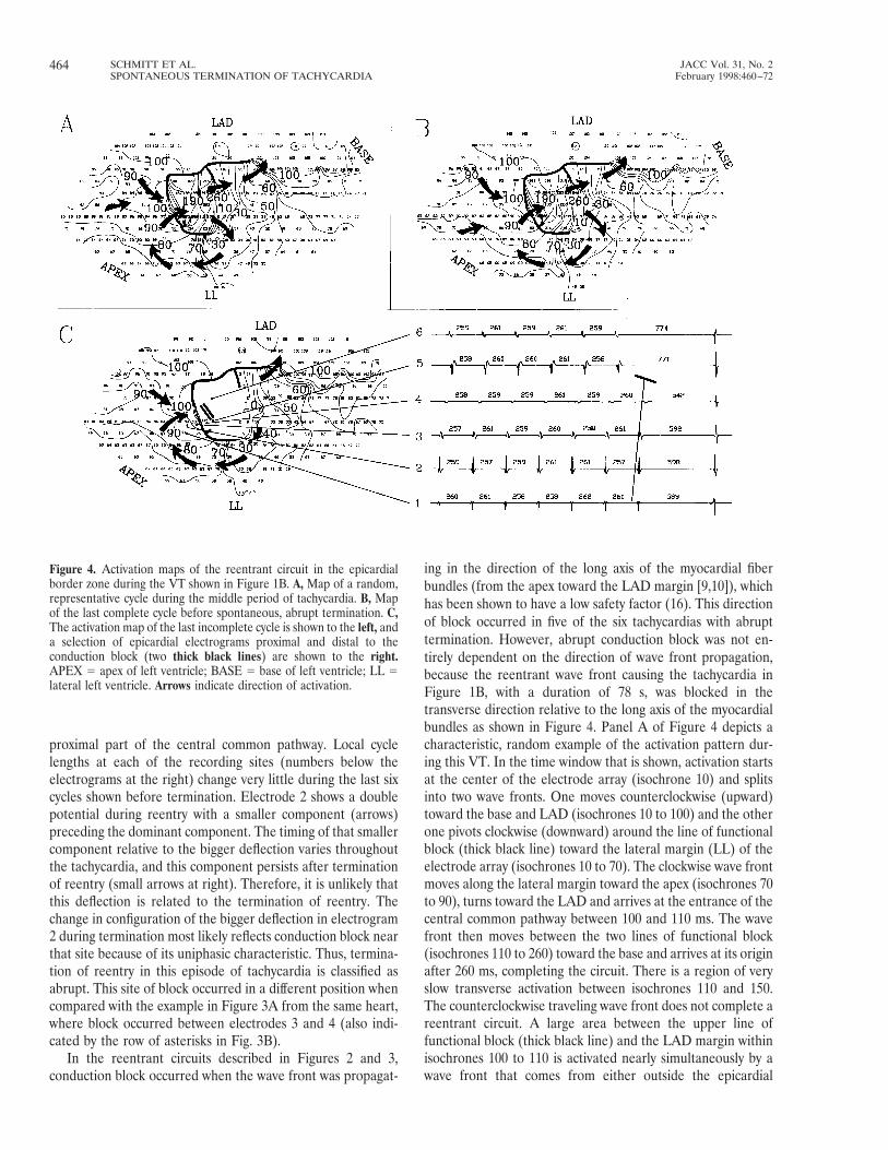
ing in the direction of the long axis of the myocardial fiberbundles (from the apex toward the LAD margin [9,10]), whichhas been shown to have a low safety factor (16). This directionof block occurred in five of the six tachycardias with abrupttermination. However, abrupt conduction block was not en-tirely dependent on the direction of wave front propagation,because the reentrant wave front causing the tachycardia inFigure 1B, with a duration of 78 s, was blocked in thetransverse direction relative to the long axis of the myocardialbundles as shown in Figure 4. Panel A of Figure 4 depicts acharacteristic, random example of the activation pattern dur-ing this VT. In the time window that is shown, activation startsat the center of the electrode array (isochrone 10) and splitsinto two wave fronts. One moves counterclockwise (upward)toward the base and LAD (isochrones 10 to 100) and the otherone pivots clockwise (downward) around the line of functionalblock (thick black line) toward the lateral margin (LL) of theelectrode array (isochrones 10 to 70). The clockwise wave frontmoves along the lateral margin toward the apex (isochrones 70to 90), turns toward the LAD and arrives at the entrance of thecentral common pathway between 100 and 110 ms. The wavefront then moves between the two lines of functional block(isochrones 110 to 260) toward the base and arrives at its originafter 260 ms, completing the circuit. There is a region of veryslow transverse activation between isochrones 110 and 150.The counterclockwise traveling wave front does not complete areentrant circuit. A large area between the upper line offunctional block (thick black line) and the LAD margin withinisochrones 100 to 110 is activated nearly simultaneously by awave front that comes from either outside the epicardial
Figure 4. Activation maps of the reentrant circuit in the epicardialborder zone during the VT shown in Figure 1B. A, Map of a random,representative cycle during the middle period of tachycardia. B, Mapof the last complete cycle before spontaneous, abrupt termination. C,The activation map of the last incomplete cycle is shown to the left, anda selection of epicardial electrograms proximal and distal to theconduction block (two thick black lines) are shown to the right.APEX 5 apex of left ventricle; BASE 5 base of left ventricle; LL 5lateral left ventricle. Arrows indicate direction of activation.
464 SCHMITT ET AL. JACC Vol. 31, No. 2SPONTANEOUS TERMINATION OF TACHYCARDIA February 1998:460–72
border zone or transmurally. In this reentrant circuit, the linesof functional block and the central common pathway that theydelineate were oriented transverse to the long axis of themyocardial fiber bundle (9,10). This pattern occurs in ,15% ofthe figure-of-eight circuits that we have mapped. Panel B ofFigure 4 shows the last complete reentrant cycle beforetermination. The activation pattern and the cycle length werevery similar compared with those in panel A. Panel C at the leftshows the activation pattern during termination of reentry(incomplete cycle). The wave front arrives at the beginning ofthe central common pathway at the same time as in panels Aand B (isochrones 10 to 100). However, during this lastincomplete cycle, conduction block occurs in this region, thearea of slow transverse conduction, indicated by the two thickblack lines. No obvious changes in the timing or spatialactivation pattern occur before termination. This is furtherillustrated by panel C at the right, where conduction blockoccurs between electrograms 4 and 5 (thick black line). Neitherchanges in the local cycle length (numbers above the tracings)nor changes in the electrogram configuration occur beforetermination of reentry.
One possible cause of the abrupt termination of reentry wasthat the head of the reentrant wave front traveled close to therefractory tail in the region where conduction block occurredwithout the presence of a fully excitable gap. Any smallchanges in conduction velocity might then cause the wave frontto become blocked in the refractory myocardium. To test thishypothesis, premature stimuli were applied in two experimentsduring other episodes of tachycardia in the same hearts withidentical reentrant patterns, before they terminated spontane-ously. For example, to evaluate the excitable gap in the centralcommon pathway at the site of spontaneous conduction blockin the experiment described in Figure 3B, single prematurestimuli with increasing prematurity were introduced every 10to 20 cycles from the stimulating electrodes at the LAD margin(pulse symbol) during another episode of sustained tachycar-
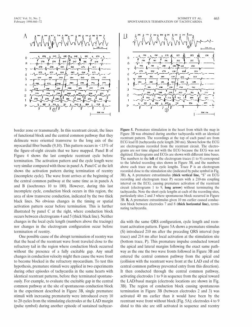
dia with the same QRS configuration, cycle length and reen-trant activation pattern. Figure 5A shows a premature stimulus(S) introduced 210 ms after the preceding QRS interval (toptrace) and 214 ms after local activation at the stimulation site(bottom trace, P). This premature impulse conducted towardthe apical and lateral margins following the exact same path-way as the one the two wave fronts followed in Figure 3B andentered the central common pathway from the apical end(collision with the reentrant wave front at the LAD end of thecentral common pathway prevented entry from this direction).It then conducted through the central common pathway,activating electrodes 1 to 9 in sequence from the apical towardthe LAD/basal margin (electrode locations are shown in Fig.3B). The region of conduction block causing spontaneoustermination in Figure 3B (between electrodes 2 and 3) wasactivated 40 ms earlier than it would have been by thereentrant wave front without block (Fig. 5A); electrodes 4 to 9distal to this site are still activated in sequence and reentry
Figure 5. Premature stimulation in the heart from which the map inFigure 3B was obtained during another tachycardia with an identicalreentrant pattern. The recordings at the top of each panel are fromECG lead II (tachycardia cycle length 280 ms). Shown below the ECGare electrograms recorded from the reentrant circuit. The electro-grams are not time aligned with the ECG because the ECG was notdigitized. Electrograms and ECGs are shown with different time bases.The numbers to the left of the electrogram traces (1 to 9) correspondto the labeled recording sites shown in Figure 3B, and the numbersabove each trace are the cycle lengths. Trace P is an electrogramrecorded close to the stimulation site (indicated by pulse symbol in Fig.3B). A, A premature extrastimulus (thick vertical line, “S” on ECGrecording and electrogram trace P) occurs with a 210-ms couplinginterval on the ECG, causing premature activation of the reentrantcircuit (electrograms 1 to 9, long arrow) without terminating thetachycardia. Note the short cycle lengths at each of the recording sites,particularly sites 2 and 3 where spontaneous block occurred in Figure3B. B, A premature extrastimulus given 10 ms earlier caused conduc-tion block between electrodes 7 and 8 (thick horizontal line), termi-nating tachycardia.
465JACC Vol. 31, No. 2 SCHMITT ET AL.February 1998:460–72 SPONTANEOUS TERMINATION OF TACHYCARDIA
continued. Shortly thereafter, another premature stimulusfrom the same site but with a prematurity of 200 ms on theECG (S) and 205 ms at the stimulus site (trace P) terminatedthe tachycardia (Fig. 5B). This premature impulse also enteredthe central common pathway from the apical end and activatedsites 2 and 3 (Fig. 3B) 49 to 52 ms earlier than they would havebeen activated by the reentrant wave front without block (Fig.5B). Block occurred between electrodes 7 and 8 (thick hori-
zontal black line above trace 7 in Fig. 5B) at the LAD end ofthe central common pathway (see Fig. 3B for location ofelectrode 7). Therefore, there was a large excitable gap in theregion where reentry stopped spontaneously in Figure 3B,between electrodes 2 and 3, because preexcitation of thisregion by as much as 50 ms did not cause block, so it is unlikelythat spontaneous termination of reentry was caused by prop-agation of the reentrant wave front into refractory myocar-dium.
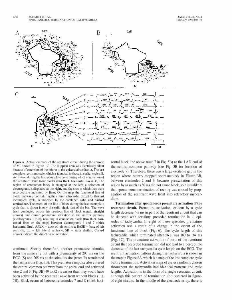
Termination after spontaneous premature activation of thereentrant circuit. Premature activation, evident by a cyclelength decrease .5 ms in part of the reentrant circuit that canbe detected with certainty, preceded termination in 11 epi-sodes of tachycardia. In eight of these episodes, prematureactivation was a result of a change in the extent of thefunctional line of block (Fig. 6). The cycle length of thistachycardia, which terminated after 56 s, was 180 to 184 ms(Fig. 1C). The premature activation of parts of the reentrantcircuit that preceded termination did not lead to a perceptibledecrease of the last tachycardia cycle length on the ECG. Thereentrant activation pattern during this tachycardia is shown inthe map in Figure 6A, which is a map of the last complete cyclebefore termination. Activation maps of cycles randomly chosenthroughout the tachycardia had identical patterns and cyclelengths. Activation is in the form of a single reentrant circuit,although this pattern of termination also occurred in figure-of-eight circuits. In the middle of the electrode array, there is
Figure 6. Activation maps of the reentrant circuit during the episodeof VT shown in Figure 1C. The stippled area was electrically silentbecause of extension of the infarct to the epicardial surface. A, The lastcomplete reentrant cycle, which is identical to those in earlier cycles. B,Activation during the last incomplete cycle during which conduction ofthe reentrant wave front blocks (two thick horizontal lines). C, Theregion of conduction block is enlarged at the left; a selection ofelectrograms is displayed at the right, and the sites at which they wererecorded are indicated by lines. On the map the functional line ofblock that was present during the entire tachycardia, except for this lastincomplete cycle, is indicated by the combined solid and dashedvertical bar. The extent of this line of block during the last incompletecycle that is shown is only the solid black part of the bar. The wavefront conducted across this previous line of block (small, straightarrows) and caused premature activation in the narrow pathway(electrograms 3 to 6), resulting in conduction block (two thick hori-zontal lines on the map) between electrograms 6 and 7 (thickhorizontal line). APEX 5 apex of left ventricle; BASE 5 base of leftventricle; LL 5 left lateral ventricle; SR 5 sinus rhythm. Curvedarrows indicate the direction of activation.
466 SCHMITT ET AL. JACC Vol. 31, No. 2SPONTANEOUS TERMINATION OF TACHYCARDIA February 1998:460–72

a large region that is electrically silent (dotted area) becauseinfarction extended in this area all the way through the leftventricular wall to the epicardial surface. The reentrant circuitis to the right of this region in the basal/lateral part of theelectrode array. The reentrant wave front moves through anarrow pathway bounded on the left side by the infarct-relatedand electrically silent epicardium and on the right side by afunctional line of block (thick black line) (isochrones 10 to110). Distal to this region, activation splits into two wavefronts. The reentrant wave front turns to the right (isochrones110 to 180) to complete the reentrant circuit. The other wavefront moves to the left (unfilled arrow) and does not lead to areentrant activation pattern in the epicardial border zone. Thefinal, incomplete cycle shown in Figure 6B is the continuationof panel A; isochrone 10 in panel B follows isochrone 180 inpanel A. It shows conduction block at the 100-ms isochrone(indicated by two thick black horizontal lines) terminatingreentry. Figure 6C reproduces and enlarges part of the map inpanel B. In panel C, the original length of the functional line ofblock during the sustained period of reentry (Fig. 6A) isindicated by the combined solid and dashed bar. During thislast cycle, the lateral extension of the line of block, indicated bythe dashed part of the bar, disappeared, leaving only a shortsegment of the original block line shown by the solid bar (Fig.6B). The general direction of activation is highlighted by thelarge, curved arrows. However, activation along the twosmaller straight arrows is important. Local activation timessuggest that in this last (incomplete) cycle, activation crossesthe distal extension of the former line of apparent block(indicated by two straight arrows) and preexcites the entrance
of the narrow pathway. This interpretation of the activationmap is supported by the electrograms at the right in Figure 6C.During continuous reentry, activation moves from the base tothe lateral region of the array (Fig. 6A). After passing elec-trodes 1 and 2, the wave front pivots around the original line ofapparent block and electrodes 2 to 5 are then activated,followed by electrodes 6 to 9 located in the narrow pathway.Electrogram 3 occurs about 67 ms after electrogram 1 and35 ms later than the sharp deflection of electrogram 2 through-out all cycles. The reason for the alternation in configuration ofelectrograms 1 and 3 is not known. During the final cycle,however, the previous, consistent and continuous sequence isaltered. Electrode 3 is activated 60 ms prematurely with a cyclelength of 121 ms, 5 ms after electrogram 1 and before electrode2, consistent with movement of the wave front across thisregion of previous block. This interpretation is also supportedby a change in electrogram configuration. Note also that asecond distinct deflection in electrogram 2 is now lost during
Figure 7. Activation maps of the reentrant circuit causing the episodeof VT in Figure 1D. A, A random, representative cycle duringtachycardia. B, The activation map is of the last complete cycle beforetermination. C, The activation map of the last incomplete cycle isshown at the left, and a selection of electrograms proximal and distalto the region of conduction block that terminated tachycardia is to theright. The dashed area on the activation map was activated prema-turely, leading to conduction block (two thick lines) in the centralcommon pathway. APEX 5 apex of left ventricle; BASE 5 base of leftventricle; LL 5 left lateral ventricle. Arrows indicate the direction ofactivation.
467JACC Vol. 31, No. 2 SCHMITT ET AL.February 1998:460–72 SPONTANEOUS TERMINATION OF TACHYCARDIA

this last cycle. Electrodes 4 to 6 are also preexcited, although toa lesser degree (by 13 to 28 ms), with some change inconfiguration, followed by conduction block between elec-trodes 6 and 7 (thick horizontal black line above electrogram 6and double horizontal black lines on the map).
Figure 7 shows another example of premature activationleading to termination of a sustained tachycardia after 104 s.
The premature activation and termination is not related toalterations in conduction across lines of functional block as inthe previous example. The corresponding surface ECG isshown in Figure 1D, which does not reflect this prematureactivation as a short last cycle. In Figure 7A, during stable VTwith a cycle length of 220 ms, two different wave fronts travelfrom the lateral margin (LL) toward the base at the right(isochrones 10 to 50) and from the apical margin toward theLAD at the left (isochrones 20 to 60). Both wave fronts uniteand enter the central common pathway between two lines offunctional block (thick black lines) (isochrones 80 to 120),returning to the site of origin in this time window to completethe reentrant activation pattern (isochrones 120 to 220). Theactivation pattern in Figure 7B, during the last completereentrant cycle before termination, shows essentially nochanges. Figure 7C at the left side shows the last incompletecycle during termination of reentry. The time window begins atthe end of the time window in panel B. The first 60 ms ofactivation of the counterclockwise circuit at the right and thefirst 40 ms of activation of the clockwise circuit at the left areessentially unchanged. The area toward the apical and LADsites of the electrode array between isochrones 40 and 60 (theregion indicated with dashes), however, is activated 10 to 20 msearlier than in previously described cycles. Because a large partof this area activates very rapidly and almost simultaneously,conduction from deeper cell layers is a possible cause. The
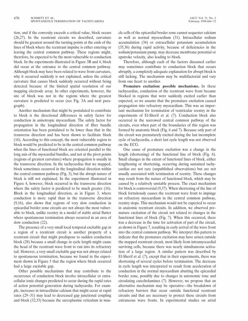
Figure 8. Activation maps of the reentrant circuit during terminationof the tachycardia shown in Figure 1E. The stippled area on the mapswas electrically silent. A, The activation pattern of a cycle duringtachycardia is shown to the left, and a selection of electrogramsrecorded at the sites indicated by lines is to the right. Numbers aboveeach electrogram trace indicate the cycle lengths. Circled numbersabove electrogram trace 6 indicate the conduction time betweenrecording sites 4 to 6 (arrows). B, The map at the left shows thetermination of reentry due to conduction block (two thick lines) in thecentral common pathway, and the electrograms recorded proximal anddistal to the site of block are shown at the right. Numbers above eachtrace are the cycle lengths, and circled numbers are conduction timesbetween recording sites 4 to 6. Conduction block occurred betweenrecording sites 5 and 6, as shown by the thick horizontal double blacklines on the map and the horizontal black line above electrogram trace6. The graph in the inset plots cycle length on the ordinate versus cyclenumber of the tachycardia at recording site 4 and shows the increase inoscillatory activity during the tachycardia. APEX 5 apex of leftventricle; BASE 5 base of left ventricle; LL 5 left lateral ventricle.Thick, curved arrows indicate the direction of activation.
468 SCHMITT ET AL. JACC Vol. 31, No. 2SPONTANEOUS TERMINATION OF TACHYCARDIA February 1998:460–72
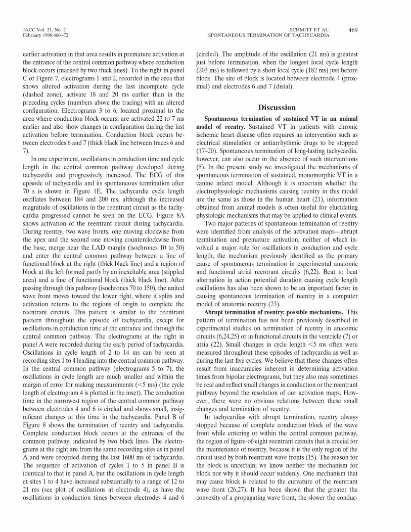
earlier activation in that area results in premature activation atthe entrance of the central common pathway where conductionblock occurs (marked by two thick lines). To the right in panelC of Figure 7, electrograms 1 and 2, recorded in the area thatshows altered activation during the last incomplete cycle(dashed zone), activate 18 and 20 ms earlier than in thepreceding cycles (numbers above the tracing) with an alteredconfiguration. Electrograms 3 to 6, located proximal to thearea where conduction block occurs, are activated 22 to 7 msearlier and also show changes in configuration during the lastactivation before termination. Conduction block occurs be-tween electrodes 6 and 7 (thick black line between traces 6 and7).
In one experiment, oscillations in conduction time and cyclelength in the central common pathway developed duringtachycardia and progressively increased. The ECG of thisepisode of tachycardia and its spontaneous termination after70 s is shown in Figure 1E. The tachycardia cycle lengthoscillates between 184 and 200 ms, although the increasedmagnitude of oscillations in the reentrant circuit as the tachy-cardia progressed cannot be seen on the ECG. Figure 8Ashows activation of the reentrant circuit during tachycardia.During reentry, two wave fronts, one moving clockwise fromthe apex and the second one moving counterclockwise fromthe base, merge near the LAD margin (isochrones 10 to 50)and enter the central common pathway between a line offunctional block at the right (thick black line) and a region ofblock at the left formed partly by an inexcitable area (stippledarea) and a line of functional block (thick black line). Afterpassing through this pathway (isochrones 70 to 150), the unitedwave front moves toward the lower right, where it splits andactivation returns to the regions of origin to complete thereentrant circuits. This pattern is similar to the reentrantpattern throughout the episode of tachycardia, except foroscillations in conduction time at the entrance and through thecentral common pathway. The electrograms at the right inpanel A were recorded during the early period of tachycardia.Oscillations in cycle length of 2 to 14 ms can be seen atrecording sites 1 to 4 leading into the central common pathway.In the central common pathway (electrograms 5 to 7), theoscillations in cycle length are much smaller and within themargin of error for making measurements (,5 ms) (the cyclelength of electrogram 4 is plotted in the inset). The conductiontime in the narrowest region of the central common pathwaybetween electrodes 4 and 6 is circled and shows small, insig-nificant changes at this time in the tachycardia. Panel B ofFigure 8 shows the termination of reentry and tachycardia.Complete conduction block occurs at the entrance of thecommon pathway, indicated by two black lines. The electro-grams at the right are from the same recording sites as in panelA and were recorded during the last 1600 ms of tachycardia.The sequence of activation of cycles 1 to 5 in panel B isidentical to that in panel A, but the oscillations in cycle lengthat sites 1 to 4 have increased substantially to a range of 12 to21 ms (see plot of oscillations at electrode 4), as have theoscillations in conduction times between electrodes 4 and 6
(circled). The amplitude of the oscillation (21 ms) is greatestjust before termination, when the longest local cycle length(203 ms) is followed by a short local cycle (182 ms) just beforeblock. The site of block is located between electrode 4 (prox-imal) and electrodes 6 and 7 (distal).
DiscussionSpontaneous termination of sustained VT in an animal
model of reentry. Sustained VT in patients with chronicischemic heart disease often requires an intervention such aselectrical stimulation or antiarrhythmic drugs to be stopped(17–20). Spontaneous termination of long-lasting tachycardia,however, can also occur in the absence of such interventions(5). In the present study we investigated the mechanisms ofspontaneous termination of sustained, monomorphic VT in acanine infarct model. Although it is uncertain whether theelectrophysiologic mechanisms causing reentry in this modelare the same as those in the human heart (21), informationobtained from animal models is often useful for elucidatingphysiologic mechanisms that may be applied to clinical events.
Two major patterns of spontaneous termination of reentrywere identified from analysis of the activation maps—abrupttermination and premature activation, neither of which in-volved a major role for oscillations in conduction and cyclelength, the mechanism previously identified as the primarycause of spontaneous termination in experimental anatomicand functional atrial reentrant circuits (6,22). Beat to beatalternation in action potential duration causing cycle lengthoscillations has also been shown to be an important factor incausing spontaneous termination of reentry in a computermodel of anatomic reentry (23).
Abrupt termination of reentry: possible mechanisms. Thispattern of termination has not been previously described inexperimental studies on termination of reentry in anatomiccircuits (6,24,25) or in functional circuits in the ventricle (7) oratria (22). Small changes in cycle length ,5 ms often weremeasured throughout these episodes of tachycardia as well asduring the last five cycles. We believe that these changes oftenresult from inaccuracies inherent in determining activationtimes from bipolar electrograms, but they also may sometimesbe real and reflect small changes in conduction or the reentrantpathway beyond the resolution of our activation maps. How-ever, there were no obvious relations between these smallchanges and termination of reentry.
In tachycardias with abrupt termination, reentry alwaysstopped because of complete conduction block of the wavefront while entering or within the central common pathway,the region of figure-of-eight reentrant circuits that is crucial forthe maintenance of reentry, because it is the only region of thecircuit used by both reentrant wave fronts (15). The reason forthe block is uncertain; we know neither the mechanism forblock nor why it should occur suddenly. One mechanism thatmay cause block is related to the curvature of the reentrantwave front (26,27). It has been shown that the greater theconvexity of a propagating wave front, the slower the conduc-
469JACC Vol. 31, No. 2 SCHMITT ET AL.February 1998:460–72 SPONTANEOUS TERMINATION OF TACHYCARDIA
tion, and if the convexity exceeds a critical value, block occurs(26,27). In the reentrant circuits we described, curvatureshould be greatest around the turning points at the ends of thelines of block where the reentrant impulse is either entering orleaving the central common pathway. These regions might,therefore, be expected to be the most vulnerable to conductionblock. In the experiments illustrated in Figure 3B and 4, blockdid occur at the entrance to the central common pathway.Although block may have been related to wave front curvature,why it occurred suddenly is not explained, unless the criticalcurvature that causes block suddenly occurred without beingdetected because of the limited spatial resolution of ourmapping electrode array. In other experiments, however, thesite of block was not in the regions where the greatestcurvature is predicted to occur (see Fig. 3A and next para-graph).
Another mechanism that might be postulated to contributeto block is the directional differences in safety factor forconduction in anisotropic myocardium. The safety factor forpropagation in the longitudinal direction of fiber bundleorientation has been postulated to be lower than that in thetransverse direction and has been shown to facilitate block(16). According to this concept, the most vulnerable region toblock would be predicted to be in the central common pathwaywhen the lines of functional block are oriented parallel to thelong axis of the myocardial bundles, and not at the pivot points(regions of greatest curvature) where propagation is usually inthe transverse direction. In the tachycardias that we mapped,block sometimes occurred in the longitudinal direction withinthe central common pathway (Fig. 3), but the abrupt nature ofblock is still not explained. In the experiment illustrated inFigure 4, however, block occurred in the transverse directionwhere the safety factor is predicted to be much greater (16).Block in the longitudinal direction, as in Figure 3, whereconduction is more rapid than in the transverse direction(9,16), also shows that regions of very slow conduction inepicardial border zone circuits are not always the most vulner-able to block, unlike reentry in a model of stable atrial flutterwhere spontaneous termination always occurred in an area ofslow conduction (22).
The presence of a very small local temporal excitable gap ina region of a reentrant circuit is another property of areentrant circuit that might predispose to sudden conductionblock (28) because a small change in cycle length might causethe head of the reentrant wave front to run into its refractorytail. However, a very small excitable gap was not always relatedto spontaneous termination, because we found in the experi-ment shown in Figure 5 that the region where block occurredhad a large excitable gap.
Other possible mechanisms that may contribute to theoccurrence of conduction block involve intracellular or extra-cellular ionic changes predicted to occur during the rapid ratesof action potential generation during tachycardia. For exam-ple, increases in intracellular calcium that might occur at rapidrates (29–31) may lead to decreased gap junctional couplingand block (32,33) because the sarcoplasmic reticulum in mus-
cle cells of the epicardial border zone cannot sequester calciumas well as normal myocardium (31). Intracellular sodiumaccumulation (34) or extracellular potassium accumulation(35,36) during rapid activity, because of deficiencies in thesodium/potassium pump, may decrease membrane potential orupstroke velocity, also leading to block.
Therefore, although each of the factors discussed earliermay sometimes contribute to conduction block that occursabruptly, a completely adequate explanation for abrupt block isstill lacking. The mechanism may be multifactorial and varyfrom one heart to another.
Premature excitation: possible mechanisms. In thesetachycardias, conduction of the reentrant wave front becameblocked in regions that were suddenly excited earlier thanexpected, so we assume that the premature excitation causedpropagation into refractory myocardium. This was an impor-tant mechanism for termination of ventricular reentry in theexperiments of El-Sherif et al. (7). Conduction block alsooccurred in the narrowed central common pathway of thecircuits, even when part of the boundaries of this region wasformed by anatomic block (Fig. 6 and 7). Because only part ofthe circuit was prematurely excited during the last incompletecycle of tachycardia, a short last cycle length was not observedon the ECG.
One cause of premature excitation was a change in thelength (shortening) of the functional line of block (Fig. 6).Small changes in the extent of functional lines of block, eitherlengthening or shortening, occurring during sustained tachy-cardias are not rare (unpublished observations) but are notusually associated with termination of reentry. These changesmay result from the nature of functional block, which may becaused by a relatively unstable process. The exact mechanismfor block is controversial (9,37). When shortening of the line ofblock fortuitously causes the reentrant wave front to impingeon refractory myocardium in the central common pathway,reentry stops. This mechanism would not be expected to occurin anatomic reentrant circuits. In addition, we observed pre-mature excitation of the circuit not related to changes in thefunctional lines of block (Fig. 7). When this occurred, therewas a decrease in the time for activation of part of the circuit,as shown in Figure 7, resulting in early arrival of the wave frontinto the central common pathway. We interpret this pattern toindicate that the premature excitation may have arisen outsidethe mapped reentrant circuit, most likely from intramyocardialsurviving cells, because there was nearly simultaneous activa-tion of a large region. A similar pattern was described byEl-Sherif et al. (7), except that in their experiments, there wasshortening of several cycles before termination. The decreasein cycle length was interpreted to result from acceleration ofconduction in the normal myocardium abutting the epicardialborder zone, possibly due to changes in autonomic tone andcirculating catecholamines (7). However, we propose that analternative mechanism may be operative—the breakdown ofrefractory barriers that occur outside functional reentrantcircuits and that are necessary to protect these circuits fromextraneous wave fronts. In experimental studies on atrial
470 SCHMITT ET AL. JACC Vol. 31, No. 2SPONTANEOUS TERMINATION OF TACHYCARDIA February 1998:460–72
reentry, secondary wave fronts leaving the original reentrantpathway have been shown to collide outside the reentrantcircuit, extinguishing each other and thus being preventedfrom entering excitable regions of the circuit, colliding with thereentrant wave front and terminating reentry. Slight alterationsof conduction, such as those observed after antiarrhythmicdrug administration, resulted in breakdown of this functionalbarrier, causing a shortcut of the secondary wave front into thereentrant pathway, leading to preexcitation and termination ofreentry (24,25,38). According to this concept, alterations ofconduction even outside the reentrant circuit might lead totermination of reentry.
In only one episode of tachycardia, which we have includedamong those terminated by premature excitation, was termi-nation possibly related to oscillatory cycle lengths in thereentrant circuit (Fig. 8). As tachycardia proceeded, oscilla-tions in conduction and cycle length increased in magnitudeand involved the termination site. Eventually conduction be-came blocked after a short cycle length that followed thelongest cycle length oscillation in the circuit (6). Block mayhave resulted from propagation of the reentrant impulse intomyocardium with a long refractory period caused by theprevious long cycle length. The reasons for the appearance ofenhanced oscillations in conduction and cycle length duringthis tachycardia are unknown. Therefore, the mechanismshown by Frame and Simson (6) to be responsible for termi-nation of reentry in atrial anatomic circuits and that shown byOrtiz et al. (22) for termination of reentry in atrial functionalcircuits may have occurred but is relatively rare in this mech-anism for ventricular reentry.
An additional pattern of activation associated with sponta-neous termination of sustained VT described by El-Sherif et al.(7), but which did not occur in any of the tachycardias in ourseries, was conduction block in the original circuit, causingtachycardia with the formation of a new circuit and continuedreentry for several more cycles.
Relation between experimental results and clinical obser-vations. The mechanisms for termination of unsustained VThave been investigated in several clinical studies, both fromanalysis of the ECG and from activation mapping (39,40).Evidence for cycle length oscillations and possible impinge-ment of the conducting wave front on refractory myocardium,causing termination of these arrhythmias, has been obtained.Similar oscillations did not occur during sustained tachycardia,although spontaneous termination was not observed. However,in one study (5) there was an increase in cycle length variabilitybefore spontaneous termination of sustained tachycardia. Thelast cycle was either shorter, longer or unchanged comparedwith the preceding cycles before termination. No clear expla-nation for termination was obvious, including impingement ofthe reentrant wave front on refractory myocardium because ofoscillations. The relation of our results to the termination ofclinical sustained VT, therefore, awaits more clinical data,especially activation maps of reentrant circuits.
References1. Wilber DJ, Garan H, Finkelstein D, et al. Out-of-hospital cardiac arrest: use
of electrophysiological testing in the prediction of long-term outcome.N Engl J Med 1988;318:19–24.
2. Richards DA, Cody DV, Denniss AR, Russell PA, Young AA, Uther JB. Anew protocol of programmed stimulation for assessment of predisposition tospontaneous ventricular arrhythmias. Eur Heart J 1983;4:376–82.
3. Brugada P, Wellens HJJ. Programmed electrical stimulation of the humanheart. In: Josephson ME, Wellens HJJ, editors. Tachycardias: Mechanismsand Management. Armonk (NY): Futura, 1984:61–90.
4. Josephson ME, Marchlinski FE, Cassidy DM, Vassallo JA, Almendral JM,Grogan W. Sustained ventricular tachycardia in coronary artery disease—evidence for reentrant mechanism. In: Zipes DP, Jalife J, editors. CardiacElectrophysiology and Arrhythmias. Orlando (FL): Grune & Stratton,1985:409.
5. Callans DJ, Marchlinski FE. Characterization of spontaneous termination ofsustained ventricular tachycardia associated with coronary artery disease.Am J Cardiol 1991;67:50–4.
6. Frame LH, Simson MB. Oscillations of conduction, action potential dura-tion, and refractoriness: a mechanism for spontaneous termination ofreentrant tachycardias. Circulation 1988;78:1277–87.
7. El-Sherif N, Yin H, Caref EB, Restivo M. Electrophysiological mechanismsof spontaneous termination of sustained monomorphic reentrant ventriculartachycardia in the canine postinfarction heart. Circulation 1996;93:1567–78.
8. El-Sherif N, Smith RA, Evans K. Canine ventricular arrhythmias in the latemyocardial infarction period. 8. Epicardial mapping of reentrant circuits.Circ Res 1981;49:255–65.
9. Dillon SM, Allessie MA, Ursell PC, Wit AL. Influences of anisotropic tissuestructure on reentrant circuits in the epicardial border zone of subacutecanine infarcts. Circ Res 1988;63:182–206.
10. Waldecker B, Coromilas J, Saltman AE, Dillon SM, Wit AL. Overdrivestimulation of functional reentrant circuits causing ventricular tachycardia inthe infarcted canine heart: resetting and entrainment. Circulation 1993;87:1286–1305.
11. Cardinal R, Savard P, Carson DL, Perry JB, Page P. Mapping of ventriculartachycardia induced by programmed stimulation in canine preparations ofmyocardial infarction. Circulation 1984;70:136–48.
12. Waldecker B, Schmitt H, Coromilas J, Wit AL. Mechanisms of spontaneoustermination of sustained ventricular tachycardia in infarcted canine hearts[abstract]. Circulation 1993;88 Suppl I:I-621.
13. Harris AS. Delayed development of ventricular ectopic rhythms followingexperimental coronary occlusion. Circulation 1950;1:1318–28.
14. Coromilas J, Saltman AE, Waldecker B, Dillon SM, Wit AL. Electrophysi-ological effects of flecainide on anisotropic conduction and reentry ininfarcted canine hearts. Circulation 1995;91:2245–63.
15. El-Sherif N. The figure 8 model of reentrant excitation in the caninepostinfarction heart. In: Zipes DP, Jalife J, editors. Cardiac Electrophysiol-ogy and Arrhythmias. New York: Grune & Stratton, 1985:363–78.
16. Spach MS, Miller WT, Geselowitz DB, Barr RC, Kootsey JM, Johnson EA.The discontinuous nature of propagation in normal canine cardiac muscle:evidence for recurrent discontinuities of intracellular resistance that affectthe membrane currents. Circ Res 1981;48:39–54.
17. Waldo AL, Henthorn RW, Plumb VJ, MacLean WAH. Demonstration ofthe mechanism of transient entrainment and interruption of ventriculartachycardia with rapid atrial pacing. J Am Coll Cardiol 1984;3:422–30.
18. Gorgels AP, van den Dool A, Hofs A, et al. Comparison of procainamideand lidocaine in terminating sustained monomorphic ventricular tachycar-dia. Am J Cardiol 1996;78:43–6.
19. Ho DS, Zecchin RP, Richards DA, Uther JB, Ross DL. Double-blind trial oflignocaine versus sotalol for acute termination of spontaneous sustainedventricular tachycardia. Lancet 1994;344:18–23.
20. Charos GS, Haffajee CI, Gold RL, Bishop RL, Berkovits BV, Alpert JS. Atheoretically and practically more effective method for interruption ofventricular tachycardia: self-adapting autodecremental overdrive pacing.Circulation 1986;73:309–15.
21. Wit AL, Janse MJJ. The Ventricular Arrhythmias of Ischemia and Infarc-tion: Electrophysiological Mechanisms. Armonk (NY): Futura, 1993.
22. Ortiz J, Igarashi M, Gonzalez HX, Laurita K, Rudy Y, Waldo AL.Mechanism of spontaneous termination of stable atrial flutter in the caninesterile pericarditis model. Circulation 1993;88:1866–77.
23. Rudy Y, Quan WL. A model study of the effects of the discrete cellular
471JACC Vol. 31, No. 2 SCHMITT ET AL.February 1998:460–72 SPONTANEOUS TERMINATION OF TACHYCARDIA
structure on electrical propagation in cardiac tissue. Circ Res 1987;61:815–23.
24. Pinto JM, Graziano JN, Boyden PA. Endocardial mapping of reentry aroundan anatomical barrier in the canine right atrium: observations during theaction of the class IC agent, flecainide. J Cardiovasc Electrophysiol 1993;4:672–85.
25. Boyden PA, Graziano JN. Activation mapping of reentry around an ana-tomical barrier in the canine atrium: observations during the action of theclass III agent, d-sotalol. J Cardiovasc Electrophysiol 1993;4:266–79.
26. Cabo C, Pertsov AM, Baxter WT, Davidenko JM, Gray RA, Jalife J.Wave-front curvature as a cause of slow conduction and block in isolatedcardiac muscle. Circ Res 1994;75:1014–28.
27. Fast VG, Kleber AG. Role of wavefront curvature in propagation of thecardiac impulse. Cardiovasc Res 1997;33:258–71.
28. Peters N, Coromilas J, Hanna M, Josephson ME, Wit AL. Characteristics ofthe temporal and spatial excitable gap in anisotropic reentrant circuits. CircRes. In press.
29. Blinks JR, Koch-Weser J. Analysis of the effects of changes in the rate andrhythm upon myocardial contractility. J Pharmacol Exp Ther 1961;134:373–89.
30. Koretsune Y, Marban E. Cell calcium in the pathophysiology of ventricularfibrillation and in the pathogenesis of postarrhythmic contractile dysfunc-tion. Circulation 1989;80:369–79.
31. Licata A, Aggarwal R, Robinson RB, Boyden PA. Frequency dependenteffects on Ca transients and cell shortening in myocytes that survive in theinfarcted heart. Cardiovasc Res 1997;33:341–50.
32. White RL, Doeller JE, Verselis VK, Wittenberg BA. Gap junctionalconductance between pairs of ventricular myocytes is modulated synergisti-cally by H1 and Ca11. J Gen Physiol 1990;95:1061–75.
33. Noma A, Tsuboi N. Dependence of junctional conductance on proton,calcium and magnesium ions in cardiac paired cells of guinea-pig. J Physiol1987;382:193–211.
34. Cohen CJ, Fozzard HA, Sheu SS. Increase in intracellular sodium ionactivity during stimulation in mammalian cardiac muscle. Circ Res 1982;50:651–62.
35. Kline R, Morad M. Potassium efflux and accumulation in heart muscle:evidence from K1 electrode experiments. Biophys J 1976;16:367–72.
36. Kline RP, Cohen I, Falk R, Kupersmith J. Activity-dependent extracellularK1 fluctuations in canine Purkinje fibers. Nature 1980;286:68–71.
37. Restivo M, Gough WB, El-Sherif N. Ventricular arrhythmias in subacutemyocardial infarction period: high-resolution activation and refractory pat-terns of reentrant rhythms. Circ Res 1990;66:1310–27.
38. Boyden PA. Models of atrial reentry. J Cardiovasc Electrophysiol 1995;6:313–24.
39. Duff HJ, Mitchell LB, Gillis AM, et al. Electrocardiographic correlates ofspontaneous termination of ventricular tachycardia in patients with coronaryartery disease. Circulation 1993;88:1054–62.
40. Pogwizd SM, Chung MK, Cain ME. Termination of ventricular tachycardiain the human heart: insights from three-dimensional activation mapping ofnonsustained and sustained ventricular tachycardias. Circulation 1997;95:2528–40.
472 SCHMITT ET AL. JACC Vol. 31, No. 2SPONTANEOUS TERMINATION OF TACHYCARDIA February 1998:460–72