mechanisms: innate immunity and animal pathogens · summary of mechanisms in innate immunity and...
TRANSCRIPT

1
Mechanisms:
Innate immunity and animal pathogens
Updated: July 2015

2
Contents MECHANISMS.......................................................................................................................................... 3
INNATE IMMUNITY AND ANIMAL PATHOGENS .................................................................................. 3
Microorganisms and pathogens in animals ............................................................................................ 3
What is a pathogen? ........................................................................................................................... 3
Key bacterial pathogens in cattle include:6,7 ....................................................................................... 4
Key viral pathogens in cattle include:6–9 ............................................................................................. 4
Key bacterial pathogens in poultry include:10 ..................................................................................... 4
Key viral pathogens in poultry include:7,11 .......................................................................................... 4
The immune response to pathogens in animals ..................................................................................... 4
Innate immunity protects against pathogens during the early stages of infection ............................ 4
Immune evasion .................................................................................................................................. 5
Adaptive immunity provides a targeted and more robust response to pathogens that are not
eliminated by the innate immune system .......................................................................................... 6
The role of Mannheimia haemolytica in bovine respiratory disease (BRD) ........................................... 7
Bovine respiratory disease (BRD) ........................................................................................................ 7
Pathogenesis of bovine respiratory disease ....................................................................................... 8
Escherichia coli infection in poultry and other species ........................................................................... 9
Pathogenic infections can manifest in many forms ............................................................................ 9
Pathogenesis of avian colibacillosis .................................................................................................... 9
E. coli risk factors32 .............................................................................................................................. 9
Virulence factors33 .............................................................................................................................. 9
E. coli can infect a variety of livestock species .................................................................................. 10
Other types of pathogens that cause disease in livestock .................................................................... 10
Respiratory infections in poultry ...................................................................................................... 10
Gastrointestinal and urogenital infections in livestock .................................................................... 11
Mastitis and metritis in cows ............................................................................................................ 11
Summary ............................................................................................................................................... 11
Summary of mechanisms in innate immunity and animal pathogens ............................................. 11
References ............................................................................................................................................ 12

3
MECHANISMS
INNATE IMMUNITY AND ANIMAL PATHOGENS
Microorganisms and pathogens in animals
What is a pathogen?
The microorganisms that share our environment include bacteria, mycoplasma, viruses, fungi,
protozoa and helminths (Figure 1). The animal body provides a favorable habitat for many
microorganisms as it is warm and constitutes a ready supply of nutrients and water. Live animals can
resist colonization by many microorganisms; with others, a symbiotic relationship develops between
the animal and microorganism and the two live together without harming each other. Areas that are
highly colonized include the skin, respiratory and gastrointestinal tracts. Microorganisms that live
innocuously on the animal are called commensals.1
A pathogen has traditionally been thought of as a microorganism that causes or can cause damage
to the host resulting in disease. Throughout this website, the term pathogen will be used according
to this definition. However, more recently, the community has questioned the use of the term
‘pathogen’. The properties of a microorganism that allow it to grow and survive in a host, known as
virulence factors, may not always be present on a particular pathogen, meaning it can exist in a
pathogenic or non-pathogenic state. In addition, microorganisms may cause disease in some
individuals but not others, indicating that differences in the way the host immune system reacts to
the microorganism may also contribute to whether it has pathogenic consequences.1–4 Disease thus
occurs only when the balance between host immunity and microorganism virulence is disturbed.1
The presentation of a disease caused by a particular pathogen is determined by the type of
pathogen and the ways in which they cause damage to the tissues.4
Figure 1. Types of pathogen that can cause disease in animals5

4
Key bacterial pathogens in cattle include:6,7
● Mannheimia haemolytica
● Histophilus somni
● Pasteurella multocida
● Salmonella spp.
● Escherichia coli
● Staphylococcus aureus
● Streptococcus agalactiae
● Corynebacterium bovis
Key viral pathogens in cattle include:6–9
● Bovine herpesvirus-1 (BHV-1; infectious bovine
rhinotracheitis)
● Parainfluenza virus-3 (PI3V)
● Bovine coronavirus (BoCV)
● Bovine respiratory syncytial virus (BRSV)
● Bovine viral diarrhea virus (BVDV)
Key bacterial pathogens in poultry include:10
● Escherichia coli
● Salmonella spp.
● Staphylococcus spp.
● Ornithobacterium rhinotracheal
● Clostridium perfringens
Key viral pathogens in poultry include:7,11
● Avian influenza viruses
● Newcastle disease virus (NDV), synonymous with avian
paramyxovirus serotype 1 (PMV-1)
● Herpes viruses, including Marek’s disease virus (MDV)
● Coronaviruses, including infectious bronchitis virus (IBV)
The immune response to pathogens in animals
Innate immunity protects against pathogens during the early stages of infection
Physical barriers represent the first line of defense against pathogens and once these have been
breached, the cells and molecules that comprise the immune system mount a coordinated response
to destroy and remove the invading pathogen.12
Different pathogens have different lifecycles, and this means that the immune response must be
tailored to a particular pathogen. Most pathogens go through a stage where they reside outside the
cells of their host, within the tissues or on the epithelial surfaces that line body cavities. Some
pathogens, including the majority of bacteria, only ever inhabit this compartment, while other

5
pathogens enter the animal’s cells. In order to replicate, all viruses and some types of bacteria, such
as Salmonella, need to move inside their host’s cells. While pathogens are outside the cells they are
susceptible to removal by a type of cell known as a phagocyte, which surrounds and engulfs the
pathogen, then destroys it with toxic products. The phagocyte also becomes activated and can signal
to other immune cells (Figure 2). However, some bacteria are surrounded by a mucus-like layer
called a capsule that protects them from phagocytic engulfment. The complement system is a group
of proteins that can help by binding to invading microorganisms to make them easier for phagocytes
to recognize and remove. If a pathogen is not detected and removed by phagocytosis before it
enters a host cell, a type of immune cell called a natural killer (NK) cell is able to recognize signals
given out by host cells that are infected with pathogens and subsequently destroy the infected host
cell as a means of limiting pathogen spread.12
Figure 2. Phagocytosis is a receptor-regulated process and can trigger production of
inflammatory mediators13
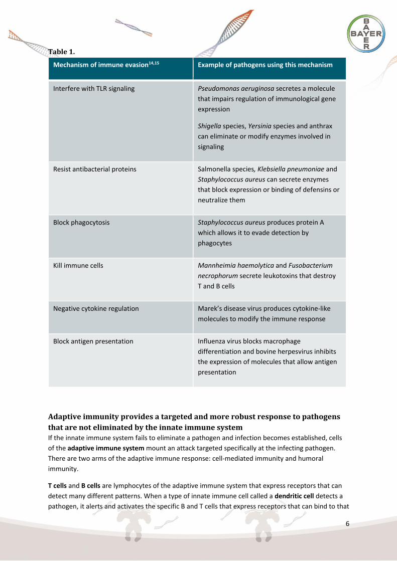
Immune evasion
Many pathogenic microorganisms can overcome innate immune defenses, such as those mentioned
in the paragraph above, and can replicate and grow within the host body.
Table 1 gives examples of mechanisms through which pathogens may evade the immune system.14
6
Table 1.
Mechanism of immune evasion14,15 Example of pathogens using this mechanism
Interfere with TLR signaling Pseudomonas aeruginosa secretes a molecule
that impairs regulation of immunological gene
expression
Shigella species, Yersinia species and anthrax
can eliminate or modify enzymes involved in
signaling
Resist antibacterial proteins Salmonella species, Klebsiella pneumoniae and
Staphylococcus aureus can secrete enzymes
that block expression or binding of defensins or
neutralize them
Block phagocytosis Staphylococcus aureus produces protein A
which allows it to evade detection by
phagocytes
Kill immune cells Mannheimia haemolytica and Fusobacterium
necrophorum secrete leukotoxins that destroy
T and B cells
Negative cytokine regulation Marek’s disease virus produces cytokine-like
molecules to modify the immune response
Block antigen presentation Influenza virus blocks macrophage
differentiation and bovine herpesvirus inhibits
the expression of molecules that allow antigen
presentation
Adaptive immunity provides a targeted and more robust response to pathogens
that are not eliminated by the innate immune system
If the innate immune system fails to eliminate a pathogen and infection becomes established, cells
of the adaptive immune system mount an attack targeted specifically at the infecting pathogen.
There are two arms of the adaptive immune response: cell-mediated immunity and humoral
immunity.
T cells and B cells are lymphocytes of the adaptive immune system that express receptors that can
detect many different patterns. When a type of innate immune cell called a dendritic cell detects a
pathogen, it alerts and activates the specific B and T cells that express receptors that can bind to that

7
particular pathogen. These cells proliferate to produce many copies of these targeted lymphocytes.
Cell-mediated immunity is one arm of the adaptive immune response. It acts via cytotoxic T cells and
phagocytes and predominantly protects against pathogens that reside inside host cells. Humoral
immunity, the other arm of the adaptive immune response in which B cells produce antibodies
against pathogens, largely protects against pathogens found outside of these cells.16,17
Activation of adaptive immunity relies on the recognition of pathogens by innate immune cells,
which subsequently alert adaptive immune cells to the presence of the invading microorganism. An
immunocompromised animal may not mount an innate or adaptive immune response that is
sufficient to completely remove the infecting pathogen.16,17
The role of Mannheimia haemolytica in bovine respiratory
disease (BRD)
Bovine respiratory disease (BRD)
BRD is associated with substantial economic losses, resulting from both the direct effects of disease
on animals, and indirect effects associated with treating and/or preventing its spread.18–22 BRD
describes a multifactorial condition that can be caused by a variety of bacterial and/or viral
pathogens.23,24 Mannheimia haemolytica is a bacterium that is commonly isolated from cattle with
BRD.25 It is also found in the respiratory tract of some healthy cattle as a commensal species.26
Signs of Bovine Respiratory disease
Clinical signs of BRD include depression, anorexia, nasal discharge and coughing. The presence of
lung lesions is also indicative of BRD, even in animals that do not display symptoms.27,28 Despite the
usage of vaccines and antibiotics, BRD continues to remain a challenge especially in feedlot cattle.
Environmental stressors are thought to contribute to BRD susceptibility, as it is most commonly seen
in young cattle within the first 2 months of arrival at the feedlot.26 Transportation and co-mingling of
cattle from multiple sources may increase both pathogen exposure and the animal’s stress level,
while procedures such as weaning, dehorning or castration, may also represent stressful events.25
Stress can lead to increased secretion of stress hormones, which can bind to receptors and lead to
secretion of cytokines, the signaling molecules of the immune system, which can alter the way in
which the immune system responds.29,30
Cattle displaying symptoms of BRD are often infected with other pathogens in addition to
M. haemolytica. These may include other bacteria, commonly Pasteurella multocida, Histophilus
somni and Mycoplasma bovis, or viruses, including bovine viral diarrheal virus (BVDV), bovine
respiratory syncytial virus (BRSV) and parainfluenza 3 virus (PI3V) (figure 3). Many cattle are
persistently infected with BVDV. The significance of this is that viral infection can modify the immune
system’s capacity to respond to bacterial infection, leaving the animal susceptible to infection and
unable to control or clear it.25

8
Figure 3. Bovine respiratory disease (BRD) in cattle describes a multifactorial
condition23,24
Pathogenesis of bovine respiratory disease
Within a bacterial or viral species, there may be subgroups known as serotypes, characterized by
slight variations in structure or features. M. haemolytica serotypes S2 and S4 are found in the
nasopharynx of healthy cattle, and generally do not cause disease. Pathogenicity results from an
opportunistic infection, usually by serotype S1. Stress or viral infection may modify the
microenvironment, allowing serotype S1 to colonize the nasal mucosa; subsequent inhalation
enables the bacteria to reach the lungs.23
A number of virulence factors allow M. haemolytica to infect the lung and contribute to the lung
pathology associated with BRD.23,31
Leukotoxin is a toxin secreted by M. haemolytica that targets the immune cells of ruminant species.
At low concentrations it can activate macrophages and neutrophils, promoting inflammation. At
higher concentrations, it creates pores in immune cell membranes, killing the cell and allowing them
to release the toxic products they use to destroy bacteria, which damages the surrounding lung
tissue.23,31
Lipopolysaccharide (LPS) is a part of the bacterial cell wall that can bind to receptors on immune
cells, such as macrophages and neutrophils. At low concentrations it inhibits phagocytosis, while at
high concentrations the cells are stimulated, contributing to inflammation. Inflammatory mediators
cause damage to the lung. LPS also binds, and is directly toxic to, the pulmonary endothelium. It
binds to epithelial cells causing blood vessel dilation, permitting immune cells and fluid to leave the
blood vessels, which can lead to a life-threatening fall in blood pressure.23

9
Escherichia coli infection in poultry and other species
Pathogenic infections can manifest in many forms
The bacterium Escherichia coli is a commensal microorganism in healthy poultry. However, certain
strains, known as avian pathogenic E. coli (APEC), can move from the intestinal lining to invade
various internal organs. Antibiotic resistance is common among APEC and resistance traits are often
carried on DNA molecules that are separate from the main genome and can be easily transferred
between bacteria. This makes APEC difficult to treat. Acute colibacillosis due to E. coli causes
septicemia and death, while in its subacute form, infected birds may exhibit a whole host of diseases
and conditions including, but not limited to (figure 4):32,33
E. coli infections in poultry
● Septicemia: may be associated with pericarditis, peritonitis and bile-staining and necrotic
foci in the liver33
● Salpingitis: infection of the oviduct may occur via the left abdominal airsac causing
salpingitis, which compromises a bird’s egg-laying capacity33
● Swollen head syndrome: causes edema of the cranial and periorbital skin but has a low
mortality rate and minimal effect on egg production.32 It is thought to occur as an
opportunistic infection following a viral infection32,33
● Respiratory tract infection: primary site of infection, occurs through inhalation of fecal-
contaminated dust. Bacteria may reach the blood, causing stunted growth or death32,33
● Yolk sac infection: often associated with fecal contamination of the egg surface and can lead
to embryonic mortality or death during the first few weeks of life33
● Cellulitis: chronic inflammation of the tissue of the abdomen and thighs32
● Coligranuloma: leads to development of granulomas in liver, caeca, duodenum and
mesenterium and is associated with a high mortality rate32
Pathogenesis of avian colibacillosis
● The mechanism by which APEC causes infection is not well understood, however a number
of risk factors and pathogen virulence factors have been identified32,33
E. coli risk factors32
● Housing: poor housing hygiene, e.g. excess ammonia or dust, overcrowding, temperature
● Pathogen: virulence of the strain, duration of pathogen exposure
● Immune status of the bird: infection with other pathogens, such as Newcastle disease virus,
infectious bronchitis virus and Mycoplasma gallisepticum may increase the risk of
colibacillosis, and damage to the respiratory system may also increase susceptibility
Virulence factors33
● Outer membrane proteins and/or capsules: help resist bactericidal effects of complement
● Cytotoxins: have been identified in some strains of APEC, although this is not widespread
● Mechanisms to acquire iron: as iron availability is restricted in the animal’s bodily fluids,
some bacteria have developed systems to compete with their hosts for iron
● Fimbriae: help improve bacterial attachment to epithelial cells

10
Figure 4. Disease caused by infection with APEC in poultry34
E. coli can infect a variety of livestock species
As in cattle, stress or viral infection can lead to immunosuppression in poultry. In addition to causing
disease in young chicks, infectious bursal disease virus (IBDV) also damages immune tissues such as
the bursa of Fabricus, reducing the bird’s ability to fight infection by other pathogens.35
Furthermore, pathogens such as E. coli can infect multiple species. Colibacillosis is an animal disease
caused by E. coli that can affect mammals (including cattle, sheep and pigs), as well as avian species.
The avian form is typically a localized or systemic disease, occurring secondarily when host defenses
have been impaired or overwhelmed by virulent E. coli strains. Conversely, the mammalian form is
most often a primary enteric or urinary tract disease34
Other types of pathogens that cause disease in livestock
Respiratory infections in poultry

11
Gastrointestinal and urogenital infections in livestock
Gastroenteritis can result from infection with various bacteria, e.g. E. coli34 or Campylobacter spp.7
Salmonellosis typically presents as enteritis (systemic septicemia – or typhoid – is the other major
form). The serotypes of Salmonella typically have a narrow range of host species (‘serovar-host
specificity’)7
● Common pathogens causing gastrointestinal (GI) infections include: Salmonella
typhimurium, S. dublin, S. Newport (cattle); S. typhimurium, S. choleraesuis (pigs); S.
typhimurium, S. dublin, S. abortusovis, S. anatum, S. Montevideo (sheep); S. enteritidis, S.
typhimurium, S. gallinarum, S. pullorum (poultry)
Cattle, sheep and pigs are susceptible to cystitis (inflammation of the urinary bladder) and
pyelonephritis (infection of the kidneys)7
● Bacteria that cause these infections include: Corynebacterium renale (C. renale, C. cystitidis,
and C. pilosum), E. coli, Trueperella pyogenes, Streptococcus spp., and Staphylococcus spp.
Mastitis and metritis in cows
Mastitis (inflammation of the mammary gland) occurs in almost all domestic mammals, but is of
greatest frequency and economic importance in dairy cattle7
● Many microorganisms can opportunistically invade tissue and cause mastitis. Most infections
are caused by species of streptococci, staphylococci, and Gram-negative rod bacteria7
Metritis (inflammation of the uterus) occurs in cattle, typically after calving37
● Bacteria that cause these infections include: Arcanobacterium pyogenes, Fusobacterium
necrophorum, E. coli, Streptococcus spp., Staphylococcus spp., and Pseudomonas spp38
Summary
Summary of mechanisms in innate immunity and animal pathogens
● Animals and microorganisms have evolved to live together over a long time, and in many
cases microorganisms can inhabit the animal body without causing disease39
● The term pathogen is used to describe a microorganism that causes or can cause damage to
the host resulting in disease39
● Infection by a pathogen may occur if a microorganism possesses features that enhance its
ability to live in the tissues or cells of an animal, known as virulence factors, or if the immune
response mounted by the animal is insufficiently strong to remove the invading pathogen2,3
● When immunity is suppressed, for example by stress or viral infection, animals may be more
susceptible to diseases such as BRD or colibacillosis25,35
● Once physical barriers protecting the animal are breached (e.g. skin), the innate immune
system tries to fight the pathogen, predominantly through phagocytosis25
● Infection may manifest in many ways and depends on the type of pathogen and the location
of the infection40

12
● Some microorganisms have evolved mechanisms that allow them to evade the innate
immune system, such as interfering with cellular signaling39,40
● Adaptive immunity is alerted to the presence of an infection by innate immune cells and
offers a more targeted response to pathogens39,40
References 1. Tizard IR. Veterinary Immunology, 9th edn, 2013:3,4.
2. Pirofski L & Casadevall A. BMC Biology 2012;10:6.
3. Casadevall A & Pirofski L. Infect Immun 2000;68:12, 6511–8.
4. Murphy K. Janeway’s Immunobiology, 8th edn, 2011:38,25,40–42.
5. Tizard IR. Veterinary Immunology, 9th edn, 2013:85–86,313–21.
6. Ackermann MR, et al. Vet Clin North Am Food Anim Pract 2010;26:215–28.
7. Merck Veterinary Manual. Available at: http://www.merckmanuals.com/vet (accessed 09
March 2015).
8. Fulton RW, et al. Can J Vet Res 2011;75:191–9.
9. Zientara S & Ponsart C. Reprod Fertil Dev 2014;27:63–71.
10. Agunos A et al. Can Vet J 2013;54:1041–52.
11. Couteaudier M & Denesvre C. Vet Res 2014;45:36.
12. Murphy K. Janeway’s Immunobiology, 8th edn, 2011:38–43.
13. Tizard IR. Veterinary Immunology, 9th edn, 2013:35–39.
14. Murphy K. Janeway’s Immunobiology, 8th edn, 2011:490,516.
15. Tizard IR. Veterinary Immunology, 9th edn, 2013:288–90,302–5.
16. Tizard IR. Veterinary Immunology, 9th edn, 2013:95,127,138,143,
17. Murphy K. Janeway’s Immunobiology, 8th edn, 2011:12,13,26–8,276–8.
18. Griffin D. Vet Clin North Am Food Anim Pract 1997;13:367–77.
19. National Animal Disease Information Service. Respiratory Disease in Dairy and Beef Rearer
Units. Available at: http://www.nadis.org.uk/bulletins/respiratory-disease-in-dairy-and-beef-
rearer-units.aspx. Accessed April 2015.
20. Sackett D, et al. Meat and Livestock Australia; 2006. Available at:
http://www.mla.com.au/CustomControls/PaymentGateway/ViewFile.aspx?mFDUp1AYI9VUf
+h/ZH4CYhopVLs5O3WvlD8Tvjx4WwqIhJCS8/UdRwc9AKswn/HN3EYMKKAfsht7d1Tnt3BqiA=
=. Accessed April 2015
21. Schneider MJ, et al. J Anim Sci 2009;87:1821–7.
22. International Federation for Animal Health. The Cost of Animal Disease. Oxford Analytica;
2012. Available from: http://www.bft-online.de/fileadmin/bft/publikationen/IFAH_Oxford-
Analytica_The-Costs-of-Animal-Disease_October2012.pdf. Accessed April 2015.
23. Singh K, et al. Vet Pathol 2011;48:338–48.
24. Klima CL, et al. J Clin Microbiol 2014;52:438–48.
25. Taylor JD, et al. Can Vet J 2010;51:1095–102.
26. Klima CL, et al. Can J Vet Res 2014;78:38–45.
27. White BJ, et al. J Vet Diagn Invest 2009;21:446–53.
28. Gifford CA, et al. J Anim Sci 2012;90:1438–51.
29. Burdick NC, et al. Int J Zoology 2011;373197.
30. Tizard IR. Veterinary Immunology, 9th edn, 2013:222–4.

13
31. Thumbikat P, et al. Vet Res 2005;36:771–86.
32. Lutful Kabir SM. Int J Environ Res Public Health 2010;7:89–114.
33. Dho-Moulin M & Morris Fairbrother JM. Vet Res 1999;30:299–316.
34. Barnes et al. Colibacillosis. Diseases of Poultry. Saif YM. Iowa, Blackwell Publishing
Professional. 2008;12:716–62. Available from:
https://himakahaunhas.files.wordpress.com/2013/03/disease-of-poultry.pdf. Accessed
March 2015.
35. Schat KA & Skinner MA. In: Avian Immunology. Schat KA (ed.). Elsevier, San Diego, CA, US,
2014.
36. Gupta A & Ezzeldin T. Mycoplasma complicated chronic respiratory disease (CCRD) – a
Review. Available from: http://en.engormix.com/MA-poultry-
industry/health/articles/mycoplasma-complicated-chronic-respiratory-t1427/165-p0.htm.
Accessed April 2015.
37. National Animal Disease Information Service. Fertility in dairy herds – uterine infection.
Available from: http://www.nadis.org.uk/bulletins/fertility-in-dairy-herds/part-7-uterine-
infection.aspx?altTemplate=PDF. Accessed April 2015.
38. Azawi OI. Buffalo Bulletin 2013;32:1–17.
39. Tizard IR. Veterinary Immunology, 9th edn, 2013:3,4,94,97,288,289,303–5.
40. Murphy K. Janeway’s Immunobiology, 8th edn, 2011:12,13,39–41.