mechanistic insights and progress on the galnac … insights and progress on the galnac-sirna...
TRANSCRIPT
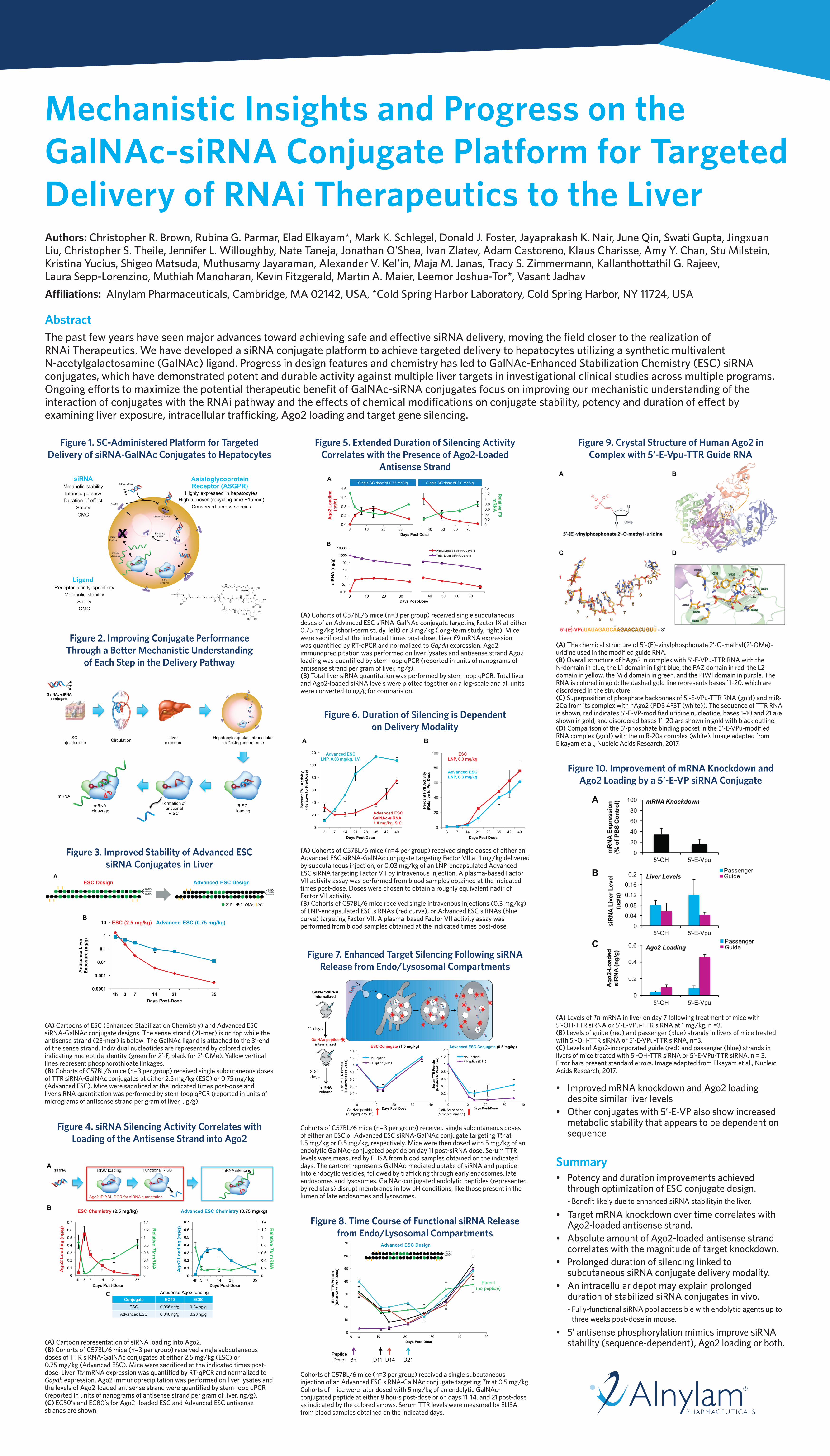
Mechanistic Insights and Progress on the GalNAc-siRNA Conjugate Platform for Targeted Delivery of RNAi Therapeutics to the LiverAuthors: Christopher R. Brown, Rubina G. Parmar, Elad Elkayam*, Mark K. Schlegel, Donald J. Foster, Jayaprakash K. Nair, June Qin, Swati Gupta, Jingxuan Liu, Christopher S. Theile, Jennifer L. Willoughby, Nate Taneja, Jonathan O’Shea, Ivan Zlatev, Adam Castoreno, Klaus Charisse, Amy Y. Chan, Stu Milstein, Kristina Yucius, Shigeo Matsuda, Muthusamy Jayaraman, Alexander V. Kel’in, Maja M. Janas, Tracy S. Zimmermann, Kallanthottathil G. Rajeev, Laura Sepp-Lorenzino, Muthiah Manoharan, Kevin Fitzgerald, Martin A. Maier, Leemor Joshua-Tor*, Vasant Jadhav
Affiliations: Alnylam Pharmaceuticals, Cambridge, MA 02142, USA, *Cold Spring Harbor Laboratory, Cold Spring Harbor, NY 11724, USA
AbstractThe past few years have seen major advances toward achieving safe and effective siRNA delivery, moving the field closer to the realization of RNAi Therapeutics. We have developed a siRNA conjugate platform to achieve targeted delivery to hepatocytes utilizing a synthetic multivalent N-acetylgalactosamine (GalNAc) ligand. Progress in design features and chemistry has led to GalNAc-Enhanced Stabilization Chemistry (ESC) siRNA conjugates, which have demonstrated potent and durable activity against multiple liver targets in investigational clinical studies across multiple programs. Ongoing efforts to maximize the potential therapeutic benefit of GalNAc-siRNA conjugates focus on improving our mechanistic understanding of the interaction of conjugates with the RNAi pathway and the effects of chemical modifications on conjugate stability, potency and duration of effect by examining liver exposure, intracellular trafficking, Ago2 loading and target gene silencing.
Figure 1. SC-Administered Platform for Targeted Delivery of siRNA-GalNAc Conjugates to Hepatocytes
Figure 2. Improving Conjugate Performance Through a Better Mechanistic Understanding
of Each Step in the Delivery Pathway
Figure 3. Improved Stability of Advanced ESC siRNA Conjugates in Liver
(A) Cartoons of ESC (Enhanced Stabilization Chemistry) and Advanced ESC siRNA-GalNAc conjugate designs. The sense strand (21-mer) is on top while the antisense strand (23-mer) is below. The GalNAc ligand is attached to the 3’-end of the sense strand. Individual nucleotides are represented by colored circles indicating nucleotide identity (green for 2’-F, black for 2’-OMe). Yellow vertical lines represent phosphorothioate linkages. (B) Cohorts of C57BL/6 mice (n=3 per group) received single subcutaneous doses of TTR siRNA-GalNAc conjugates at either 2.5 mg/kg (ESC) or 0.75 mg/kg (Advanced ESC). Mice were sacrificed at the indicated times post-dose and liver siRNA quantitation was performed by stem-loop qPCR (reported in units of micrograms of antisense strand per gram of liver, ug/g).
Figure 4. siRNA Silencing Activity Correlates with Loading of the Antisense Strand into Ago2
(A) Cartoon representation of siRNA loading into Ago2. (B) Cohorts of C57BL/6 mice (n=3 per group) received single subcutaneous doses of TTR siRNA-GalNAc conjugates at either 2.5 mg/kg (ESC) or 0.75 mg/kg (Advanced ESC). Mice were sacrificed at the indicated times post-dose. Liver Ttr mRNA expression was quantified by RT-qPCR and normalized to Gapdh expression. Ago2 immunoprecipitation was performed on liver lysates and the levels of Ago2-loaded antisense strand were quantified by stem-loop qPCR (reported in units of nanograms of antisense strand per gram of liver, ng/g). (C) EC50’s and EC80’s for Ago2 -loaded ESC and Advanced ESC antisense strands are shown.
Figure 5. Extended Duration of Silencing Activity Correlates with the Presence of Ago2-Loaded
Antisense Strand
(A) Cohorts of C57BL/6 mice (n=3 per group) received single subcutaneous doses of an Advanced ESC siRNA-GalNAc conjugate targeting Factor IX at either 0.75 mg/kg (short-term study, left) or 3 mg/kg (long-term study, right). Mice were sacrificed at the indicated times post-dose. Liver F9 mRNA expression was quantified by RT-qPCR and normalized to Gapdh expression. Ago2 immunoprecipitation was performed on liver lysates and antisense strand Ago2 loading was quantified by stem-loop qPCR (reported in units of nanograms of antisense strand per gram of liver, ng/g). (B) Total liver siRNA quantitation was performed by stem-loop qPCR. Total liver and Ago2-loaded siRNA levels were plotted together on a log-scale and all units were converted to ng/g for comparision.
Figure 6. Duration of Silencing is Dependent on Delivery Modality
(A) Cohorts of C57BL/6 mice (n=4 per group) received single doses of either an Advanced ESC siRNA-GalNAc conjugate targeting Factor VII at 1 mg/kg delivered by subcutaneous injection, or 0.03 mg/kg of an LNP-encapsulated Advanced ESC siRNA targeting Factor VII by intravenous injection. A plasma-based Factor VII activity assay was performed from blood samples obtained at the indicated times post-dose. Doses were chosen to obtain a roughly equivalent nadir of Factor VII activity. (B) Cohorts of C57BL/6 mice received single intravenous injections (0.3 mg/kg) of LNP-encapsulated ESC siRNAs (red curve), or Advanced ESC siRNAs (blue curve) targeting Factor VII. A plasma-based Factor VII activity assay was performed from blood samples obtained at the indicated times post-dose.
Figure 7. Enhanced Target Silencing Following siRNA Release from Endo/Lysosomal Compartments
Cohorts of C57BL/6 mice (n=3 per group) received single subcutaneous doses of either an ESC or Advanced ESC siRNA-GalNAc conjugate targeting Ttr at 1.5 mg/kg or 0.5 mg/kg, respectively. Mice were then dosed with 5 mg/kg of an endolytic GalNAc-conjugated peptide on day 11 post-siRNA dose. Serum TTR levels were measured by ELISA from blood samples obtained on the indicated days. The cartoon represents GalNAc-mediated uptake of siRNA and peptide into endocytic vesicles, followed by trafficking through early endosomes, late endosomes and lysosomes. GalNAc-conjugated endolytic peptides (represented by red stars) disrupt membranes in low pH conditions, like those present in the lumen of late endosomes and lysosomes.
Figure 8. Time Course of Functional siRNA Release from Endo/Lysosomal Compartments
Cohorts of C57BL/6 mice (n=3 per group) received a single subcutaneous injection of an Advanced ESC siRNA-GalNAc conjugate targeting Ttr at 0.5 mg/kg. Cohorts of mice were later dosed with 5 mg/kg of an endolytic GalNAc-conjugated peptide at either 8 hours post-dose or on days 11, 14, and 21 post-dose as indicated by the colored arrows. Serum TTR levels were measured by ELISA from blood samples obtained on the indicated days.
Figure 9. Crystal Structure of Human Ago2 in Complex with 5’-E-Vpu-TTR Guide RNA
(A) The chemical structure of 5’-(E)-vinylphosphonate 2’-O-methyl(2’-OMe)-uridine used in the modified guide RNA. (B) Overall structure of hAgo2 in complex with 5’-E-VPu-TTR RNA with the N-domain in blue, the L1 domain in light blue, the PAZ domain in red, the L2 domain in yellow, the Mid domain in green, and the PIWI domain in purple. The RNA is colored in gold; the dashed gold line represents bases 11–20, which are disordered in the structure. (C) Superposition of phosphate backbones of 5’-E-VPu-TTR RNA (gold) and miR-20a from its complex with hAgo2 (PDB 4F3T (white)). The sequence of TTR RNA is shown, red indicates 5’-E-VP-modified uridine nucleotide, bases 1–10 and 21 are shown in gold, and disordered bases 11–20 are shown in gold with black outline. (D) Comparison of the 5’-phosphate binding pocket in the 5’-E-VPu-modified RNA complex (gold) with the miR-20a complex (white). Image adapted from Elkayam et al., Nucleic Acids Research, 2017.
Figure 10. Improvement of mRNA Knockdown and Ago2 Loading by a 5’-E-VP siRNA Conjugate
(A) Levels of Ttr mRNA in liver on day 7 following treatment of mice with 5’-OH-TTR siRNA or 5’-E-VPu-TTR siRNA at 1 mg/kg, n =3. (B) Levels of guide (red) and passenger (blue) strands in livers of mice treated with 5’-OH-TTR siRNA or 5’-E-VPu-TTR siRNA, n=3. (C) Levels of Ago2-incorporated guide (red) and passenger (blue) strands in livers of mice treated with 5’-OH-TTR siRNA or 5’-E-VPu-TTR siRNA, n = 3. Error bars present standard errors. Image adapted from Elkayam et al., Nucleic Acids Research, 2017.
• Improved mRNA knockdown and Ago2 loading despite similar liver levels
• Other conjugates with 5’-E-VP also show increased metabolic stability that appears to be dependent on sequence
Summary• Potency and duration improvements achieved
through optimization of ESC conjugate design. - Benefit likely due to enhanced siRNA stabilityin the liver.
• Target mRNA knockdown over time correlates with Ago2-loaded antisense strand.
• Absolute amount of Ago2-loaded antisense strand correlates with the magnitude of target knockdown.
• Prolonged duration of silencing linked to subcutaneous siRNA conjugate delivery modality.
• An intracellular depot may explain prolonged duration of stabilized siRNA conjugates in vivo.
- Fully-functional siRNA pool accessible with endolytic agents up to three weeks post-dose in mouse.
• 5’ antisense phosphorylation mimics improve siRNA stability (sequence-dependent), Ago2 loading or both.