mechanotransduction: use the force(s) -...
TRANSCRIPT

Paluch et al. BMC Biology (2015) 13:47 DOI 10.1186/s12915-015-0150-4
FORUM Open Access
Mechanotransduction: use the force(s)
Ewa K. Paluch1*, Celeste M. Nelson2, Nicolas Biais3, Ben Fabry4, Jens Moeller5, Beth L. Pruitt6, Carina Wollnik7,Galina Kudryasheva7, Florian Rehfeldt7 and Walter Federle8Abstract
Mechanotransduction - how cells sense physical forcesand translate them into biochemical and biologicalresponses - is a vibrant and rapidly-progressing field,and is important for a broad range of biologicalphenomena. This forum explores the role ofmechanotransduction in a variety of cellular activitiesand highlights intriguing questions that deserve furtherattention.
plated on a compliant substratum (or within a hydrogel
Four centuries of cells and mechanical forcesCeleste M NelsonCells were first observed under the microscope by RobertHooke in 1665. That these tiny objects are actually themost fundamental units of life would not be appreciateduntil the early 1800s, but Hooke’s seminal discovery isstriking to us now in the early 21st century for anotherreason. Amongst physical scientists, Hooke is betterknown for ‘Hooke’s Law’, the principle of physics thatstates that the force F needed to change the length of anelastic spring by some distance x is linearly proportionalto that distance (F = kx), where k is the proportionalityconstant that describes the stiffness of the spring. Hookethus made the earliest discoveries that would eventuallyfather two seemingly disparate fields. We now know, ofcourse, that physical forces are fundamental to cell biology.That cells are subject to the laws of physics - of me-
chanics - was first postulated by Wilhelm His in the late1800s [1]. The physical nature of cells and tissues wasembraced by embryologists and early cell biologists, asthe only tools to interrogate their behavior were mech-anical in nature. The discovery of the structure of DNAby Watson and Crick in 1953 ushered in an excitingnew era of molecular biology - instead of being considereda physical material, the cell was viewed as a container ofgenetic material and enzymes. The past 20 years have seen
* Correspondence: [email protected] Laboratory for Molecular Cell Biology, University College London,Gower Street, London WC1E 6BT, UKFull list of author information is available at the end of the article
© 2015 Paluch et al. This is an Open Access a(http://creativecommons.org/licenses/by/4.0),provided the original work is properly creditedcreativecommons.org/publicdomain/zero/1.0/
a resurgence of mechanics in cell biology, with new para-digms emerging that have changed our understanding ofalmost every fundamental cellular process, from cell div-ision to differentiation to morphogenesis.This new age of enlightenment in mechanobiology has
been enabled by technological breakthroughs resultingfrom collaborations between biologists, physicists, andengineers. We can now estimate the forces that cellsexert on their surroundings. Traction force microscopy[2] is one approach to perform this estimation: cells are
[3]) that contains beads that act as fiducial markers. Asthe cell exerts force on the substratum, the resultingmotion of the beads is tracked. The measured bead dis-placements can then be used to estimate the forceexerted by the cells à la Hooke’s Law; the actual mathinvolved for a quantitative understanding is more com-plicated than the equation described above since thephysical situation is significantly more complex than thestretching of a spring, but the spirit of Hooke’s equationholds. It is important to note that force is not measuredhere - it is calculated, and the accuracy of the calculationdepends on the resolution of the measurements, the ma-terial properties of the substratum, and the validity ofthe underlying mathematical model.Other force measurement calculation techniques in-
clude micropost arrays and atomic force microscopy(AFM). Micropost arrays actually use Hooke’s Law tocalculate the forces exerted by cells on the underlyingposts, provided that the deformations of the substratuminduced by the cells are small [4]. In AFM, what arereally being measured are the mechanical properties ofthe cell, not the force that the cell exerts. A cantileverprobe is used to tap gently on the surface of the cell; thedeflection of the cantilever is proportional to the stiff-ness of the region being tapped. In the mechanobiologyliterature, these readouts are often mistakenly referredto as ‘tension’.To detect tension within the cell, advances have been
made using molecular sensors. Physically, tension is thepulling force exerted when a one-dimensional chain of
rticle distributed under the terms of the Creative Commons Attribution Licensewhich permits unrestricted use, distribution, and reproduction in any medium,. The Creative Commons Public Domain Dedication waiver (http://) applies to the data made available in this article, unless otherwise stated.
Paluch et al. BMC Biology (2015) 13:47 Page 2 of 14
objects is pulled apart (the opposite of compression).The recently developed fluorescence-resonance energytransfer (FRET)-based force sensors are intracellularprobes that measure tension (not force, per se). Theseinclude clever systems that rely on the unfolding of pro-teins at strategic locations in the cell, including vinculinat focal adhesions [5] and cadherin at adherens junctions[6, 7]. Again, the assumption with these molecular sensorsis that the protein behaves as a linear spring, followingHooke’s Law. The validity of this assumption remains tobe verified for most cellular contexts.Almost 400 years after Hooke’s original discoveries,
the field is now poised to detail precisely how cells exertphysical forces as well as how physical forces alter signal-ing within cells, a process known as mechanotransduction.
Forces on cells of all domains of life:mechanotransduction as a common languageNicolas BiaisAny assemblage of building blocks - whether animate orinanimate, whether a rock or a human being - needsphysical forces to hold itself together. Without the at-tractive and repulsive forces between atoms, any objectwe know will just crumble to a nondescript pile of matter.Similarly, without the mechanical interactions between itscells, any multicellular organism would lose its form, func-tions, and any of the attributes we usually recognize it for.Ever since the seminal work of D’Arcy Thompson [8],there is no denying that physical forces and mechanics areof paramount importance in shaping biological entities,and that importance goes beyond the structural roleplayed by mechanics to hold cells together. The incrediblesuccess of molecular biology and the effective explanatorypower of reaction–diffusion models have imposed a verychemical mindset to most of our explanations of bio-logical phenomena. Molecular recognition of diffusingcognate molecules (protein-protein or protein-small mol-ecule) is a tenet of biology, but in recent years it has beenmore and more obvious that the colocalization in timeand space of molecules was not always enough to trigger agiven biological outcome. In many cases, the existence offorces acting directly on molecules or cells is required inorder to trigger the correct biological response. This is inessence what mechanotransduction is: the ability to alterbiological outcomes through mechanical forces.One of the most interesting features of mechanotrans-
duction is that it reveals a new layer of modulation ofthe interactions between molecules, and a potential globalguiding principle for organizing biological entities frommolecules to cells. At the same time, as new technologicaladvances have enabled us to measure and apply forces oncells and molecules (optical tweezers, magnetic tweezers,and lithography to name a few examples), we have cometo realize how pervasive the role of physical forces is.
Mechanotransduction, defined as the modulation of bio-logical fates by physical forces, has been found to occur inall corners of the biological realm and with an extremelyrich and diverse set of mechanisms. Some of these mecha-nisms are very similar across all domains of life, as in thecase of the mechanosensitive channels that allow physicalstimuli on or across membranes to control the flow ofmolecules across these membranes: flow that can in turnrelease osmotic pressure or trigger another signaling path-way [9–11]. Some are more specific to a given subset ofcells. As an example, the role of the mammalian cell cyto-skeleton in responding to physical cues such as the rigidityof its environment is one of the most studied examples ofmechanotransduction.Thanks to their cytoskeleton, mammalian cells can
easily exert forces in the nanoNewton range on theirsurroundings and sense the mechanics of cells or sub-strates around them [12]. For mammalian cells, physicalforces play a direct role in important biological choicessuch as stem cell differentiation, motility or tumor for-mation [13–15]. Only some of the mechanisms of thiscomplex system have been elucidated. Some exemplifydirect coupling between chemical signaling and mechan-ical forces: stretching of some molecules of the focal ad-hesion exposes either cryptic binding sites or crypticphosphorylation sites, thus triggering signaling pathways[16, 17]. Others represent responses to physical forcesthat allow for adaptation of a cell and its cytoskeletalnetwork to external changes of stiffness in less than100 ms [18]. The physical tension of the plasma mem-brane can also play a role as an orchestrator of manycellular events [19]. Note that in all instances, the originof the forces is not important, just that these forces arepresent. For instance, in the case of the development ofthe Drosophila embryo, forces resulting from internalmotions of cells control cellular fate and expression ofdevelopmental genes. By altering these forces, one canalter cellular differentiation [20].Going back to the case of the response of mammalian
cells’ cytoskeletons, the recruitment of actin seen at focaladhesion points can also be at least partially recapitu-lated by artificially exerting forces on other locations ofthe cells [21]. If it now seems obvious that mechanicalcues from mammalian cells’ surroundings and betweenthese cells are crucial for short- and long-term normalbiological behavior, the potential mechanical impact ofthe cells of many bacterial species is largely overlooked.We have known for a long time that we humans are out-weighed 10 to 1 in numbers of cells by the microbiomethat we carry with us [22]. We now know that we arealso outweighed 25 thousand to 5 million in terms ofgenes [23]. And the data about the modes of interactionsof all these bacteria cells with our own cells is still quitescarce. So could it be that many bacteria are using
Paluch et al. BMC Biology (2015) 13:47 Page 3 of 14
mechanotransduction to interact with our cells? Neis-seria gonorrhoeae, the causative agent of gonorrhea, hasemerged as a paradigm of mechano-micro-biology: thestudy of the role of physical forces in microbes. Thelong retractable polymers that emanate from the bodiesof these bacteria, named type IV pili, enable them toexert physical forces reaching the nanoNewton rangeon their surroundings, the same amplitude of forcesthat mammalian cells exert on their own surroundings[24]. Similarly to the recruitment of actin and othermolecules at focal adhesion points, the forces exertedby N. gonorrhoeae cells trigger accumulation of actinand other proteins, events critical to colonization of thehost [25, 26]. Thus, Neisseria and human cells appear tobe engaged in a physical cross-talk where bacterial cellshave at least partially co-opted mechanotransductionpathways from human cells.Despite our often detailed understanding of the bio-
chemical reactions that control cellular fates, or maybebecause of it, we may overlook the fact that mechanicalforces are a powerful means to modulate or overridemany of these biochemical reactions. Whether membersof the eukarya, bacteria or archaea domain of life, allcells share a genetic material made of DNA, but alsoneed to interact physically with their surroundings.Through evolution, many different types of cells havedeveloped mechanisms that can intertwine mechanicalforces and biochemical reactions. Not only are thosemechanisms essential for the survival of the cells, butthey also provide an incredible platform for interactionbetween cells of all domains. As soon as a cell possessesa way to exert mechanical forces, it will have the abilityto modulate the functions of other cells (as we have seenin the case of Neisseria). There are many examples ofcells from one domain (for instance bacteria) which haveevolved toxins or effectors with the ability to hijackcomplex molecular machinery from cells from anotherdomain (for instance eukarya), but these modes of inter-actions rely on specific molecular interactions and re-quire a long evolution to be put in place. On the otherhand, mechanical forces constitute a natural commonlanguage between cells of all domains that can easily bemodified. Studying the mechanical interactions betweencells of different domains that have co-evolved for a longtime, as, for instance, human cells and the members ofthe human microbiota, will help us to delineate the fullmodalities of mechanotransduction.
Force transmission via non-specific frictionEwa K PaluchSpecific attachments of cells to their substrate, mediatedby dedicated proteins such as integrins or cadherins, havelong been considered paramount for cell migration. Yet,recent studies demonstrate that effective cell movement
can occur in the absence of such specific attachmentpoints. In a seminal paper in 2008, Lammermann et al.[27] showed that dendritic cells can migrate in the lymphnode or in collagen matrices in the complete absence ofintegrins, demonstrating that, in three-dimensional envi-ronments, low non-specific adhesion can be sufficient todrive migration. More recently, two experimental studiesidentified myosin activity and confinement as key parame-ters favoring a switch to low-adhesion modes of migrationin cultured cells and in vivo [28, 29].Several theoretical studies have explored possible mech-
anical bases of force transmission during migration with-out specific adhesions. An important requirement for thistype of movement is three-dimensional confinement. In-deed, without confinement, thermal fluctuations wouldprevent sustained contact between the cell and the sub-strate, which is required for force transmission, and thecell would ‘drift away’, as no specific adhesions are there toanchor it to the substrate [30]. Various theoretical mecha-nisms of force generation and transmission have been pro-posed, including chimneying, where cells push themselvesoff the substrate like an alpinist climbing a rock cleft [31];intercalation, where lateral protrusions insert into gaps in adiscontinuous three-dimensional matrix, thus providinganchors for traction force generation [32]; and non-specific substrate friction that could mediate intracellularforces [33]. In a recent study, we combined theory and ex-periments to investigate the origin and magnitude of theforces involved in the migration of Walker carcinosarcomacells, which do not rely on specific adhesions and displayactive migration in confinement [34]. We could show thatWalker cells move using non-specific friction that trans-mits to the substrate forces generated by contractile acto-myosin flows at the cell cortex. Interestingly, we found thatthe forces involved are orders of magnitude lower thanduring specific-adhesion-based migration [34]. Even inconditions of high substrate friction, Walker cells exertstresses lower than 1 Pa, and rapid cell movement is stillobserved with stresses of a few mPa, strikingly less thanthe 0.1-5 kPa stresses typically exerted at integrin-mediatedadhesions (see also the piece by Ben Fabry in this forum).As the conditions under which cells display adhesion-
independent migration are progressively being unveiled,many important questions arise. For instance, it remainsunclear whether any migratory cell can migrate withoutspecific adhesions or if friction-based migration is re-stricted to certain cell types. From a mechanical stand-point, the finding that the forces driving friction-basedmigration are orders of magnitude lower than the forcesinvolved in integrin-mediated migration raises the puz-zling question of the biological function of the strongforces exerted at integrin-mediated adhesions. One canspeculate that these forces primarily function to sensesubstrate stiffness, which is the basis of durotaxis and

Paluch et al. BMC Biology (2015) 13:47 Page 4 of 14
can guide differentiation of stem cells (see also thepieces by Ben Fabry and Carina Wollnik et al. in thisforum). Furthermore, strong attachment forces may berequired for cells migrating against a flow, such as inblood vessels. Another key question is the molecularbasis of friction. It is unclear whether it depends on thechemistry of the cell surface and the substrate only, or ifgeometric features such as substrate rugosity and geom-etry might also play a role. Finally, it will be exciting toelucidate in what physiological contexts friction-basedmigration occurs in vivo.
Acto-myosin cycling kinetics and focal adhesionreinforcement drives cellular durotaxisBen FabryDurotaxis describes the movement of cells along a stiff-ness gradient of the substrate. Similar to chemotaxiswhere cells migrate towards a concentration gradient ofchemokines, durotaxis is thought to be important for tis-sue formation during embryogenesis, or the migration ofcells during wound healing, inflammation, and metastasis.Obviously, durotaxis cannot be fully understood without abasic understanding of cell migration. Migration, in turn,cannot be understood without some understanding of theunderlying fundamental processes: cell adhesion (and de-adhesion), spreading, and contraction. The canonical pic-ture of how cells crawl on a planar substrate is that of asequential four-step process [35]. First, cells form protru-sions at the leading edge, driven by actin polymerization.Second, these protrusions are attached to the substratethrough the formation of focal adhesions. Third, thesefocal adhesions are connected to actin stress fibers thatare tensed through the contractile activity of myosin mo-tors. Finally, the focal adhesions at the rear end of the cellsde-adhere under the influence of time and contractileforce. Each of these processes can contribute to durotaxis.Focal adhesions generate friction between the cell and
the substrate. Stronger and longer-lasting adhesions giverise to higher friction and thus result in a lower speed ofcell migration. As discussed in more detail below, cellsform stronger adhesions and consequently migrate moreslowly on stiffer substrates [36]. If we consider that cellmigration on a mechanically isotropic substrate is a dir-ectionally random process, it follows that cells spend lesstime in regions with low stiffness, and thus more time inregions with higher stiffness. The net effect is durotaxis,and to understand this, we need to understand why ad-hesions become stronger on stiffer substrates.For reasons that are still not fully known, adhesions
are reinforced (they become larger and stronger) undermechanical load [37, 38]. The mechanical load equals theinternal contractile stress of the cytoskeleton and at thesame time the external substrate tractions. Cells usuallygenerate higher tractions on stiffer substrates [39];
hence, adhesions also become stronger on stiffer sub-strates [36]. Thus, we next need to understand whytractions, and cell contractility, are stiffness-dependent.As cells contract, the resulting traction forces deform
the underlying substrate. The softer the substrate is, themore it deforms. Large deformations, however, pose aproblem for the cell for two reasons, both of which werefirst discovered in muscle tissue. First, the force-generatingcontractile apparatus of the cells has to shorten, whichreduces the force it can generate because the overlapbetween actin and myosin filaments becomes suboptimal[40]. Second, larger deformations require a larger speed ofcontraction, and more and more of the myosin-generatedforces are wasted to overcome the internal friction withactin [41]. Thus, cells cannot keep up large traction forceson soft substrates, with the consequence that focal adhe-sions become instable, which allows the cell to migratefaster until it reaches a region of higher substrate stiffness.This physical picture of durotaxis as presented here is
of course highly simplified and neglects important bio-logical details. For example, myosin activation is not con-stant in a cell but is actively controlled by force-dependentsignaling cascades that originate at focal adhesions [42].Nonetheless, key aspects of durotaxis can be explainedwithout such complex biochemical signaling events. Allthat is needed are two force-sensitive processes. One ap-pears to be the dependence of myosin-generated cytoskel-etal forces on the sliding speed, which is governed byacto-myosin crossbridge cycling kinetics [41], and theother appears to be the stress-dependent reinforcement offocal adhesions, which may also be governed by a simplephysical mechanism, namely the catch-bond kinetics re-ported for focal adhesion proteins [43]. Given the funda-mental importance of durotaxis for essential cell behaviorin the living organism, it may be sensible that it relies noton complex signaling cascades that can be easily deregu-lated, but on robust physical principles.
May the force (deformation) be with youJens Moeller and Beth L PruittDo cells sense and respond to forces or deformations?Cells within tissues are subjected to exogenous, physio-logical forces, including fluid shear stress or mechanicalload, while at the same time cells exert acto-myosin-generated contractile forces to the extracellular matrix(ECM) and to neighboring cells via cell-ECM and cell-cell adhesions [44]. Hooke’s Law and Newton’s Laws ofequilibrium readily relate the linear extension of a ‘spring’to forces, and using appropriate material models we canfurther relate forces to stresses (force/area). All mechan-ical measurements revolve around exquisitely precise dis-placement measurements, yet these displacement datamust be converted to estimate force via a set of materialdeformation models [45]. By necessity, these models are
Paluch et al. BMC Biology (2015) 13:47 Page 5 of 14
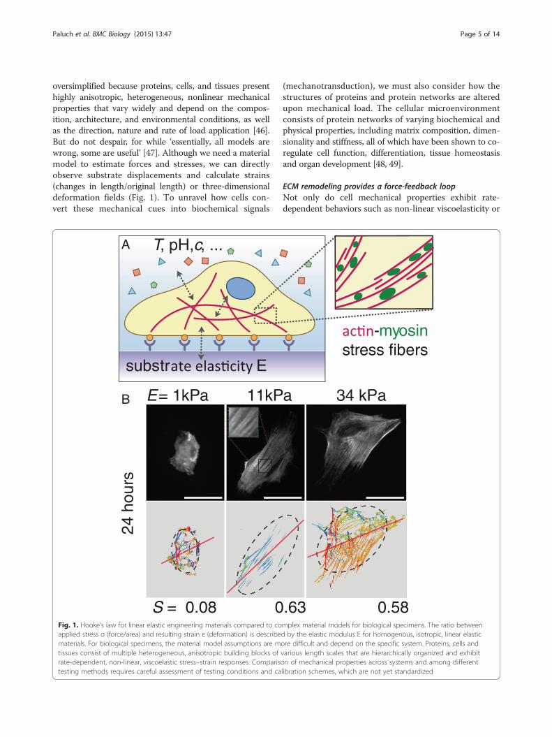
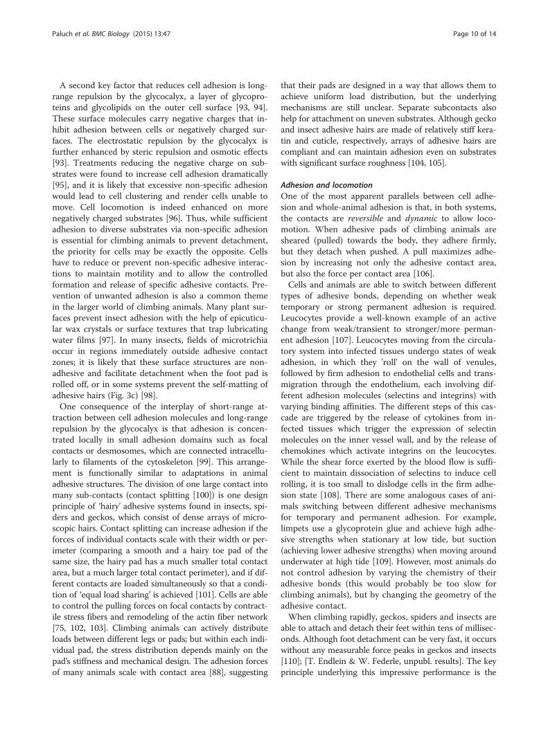
oversimplified because proteins, cells, and tissues presenthighly anisotropic, heterogeneous, nonlinear mechanicalproperties that vary widely and depend on the compos-ition, architecture, and environmental conditions, as wellas the direction, nature and rate of load application [46].But do not despair, for while ‘essentially, all models arewrong, some are useful’ [47]. Although we need a materialmodel to estimate forces and stresses, we can directlyobserve substrate displacements and calculate strains(changes in length/original length) or three-dimensionaldeformation fields (Fig. 1). To unravel how cells con-vert these mechanical cues into biochemical signals
S = 0.08 0
E = 1kPa 11kPB
substr E
T, pH, c, ...A
24 h
ours
Fig. 1. Hooke's law for linear elastic engineering materials compared to coapplied stress σ (force/area) and resulting strain ε (deformation) is describematerials. For biological specimens, the material model assumptions are mtissues consist of multiple heterogeneous, anisotropic building blocks ofrate-dependent, non-linear, viscoelastic stress–strain responses. Comparistesting methods requires careful assessment of testing conditions and ca
(mechanotransduction), we must also consider how thestructures of proteins and protein networks are alteredupon mechanical load. The cellular microenvironmentconsists of protein networks of varying biochemical andphysical properties, including matrix composition, dimen-sionality and stiffness, all of which have been shown to co-regulate cell function, differentiation, tissue homeostasisand organ development [48, 49].
ECM remodeling provides a force-feedback loopNot only do cell mechanical properties exhibit rate-dependent behaviors such as non-linear viscoelasticity or
.63 0.58
34 kPaa
-myosinstress fibers
mplex material models for biological specimens. The ratio betweend by the elastic modulus E for homogenous, isotropic, linear elasticore difficult and depend on the specific system. Proteins, cells andvarious length scales that are hierarchically organized and exhibiton of mechanical properties across systems and among differentlibration schemes, which are not yet standardized
Paluch et al. BMC Biology (2015) 13:47 Page 6 of 14
thermodynamic instabilities, but the substrate itself alsochanges with time. Cells secrete and remodel the ECMthat comprises their tissue microenvironment in a loadhistory-dependent manner. For example, bone resorptionby cell-secreted proteases as in microgravity results inmore porous, weaker ECM networks while bone growthby cell-secreted and remodeled ECM reinforcement oc-curs with weight-bearing exercise [50]. Variations in ECMbiophysical properties within or across a tissue are notonly graded in their composition, crosslinking, dimension-ality and stiffness by the cells that created them, but theseproperties also feedback on cell responses via mechanic-ally mediated cell signaling pathways and enable long-range signaling by cells through the ECM. The biochem-ical and physical properties of cell-generated ECM proteinnetworks (for example, collagens and laminins in base-ment membranes or collagens, fibronectin and elastins inblood vessels) co-regulate cell functions such as motility,proliferation, apoptosis [51], stem cell differentiation [13]and organ development [48]. Different cell types showpreferences for substrates of different rigidity, which inturn can elicit different cell-ECM traction forces [52].Unlike inert materials with fixed properties, livingcells remodel themselves and their environment underchronic loading and also actively generate mechanicalforces through actomyosin-generated tension in thecytoskeleton.
How do molecular mechanisms integrate to cellularmechanotransduction?Even though we can relate the deformations of the cellsand the cellular microenvironment to forces, mechanic-ally mediated signaling at the molecular level is also fun-damentally governed by deformation and rate sensitivity.Binding kinetics, protein crosslinking and availability ofbinding sites depend on both the protein sequence andprotein conformation, a function of thermodynamic en-ergy. Changing protein conformations can encode distinctfunctional states with different ligand binding affinities orbinding availability by exposing cryptic binding sites forother binding partners. For example, the rod domain oftalin, a focal adhesion protein linking integrins to the actincytoskeleton, undergoes large conformational changesunder acto-myosin-generated forces and exposes crypticbinding sites for vinculin [53]. Vinculin recruitment inturn contributes to a reinforcement of the focal adhesioncomplex to transduce higher load between the cells andthe ECM [54]. Similarly, actomyosin-generated tension fa-cilitates vinculin binding to a cryptic binding site in α-catenin in cell-cell adherens junctions to regulate tissueorganization [55]. Within the fibrous ECM, fibronectinunraveling is controlled by Rho-mediated cell contractilityto expose cryptic self-assembly sites and binding sites for
other proteins and growth factors [56, 57]. All thosemechanosensitive proteins consist of multiple domainswith a range of threshold unfolding loads. At appropriateforce thresholds, stiff protein domains (β-sheets, α-helices,barrels) first reorient in the direction of loading as flexiblelinker chains connecting them (turns, loops, hairpins)stretch and rotate; the individual domains unfold in theorder of their stiffness and thus contribute to highly non-linear force-displacement behavior as proteins can unfoldup to >10 times their equilibrium length. Most of theprotein force spectroscopy studies quantified forcethresholds for these phenomena through a complex setof assumptions both about the measurement tools andthe sample to arrive at forces at the scale of picoNew-tons in such load-bearing proteins as cadherin [6] andvinculin [5]. While pN molecular forces may be suffi-cient for mechanically switching individual proteinfunctions or binding affinities, aggregated forces mea-sured at the cellular level are much higher and result indeformation on the surrounding ECM on the order ofseveral cell lengths. Indeed, fibrous scaffolds transmittension over long distances through their rope-like in-terconnections [58].
How can we measure deformations (forces)?Applying or measuring displacements (and inferringforces from these measurements) across the lengthscales of proteins, cells, and tissues requires a range oftechniques and several biochemical sensors and micro-fabricated devices have been developed for this purpose.Optical and magnetic tweezers, Förster Resonance EnergyTransfer (FRET) molecular tensions sensors, and atomicforce microscopy (AFM) are widely used to study con-formational changes of individual mechanosensitive pro-teins under mechanical load [59], while optical stretchers,micropipette aspiration, AFM and microelectromechani-cal systems (MEMS) enable single cell mechanobiologicalstudies (see [60] for a review). Meanwhile, traction forcemicroscopy of fiducial markers embedded in compliantsubstrates and microfabricated post arrays are commonlyused to measure the displacement fields of single cells andmicrotissues [61]. Given the dynamic nature of the stateof cells, ECM and proteins, estimates of small forces orheterogeneous mechanical properties are not easilycompared between labs using the same method, letalone across methods, and standardized calibrationand measurement schemes are needed. Nevertheless,the variability of life is perhaps greater still, and thusmechanobiologists can learn a great deal from appro-priately designed experiments and controls to look forrelative changes in a consistent framework of measure-ments and models.

Paluch et al. BMC Biology (2015) 13:47 Page 7 of 14
Mechano-guided differentiation of humanmesenchymal stem cells: actomyosin stress fibersas collective mechanosensorsCarina Wollnik, Galina Kudryasheva and Florian RehfeldtIt is nowadays well acknowledged that mechanical stim-uli can be as important for cells as traditional biochem-ical cues [62]. They also impact the efficacy of drugs [63]and can influence the morphology and growth phase ofcellular aggregates [64]. Especially striking are the exper-iments by Engler et al., demonstrating that substrateelasticity can direct differentiation of human mesenchy-mal stem cells (hMSCs) towards various linages (neural,muscle, bone) [13]. Here, it is particularly interestingthat mimicking Young’s modulus of the in vivo environ-ment drives naïve adult stem cells towards the respectivecell type. While the initial cue (elastic properties of thematrix) and the overall outcome (changes in transcrip-tion) are well-defined, the integration of the mechanicalstimuli into biochemical signaling pathways is still notfully understood. Recently, there is mounting evidencethat direct mechanical coupling to and perturbation ofthe nuclear envelope and the nucleus might be an alter-native or additional route to alter gene regulation [65].
epithelium
L
F
L
engineering materials
homogeneousisotropiclinear elasticsmall deformations
material model assumptions
proteins, cells, tissues
heterogeneousanisotropic, hierarchicalviscoelasticlarge deformations
material model assumptions
tissufibro
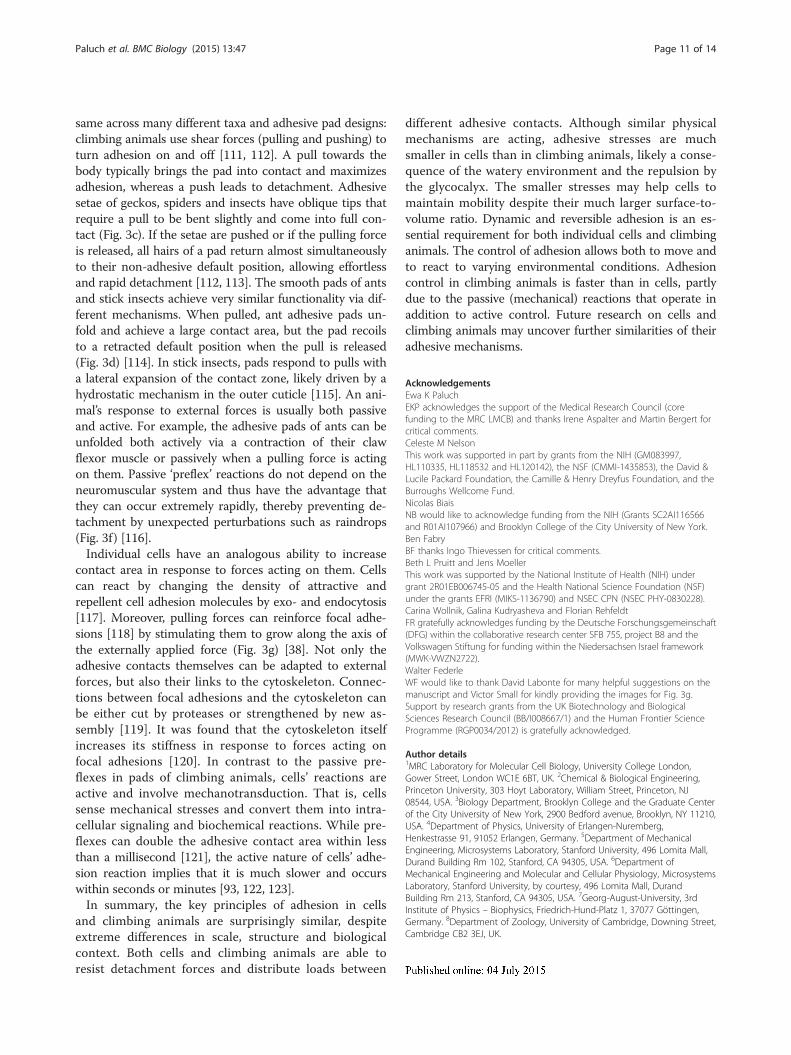
Fig. 2. Acto-myosin stress fibers are key mechanical regulators in cell-matrixActomyosin stress fibers (magnified in the inset zoom) are connected via focaand generate contractile forces that enable the cell to sense the mechanical pnuclear lamina, thus providing a direct mechanical route to gene regulation. Ab Non-monotonic dependence of stress fiber structure quantified by an ordebe used as early morphological marker for mechano-guided differentiation. SPublishing Group
To explain such a mechanical pathway, it is essentialto understand how forces can be transmitted to the nu-cleus. Actomyosin stress fibers are key players in cell ad-hesion and cell-matrix interactions as they anchor at focaladhesion sites and create cellular contractility (Fig. 2a)[66, 67]. Because they also connect directly to the nuclearlamina [68, 69], stress fibers are able to pass on stress andstrain and deform the nucleus [65]. A closer look at theactomyosin filaments in hMSCs revealed that quantifica-tion of their structure and organization by means of anorder parameter S showed significant differences withrespect to the substrate elasticity at an early stage ofmechano-induced differentiation (24 hours) [70, 71]. Onsoft (1 kPa) and rigid (34 kPa) substrates the stress fibersare organized more isotropically (S = 0.08 and 0.58, re-spectively), while on 11 kPa substrates (an intermediateelasticity and matching the in vivo stiffness of relaxedmuscle) the actomyosin bundles were parallel aligned andshowed high anisotropy as indicated by an order param-eter S = 0.63 (Fig. 2b). This early morphological markercan be understood in terms of a collective mechanosensorand is experimentally observable long before lineage-specific genes are upregulated, a process that usually takes
stress (σ) =
strain (ε) =L - L (Δlength)
A (area)F (force)
L (length)
σ = E ε
Hooke’s Law
... elastic modulusE σ (
Pa)
ε (%)
shear stresscirculating cells
endothelium
e cells in us 3D extracellular matrix
E const.
σ (
Pa)
ε (%)
mechanosensing. a Sketch of a cell adhering to a substrate of elasticity E.l adhesions and extracellular matrix proteins to the micro-environmentroperties of the substrate. The cytoskeleton is also connected to thedapted from [67] with permission from The Royal Society of Chemistry.r parameter S of hMSCs grown on substrates of different elasticity E cancale bar is 50 μm. Adapted from [70] with permission from the Nature

Paluch et al. BMC Biology (2015) 13:47 Page 8 of 14
several days [72]. Another recent study used this quantita-tive order parameter analysis to determine the effect ofsubstrate elasticity on differentiating myoblasts [73], indi-cating that mechanical stimuli and respective change ofcytoskeleton structure do play a role in differentiation ofmore than one cell type.Such significant differences in macroscopic or global
structures are most likely induced by distinct molecularcompositions of the actomyosin stress fibers on thenano-scale, a hypothesis that might be confirmed in thenear future by super-resolution microscopy methodsnow available. It is important to note that this type ofanalysis is now done with fixed cells at distinct times,just like one would do for protein or transcription ana-lysis. To get a better insight into the complex and alsohighly dynamic processes it will be of paramount im-portance to follow the temporal evolution of stress-fiberstructure and organization followed by live cell imagingand quantitatively extract parameters of the kinetics.Analyzing transcription profiles and biochemical sig-
naling cascades is essential to finally elucidate the com-plex processes underlying mechanosensory phenomena.However, the micro- and nano-structural properties ofthose mechanically active structures contain valuable in-formation on the route from outside mechanical signalsto inside biochemical regulation. In the same way thatcell biology relied for a long time on descriptive pheno-typing by analyzing global cell shape, we should use theadditional information of the morphology of stress fibersto complete our picture of mechano-guided differenti-ation of hMSCs.
Reversible adhesion in climbing animals - is itsimilar to cell adhesion?Walter FederleNot only cells but also many climbing animals such asinsects, spiders and geckos are able to move around intheir environment, yet have to resist detachment forcesby forming adhesive contacts. These animals possessspecial attachment structures on their feet that allowthem to cling to substrates. When climbing on trees theymay be challenged by the forces of gravity and wind, orby predators trying to dislodge them [74]. Cells, in turn,can be exposed to shear flows in blood vessels or to ten-sile stresses within tissues. Many animals can climb in athree-dimensional environment such as the forest can-opy by repeatedly attaching and detaching their feet,whereas cells can migrate on substrates and within tis-sues by forming new adhesive contacts at their front andreleasing them again at their trailing edge [75]. By wayof these functional similarities, it is perhaps natural tocompare cell adhesion with the adhesion of climbing an-imals. Is cell adhesion similar to the adhesion of insects,spiders and geckos?
Comparison of adhesive strengthStarting with the physical mechanism of adhesion, bothcells and climbing animals take advantage of van derWaals forces [76, 77]. These forces only become signifi-cant when two objects are in intimate contact with sep-aration distances less than approximately 10 nm [78].The contacts formed by cell adhesion molecules such asintegrin or cadherin and those of gecko adhesive hairsare likely within this range [79, 80]. In addition to vander Waals forces, cell adhesion is strongly dependent onelectrostatic forces comprising hydrogen bonds, doublelayer forces and forces between charged domains ofinteracting proteins [79]. An important role of electro-static forces via contact electrification has also been pro-posed recently for the adhesion of geckos [81], andsimilar mechanisms are possible for insects [82]. Al-though more evidence is needed to confirm the extentto which contact electrification contributes to animal ad-hesion, both cells and climbing animals are probably af-fected by electrostatic interactions. In addition, theadhesion of many climbing animals involves capillaryforces, arising from tiny amounts of fluid secreted intothe adhesive contact zone [83–87]. As cells live in awatery environment, this adhesive mechanism is usuallyabsent in cells.Given that both cells and whole animals adhere by van
der Waals forces and probably electrostatic forces, onemight expect their adhesive stresses to be of similarmagnitude. Let us briefly consider the consequences ifthis were indeed the case: depending on the geometry,adhesion forces scale with the length or the area of ad-hesive contacts [88]. For example, while the pull-offforce for a suction cup is proportional to its area, theforce needed to peel off a piece of Scotch tape dependson its width. For isometric organisms, weight increaseswith the cube of linear dimensions, and therefore fasterthan area- or length-specific adhesion. As a consequence,adhesion per body weight is expected to decrease forlarger animals. Despite their relatively large body size,however, geckos can easily hang from a single toe, andweaver ants can carry more than 100 times their ownbody weight whilst walking upside down on a smoothsurface (Fig. 3a, b). Clearly, these animals use only asmall fraction of their body surface area (that is, the ad-hesive organs on their feet) for attachment, and at leastthe gecko does not seem to employ any specific adhe-sion molecules to achieve high levels of forces.Cells have an average mass of approximately 1 ng [89]
and are thus 5 to 11 orders of magnitude lighter thangeckos and insects. Their weight-specific adhesion shouldthus be enormous in comparison to animals. Moreover,about half of a cell’s surface is in relatively close contactwith the substrate, a much larger proportion than for aclimbing animal. Thus, one might expect cells to adhere

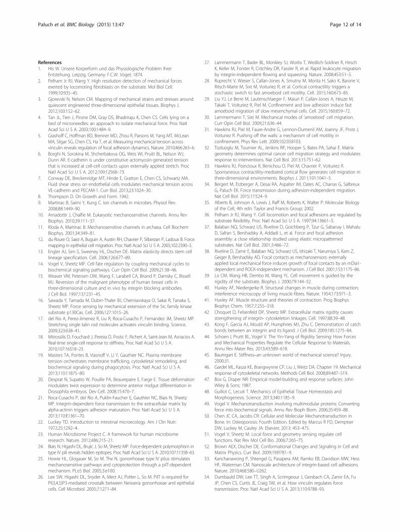
Fig. 3. Surface adhesion in climbing animals and cells. a Weaver ant (Oecophylla smaragdina) carrying more than 100 times its body weightupside-down on a smooth surface (photo: Thomas Endlein). b Tokay gecko (Gekko gecko) attached by a single toe to a tilted glass surface. Reproducedfrom [130] with permission from the Journal of Experimental Biology. c Lateral view of adhesive setae in a longhorn beetle (Clytus arietis)showing non-adhesive orientation of seta tips and anti-adhesive corrugations on the dorsal side. Reproduced from [131] with permissionfrom the Journal of Experimental Biology. d Weaver ant adhesive pad in the retracted (top) and the extended position (bottom). Reproducedfrom [114]. e Adherent cell on a deformable substrate. Inward forces are transmitted via the cytoskeleton and the focal adhesions to thesubstrate. Adapted from [75]. f Rapid increase in adhesive contact area in stick insects (Carausius morosus) in response to a rapid displacement of thesubstrate. Adapted from [121]. g B16 melanoma cell (expressing fluorescent marker for focal adhesions) before and 5 minutes after displacement of cellbody by a microneedle (direction shown by arrow), showing growth of peripheral focal contacts in the region opposite the cell body (enlarged ininsets), stimulated by tension. Reproduced from [123] with permission from the Journal of Cell Science
Table 1 Shear and adhesive strength of animal adhesive padsin comparison with single cells
Strength (kPa) Source
Shear forces
Gecko seta: real contact area 53,300 [124, 125]
Gecko seta: projected contact area 2,880
Beetle pad: real contact area 681 [112]
Beetle pad: projected contact area 259
Weaver ants 405 [126]
Stick insects 299 [112]
Barnacles 10-300 [127]
Fibroblast cells (whole) 0.048 [128]
Fibroblast cells (focal contacts) 5.5 [103]
Adhesion
Gecko seta: real contact area 10,700 [124, 125]
Gecko seta: projected contact area 576
Beetle pad: real contact area 86.9 [112]
Beetle pad: projected contact area 35.5
Stick insect pad 44.6 [112]
Barnacles 100-1000 [90]
Ants ~50 [126]
Endothelial cells 0.56-1.1 [129]
Paluch et al. BMC Biology (2015) 13:47 Page 9 of 14
very firmly to any substrate and to one another, even with-out any specific adhesion molecules, potentially limitingor preventing locomotion. Are the adhesive stresses ofcells and climbing animals indeed comparable, and howdo they compare with typical levels for van der Waalsforces?van der Waals forces are considered weak intermo-
lecular forces, but they still can produce maximum con-tact strengths of the order of 100 MPa, sufficient for a1 cm2 contact to support the weight of a small familycar [76]. Table 1 shows that the stresses measured foranimal adhesives are at least two orders of magnitudebelow these theoretical strength levels, probably a resultof stress concentrations and surface contamination.However, adhesive and shear stresses of cells are muchsmaller still, by several orders of magnitude. Why arestresses so much smaller for cells, although they usesimilar molecular forces for attachment?Firstly, cells live in a watery environment. Water not
only provides viscous ‘squeeze-out’ resistance that makesit harder for objects to come into close contact, but italso shields surface charges, and intervening layers ofwater reduce van der Waals forces [76]. However, thereare many examples of marine organisms that achievehigh adhesion strengths under water (e.g., barnacles andmussels) [90–92], so that submersion alone may notfully explain the low stress levels of cells.
Paluch et al. BMC Biology (2015) 13:47 Page 10 of 14
A second key factor that reduces cell adhesion is long-range repulsion by the glycocalyx, a layer of glycopro-teins and glycolipids on the outer cell surface [93, 94].These surface molecules carry negative charges that in-hibit adhesion between cells or negatively charged sur-faces. The electrostatic repulsion by the glycocalyx isfurther enhanced by steric repulsion and osmotic effects[93]. Treatments reducing the negative charge on sub-strates were found to increase cell adhesion dramatically[95], and it is likely that excessive non-specific adhesionwould lead to cell clustering and render cells unable tomove. Cell locomotion is indeed enhanced on morenegatively charged substrates [96]. Thus, while sufficientadhesion to diverse substrates via non-specific adhesionis essential for climbing animals to prevent detachment,the priority for cells may be exactly the opposite. Cellshave to reduce or prevent non-specific adhesive interac-tions to maintain motility and to allow the controlledformation and release of specific adhesive contacts. Pre-vention of unwanted adhesion is also a common themein the larger world of climbing animals. Many plant sur-faces prevent insect adhesion with the help of epicuticu-lar wax crystals or surface textures that trap lubricatingwater films [97]. In many insects, fields of microtrichiaoccur in regions immediately outside adhesive contactzones; it is likely that these surface structures are non-adhesive and facilitate detachment when the foot pad isrolled off, or in some systems prevent the self-matting ofadhesive hairs (Fig. 3c) [98].One consequence of the interplay of short-range at-
traction between cell adhesion molecules and long-rangerepulsion by the glycocalyx is that adhesion is concen-trated locally in small adhesion domains such as focalcontacts or desmosomes, which are connected intracellu-larly to filaments of the cytoskeleton [99]. This arrange-ment is functionally similar to adaptations in animaladhesive structures. The division of one large contact intomany sub-contacts (contact splitting [100]) is one designprinciple of ‘hairy’ adhesive systems found in insects, spi-ders and geckos, which consist of dense arrays of micro-scopic hairs. Contact splitting can increase adhesion if theforces of individual contacts scale with their width or per-imeter (comparing a smooth and a hairy toe pad of thesame size, the hairy pad has a much smaller total contactarea, but a much larger total contact perimeter), and if dif-ferent contacts are loaded simultaneously so that a condi-tion of ‘equal load sharing’ is achieved [101]. Cells are ableto control the pulling forces on focal contacts by contract-ile stress fibers and remodeling of the actin fiber network[75, 102, 103]. Climbing animals can actively distributeloads between different legs or pads; but within each indi-vidual pad, the stress distribution depends mainly on thepad’s stiffness and mechanical design. The adhesion forcesof many animals scale with contact area [88], suggesting
that their pads are designed in a way that allows them toachieve uniform load distribution, but the underlyingmechanisms are still unclear. Separate subcontacts alsohelp for attachment on uneven substrates. Although geckoand insect adhesive hairs are made of relatively stiff kera-tin and cuticle, respectively, arrays of adhesive hairs arecompliant and can maintain adhesion even on substrateswith significant surface roughness [104, 105].
Adhesion and locomotionOne of the most apparent parallels between cell adhe-sion and whole-animal adhesion is that, in both systems,the contacts are reversible and dynamic to allow loco-motion. When adhesive pads of climbing animals aresheared (pulled) towards the body, they adhere firmly,but they detach when pushed. A pull maximizes adhe-sion by increasing not only the adhesive contact area,but also the force per contact area [106].Cells and animals are able to switch between different
types of adhesive bonds, depending on whether weaktemporary or strong permanent adhesion is required.Leucocytes provide a well-known example of an activechange from weak/transient to stronger/more perman-ent adhesion [107]. Leucocytes moving from the circula-tory system into infected tissues undergo states of weakadhesion, in which they 'roll' on the wall of venules,followed by firm adhesion to endothelial cells and trans-migration through the endothelium, each involving dif-ferent adhesion molecules (selectins and integrins) withvarying binding affinities. The different steps of this cas-cade are triggered by the release of cytokines from in-fected tissues which trigger the expression of selectinmolecules on the inner vessel wall, and by the release ofchemokines which activate integrins on the leucocytes.While the shear force exerted by the blood flow is suffi-cient to maintain dissociation of selectins to induce cellrolling, it is too small to dislodge cells in the firm adhe-sion state [108]. There are some analogous cases of ani-mals switching between different adhesive mechanismsfor temporary and permanent adhesion. For example,limpets use a glycoprotein glue and achieve high adhe-sive strengths when stationary at low tide, but suction(achieving lower adhesive strengths) when moving aroundunderwater at high tide [109]. However, most animals donot control adhesion by varying the chemistry of theiradhesive bonds (this would probably be too slow forclimbing animals), but by changing the geometry of theadhesive contact.When climbing rapidly, geckos, spiders and insects are
able to attach and detach their feet within tens of millisec-onds. Although foot detachment can be very fast, it occurswithout any measurable force peaks in geckos and insects[110]; [T. Endlein & W. Federle, unpubl. results]. The keyprinciple underlying this impressive performance is the
Paluch et al. BMC Biology (2015) 13:47 Page 11 of 14
same across many different taxa and adhesive pad designs:climbing animals use shear forces (pulling and pushing) toturn adhesion on and off [111, 112]. A pull towards thebody typically brings the pad into contact and maximizesadhesion, whereas a push leads to detachment. Adhesivesetae of geckos, spiders and insects have oblique tips thatrequire a pull to be bent slightly and come into full con-tact (Fig. 3c). If the setae are pushed or if the pulling forceis released, all hairs of a pad return almost simultaneouslyto their non-adhesive default position, allowing effortlessand rapid detachment [112, 113]. The smooth pads of antsand stick insects achieve very similar functionality via dif-ferent mechanisms. When pulled, ant adhesive pads un-fold and achieve a large contact area, but the pad recoilsto a retracted default position when the pull is released(Fig. 3d) [114]. In stick insects, pads respond to pulls witha lateral expansion of the contact zone, likely driven by ahydrostatic mechanism in the outer cuticle [115]. An ani-mal’s response to external forces is usually both passiveand active. For example, the adhesive pads of ants can beunfolded both actively via a contraction of their clawflexor muscle or passively when a pulling force is actingon them. Passive ‘preflex’ reactions do not depend on theneuromuscular system and thus have the advantage thatthey can occur extremely rapidly, thereby preventing de-tachment by unexpected perturbations such as raindrops(Fig. 3f) [116].Individual cells have an analogous ability to increase
contact area in response to forces acting on them. Cellscan react by changing the density of attractive andrepellent cell adhesion molecules by exo- and endocytosis[117]. Moreover, pulling forces can reinforce focal adhe-sions [118] by stimulating them to grow along the axis ofthe externally applied force (Fig. 3g) [38]. Not only theadhesive contacts themselves can be adapted to externalforces, but also their links to the cytoskeleton. Connec-tions between focal adhesions and the cytoskeleton canbe either cut by proteases or strengthened by new as-sembly [119]. It was found that the cytoskeleton itselfincreases its stiffness in response to forces acting onfocal adhesions [120]. In contrast to the passive pre-flexes in pads of climbing animals, cells’ reactions areactive and involve mechanotransduction. That is, cellssense mechanical stresses and convert them into intra-cellular signaling and biochemical reactions. While pre-flexes can double the adhesive contact area within lessthan a millisecond [121], the active nature of cells’ adhe-sion reaction implies that it is much slower and occurswithin seconds or minutes [93, 122, 123].In summary, the key principles of adhesion in cells
and climbing animals are surprisingly similar, despiteextreme differences in scale, structure and biologicalcontext. Both cells and climbing animals are able toresist detachment forces and distribute loads between
different adhesive contacts. Although similar physicalmechanisms are acting, adhesive stresses are muchsmaller in cells than in climbing animals, likely a conse-quence of the watery environment and the repulsion bythe glycocalyx. The smaller stresses may help cells tomaintain mobility despite their much larger surface-to-volume ratio. Dynamic and reversible adhesion is an es-sential requirement for both individual cells and climbinganimals. The control of adhesion allows both to move andto react to varying environmental conditions. Adhesioncontrol in climbing animals is faster than in cells, partlydue to the passive (mechanical) reactions that operate inaddition to active control. Future research on cells andclimbing animals may uncover further similarities of theiradhesive mechanisms.
AcknowledgementsEwa K PaluchEKP acknowledges the support of the Medical Research Council (corefunding to the MRC LMCB) and thanks Irene Aspalter and Martin Bergert forcritical comments.Celeste M NelsonThis work was supported in part by grants from the NIH (GM083997,HL110335, HL118532 and HL120142), the NSF (CMMI-1435853), the David &Lucile Packard Foundation, the Camille & Henry Dreyfus Foundation, and theBurroughs Wellcome Fund.Nicolas BiaisNB would like to acknowledge funding from the NIH (Grants SC2AI116566and R01AI107966) and Brooklyn College of the City University of New York.Ben FabryBF thanks Ingo Thievessen for critical comments.Beth L Pruitt and Jens MoellerThis work was supported by the National Institute of Health (NIH) undergrant 2R01EB006745-05 and the Health National Science Foundation (NSF)under the grants EFRI (MIKS-1136790) and NSEC CPN (NSEC PHY-0830228).Carina Wollnik, Galina Kudryasheva and Florian RehfeldtFR gratefully acknowledges funding by the Deutsche Forschungsgemeinschaft(DFG) within the collaborative research center SFB 755, project B8 and theVolkswagen Stiftung for funding within the Niedersachsen Israel framework(MWK-VWZN2722).Walter FederleWF would like to thank David Labonte for many helpful suggestions on themanuscript and Victor Small for kindly providing the images for Fig. 3g.Support by research grants from the UK Biotechnology and BiologicalSciences Research Council (BB/I008667/1) and the Human Frontier ScienceProgramme (RGP0034/2012) is gratefully acknowledged.
Author details1MRC Laboratory for Molecular Cell Biology, University College London,Gower Street, London WC1E 6BT, UK. 2Chemical & Biological Engineering,Princeton University, 303 Hoyt Laboratory, William Street, Princeton, NJ08544, USA. 3Biology Department, Brooklyn College and the Graduate Centerof the City University of New York, 2900 Bedford avenue, Brooklyn, NY 11210,USA. 4Department of Physics, University of Erlangen-Nuremberg,Henkestrasse 91, 91052 Erlangen, Germany. 5Department of MechanicalEngineering, Microsystems Laboratory, Stanford University, 496 Lomita Mall,Durand Building Rm 102, Stanford, CA 94305, USA. 6Department ofMechanical Engineering and Molecular and Cellular Physiology, MicrosystemsLaboratory, Stanford University, by courtesy, 496 Lomita Mall, DurandBuilding Rm 213, Stanford, CA 94305, USA. 7Georg-August-University, 3rdInstitute of Physics – Biophysics, Friedrich-Hund-Platz 1, 37077 Göttingen,Germany. 8Department of Zoology, University of Cambridge, Downing Street,Cambridge CB2 3EJ, UK.
Paluch et al. BMC Biology (2015) 13:47 Page 12 of 14
References1. His W. Unsere Korperform und das Physiologische Problem Ihrer
Entstehung. Leipzig, Germany: F.C.W. Vogel; 1874.2. Pelham Jr RJ, Wang Y. High resolution detection of mechanical forces
exerted by locomoting fibroblasts on the substrate. Mol Biol Cell.1999;10:935–45.
3. Gjorevski N, Nelson CM. Mapping of mechanical strains and stresses aroundquiescent engineered three-dimensional epithelial tissues. Biophys J.2012;103:152–62.
4. Tan JL, Tien J, Pirone DM, Gray DS, Bhadriraju K, Chen CS. Cells lying on abed of microneedles: an approach to isolate mechanical force. Proc NatlAcad Sci U S A. 2003;100:1484–9.
5. Grashoff C, Hoffman BD, Brenner MD, Zhou R, Parsons M, Yang MT, McLeanMA, Sligar SG, Chen CS, Ha T, et al. Measuring mechanical tension acrossvinculin reveals regulation of focal adhesion dynamics. Nature. 2010;466:263–6.
6. Borghi N, Sorokina M, Shcherbakova OG, Weis WI, Pruitt BL, Nelson WJ,Dunn AR. E-cadherin is under constitutive actomyosin-generated tensionthat is increased at cell-cell contacts upon externally applied stretch. ProcNatl Acad Sci U S A. 2012;109:12568–73.
7. Conway DE, Breckenridge MT, Hinde E, Gratton E, Chen CS, Schwartz MA.Fluid shear stress on endothelial cells modulates mechanical tension acrossVE-cadherin and PECAM-1. Curr Biol. 2013;23:1024–30.
8. Thompson D. On Growth and Form. 1942.9. Martinac B, Saimi Y, Kung C. Ion channels in microbes. Physiol Rev.
2008;88:1449–90.10. Arnadottir J, Chalfie M. Eukaryotic mechanosensitive channels. Annu Rev
Biophys. 2010;39:111–37.11. Kloda A, Martinac B. Mechanosensitive channels in archaea. Cell Biochem
Biophys. 2001;34:349–81.12. du Roure O, Saez A, Buguin A, Austin RH, Chavrier P, Silberzan P, Ladoux B. Force
mapping in epithelial cell migration. Proc Natl Acad Sci U S A. 2005;102:2390–5.13. Engler AJ, Sen S, Sweeney HL, Discher DE. Matrix elasticity directs stem cell
lineage specification. Cell. 2006;126:677–89.14. Vogel V, Sheetz MP. Cell fate regulation by coupling mechanical cycles to
biochemical signaling pathways. Curr Opin Cell Biol. 2009;21:38–46.15. Weaver VM, Petersen OW, Wang F, Larabell CA, Briand P, Damsky C, Bissell
MJ. Reversion of the malignant phenotype of human breast cells inthree-dimensional culture and in vivo by integrin blocking antibodies.J Cell Biol. 1997;137:231–45.
16. Sawada Y, Tamada M, Dubin-Thaler BJ, Cherniavskaya O, Sakai R, Tanaka S,Sheetz MP. Force sensing by mechanical extension of the Src family kinasesubstrate p130Cas. Cell. 2006;127:1015–26.
17. del Rio A, Perez-Jimenez R, Liu R, Roca-Cusachs P, Fernandez JM, Sheetz MP.Stretching single talin rod molecules activates vinculin binding. Science.2009;323:638–41.
18. Mitrossilis D, Fouchard J, Pereira D, Postic F, Richert A, Saint-Jean M, Asnacios A.Real-time single-cell response to stiffness. Proc Natl Acad Sci U S A.2010;107:16518–23.
19. Masters TA, Pontes B, Viasnoff V, Li Y, Gauthier NC. Plasma membranetension orchestrates membrane trafficking, cytoskeletal remodeling, andbiochemical signaling during phagocytosis. Proc Natl Acad Sci U S A.2013;110:11875–80.
20. Desprat N, Supatto W, Pouille PA, Beaurepaire E, Farge E. Tissue deformationmodulates twist expression to determine anterior midgut differentiation inDrosophila embryos. Dev Cell. 2008;15:470–7.
21. Roca-Cusachs P, del Rio A, Puklin-Faucher E, Gauthier NC, Biais N, SheetzMP. Integrin-dependent force transmission to the extracellular matrix byalpha-actinin triggers adhesion maturation. Proc Natl Acad Sci U S A.2013;110:E1361–70.
22. Luckey TD. Introduction to intestinal microecology. Am J Clin Nutr.1972;25:1292–4.
23. Human Microbiome Project C. A framework for human microbiomeresearch. Nature. 2012;486:215–21.
24. Biais N, Higashi DL, Brujic J, So M, Sheetz MP. Force-dependent polymorphism intype IV pili reveals hidden epitopes. Proc Natl Acad Sci U S A. 2010;107:11358–63.
25. Howie HL, Glogauer M, So M. The N. gonorrhoeae type IV pilus stimulatesmechanosensitive pathways and cytoprotection through a pilT-dependentmechanism. PLoS Biol. 2005;3:e100.
26. Lee SW, Higashi DL, Snyder A, Merz AJ, Potter L, So M. PilT is required forPI(3,4,5)P3-mediated crosstalk between Neisseria gonorrhoeae and epithelialcells. Cell Microbiol. 2005;7:1271–84.
27. Lammermann T, Bader BL, Monkley SJ, Worbs T, Wedlich-Soldner R, HirschK, Keller M, Forster R, Critchley DR, Fassler R, et al. Rapid leukocyte migrationby integrin-independent flowing and squeezing. Nature. 2008;453:51–5.
28. Ruprecht V, Wieser S, Callan-Jones A, Smutny M, Morita H, Sako K, Barone V,Ritsch-Marte M, Sixt M, Voituriez R, et al. Cortical contractility triggers astochastic switch to fast amoeboid cell motility. Cell. 2015;160:673–85.
29. Liu YJ, Le Berre M, Lautenschlaeger F, Maiuri P, Callan-Jones A, Heuze M,Takaki T, Voituriez R, Piel M. Confinement and low adhesion induce fastamoeboid migration of slow mesenchymal cells. Cell. 2015;160:659–72.
30. Lammermann T, Sixt M. Mechanical modes of 'amoeboid' cell migration.Curr Opin Cell Biol. 2009;21:636–44.
31. Hawkins RJ, Piel M, Faure-Andre G, Lennon-Dumenil AM, Joanny JF, Prost J,Voituriez R. Pushing off the walls: a mechanism of cell motility inconfinement. Phys Rev Lett. 2009;102:058103.
32. Tozluoglu M, Tournier AL, Jenkins RP, Hooper S, Bates PA, Sahai E. Matrixgeometry determines optimal cancer cell migration strategy and modulatesresponse to interventions. Nat Cell Biol. 2013;15:751–62.
33. Hawkins RJ, Poincloux R, Benichou O, Piel M, Chavrier P, Voituriez R.Spontaneous contractility-mediated cortical flow generates cell migration inthree-dimensional environments. Biophys J. 2011;101:1041–5.
34. Bergert M, Erzberger A, Desai RA, Aspalter IM, Oates AC, Charras G, SalbreuxG, Paluch EK. Force transmission during adhesion-independent migration.Nat Cell Biol. 2015;17:524–9.
35. Alberts B, Johnson A, Lewis J, Raff M, Roberts K, Walter P: Molecular Biologyof the Cell, 4th edn: Taylor and Francis Group; 2002.
36. Pelham Jr RJ, Wang Y. Cell locomotion and focal adhesions are regulated bysubstrate flexibility. Proc Natl Acad Sci U S A. 1997;94:13661–5.
37. Balaban NQ, Schwarz US, Riveline D, Goichberg P, Tzur G, Sabanay I, MahaluD, Safran S, Bershadsky A, Addadi L, et al. Force and focal adhesionassembly: a close relationship studied using elastic micropatternedsubstrates. Nat Cell Biol. 2001;3:466–72.
38. Riveline D, Zamir E, Balaban NQ, Schwarz US, Ishizaki T, Narumiya S, Kam Z,Geiger B, Bershadsky AD. Focal contacts as mechanosensors: externallyapplied local mechanical force induces growth of focal contacts by an mDia1-dependent and ROCK-independent mechanism. J Cell Biol. 2001;153:1175–86.
39. Lo CM, Wang HB, Dembo M, Wang YL. Cell movement is guided by therigidity of the substrate. Biophys J. 2000;79:144–52.
40. Huxley AF, Niedergerke R. Structural changes in muscle during contraction;interference microscopy of living muscle fibres. Nature. 1954;173:971–3.
41. Huxley AF. Muscle structure and theories of contraction. Prog BiophysBiophys Chem. 1957;7:255–318.
42. Choquet D, Felsenfeld DP, Sheetz MP. Extracellular matrix rigidity causesstrengthening of integrin- cytoskeleton linkages. Cell. 1997;88:39–48.
43. Kong F, Garcia AJ, Mould AP, Humphries MJ, Zhu C. Demonstration of catchbonds between an integrin and its ligand. J Cell Biol. 2009;185:1275–84.
44. Schoen I, Pruitt BL, Vogel V. The Yin-Yang of Rigidity Sensing: How Forcesand Mechanical Properties Regulate the Cellular Response to Materials.Annu Rev Mater Res. 2013;43:589–618.
45. Baumgart E. Stiffness–an unknown world of mechanical science? Injury.2000;31.
46. Gardel ML, Kasza KE, Brangwynne CP, Liu J, Weitz DA. Chapter 19: Mechanicalresponse of cytoskeletal networks. Methods Cell Biol. 2008;89:487–519.
47. Box G, Draper NR: Empirical model-building and response surfaces: JohnWiley & Sons; 1987.
48. Guillot C, Lecuit T. Mechanics of Epithelial Tissue Homeostasis andMorphogenesis. Science. 2013;340:1185–9.
49. Vogel V. Mechanotransduction involving multimodular proteins: Convertingforce into biochemical signals. Annu Rev Bioph Biom. 2006;35:459–88.
50. Chen JC CA, Jacobs CR: Cellular and Molecular Mechanotransduction inBone. In: Osteoporosis: Fourth Edition. Edited by Marcus R FD, DemptserDW, Luckey M, Cauley JA: Elsevier; 2013: 453–475.
51. Vogel V, Sheetz M. Local force and geometry sensing regulate cellfunctions. Nat Rev Mol Cell Bio. 2006;7:265–75.
52. Brown AEX, Discher DE. Conformational Changes and Signaling in Cell andMatrix Physics. Curr Biol. 2009;19:R781–9.
53. Kanchanawong P, Shtengel G, Pasapera AM, Ramko EB, Davidson MW, HessHF, Waterman CM. Nanoscale architecture of integrin-based cell adhesions.Nature. 2010;468:580–U262.
54. Dumbauld DW, Lee TT, Singh A, Scrimgeour J, Gersbach CA, Zamir EA, FuJP, Chen CS, Curtis JE, Craig SW, et al. How vinculin regulates forcetransmission. Proc Natl Acad Sci U S A. 2013;110:9788–93.

Paluch et al. BMC Biology (2015) 13:47 Page 13 of 14
55. Nelson WJ, Dickinson DJ, Weis WI. Roles of Cadherins and Catenins in CellCell Adhesion and Epithelial Cell Polarity. Prog Mol Biol Transl.2013;116:3–23.
56. Baneyx G, Baugh L, Vogel V. Fibronectin extension and unfolding within cellmatrix fibrils controlled by cytoskeletal tension. Proc Natl Acad Sci U S A.2002;99:5139–43.
57. Zhong CL, Chrzanowska-Wodnicka M, Brown J, Shaub A, Belkin AM, BurridgeK. Rho-mediated contractility exposes a cryptic site in fibronectin andinduces fibronectin matrix assembly. J Cell Biology. 1998;141:539–51.
58. Rudnicki MS, Cirka HA, Aghvami M, Sander EA, Wen Q, Billiar KL. NonlinearStrain Stiffening Is Not Sufficient to Explain How Far Cells Can Feel onFibrous Protein Gels. Biophys J. 2013;105:11–20.
59. Neuman KC, Nagy A. Single-molecule force spectroscopy: optical tweezers,magnetic tweezers and atomic force microscopy. Nat Methods.2008;5:491–505.
60. Van Vliet KJ, Bao G, Suresh S. The biomechanics toolbox: experimentalapproaches for living cells and biomolecules. Acta Mater. 2003;51:5881–905.
61. Taylor RE, Mukundan V, Pruitt BL. Tools for Studying BiomechanicalInteractions in Cells. In: Mechanobiology of Cell-Cell and Cell-MatrixInteractions. 2011. p. 233–65.
62. Discher DE, Janmey P, Wang YL. Tissue cells feel and respond to thestiffness of their substrate. Science. 2005;310:1139–43.
63. Rehfeldt F, Engler AJ, Eckhardt A, Ahmed F, Discher DE. Cell responses tothe mechanochemical microenvironment - Implications for regenerativemedicine and drug delivery. Adv Drug Deliver Rev. 2007;59:1329–39.
64. Kaliman S, Jayachandran C, Rehfeldt F, Smith A-S. Novel Growth Regime ofMDCK II Model Tissues on Soft Substrates. Biophys J. 2014;106:L25–8.
65. Swift J, Ivanovska IL, Buxboim A, Harada T, Dingal PCDP, Pinter J, PajerowskiJD, Spinler KR, Shin J-W, Tewari M, et al. Nuclear Lamin-A Scales with TissueStiffness and Enhances Matrix-Directed Differentiation. Science.2013;341:6149.
66. Pellegrin S, Mellor H. Actin stress fibres. J Cell Sci. 2007;120:3491–9.67. Rehfeldt F, Brown AEX, Raab M, Cai SS, Zajac AL, Zemel A, Discher DE.
Hyaluronic acid matrices show matrix stiffness in 2D and 3D dictatescytoskeletal order and myosin-II phosphorylation within stem cells. IntegrBiol-UK. 2012;4:422–30.
68. Maniotis AJ, Chen CS, Ingber DE. Demonstration of mechanical connectionsbetween integrins cytoskeletal filaments, and nucleoplasm that stabilizenuclear structure. Proc Natl Acad Sci U S A. 1997;94:849–54.
69. Herrmann H, Aebi U. Intermediate filaments: Molecular structure, assemblymechanism, and integration into functionally distinct intracellular scaffolds.Annu Rev Biochem. 2004;73:749–89.
70. Zemel A, Rehfeldt F, Brown AEX, Discher DE, Safran SA. Optimal matrixrigidity for stress-fibre polarization in stem cells. Nat Phys. 2010;6:468–73.
71. Zemel A, Rehfeldt F, Brown AEX, Discher DE, Safran SA. Cell shape,spreading symmetry, and the polarization of stress-fibers in cells.J Phys-Condens Mat. 2010;22:19.
72. Engler AJ, Sen S, Sweeney HL, Discher DE. Matrix Elasticity Directs Stem CellLineage Specification. Cell. 2006;126:677–89.
73. Yoshikawa HY, Kawano T, Matsuda T, Kidoaki S, Tanaka M. Morphology andAdhesion Strength of Myoblast Cells on Photocurable Gelatin under Native andNon-native Micromechanical Environments. J Phys Chem B. 2013;117:4081–8.
74. Scherge M, Gorb SN. Biological micro- and nanotribology: nature's solutions.Berlin, New York: Springer; 2001.
75. Ladoux B, Nicolas A. Physically based principles of cell adhesionmechanosensitivity in tissues. Rep Prog Phys. 2012;75:116601.
76. Kendall K, Kendall M, Rehfeldt F. Adhesion of cells, viruses andnanoparticles. Dordrecht Heidelberg London New York: Springer; 2011.
77. Loskill P, Puthoff J, Wilkinson M, Mecke K, Jacobs K, Autumn K. Macroscaleadhesion of gecko setae reflects nanoscale differences in subsurfacecomposition. J R Soc Interface. 2013;10:20120587.
78. Israelachvili J. Intermolecular and surface forces. London: AcademicPress; 1992.
79. Leckband D, Israelachvili J. Intermolecular forces in biology. Quart RevBiophys. 2001;34:105–267.
80. Tian Y, Pesika N, Zeng H, Rosenberg K, Zhao B, McGuiggan P, Autumn K,Israelachvili J. Adhesion and friction in gecko toe attachment anddetachment. Proc Natl Acad Sci U S A. 2006;103:19320–5.
81. Izadi H, Stewart KME, Penlidis A. Role of contact electrification andelectrostatic interactions in gecko adhesion. J R Soc Interface.2014;11:20140371.
82. McGonigle DF, Jackson CW, Davidson JL. Triboelectrification of houseflies(Musca domestica L.) walking on synthetic dielectric surfaces. J Electrostatics.2002;54:167–77.
83. Ghazi-Bayat A, Hasenfuss I. On the origin of the adhesive fluid of the tarsaladhesive pads in Pentatomidae (Heteroptera). Zool Anz. 1980;204:13–8.
84. Federle W, Riehle M, Curtis ASG, Full RJ. An integrative study of insectadhesion: mechanics and wet adhesion of pretarsal pads in ants. IntegrComp Biol. 2002;42:1100–6.
85. Edwards JS, Tarkanian M. The adhesive pads of Heteroptera: a re-examination.Proc R Entom Soc London A. 1970;45:1–5.
86. Emerson SB, Diehl D. Toe pad morphology and mechanisms of sticking infrogs. Biol J Linn Soc. 1980;13:199–216.
87. Peattie AM, Dirks J-H, Henriques S, Federle W. Arachnids secrete a fluid overtheir adhesive pads. PLoS One. 2011;6, e20485.
88. Labonte D, Federle W: Scaling and biomechanics of surface attachment inclimbing animals. Phil Trans R Soc B 2015;20140027.
89. Guertin DA, Sabatini DM. Cell size control. In: eLS. Chichester: John Wiley &Sons Ltd; 2006.
90. Crisp DJ, Walker G, Young GA, Yule AB. Adhesion and Substrate Choice inMussels and Barnacles. J Colloid Interf Sci. 1985;104:40–50.
91. Aldred N, Clare AS. The adhesive strategies of cyprids and development ofbarnacle-resistant marine coatings. Biofouling. 2008;24:351–63.
92. Smith AM. Negative-Pressure Generated by Octopus Suckers - a Study ofthe Tensile-Strength of Water in Nature. J Exp Biol. 1991;157:257–71.
93. Sackmann E, Smith A-S. Physics of cell adhesion: some lessons fromcell-mimetic systems. Soft Matt. 2014;10:1644–59.
94. Bell GI, Dembo M, Bongrand P. Cell adhesion, Competition betweennonspecific repulsion and specific bonding. Biophys J. 1984;45:1051–64.
95. Gingell D, Todd I. Red blood cell adhesion, II. Interferometric examination ofthe interaction with hydrocarbon oil and glass. J Cell Sci. 1980;41:135–49.
96. Sugimoto Y. Effect on the adhesion and locomotion of mouse fibroblastsby their interacting with differently charged substrates: A quantitative studyby ultrastructural method. Exp Cell Res. 1981;135:39–45.
97. Whitney HM, Federle W. Biomechanics of plant-insect interactions. CurrOpin Plant Biol. 2013;16:105–11.
98. Stork NE. The Adherence of Beetle Tarsal Setae to Glass. J Nat Hist.1983;17:583–97.
99. Bongrand P: Adhesion of cells. In: Handbook of Biological Physics.Edited by Lipowsky R, Sackmann E, vol. 1: Elsevier Science B.V.; 1995:Chapter 16, 755–803.
100. Arzt E, Gorb S, Spolenak R. From micro to nano contacts in biologicalattachment devices. Proc Natl Acad Sci U S A. 2003;100:10603–6.
101. Hui C-Y, Glassmaker NJ, Tang T, Jagota A. Design of biomimetic fibrillarinterfaces: 2, Mechanics of enhanced adhesion. J R Soc Interface.2004;1:35–48.
102. Fletcher DA, Mullins RD. Cell mechanics and the cytoskeleton. Nature.2010;463:485–92.
103. Balaban NQ, Schwarz US, Riveline D, Goichberg P, Tzur G, Sabanay I, MahaluD, Safran S, Bershadsky A, Addadi L, et al. Force and focal adhesionassembly: a close relationship studied using elastic micropatternedsubstrates. Nat Cell Biol. 2001;3:466–72.
104. Hui C-Y, Glassmaker NJ, Jagota A. How compliance compensates for surfaceroughness in fibrillar adhesion. J Adhesion. 2005;81:699–721.
105. Autumn K, Majidi C, Groff RE, Dittmore A, Fearing R. Effective elasticmodulus of isolated gecko setal arrays. J Exp Biol. 2006;209:3558–68.
106. Labonte D, Federle W. Functionally different pads on the same foot allowcontrol of attachment: stick insects have load-sensitive "heel" pads for frictionand shear-sensitive "toe" pads for adhesion. PLoS One. 2013;8, e81943.
107. Granger DN, Schmid-Schönbein GW: Physiology and pathophysiology ofleukocyte adhesion. Oxford Oxford University Press; 1995.
108. Dufrêne Y (ed.): Life at the nanoscale: atomic force microscopy of live cells:Pan Stanford Publishing Pte. Ltd.; 2011.
109. Smith AM. Alternation between Attachment Mechanisms by Limpets in theField. J Exp Mar Biol Ecol. 1992;160:205–20.
110. Autumn K, Hsieh ST, Dudek DM, Chen J, Chitaphan C, Full RJ. Dynamics ofgeckos running vertically. J Exp Biol. 2006;209:260–72.
111. Autumn K, Dittmore A, Santos D, Spenko M, Cutkosky M. Frictionaladhesion: a new angle on gecko attachment. J Exp Biol. 2006;209:3569–79.
112. Bullock J, Drechsler P, Federle W. Comparison of smooth and hairyattachment pads in insects: friction, adhesion and mechanisms fordirection-dependence. J Exp Biol. 2008;211:3333–43.

Paluch et al. BMC Biology (2015) 13:47 Page 14 of 14
113. Autumn K, Hansen W. Ultrahydrophobicity indicates a non-adhesive defaultstate in gecko setae. J Comp Physiol A. 2006;192:1205–12.
114. Federle W, Brainerd EL, McMahon TA, Hölldobler B. Biomechanics of themovable pretarsal adhesive organ in ants and bees. Proc Natl Acad SciU S A. 2001;98:6215–20.
115. Dirks J-H, Li M, Kabla A, Federle W. In vivo dynamics of the internal fibrousstructure in smooth adhesive pads of insects. Acta Biomater. 2012;8:2730–6.
116. Endlein T, Federle W. Rapid preflexes in smooth adhesive pads of insectsprevent sudden detachment. P Roy Soc B-Biol Sci. 2013;280:1757.
117. Ezratty EJ, Bertaux C, Marcantonio EE, Gundersen GG. Clathrin mediatesintegrin endocytosis for focal adhesion disassembly in migrating cells. J CellBiol. 2009;187:733–47.
118. Choquet D, Felsenfeld DP, Sheetz MP. Extracellular matrix rigidity causesstrengthening of Integrin–cytoskeleton linkages. Cell. 1997;88:39–48.
119. Stehbens SJ, Paszek M, Pemble H, Ettinger A, Gierke S, Wittmann T. CLASPslink focal-adhesion-associated microtubule capture to localized exocytosisand adhesion site turnover. Nat Cell Biol. 2014;16:558–70.
120. Wang N, Butler J, Ingber D. Mechanotransduction across the cell surfaceand through the cytoskeleton. Science. 1993;260:1124–7.
121. Endlein T, Federle W. Rapid preflexes in smooth adhesive pads of insectsprevent sudden detachment. Proc R Soc B. 2013;280:20122868.
122. Riveline D, Zamir E, Balaban NQ, Schwarz US, Ishizaki T, Narumiya S, Kam Z,Geiger B, Bershadsky AD. Focal contacts as mechanosensors: externallyapplied local mechanical force induces growth of focal contacts by anMdia1-dependent and Rock-independent mechanism. J Cell Biol.2001;153:1175–86.
123. Kaverina I, Krylyshkina O, Beningo K, Anderson K, Wang YL, Small JV. Tensilestress stimulates microtubule outgrowth in living cells. J Cell Sci.2002;115:2283–91.
124. Autumn K, Liang YA, Hsieh ST, Zesch W, Chan WP, Kenny TW, Fearing R, FullRJ. Adhesive force of a single gecko foot-hair. Nature. 2000;405:681–5.
125. Autumn K. Properties, principles, and parameters of the gecko adhesivesystem. In: Smith AM, Callow JA, editors. Biological Adhesives. Chapter 12thed. Berlin, Heidelberg: Springer-Verlag; 2006. p. 225–56.
126. Federle W, Baumgartner W, Hölldobler B. Biomechanics of ant adhesivepads: frictional forces are rate- and temperature-dependent. J Exp Biol.2004;207:67–74.
127. Sun Y, Guo S, Walker G, Kavanagh C, Swain G. Surface elastic modulus ofbarnacle adhesive and release characteristics from silicone surfaces.Biofouling. 2004;20:279–89.
128. Gallant ND, Michael KE, García AJ. Cell adhesion strengthening:contributions of adhesive area, integrin binding, and focal adhesionassembly. Mol Biol Cell. 2005;16:4329–40.
129. Takamizawa K, Shoda K, Matsuda T. Pull-out mechanical measurement oftissue-substrate adhesive strength: endothelial cell monolayer sheet formedon a thermoresponsive gelatin layer. J Biomater Sci Polym Ed.2002;13:81–94.
130. Autumn K, Dittmore A, Santos D, Spenko M, Cutkosky M. Frictionaladhesion: a new angle on gecko attachment. J Exp Biol. 2006;209:3569–79.
131. Federle W. Why are so many adhesive pads hairy? J Exp Biol.2006;209:2611–21.