med chem 535p – diagnostic medicinal chemistry …courses.washington.edu/medch535/new/pdfs/7....
TRANSCRIPT
Med Chem 535P – Diagnostic Medicinal Chemistry
Blood Chemistry ~ Lipids and Glucose
I. Cholesterol and Lipids
A. Biology and Chemistry
B. Dietary Uptake
C. Blood Transport
D. Lipoprotein Disorders
II. Fasting Lipid Panel
A. Triglycerides (TG)*
B. Cholesterol
1. Total Cholesterol (TC)*
2. Low Density Lipoprotein Cholesterol (LDL-C)*
3. High Density Lipoprotein Cholesterol (HDL-C)*
4. TC:HDL and LDL:HDL ratios
C. Homocysteine
D. C-Reactive Protein
E. Home and Point of Care Testing
III. Drug Effects and Lipids
BLOOD CHEMISTRY ~ LIPIDS AND GLUCOSE
I. Cholesterol and Lipids
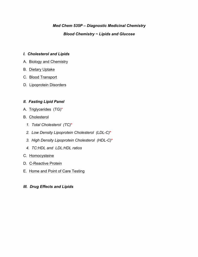
A. Biology and Chemistry. The primary lipids in the body are cholesterol, triglycerides, and phospholipids. They make up biological membranes (phospholipids and cholesterol), serve as precursors to secondary messengers (arachidonic acid), and as precursors to a variety of steroid hormones and bile acids (cholesterol). As such, they are strictly required for the human organism; however, hypercholesterolemia and hyperlipidemia are responsible for significant morbidity and mortality in the human population (coronary artery disease and peripheral vascular disease).
Triglycerides are esters of fatty acids and glycerol and represent the major lipid storage form in humans. Fatty acids are used as biological fuel (β-oxidation). Triglycerides can be absorbed from the diet (exogenous triglycerides) or synthesized in the liver (endogenous triglycerides).
Short Chain Fatty Acid: < 6 carbon chain Medium Chain Fatty Acid: 6 – 12 carbon chain Long Chain Fatty Acid: 13 - 21 carbon chain Very Long Chain Fatty Acid: > 22 carbons
Cholesterol affects membrane fluidity and is the precursor to the bile acids and to a variety of steroid hormones. It can be obtained in the diet, but ~ 90% is synthesized in the liver.
Mammalian membranes are composed of phospholipids, sphingolipids, and cholesterol.
Phosphatidyl choline Phosphatidyl ethanolamine Phosphatidyl inositol
H2C
HC
H2C
O
O
O
R1
R2
R3
O
O
O
H2C
HC
H2C
OH
O
OH
R2
OR1
O
HO
R3
O
HO
Lipase
CH3
CH3
HO
OH
HO OH
O
OH
cholesterol cholic acid
OHO
O
aldosterone
HOCH
O
H2C
HC
H2C
O
O
O
R1
R2
O
O
P
O
O
O-
X
B. Dietary Uptake
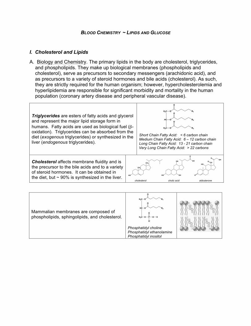
Lingual and pancreatic lipases degrade triglycerides to free fatty acids and monoglycerides. Short and medium chain fatty acids are absorbed by simple diffusion and are transported in the blood by albumin.
Bile acids emulsify long chain fatty acids, monoglycerides, and cholesterol, which then diffuse into the endothelial cells.
In the ER, triglycerides are re-synthesized and cholesterol is esterified with fatty acids for transport through the bloodstream.
Triglycerides, cholesterol, esterified cholesterol and apolipoproteins are packaged into cholymicrons in the ER of the cell. They are secreted into the lymph and finally into the bloodstream, which turns a milky-white color. Indeed, the word "chylomicron" is made up of "chylo-", milky + "micron", small. = small milky (globules).
C. Blood Transport
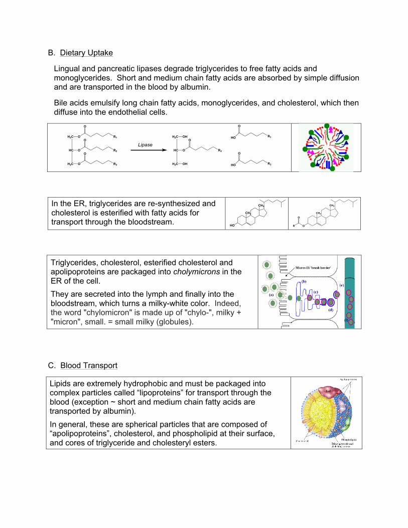
Lipids are extremely hydrophobic and must be packaged into complex particles called “lipoproteins” for transport through the blood (exception ~ short and medium chain fatty acids are transported by albumin). In general, these are spherical particles that are composed of “apolipoproteins”, cholesterol, and phospholipid at their surface, and cores of triglyceride and cholesteryl esters.
H2C
HC
H2C
O
O
O
R1
R2
R3
O
O
O
H2C
HC
H2C
OH
O
OH
R2
OR1
O
HO
R3
O
HO
Lipase
CH3
CH3
HO
CH3
CH3
OR
O
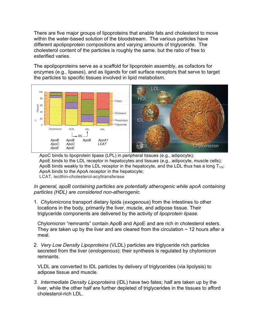
There are five major groups of lipoproteins that enable fats and cholesterol to move within the water-based solution of the bloodstream. The various particles have different apolipoprotein compositions and varying amounts of triglyceride. The cholesterol content of the particles is roughly the same, but the ratio of free to esterified varies.
The apolipoproteins serve as a scaffold for lipoprotein assembly, as cofactors for enzymes (e.g., lipases), and as ligands for cell surface receptors that serve to target the particles to specific tissues involved in lipid metabolism.
ApoB ApoB ApoB ApoA1 ApoC ApoC LCAT ApoE ApoE
ApoC binds to lipoprotein lipase (LPL) in peripheral tissues (e.g., adipocyte); ApoE binds to the LDL receptor in hepatocytes and tissues (e.g., adipocyte, muscle cells); ApoB binds weakly to the LDL receptor in the hepatocyte, and the LDL thus has a long T1/2; ApoA binds to the ApoA receptor in the hepatocyte; LCAT, lecithin-cholesterol-acyltransferase
In general, apoB containing particles are potentially atherogenic while apoA containing particles (HDL) are considered non-atherogenic.
1. Chylomicrons transport dietary lipids (exogenous) from the intestines to other locations in the body, primarily the liver, muscle, and adipose tissue. Their triglyceride components are delivered by the activity of lipoprotein lipase.
Chylomicron “remnants” contain ApoB and ApoE and are rich in cholesterol esters. They are taken up by the liver and are cleared from the circulation ~ 12 hours after a meal.
2. Very Low Density Lipoproteins (VLDL) particles are triglyceride rich particles secreted from the liver (endogenous); their synthesis is regulated by chylomicron remnants.
VLDL are converted to IDL particles by delivery of triglycerides (via lipolysis) to adipose tissue and muscle.
3. Intermediate Density Lipoproteins (IDL) have two fates; half are taken up by the liver, while the other half are further depleted of triglycerides in the tissues to afford cholesterol-rich LDL.
IDL
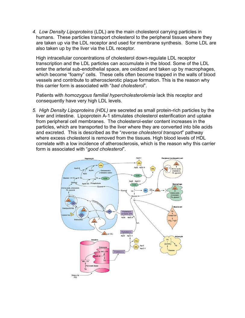
4. Low Density Lipoproteins (LDL) are the main cholesterol carrying particles in humans. These particles transport cholesterol to the peripheral tissues where they are taken up via the LDL receptor and used for membrane synthesis. Some LDL are also taken up by the liver via the LDL receptor.
High intracellular concentrations of cholesterol down-regulate LDL receptor transcription and the LDL particles can accumulate in the blood. Some of the LDL enter the arterial sub-endothelial space, are oxidized and taken up by macrophages, which become “foamy” cells. These cells often become trapped in the walls of blood vessels and contribute to atherosclerotic plaque formation. This is the reason why this carrier form is associated with “bad cholesterol”.
Patients with homozygous familial hypercholesterolemia lack this receptor and consequently have very high LDL levels.
5. High Density Lipoproteins (HDL) are secreted as small protein-rich particles by the liver and intestine. Lipoprotein A-1 stimulates cholesterol esterification and uptake from peripheral cell membranes. The cholesterol-ester content increases in the particles, which are transported to the liver where they are converted into bile acids and excreted. This is described as the “reverse cholesterol transport” pathway where excess cholesterol is removed from the tissues. High blood levels of HDL correlate with a low incidence of atherosclerosis, which is the reason why this carrier form is associated with “good cholesterol”.
148
II) The lipoproteins are used to move hydrophobic insoluble lipid materials such as TAG and
cholesterol from tissue to tissue using the circulatory system.
A) TAG transport is important in energy metabolism and storage of lipid based energy in adipose.
B) Cholesterol is important in membrane function and atherosclerotic disease.
C) We will look at how the lipoproteins do their job by following their fate in the circulatory
system.
D) The function of the protein components (the apo proteins) is important for targeting lipoprotein
content to various tissues, for cholesterol trafficking and for structural integrity of the
lipoproteins. As we will see some of the proteins (the B proteins) remain with the lipoprotein
for its entire lifetime while other proteins are passed amongst the lipoproteins to carry out
particular functions.
D. Lipoprotein Disorders
There is a strong correlation between hypercholesterolemia/hyperlipidemia and the development of atherosclerotic vascular disease (coronary heart disease, stroke, peripheral vascular disease).
Dyslipidemia can be defined as (i) elevated triglycerides, total cholesterol (TC), or LDL-C, or (ii) as low HDL-C concentration, or some combination of the two.
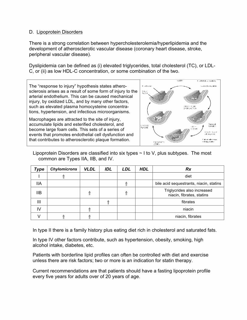
The “response to injury” hypothesis states athero-sclerosis arises as a result of some form of injury to the arterial endothelium. This can be caused mechanical injury, by oxidized LDL, and by many other factors, such as elevated plasma homocysteine concentra-tions, hypertension, and infectious microorganisms.
Macrophages are attracted to the site of injury, accumulate lipids and esterified cholesterol, and become large foam cells. This sets of a series of events that promotes endothelial cell dysfunction and that contributes to atherosclerotic plaque formation.
Lipoprotein Disorders are classified into six types ~ I to V, plus subtypes. The most common are Types IIA, IIB, and IV.
Type Chylomicrons VLDL IDL LDL HDL Rx I ⇑ diet
IIA ⇑ bile acid sequestrants, niacin, statins
IIB ⇑ ⇑ Triglycrides also increased niacin, fibrates, statins
III ⇑ fibrates
IV ⇑ niacin
V ⇑ ⇑ niacin, fibrates
In type II there is a family history plus eating diet rich in cholesterol and saturated fats.
In type IV other factors contribute, such as hypertension, obesity, smoking, high alcohol intake, diabetes, etc.
Patients with borderline lipid profiles can often be controlled with diet and exercise unless there are risk factors; two or more is an indication for statin therapy.
Current recommendations are that patients should have a fasting lipoprotein profile every five years for adults over of 20 years of age.
II. Fasting Lipid Profile: Triglycerides, total cholesterol, LDL, HDL. Ranges are for adults > 20 years old.
A. Triglycerides (TG)*
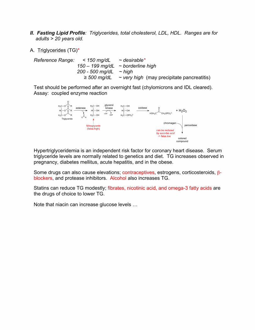
Reference Range: < 150 mg/dL ~ desirable* 150 – 199 mg/dL ~ borderline high 200 - 500 mg/dL ~ high ≥ 500 mg/dL ~ very high (may precipitate pancreatitis)
Test should be performed after an overnight fast (chylomicrons and IDL cleared). Assay: coupled enzyme reaction
Hypertriglyceridemia is an independent risk factor for coronary heart disease. Serum triglyceride levels are normally related to genetics and diet. TG increases observed in pregnancy, diabetes mellitus, acute hepatitis, and in the obese.
Some drugs can also cause elevations; contraceptives, estrogens, corticosteroids, β-blockers, and protease inhibitors. Alcohol also increases TG.
Statins can reduce TG modestly; fibrates, nicotinic acid, and omega-3 fatty acids are the drugs of choice to lower TG.
Note that niacin can increase glucose levels …
esteraseH2C
HC
H2C
O
O
O
R
R
R
O
O
O
Triglyceride-O R
O
H2C
HC
H2C
OH
OH
OH
H2C
HC
H2C
OH
OH
OPO32-
glycerolkinase
ATPADP
oxidase
HOH2CC
CH2OPO32-
O+ H2O2
peroxidasechromagen
coloredcompound
can be reducedby ascorbic acid -> false low
Nitroglycerin(false high)
B. Cholesterol
1. Total Serum Cholesterol (TC; free plus cholesterol esters)
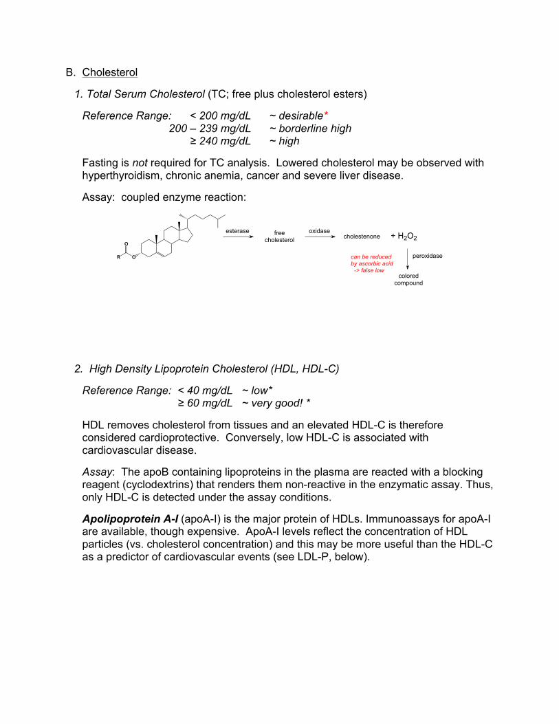
Reference Range: < 200 mg/dL ~ desirable* 200 – 239 mg/dL ~ borderline high ≥ 240 mg/dL ~ high
Fasting is not required for TC analysis. Lowered cholesterol may be observed with hyperthyroidism, chronic anemia, cancer and severe liver disease.
Assay: coupled enzyme reaction:
2. High Density Lipoprotein Cholesterol (HDL, HDL-C)
Reference Range: < 40 mg/dL ~ low* ≥ 60 mg/dL ~ very good! *
HDL removes cholesterol from tissues and an elevated HDL-C is therefore considered cardioprotective. Conversely, low HDL-C is associated with cardiovascular disease.
Assay: The apoB containing lipoproteins in the plasma are reacted with a blocking reagent (cyclodextrins) that renders them non-reactive in the enzymatic assay. Thus, only HDL-C is detected under the assay conditions.
Apolipoprotein A-I (apoA-I) is the major protein of HDLs. Immunoassays for apoA-I are available, though expensive. ApoA-I levels reflect the concentration of HDL particles (vs. cholesterol concentration) and this may be more useful than the HDL-C as a predictor of cardiovascular events (see LDL-P, below).
OR
O
esterase freecholesterol
oxidasecholestenone + H2O2
peroxidase
coloredcompound
can be reducedby ascorbic acid -> false low
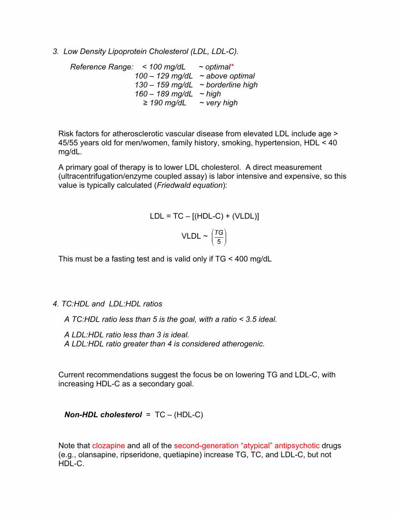
3. Low Density Lipoprotein Cholesterol (LDL, LDL-C).
Reference Range: < 100 mg/dL ~ optimal* 100 – 129 mg/dL ~ above optimal 130 – 159 mg/dL ~ borderline high 160 – 189 mg/dL ~ high ≥ 190 mg/dL ~ very high
Risk factors for atherosclerotic vascular disease from elevated LDL include age > 45/55 years old for men/women, family history, smoking, hypertension, HDL < 40 mg/dL.
A primary goal of therapy is to lower LDL cholesterol. A direct measurement (ultracentrifugation/enzyme coupled assay) is labor intensive and expensive, so this value is typically calculated (Friedwald equation):
LDL = TC – [(HDL-C) + (VLDL)]
VLDL ~ TG5
!
"#
$
%&
This must be a fasting test and is valid only if TG < 400 mg/dL
4. TC:HDL and LDL:HDL ratios
A TC:HDL ratio less than 5 is the goal, with a ratio < 3.5 ideal.
A LDL:HDL ratio less than 3 is ideal. A LDL:HDL ratio greater than 4 is considered atherogenic.
Current recommendations suggest the focus be on lowering TG and LDL-C, with increasing HDL-C as a secondary goal.
Non-HDL cholesterol = TC – (HDL-C)
Note that clozapine and all of the second-generation “atypical” antipsychotic drugs (e.g., olansapine, ripseridone, quetiapine) increase TG, TC, and LDL-C, but not HDL-C.
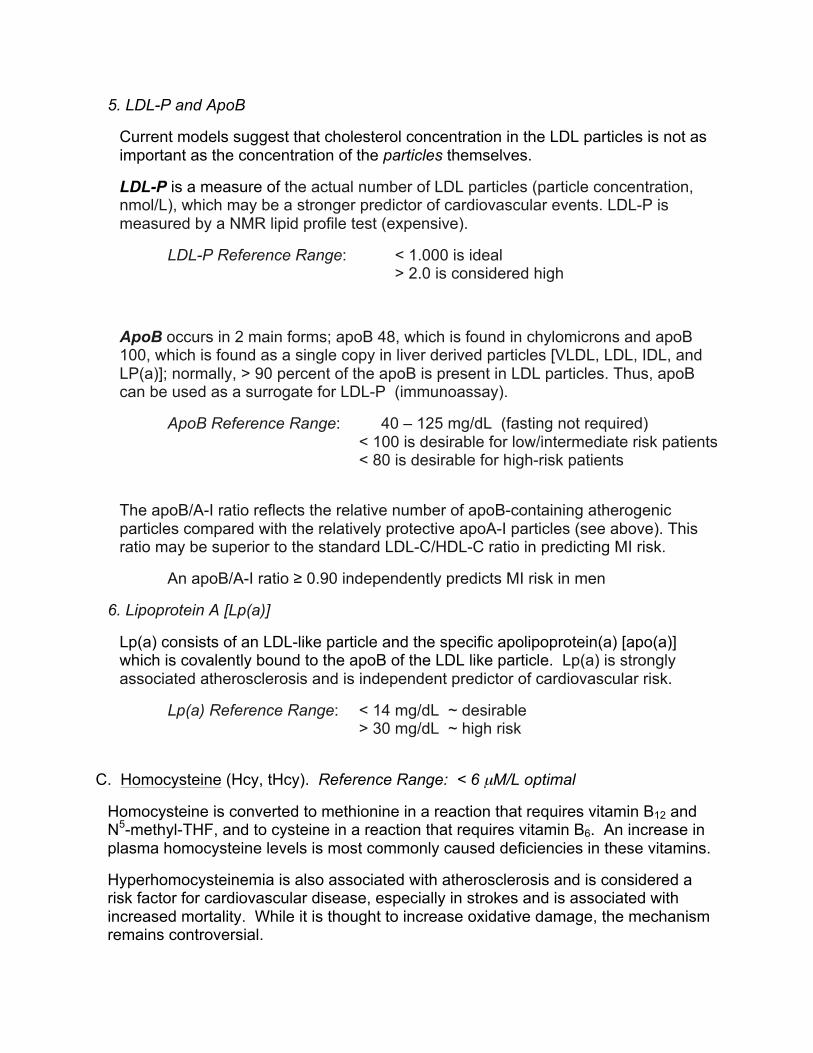
5. LDL-P and ApoB
Current models suggest that cholesterol concentration in the LDL particles is not as important as the concentration of the particles themselves.
LDL-P is a measure of the actual number of LDL particles (particle concentration, nmol/L), which may be a stronger predictor of cardiovascular events. LDL-P is measured by a NMR lipid profile test (expensive).
LDL-P Reference Range: < 1.000 is ideal > 2.0 is considered high
ApoB occurs in 2 main forms; apoB 48, which is found in chylomicrons and apoB 100, which is found as a single copy in liver derived particles [VLDL, LDL, IDL, and LP(a)]; normally, > 90 percent of the apoB is present in LDL particles. Thus, apoB can be used as a surrogate for LDL-P (immunoassay).
ApoB Reference Range: 40 – 125 mg/dL (fasting not required) < 100 is desirable for low/intermediate risk patients < 80 is desirable for high-risk patients
The apoB/A-I ratio reflects the relative number of apoB-containing atherogenic particles compared with the relatively protective apoA-I particles (see above). This ratio may be superior to the standard LDL-C/HDL-C ratio in predicting MI risk.
An apoB/A-I ratio ≥ 0.90 independently predicts MI risk in men
6. Lipoprotein A [Lp(a)]
Lp(a) consists of an LDL-like particle and the specific apolipoprotein(a) [apo(a)] which is covalently bound to the apoB of the LDL like particle. Lp(a) is strongly associated atherosclerosis and is independent predictor of cardiovascular risk.
Lp(a) Reference Range: < 14 mg/dL ~ desirable > 30 mg/dL ~ high risk
C. Homocysteine (Hcy, tHcy). Reference Range: < 6 µM/L optimal
Homocysteine is converted to methionine in a reaction that requires vitamin B12 and N5-methyl-THF, and to cysteine in a reaction that requires vitamin B6. An increase in plasma homocysteine levels is most commonly caused deficiencies in these vitamins.
Hyperhomocysteinemia is also associated with atherosclerosis and is considered a risk factor for cardiovascular disease, especially in strokes and is associated with increased mortality. While it is thought to increase oxidative damage, the mechanism remains controversial.
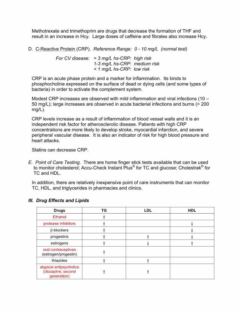
Methotrexate and trimethoprim are drugs that decrease the formation of THF and result in an increase in Hcy. Large doses of caffeine and fibrates also increase Hcy.
D. C-Reactive Protein (CRP). Reference Range: 0 - 10 mg/L (normal test)
For CV disease: > 3 mg/L hs-CRP: high risk 1-3 mg/L hs-CRP: medium risk < 1 mg/L hs-CRP: low risk
CRP is an acute phase protein and a marker for inflammation. Its binds to phosphocholine expressed on the surface of dead or dying cells (and some types of bacteria) in order to activate the complement system.
Modest CRP increases are observed with mild inflammation and viral infections (10 – 50 mg/L); large increases are observed in acute bacterial infections and burns (> 200 mg/L).
CRP levels increase as a result of inflammation of blood vessel walls and it is an independent risk factor for atherosclerotic disease. Patients with high CRP concentrations are more likely to develop stroke, myocardial infarction, and severe peripheral vascular disease. It is also an indicator of risk for high blood pressure and heart attacks.
Statins can decrease CRP.
E. Point of Care Testing. There are home finger stick tests available that can be used to monitor cholesterol; Accu-Check Instant Plus® for TC and glucose; Cholestrak® for TC and HDL.
In addition, there are relatively inexpensive point of care instruments that can monitor TC, HDL, and triglycerides in pharmacies and clinics.
III. Drug Effects and Lipids
Drugs TG LDL HDL Ethanol ⇑
protease inhibitors ⇑ ⇓
β-blockers ⇑ ⇓ progestins ⇑ ⇑ ⇓ estrogens ⇑ ⇓ ⇑
oral contraceptives (estrogen/progestin) ⇑
thiazides ⇑ ⇑
atypical antipsychotics (clozapine; second
generation) ⇑ ⇑
Lecture 6 ~ Lipids Terms You Need to Know Atherosclerosis Lipoprotein lipase Foamy cells You should be prepared to briefly describe the primary lipids in mammals; what is the difference between phospholipids, triglycerides, and cholesterol and what are their primary roles? Be prepared to describe how cholesterol and triglycerides are absorbed from the GI track; what is the role of bile acids, albumin and chylomicrons in the process? Be prepared to describe the five major groups of lipoproteins; how are they differentiated? You should know their relative lipid/protein content and their biological role in humans, as described in class and in the notes. Be prepared to describe the basic mechanism for the formation of an atherosclerotic plaque to your patient. What is the connection between LDL, HDL, and atherosclerotic plaque formation? Be prepared to discuss the essential features of Type II and Type IV lipid disorders. Be prepared to discuss how the lipid assays work and the effects of nitroglycerine and ascorbic acid on the triglyceride, TC, and HDL assays. How do the TC and HDL-C assays differ and how do you obtain LDL-C? What is the limitation on the Friedwald equation … You should be prepared to discuss which lipid levels require an overnight fast and which do not. You should be prepared to discuss the relevance of TC:HDL and LDL:HDL ratios with your patient. What is the primary goal of anti-lipid therapy? Be prepared to discuss the significance of elevated LDL-P, ApoB, and ApoA-1 levels. How do they relate to LDL-C and HDL-C? Be prepared to discuss the significance of elevated Lp(a), Hcy, and CRP levels in the context of risk factors for atherosclerosis and why. You should be prepared to describe the effects of the following drugs on lipid levels: contraceptives, alcohol, clozapine, second-generation atypical antipsychotic agents, methotrexate, and trimethoprim.

You should be prepared to describe the effects of the following drugs on lipid levels: fibrates, nicotinic acid, omega-3 fatty acids, the “statins”. Be prepared to discuss any drug colored in red in any of the lecture notes …