mediastinal histoplasmosis: report of the first two
TRANSCRIPT
Rev. Inst. Med. trop. S. Paulo47(2):103-105, March-April, 2005
(1) Pesquisador 1C do CNPq; Departamento de Medicina Interna, FAMED, UFRGS, Porto Alegre, RS, Brazil.(2) Disciplina de Pneumologia, FAMED, UFBA, Salvador, BA, Brazil.(3) Disciplina de Doenças Infecciosas, FAMED, UFPE, Recife, PE, Brazil.Correspondence to: Dr. Luiz Carlos Severo, Laboratório de Micologia, Hospital Santa Rita, Santa Casa-Complexo Hospitalar, Annes Dias 285, 90020-090 Porto Alegre, RS, Brasil. Fax (51)
32148435. E- mail: [email protected]
MEDIASTINAL HISTOPLASMOSIS: REPORT OF THE FIRST TWO BRAZILIAN CASES OFMEDIASTINAL GRANULOMA
Luiz Carlos SEVERO(1), Antonio Carlos Moreira LEMOS(2) & Heloisa Ramos LACERDA(3)
SUMMARY
This report documents the first two Brazilian cases of mediastinal granuloma due to histoplasmosis, presenting selectedaspects on the diagnosis. Tissue samples revealing histoplasmosis were obtained from each of the patients by mediastinoscopy andthoracotomy. In the second patient, a subcarinal calcified mass eroded into the bronchial tree, leading to secondary bilateralaspiration pneumonitis one week after thoracotomy.
Although rare, histoplasmosis should be included in the differential diagnosis of mediastinal granuloma, specially if there arecalcifications greater than 10 mm in dimension.
KEYWORDS: Histoplasmosis; Histoplasma capsulatum var. capsulatum; Mediastinal histoplasmosis; Mediastinal granuloma.
INTRODUCTION
Classic histoplasmosis is caused by the thermal dimorphic fungusHistoplasma capsulatum var. capsulatum (H. capsulatum). The usualmanifestation is an acute, self-limited, flu-like respiratory illness,however, depending on the host’s defenses and extent of inoculation, avariety of clinical manifestations may result5.
The resolution process of granulomatous lymphadenitis due to H.capsulatum may cause mediastinal disease. Mediastinal granulomaand fibrosing mediastinitis are both well-documented, thoughuncommon, mediastinal sequela of infection. Their formation dependson the lymph node caseation and the degree of fibrotic response incitedto the organism7.
There are three clinical syndromes associated with mediastinalhistoplasmosis: mediastinal adenitis, mediastinal granuloma andfibrosing mediastinitis14. In Brazil, only the latter has been previouslydocumented8. To present the first two cases of mediastinal granulomawarrant this report. A brief review of the pertinent literature is presented.
CASE REPORTS
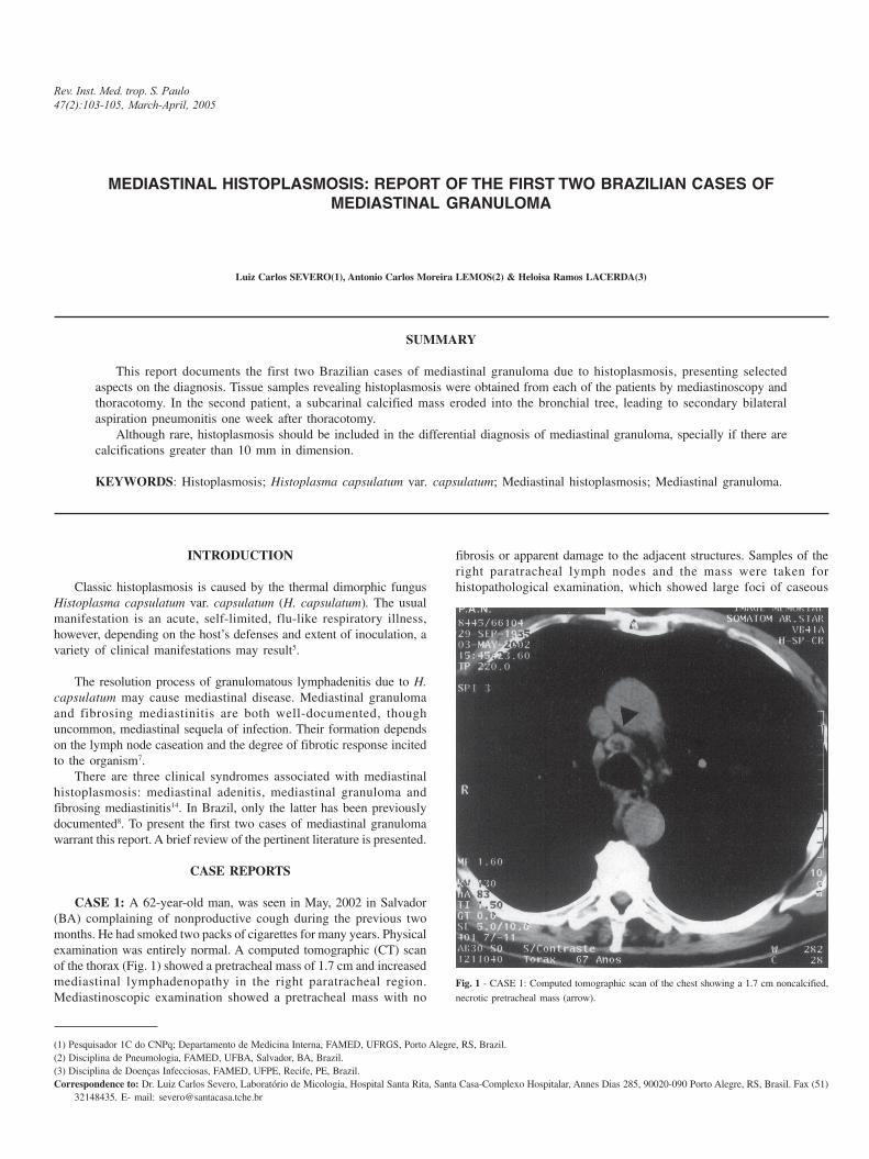
CASE 1: A 62-year-old man, was seen in May, 2002 in Salvador(BA) complaining of nonproductive cough during the previous twomonths. He had smoked two packs of cigarettes for many years. Physicalexamination was entirely normal. A computed tomographic (CT) scanof the thorax (Fig. 1) showed a pretracheal mass of 1.7 cm and increasedmediastinal lymphadenopathy in the right paratracheal region.Mediastinoscopic examination showed a pretracheal mass with no
fibrosis or apparent damage to the adjacent structures. Samples of theright paratracheal lymph nodes and the mass were taken forhistopathological examination, which showed large foci of caseous
Fig. 1 - CASE 1: Computed tomographic scan of the chest showing a 1.7 cm noncalcified,
necrotic pretracheal mass (arrow).

104
SEVERO, L.C.; LEMOS, A.C.M. & LACERDA, H.R. - Mediastinal histoplasmosis: report of the first two Brazilian cases of mediastinal granuloma. Rev. Inst. Med. trop. S. Paulo, 47(2):103-105, 2005.
necrosis and fibrosis. A Mantoux skin test was applied, developing 13mm induration. The patient was referred for treatment of a presumedtuberculosis. However, microscopic examination of specially stainedspecimens was performed. The test for acid-fast organisms wasnegative. On Gomori-Grocott methenamine silver stain (GMS)organisms similar to H. capsulatum were present within caseous areas.Treatment with itraconazole, 100 mg BID was initiated; after six monthsthe patient was asymptomatic.
CASE 2: A 23-year-old man was seen on September 23, 2002 inRecife (PE) complaining of fever, substernal chest pain, and coughlasting one month. During the period of clinical investigation the patienthad an episode of foul sputum productive cough. Chest CT demonstrateda 4.5 cm subcarinal mass with scattered calcifications (Fig. 2). OnOctober 6, an exploratory thoracotomy was performed. Microscopicexamination of the biopsy specimen disclosed calcified necrotic debrisand chronic inflammation with a border of palisading histiocytes andLanghans’ giant cells. Slides stained with GMS showed many yeastcells resembling H. capsulatum within the areas of caseous necrosis.Ziehl-Neelsen stain for acid-fast bacilli was negative. Serologic test tohistoplasmosis (immunodiffusion) was positive.
One week after thoracotomy the patient was readmitted becausehe developed another episode of productive cough with foul-smellingpurulent material associated with aspiration. On physical examinationhe appeared acutely ill. A chest radiograph showed bilateral aspirationpneumonitis. He was treated with a 3-week course of liposomalamphotericin B and broad-spectrum antibiotics. His pulmonarysymptoms and chest radiographic abnormalities resolved. AmphotericinB and the antibiotics were stopped, 200 mg/day oral itraconazol wasinitiated and the patient was discharged. He remained in treatment for
one year. Along with his clinical improvement, the immunodiffusionassay became negative. The patient has remained asymptomatic.
DISCUSSION
Relatively few cases of histoplasmosis with mediastinalinvolvement have been reported, and among these, only a very fewhave had discrete granulomas not complicated by surrounding fibrosis7
as was seen in the present cases. These complications occur when acaseous lymph node or the hyperreactive fibrous response involved inhealing damages contiguous structures, leading to constrictivepericarditis, obstruction of the superior vena cava, tracheobronchialtree, pulmonary artery and veins, and esophagus, with formation offistulas, commonly bronchopleural3,4. Occasionally, the caseous centermay become liquefied and dissect into the tracheobronchial tree6,9,14,as observed in our second case.
Computed tomography is the preferred imaging technique indemonstrating mediastinal calcifications1. Calcification in associationwith histoplasmosis tends to be focal and scattered, greater than 10mm in diameter, most often found in the right tracheal area, and isseen more than 10 years after initial infection6, as in the case of thesecond patient, who revealed close contact with chicken coop duringhis childhood.
In both of our cases, CT scans effectively demonstrated andlocalized the mediastinal granulomas11; however, the ultimate diagnosiswas not suspected until the biopsies were analyzed. In patients withesophageal symptoms (dysphagia or odynophagia) mediastinal massescan be diagnosed by endosonography and endoscopic fine-needleaspiration biopsy13,15. Histologic analysis reveals granulomatousinflammation with centers of caseating necrosis. The specific diagnosisis made by methenamine silver staining that demonstrates yeast formsresembling H. capsulatum in small numbers in necrotic areas. Cultureis usually negative14. Fungal serology may be of value in establishingthe diagnosis, as observed in our second case, and can be used as ascreening test12,14. However, a negative result does not excludehistoplasmosis because of the low rate of serological positivity inmediastinal granuloma13. Differential diagnosis includes benign orslowly growing neoplasm, mediastinal cyst, tuberculosis and, rarely,sarcoidosis10,13. The presence of calcification supports the diagnosis ofgranulomatous infection11. Althought rare2,9, histoplasmosis should beincluded in the differential diagnosis of mediastinal granulomas,although tuberculosis is more likely in regions non-endemic to themycosis1.
RESUMO
Histoplasmose mediastinal: relato dos dois primeiros casosbrasileiros de granuloma mediastinal
São relatados os dois primeiros casos de granuloma mediastinalpor histoplasmose no Brasil, apresentando aspectos selecionados sobredignóstico. O diagnóstico tecidual de histoplasmose foi obtido pormediastinoscopia e toracotomia, respectivamente. Em um paciente amassa calcificada subcarinal erodiu na árvore brônquica compneumonite de aspiração bilateral uma semana após a toracotomia.
Embora rara, histoplasmose deve ser incluída no diagnóstico
Fig. 2 - CASE 2: Contrast-enhanced computed tomographic scan of the chest demonstrates
a 4.5 cm subcarinal mass with scattered calcifications (arrow).

SEVERO, L.C.; LEMOS, A.C.M. & LACERDA, H.R. - Mediastinal histoplasmosis: report of the first two Brazilian cases of mediastinal granuloma. Rev. Inst. Med. trop. S. Paulo, 47(2):103-105, 2005.
105
diferencial de granuloma mediastinal especialmente com calcificaçãomaior do que 10 mm de diâmetro.
REFERENCES
1. AKMAN, C.; KANTARCI, F. & CETINKAYA, S. - Imaging in mediastinitis: a systematicreview based on aetiology. Clin. Radiol., 59: 573-585, 2004.
2. BERNARD, A.C. & MULLETT, T.W. - Mediastinal granuloma complicatinghistoplasmosis J. Ky med. Ass., 101: 12-14, 2003.
3. DAVIS, A.M.; PIERSON, R.N. & LOYD, J.E. - Mediastinal fibrosis. Semin. resp. Infect.,16: 119-130, 2001.
4. GILLILAND, M.D.; SCOTT, L.D. & WALKER, W.E. - Esophageal obstruction causedby mediastinal histoplasmosis: beneficial results of operation. Surgery, 95: 59-62,1984.
5. GOODWIN Jr., R.A. & DES PREZ, R.M. - State of the art: histoplasmosis. Amer. Rev.resp. Dis., 117: 929-956, 1978.
6. GOODWIN Jr., R.A.; LOYD, J.E. & DES PREZ, R.M. - Histoplasmosis in normal hosts.Medicine (Baltimore), 60: 231-266, 1981.
7. GOODWIN Jr., R.A.; NICKELL, J.A. & DES PREZ, R.M. - Mediastinal fibrosiscomplicating healed primary histoplasmosis and tuberculosis. Medicine (Baltimore),51: 227-246, 1972.
8. JERÔNIMO, A.L.C.; CORREA, J.C.; GONÇALVES, A.J.R.; BEZERRA, C.M.F. &CUNHA, R.Q. - Mediastinite fibrosante por histoplasmose. Arq. bras. Med., 60:129-131, 1986.
9. KAUFFMAN, C.A. - Pulmonary histoplasmosis. Curr. infect. Dis. Rep., 3: 279-285,2001.
10. KERN, J.A.; DANIEL, T.M.; TRIBBLE, C.G.; SILEN, M.L. & RODGERS, B.M. -Thoracoscopic diagnosis and treatment of mediastinal masses. Ann. thorac. Surg.,56: 92-96, 1993.
11. LANDAY, M.J. & ROLLINS, N.K. - Mediastinal histoplasmosis granuloma: evaluationwith CT. Radiology, 172: 657-659, 1989.
12. MATHISEN, D.J. & GRILLO, H.C. - Clinical manifestation of mediastinal fibrosis andhistoplasmosis. Ann. thorac. Surg., 54: 1053-1057, 1992.
13. SAVIDES, T.J.; GRESS, F.G.; WHEAT, L.J.; IKENBERRY, S. & HAWES, R.H. -Dysphagia due to mediastinal granulomas: diagnosis with endoscopic ultra-sonography. Gastroenterology, 109: 366-373, 1995.
14. WHEAT, L.J.; CONCES, D.; ALLEN, S.D.; BLUE-HNIDY, D. & LOYD, J. – Pulmonaryhistoplasmosis syndromes: recognition, diagnosis, and management. Semin. resp.crit. care Med., 25: 129-144, 2004.
15. WIERSEMA, M.J.; CHAK, A. & WIERSEMA, L.M. – Mediastinal histoplasmosis:evaluation with endosonography and endoscopic fine-needle aspiration biopsy.Gastroint. Endosc., 40: 78-81, 1994.
Received: 27 August 2004Accepted: 18 January 2005