medical bacteriology lab manual (mls3430c) fall...
TRANSCRIPT

Medical Bacteriology Lab Manual (MLS3430C) Fall 2016
0

Medical Bacteriology Lab Manual (MLS3430C) Fall 2016
1
Table of Contents
Table of Contents ....................................................................................................................... 1
Table of Figures: ......................................................................................................................... 4
Preface ....................................................................................................................................... 5
Week 1: General Guidelines about Medical Bacteriology Lab .................................................. 6
Week 2 & 3: Culture Media Inoculation & Colony Isolation .................................................... 16
Week 4: Spore-Forming Gram-Positive Bacilli (Bacillus & Clostridium Species) ..................... 21
Week 5: Non-Spore-Forming Gram-Positive Bacilli: Corynebacterium, Propionibacterium
Corynebacterium diphtheria .................................................................................................... 26
Week 6: Staphylococci & Streptococci .................................................................................... 30
Week 7 & 8: Enteric Gram-Negative Rodes (Enterobacteriaceae) .......................................... 40
Week 9: Vibrios, Campylobacters, Helicobacter & Associated Bacteria .................................. 43
Week 10: Pseudomonads & Anaerobic Bacteria ...................................................................... 46
Week 11: Haemophilus, Bordetelia & Legioneliae Haemophilus genus .................................. 48
Week 12: Brucelia, Yersinia, Franciselia & Pasteurelia Brucella genus .................................... 52
Week 13: The Neisseriae & Unusual Bacterial Pathogens Neisseriae genus & Mycobacteria 56
Week 14: The Analytical Profile Index (API) ............................................................................ 60
A / Staining Techniques ........................................................................................................... 61
Staining Technique 1. Gram’s Stain Procedure: .................................................................... 61
Staining Technique 2. Endospore Stain Procedure ............................................................... 62
Staining Technique 3. Capsule Stain Procedure: .................................................................. 63
Staining Technique 4. Acid Fast Stain Procedure: ................................................................. 64
B / Metabolic Activities of Bacteria ......................................................................................... 65
Simple Carbohydrate Fermentations ............................................................. 66
Starch Hydrolysis ............................................................................................ 69

Medical Bacteriology Lab Manual (MLS3430C) Fall 2016
2
Oxidation-Fermentation (OF) test .................................................................. 71
Indole test ...................................................................................................... 73
Methyl Red (MR) test ..................................................................................... 75
Voges-Proskauer (VP) test .............................................................................. 77
Citrate test ...................................................................................................... 79
Production of Indole and Hydrogen Sulphide, and Motility .......................... 82
Kligler Iron Agar (KIA) ..................................................................................... 86
C / Activities of Bacterial Enzymes ........................................................................................... 90
Activity of Catalase ......................................................................................... 91
Activity of Coagulase ...................................................................................... 94
Activity of Oxidase .......................................................................................... 97
Activity of Urease ........................................................................................... 99
Activity of Gelatinase ................................................................................... 101
Phenylalaninedeaminase test / Phenylpyruvic acid (PPA) test .................... 104
API & AST tests + Lab Automation ............................................................... 106
Antibiotic Susceptibility Tests for Identification .......................................... 109
Some Culture Media Ingredients: .......................................................................................... 110

Medical Bacteriology Lab Manual (MLS3430C) Fall 2016
3
Medical Bacteriology Lab Manual (MLS3430C) Fall 2016
4
Table of Figures:
Figure 1 Culture Charesteristics ....................................................................................................................................................................... 18
Figure 2 Different Bacterial Colonial Morphologies......................................................................................................................................... 19
Figure 3 Streak Plate Showing Isolation of Two Different Bacterial Types ...................................................................................................... 19
Figure 4 Bacterial growth characteristics in broth. From LEFT to RIGHT: uninoculated tube, precipitation reaction, turbidity, flocculation,
pellicle formation. ........................................................................................................................................................................................... 20
Figure 5 Bacillus anthracis Colonies ................................................................................................................................................................. 23
Figure 6 Bacillus cereus Colonies ..................................................................................................................................................................... 23
Figure 7 Endospores ........................................................................................................................................................................................ 23
Figure 8 Bacillus mycoides Colonies ................................................................................................................................................................ 24
Figure 9 Clostridium perfringens ..................................................................................................................................................................... 24
Figure 10 Corynebacterium diphtheriae Stained Cells LEFT & Colonies RIGHT ............................................................................................... 27
Figure 11 Listeria monocytogenes Stained Cells LEFT & Colonies RIGHT......................................................................................................... 27
Figure 12 P. acnes Stained Cells LEFT & Colonies RIGHT ................................................................................................................................. 27
Figure 13 Staphylococci Cell Morphology ........................................................................................................................................................ 32
Figure 14 Staphylococci Colonies in Nutreint & Blood Agar ............................................................................................................................ 33
Figure 15 Manitol Fermentation RIGHT & Non-fermenting Staphylococci ...................................................................................................... 33
Figure 16 LEFT to RIGHT Alpha, Beta & Gamma Hemolysis ............................................................................................................................. 34
Figure 17 Stained Streptococci Cells ................................................................................................................................................................ 34
Figure 18 β, α and γ Hemolysis of Streptococci ............................................................................................................................................... 36
Figure 19 CAMP Test ....................................................................................................................................................................................... 36
Figure 20 When Durham tubes are placed inside broth tubes, gas produced by fermentation of carbohydrates in the medium can be
visualized as a bubble in the inner tube (right). The organism on the middle does not produce gas when fermenting carbohydrates. Light
red colour of the tube on the left indicates no fermentation.......................................................................................................................... 66
Figure 21 Clear Zone of Unstained Starch Culture Media due to Bacterial Utilization .................................................................................... 70
Figure 22 Indole Test; Positive [LEFT] & Negative [RIGHT] ............................................................................................................................. 74
Figure 23 Methyl Red Test; Positive [LEFT] & Negative [RIGHT] ...................................................................................................................... 76
Figure 24 Voges Proskauer Test ...................................................................................................................................................................... 78
Figure 25 Positive Citrate Utilization Test [RIGHT] & Negative [LEFT] ............................................................................................................. 80
Figure 26 IMViC [Indole, Methyl Red, Voges-Proskauer, Citrate] set of reactions used to differentiate between E. coli with reaction pattern
of [+, +, -, -] & Enterobacter aerogenes with [-, -, +, +] .................................................................................................................................... 81
Figure 27 Hydrogen Sulfide Production and/or Motility in SIM Media ............................................................................................................ 83
Figure 28 Reactions in Sulfide Indole Motility medium [SIM] RIGHT negative for all, MIDDLE is positive for all whilst LEFT is positive for
Motility and Indole, but negative for Sulfide ................................................................................................................................................... 85
Figure 29 Kligler Iron Agar Reactions ............................................................................................................................................................... 88
Figure 30 KIA reactions. ................................................................................................................................................................................... 89
Figure 31 Catalase Test [Notice the Bubbles; Oxygen libration] ...................................................................................................................... 92
Figure 32 Coagulase Positive [LEFT] & Negative [RIGHT] ................................................................................................................................. 96
Figure 33 Oxidase Positive [RIGHT] & Negative [LEFT] .................................................................................................................................... 98
Figure 34 Urease Positive [LEFT] & Negative [RIGHT] .................................................................................................................................... 100
Figure 35 Gelatinase test by tube [coloured] and strip method [Black & white] ........................................................................................... 102
Figure 36 Phenylalanine Deamination. .......................................................................................................................................................... 105
Figure 37 Reactions in API 20E. ..................................................................................................................................................................... 107
Figure 38 Novobiosin sensitive Staph. epidermidis RIGHT vs. resisting Staph. saprophyticus ....................................................................... 109

Medical Bacteriology Lab Manual (MLS3430C) Fall 2016
5
Preface
This lab manual is prepared to direct MLS students to acquire an in-depth knowledge of
diagnostic strategies in Medical Bacteriology. This field is the culmination of several years of
study. It is an exciting time for students, and offers unique experiences in the medical
laboratory setting. Students will achieve from this experience benefits comparable to the
effort they put forth.
The lab manual preparation process was done in light of Harley & Prescott: Laboratory
Exercises in Microbiology, Fifth Edition, 2002, Morello et al., Laboratory Manual and
Workbook in Microbiology, Seventh Edition, 2003, Kayser et al., Medical Microbiology, 2005
and Gillespie−Bamford Medical Microbiology and Infection at a Glance, Fourth Edition, 2012
books.
The first three labs of the manual are devoted to essential microbiology where aseptic
techniques, staining and culture media preparations are covered. Then metabolic and
enzymatic principles of bacterial diagnosis are explained followed by grouping of medically
important bacteria with regard to their sources of sample collection.
Procedures listed from Week 2 to Week 14 are referring to four main staining techniques
and different biochemical and metabolic activity experiments 1 to17.
The last lab session is to familiarize students with lab automation along with antimicrobial
susceptibility tests.
Dr Sirwan M Muhammed & Alan Ahmed
Spring 2016

Medical Bacteriology Lab Manual (MLS3430C) Fall 2016
6
Week 1: General Guidelines about Medical Bacteriology Lab
Lab Safety
1. All materials and clothes other than those needed for the laboratory are to be kept away from
the work area.
2. A lab coat or other protective clothing must be worn during lab. The lab clothing is not to be
worn outside of the laboratory.
3. Clean the lab table before and after lab with the disinfectant solution provided.
4. Wash hands before leaving lab.
5. Any item contaminated with bacteria or body fluids must be disposed of properly. Disposable
items are to be placed in the BIOHAZARD container. Reusable items are to be placed in the
designated area for autoclaving prior to cleaning. Sharps are to be disposed of in the
appropriate container.
6. Reusable items should have all tape and marks removed by the student before being
autoclaved.
7. Because organisms used in this class are potentially pathogenic, aseptic technique must be
observed at all times. NO eating, drinking, application of cosmetics or smoking is allowed.
Mouth pipetting is not allowed.
8. Cuts and scratches must be covered with Band-Aids. Disposable gloves will be provided on
request.
9. Long hair should be tied back while in lab.
10. All accidents, cuts, and any damaged glassware or equipment should be reported to the lab
instructor immediately.
11. Sterilization techniques will involve the use of Bacticinerators that are fire and burn hazards.
12. Bacticinerators reach an internal temperature of 850oC or 1500oF. Keep all combustibles
(capable of catching fire) away from the Bacticinerators. Do not leave inoculating loops or
needles propped in the Bacticinerator.
13. Microscopes and other instruments are to be cared for as directed by the instructor.
14. It is the responsibility of the student to know the location and use of all safety equipment in
the lab (eyewash, fire extinguisher, etc).
15. Cultures may not be removed from the lab. Visitors are not allowed in the lab.
16. Doors and windows are to be kept closed at all times.
17. For the best lab experience, read labs before coming to class. Make notes as necessary. Wait
for a laboratory Background by the instructor before starting work.

Medical Bacteriology Lab Manual (MLS3430C) Fall 2016
7
Every student must sign and return a copy of these Laboratory Safety Rules to the laboratory instructor
at the end of class.
I have read and understand the above rules and agree to follow them.
Signed……………………………………………Date…………………………………. Name
…………………………………………………………….

Medical Bacteriology Lab Manual (MLS3430C) Fall 2016
8
Professionalism
The student is expected to conduct himself/herself in a professional manner at all times. The ability
to communicate in a respectful manner under all circumstances is an expectation of a professional.
The student must remember that all patient information is privileged and as such strict confidentially
must be maintained. The student should realize that in some ways his/her education is just beginning,
and to remain current during the work years ahead, it is important to participate in continuing
education activities on a routine basis.
A Professional Medical Laboratory Science student will:
1. Demonstrate the ability to ask pertinent questions or for assistance if needed.
2. Demonstrate the ability to work independently within student guidelines.
3. Communicate courteously, effectively and professionally with instructors, laboratory staff,
other healthcare personnel, patients, and visitors.
4. Demonstrate interest and enthusiasm for the medical laboratory science profession.
5. Accept evaluation of performance as constructive when offered by instructor laboratory
personnel, and follow through with suggestions made.
6. Adhere to laboratory safety regulations in each clinical area.
7. Maintain a clean, organized work area.
8. Utilize reagents and supplies judiciously.
9. Replenish supplies required in the laboratory work area.
10. Demonstrate self-confidence in the operation of equipment and in the performance of
laboratory procedures.
11. Report patient laboratory results only to authorized personnel.
12. Maintain the confidentiality of all privileged information.
13. Cooperate with other laboratory personnel to create a pleasant and efficient work
environment.
14. Demonstrate the ability to concentrate on the laboratory test procedure being performed and
the need to avoid distractions.
15. Demonstrate organizational skills through ability to coordinate the quantity of work needed
to be done with the time available for its completion.
16. Practice acceptable quality assurance as established for each clinical area.
17. Defend the policy of running quality control samples according to laboratory protocol.
18. Coordinate theory with laboratory analysis to appropriately judge patient data.
19. Recognize technical problems and plan possible corrective action.
20. Maintain composure and work quality under stressful conditions.
Medical Bacteriology Lab Manual (MLS3430C) Fall 2016
9
21. Demonstrate concern for professional self-image and that of the medical laboratory science
profession by practicing ethical behaviour, participating in professional activities and
attending professional seminars to maintain knowledge base.
Medical Bacteriology Lab Manual (MLS3430C) Fall 2016
10
Sample Handling
The first step in the accurate diagnosis of infectious diseases is the proper collection and
handling of specimens. These pre-analytical, analytical, and post-analytical factors are
essential for quality assessment in the laboratory.
Specimen handling involves the following steps:
1. Correct identification of patient
2. Correct specimen collection
3. Correct use of appropriate specimen containers
4. Correct labelling of forms and containers
5. Timeliness of transport
6. Correct identification of special procedures based on suspected pathogens
7. Correct handling in the laboratory with respect to selection of growth media, stains,
incubation
8. Times and temperatures
9. Correct reporting of results
10. Correct time for specimen collection – blood cultures, etc.
11. Correct identification of special handling – ice, prechilled tubes, spin immediately, etc.

Medical Bacteriology Lab Manual (MLS3430C) Fall 2016
11
Principles of Sterilization and Disinfection
Sterilization is defined as the killing or removal of all microorganisms and viruses from an object or
product. Disinfection means rendering an object, the hands or skin free of pathogens. The term asepsis
covers all measures aiming to prevent contamination of objects or wounds. Disinfection and
sterilization makes use of both physical and chemical agents.

Medical Bacteriology Lab Manual (MLS3430C) Fall 2016
12
Aseptic Techniques
Objectives
To implement aseptic techniques so that bacteriological examinations are performed accurately and
examiners are safe from being infected by pathogenic Bacteria. In other words, to prevent microbial
contamination of materials or wounds.
Background
The Medical Bacteriology laboratory, whether in a classroom or a working diagnostic laboratory, is a
place where cultures of microorganisms are handled and examined. This type of activity must be
carried out with good aseptic technique in a thoroughly clean, well-organized workplace. In aseptic
technique, all materials that are used have been sterilized to kill any microorganisms contained in or
on them, and extreme care is taken not to introduce new organisms from the environment. Even if
the microorganisms you are studying are not usually considered pathogenic (disease producing), any
culture of any organism should be handled as if it were a potential pathogen. With current medical
practices and procedures, many patients with lowered immune defences survive longer than they did
before. As a result, almost any microorganism can cause disease in them under the appropriate
circumstances. Each student must quickly learn and continuously practice aseptic laboratory
technique. It is important to prevent contamination of your hands, hair, and clothing with culture
material and also to protect your neighbours from such contamination. In addition, you must not
contaminate your work with microorganisms from the environment. The importance of asepsis and
proper disinfection is stressed throughout this manual and demonstrated by the experiments. Once
these techniques are learned in the laboratory, they apply to almost every phase of patient care,
especially to the collection and handling of specimens that are critical if the laboratory is to make a
diagnosis of infectious disease.

Medical Bacteriology Lab Manual (MLS3430C) Fall 2016
13
Principles of Diagnosis
Identification of the organism causing an infectious process is frequently essential for
effective antimicrobial and supportive therapy. Initial treatment may be empiric, based upon
the microbiologic epidemiology of the infection and the patient's symptoms. However,
definitive microbiologic diagnosis of an infectious disease usually involves one or more of the
following five basic laboratory techniques, which guide the physician along a narrowing path
of possible causative organisms:
1. Direct microscopic visualization of the organism;
2. Cultivation and identification of the organism;
3. Detection of microbial antigens [immunohematology];
4. Detection of an inflammatory or host immune response to the microorganism
[immunohematology];
5. Detection of microbial DNA or RNA [molecular biology] and
All laboratory studies must first be directed by the patient's clinical information (that is, their
history and physical examination), and then evaluated, taking into consideration the
sensitivity and specificity of the test.
Medical Bacteriology Lab Manual (MLS3430C) Fall 2016
14
DIAGNOSIS OF BACTERIAL INFECTIONS
Microscopy and Stains
Microscopic examination of stained or unstained specimens is a relatively simple and
inexpensive, but much less sensitive method than culture for detection of small numbers of
bacteria. A specimen must contain at least 105 organisms per milliliter before it is likely that
organisms will be seen on a smear.
Gram staining is a very useful procedure in diagnostic microbiology. Most specimens
submitted when bacterial infection is suspected should be smeared on glass slides, Gram-
stained, and examined microscopically. On microscopic examination, the Gram reaction
(purpleblue indicates gram-positive organisms; red, gram-negative) and morphology (shape:
cocci, rods, fusiform, or other; of bacteria should be noted.
Specimens submitted for examination for mycobacteria should be stained for acid-fast
organisms: The most sensitive fluorescent stains for mycobacteria detection, such as
auramine- rhodamine, should be used. Confirmation of a positive fluorescent stain is usually
performed using one of the nonfluorescent acid-fast stains, either Ziehl-Neelsen stain or
Kinyoun stain.
Culture Systems
For diagnostic bacteriology, it is necessary to use several types of media for routine culture,
particularly when the possible organisms include aerobic, facultatively anaerobic, and
obligately anaerobic bacteria. The standard medium for specimens is blood agar, usually
made with 5% sheep blood. Most aerobic and facultatively anaerobic organisms will grow on
blood agar. Chocolate agar, a medium containing heated blood with or without supplements,
is a second necessary medium; some organisms that do not grow on blood agar, including
pathogenic Neisseria and Haemophilus, will grow on chocolate agar. A selective medium for
enteric gram negative rods (either MacConkey agar or eosin–methylene blue [EMB] agar) is a
third type of medium used routinely. Specimens to be cultured for obligate anaerobes must
be plated on at least two additional types of media, including a highly supplemented agar
such as brucella agar with hemin and vitamin K and a selective medium containing substances
that inhibit the growth of enteric gram-negative rods and facultatively anaerobic or anaerobic
gram-positive cocci.

Medical Bacteriology Lab Manual (MLS3430C) Fall 2016
15
Broth cultures in highly enriched media are important for back-up cultures of biopsy tissues
and body fluids such as cerebrospinal fluid. Broth cultures may give positive results when
there is no growth on solid media because of the small number of bacteria present in the
inoculums.

Medical Bacteriology Lab Manual (MLS3430C) Fall 2016
16
Week 2 & 3: Culture Media Inoculation & Colony Isolation
Background:
Given that many clinical specimens contain a mixed flora of microorganisms. When these specimens
are set up for culture, if only one isolation plate were inoculated, a great deal of time would be spent
in subculturing and sorting through the bacterial species that grow out. Instead, the microbiologist
uses several types of primary media at once (i.e., a battery) to culture the specimen initially. In general,
the primary battery has three basic purposes:
1. To culture all bacterial species present and see which, if any, predominates
2. To differentiate species by certain characteristic responses to ingredients of the culture
medium
3. To selectively encourage growth of those species of interest while suppressing the normal
flora
The basic medium on which a majority of bacteria present in a clinical specimen will grow contains
agar enriched with blood and other nutrients required by pathogens. The blood, which provides
excellent enrichment, is obtained from animal sources, most often from sheep. The use of human
blood (usually obtained from outdated collections in blood banks) in culture media is not
recommended because it may contain substances such as antimicrobial agents, antibodies, and
anticoagulants that are either inhibitory to the growth of fastidious microorganisms or interfere with
characteristic reactions.
In addition to basic nutrients, differential media contain one or more components, such as a particular
carbohydrate, that can be used by some microorganisms but not by others. If the microorganism uses
the component during the incubation period, a change occurs in an indicator that is also included in
the medium.
Selective media contain one or more components that suppress the growth of some microorganisms
without seriously affecting the ability of others to grow. Such media may also contain ingredients for
differentiating among the species that do survive.
Objectives:
After completing this lab, students are expected to attain proficiency in:
Collecting sample aseptically
Culturing collected samples on different culture media
Staining and microscopy techniques
Following sample labelling
Medical Bacteriology Lab Manual (MLS3430C) Fall 2016
17
Obtaining single colony
Materials
o Nutrient agar plates
o Blood agar plates
o MacConkey or (EMB) agar plates
o Mannitol salt agar plates (MSA)
o Gram stain
o Simulated faecal suspension, containing Escherichia coli, Pseudomonas aeruginosa, and
Staphylococcus epidermidis
Procedure
1. Inoculate the simulated faecal specimen on nutrient agar, blood agar, EMB, and MSA plates.
Streak each plate for isolation of colonies. Incubate at 35°C.
2. Make a Gram stain (see page 61) of the faecal suspension and examine it.
For Culturing techniques, Microscopy, and Staining;
Refer to Introduction to Microbiology Lab Manual (prepared by Dr Belal, Fall 2015).
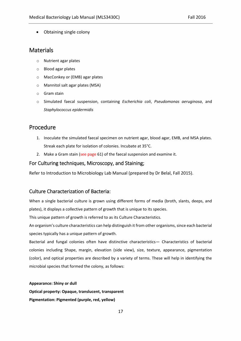
Culture Characterization of Bacteria:
When a single bacterial culture is grown using different forms of media (broth, slants, deeps, and
plates), it displays a collective pattern of growth that is unique to its species.
This unique pattern of growth is referred to as its Culture Characteristics.
An organism’s culture characteristics can help distinguish it from other organisms, since each bacterial
species typically has a unique pattern of growth.
Bacterial and fungal colonies often have distinctive characteristics— Characteristics of bacterial
colonies including Shape, margin, elevation (side view), size, texture, appearance, pigmentation
(color), and optical properties are described by a variety of terms. These will help in identifying the
microbial species that formed the colony, as follows:
Appearance: Shiny or dull
Optical property: Opaque, translucent, transparent
Pigmentation: Pigmented (purple, red, yellow)

Medical Bacteriology Lab Manual (MLS3430C) Fall 2016
18
Nonpigmented (cream, tan, white)
Texture: Rough or smooth
Figure 1 Culture Charesteristics
Medical Bacteriology Lab Manual (MLS3430C) Fall 2016
19
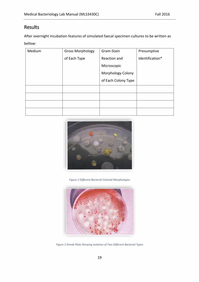
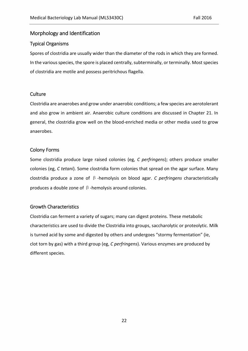
Results
After overnight incubation features of simulated faecal specimen cultures to be written as
bellow:
Medium Gross Morphology
of Each Type
Gram-Stain
Reaction and
Microscopic
Morphology Colony
of Each Colony Type
Presumptive
Identification*
Figure 2 Different Bacterial Colonial Morphologies
Figure 3 Streak Plate Showing Isolation of Two Different Bacterial Types

Medical Bacteriology Lab Manual (MLS3430C) Fall 2016
20
Figure 4 Bacterial growth characteristics in broth. From LEFT to RIGHT: uninoculated tube, precipitation reaction, turbidity,
flocculation, pellicle formation.
Review Questions
Why is MacConkey agar selective as well as differential?
Why is blood agar useful as a primary isolation medium?
Define a differential medium and discuss its purpose.

Medical Bacteriology Lab Manual (MLS3430C) Fall 2016
21
Week 4: Spore-Forming Gram-Positive Bacilli (Bacillus &
Clostridium Species)
BACILLUS SPECIES
The genus Bacillus includes large aerobic, gram-positive rods occurring in chains. Most
members of this genus are saprophytic organisms prevalent in soil, water, and air and on
vegetation, such as Bacillus cereus and Bacillus subtilis.
Morphology and identification
Typical Organisms
The typical cells, measuring 1 x 3-4 µm, have square ends and are arranged in long chains;
spores are located in the center of the nonmotile bacilli.
Culture
Colonies of B anthracis are round and have a “cut glass” appearance in transmitted light.
Hemolysis is uncommon with B anthracis but common with B cereus and the saprophytic
bacilli. Gelatin is liquefied, and growth in gelatin stabs resembles an inverted fir tree.
Growth Characteristics
The saprophytic bacilli use simple sources of nitrogen and carbon for energy and growth. The
spores are resistant to environmental changes, withstand dry heat and certain chemical
disinfectants for moderate periods, and persist for years in dry earth. Animal products
contaminated with anthrax spores (eg, hides, bristles, hair, wool, bone) can be sterilized by
autoclaving.
CLOSTRIDIUM SPECIES
The clostridia are large anaerobic, gram-positive, motile rods. Many decompose proteins or
form toxins, and some do both. Their natural habitat is the soil or the intestinal tract of
animals and humans, where they live as saprophytes. Among the pathogens are the
organisms causing botulism, tetanus, gas gangrene, and pseudomembranous colitis.
Medical Bacteriology Lab Manual (MLS3430C) Fall 2016
22
Morphology and Identification
Typical Organisms
Spores of clostridia are usually wider than the diameter of the rods in which they are formed.
In the various species, the spore is placed centrally, subterminally, or terminally. Most species
of clostridia are motile and possess peritrichous flagella.
Culture
Clostridia are anaerobes and grow under anaerobic conditions; a few species are aerotolerant
and also grow in ambient air. Anaerobic culture conditions are discussed in Chapter 21. In
general, the clostridia grow well on the blood-enriched media or other media used to grow
anaerobes.
Colony Forms
Some clostridia produce large raised colonies (eg, C perfringens); others produce smaller
colonies (eg, C tetani). Some clostridia form colonies that spread on the agar surface. Many
clostridia produce a zone of β-hemolysis on blood agar. C perfringens characteristically
produces a double zone of β-hemolysis around colonies.
Growth Characteristics
Clostridia can ferment a variety of sugars; many can digest proteins. These metabolic
characteristics are used to divide the Clostridia into groups, saccharolytic or proteolytic. Milk
is turned acid by some and digested by others and undergoes “stormy fermentation” (ie,
clot torn by gas) with a third group (eg, C perfringens). Various enzymes are produced by
different species.

Medical Bacteriology Lab Manual (MLS3430C) Fall 2016
23
Figure 5 Bacillus anthracis Colonies
Figure 6 Bacillus cereus Colonies
Figure 7 Endospores

Medical Bacteriology Lab Manual (MLS3430C) Fall 2016
24
Figure 8 Bacillus mycoides Colonies
Figure 9 Clostridium perfringens
Laboratory diagnostic techniques
Bacillus anthracis:
o Gram stain and culture of blood, respiratory secretions or lesions
o Serology
o PCR
Bacillus cereus:
Medical Bacteriology Lab Manual (MLS3430C) Fall 2016
25
o Clinical grounds
o Culture and Gram stain of implicated food
Clostridia:
Primarily a clinical diagnosis; organism is rarely isolated.
Materials
o Gram Stain
o Endospore Stain
o Starch, Blood & Nutrient Agar
Procedure
1. Inoculate your samples on blood and nutrient agar
2. Stain obtained single colonies with Gram (see page 61) and Endospore stains (see
page 62)
3. Do Starch Hydrolysis Test (see page 69)
Results
Record your result as follows:
Bacteria Sample Colony Characteristics on: Cell Characteristics with
Blood Agar Nutrient Agar Gram Stain Spore Staing
1
2
Review questions
What is the benefit of gram stain in this lab?
Why don’t you need to use MacConkey Agar Media for cultivation?
Why is iodine used to detect starch hydrolysis?
Did you see spores of Bacillus mycoides?
o If yes why?
o If not why?
Medical Bacteriology Lab Manual (MLS3430C) Fall 2016
26
Week 5: Non-Spore-Forming Gram-Positive Bacilli:
Corynebacterium, Propionibacterium Corynebacterium
diphtheria
CORYNEBACTERIUM DIPHTHERIAE
Morphology and Identification
Characteristically, they possess irregular swellings at one end that give them the “club-
shaped” appearance. Irregularly distributed within the rod (often near the poles) are
granules staining deeply with aniline dyes (metachromatic granules) that give the rod a
beaded appearance.
On blood agar, the C diphtheriae colonies are small, granular, and gray with irregular edges
and may have small zones of hemolysis. On agar containing potassium tellurite, the colonies
are brown to black with a brown-black halo because the tellurite is reduced ntracellularly. C
diphtheriae and other corynebacteria grow aerobically on most ordinary laboratory media.
On Loeffler serum medium, corynebacteria grow much more readily than other respiratory
organisms, and the morphology of organisms is typical in smears made from these colonies.
Gram-Positive Anaerobes / Gram-Positive Bacilli:
Propionibacterium
Propionibacterium species are members of the normal microbiota of the skin, oral cavity,
large intestine, conjunctiva, and external ear canal. Their metabolic products include
propionic acid, from which the genus name derives. On Gram stain, they are highly
pleomorphic, showing curved, clubbed, or pointed ends; long forms with beaded uneven
staining; and occasionally coccoid or spherical forms. Propionibacterium acnes, often
considered an opportunistic pathogen, causes the disease acne vulgaris and is associated with
a variety of inflammatory conditions.

Medical Bacteriology Lab Manual (MLS3430C) Fall 2016
27
Figure 10 Corynebacterium diphtheriae Stained Cells LEFT & Colonies RIGHT
Figure 12 P. acnes Stained Cells LEFT & Colonies RIGHT
Figure 11 Listeria monocytogenes Stained Cells LEFT & Colonies RIGHT

Medical Bacteriology Lab Manual (MLS3430C) Fall 2016
28
Laboratory diagnostic techniques
Corynebacterium diptheriae:
o Elek test to document toxin production (ELISA for toxin is now the frontline)
o Toxin produced by toxin-producing strains diffuses away from growth.
o Antitoxin diffuses away from the strip of filter paper.
o Precipitin lines form at zone of equivalence.
Listeria monocytogenes:
o Blood or CSF culture
o CSF wet mount [motility] or Gram stain
Materials
Gram Stain
Blood & Nutrient Agar
H2O2 for catalase test
Procedure
1. Inoculate swab samples on Nutrient & Blood Agar Plates and incubate them at 37oC for
24 Hours.
2. After overnight incubation, observe morphology of colonies and Gram (see page 61)
stained bacterial cells
3. Then do catalase test for the suspected bacilli forms (see page 91)
Results
Bacteria Sample Colony Characteristics on: Bacterial Cell Response with
Blood Agar Nutrient Agar Gram Stain Catalase H2O2
1
2

Medical Bacteriology Lab Manual (MLS3430C) Fall 2016
29
Review Questions
What is the substrate of the catalase reaction? Why are bubbles produced in a positive
catalase test?
Why will a false-positive catalase test result if the organisms are tested on a medium
containing blood?
Medical Bacteriology Lab Manual (MLS3430C) Fall 2016
30
Week 6: Staphylococci & Streptococci
Staphylococci
Morphology and identification
Typical Organisms
Staphylococci are spherical cells about 1 μm in diameter arranged in irregular clusters. Single
cocci, pairs, tetrads, and chains are also seen in liquid cultures. Young cocci stain strongly
gram positive; on aging, many cells become gram negative. Staphylococci are nonmotile and
do not form spores.
Culture
Staphylococci grow readily on most bacteriologic media under aerobic or microaerophilic
conditions. They grow most rapidly at 37°C but form pigment best at room temperature
(20–25°C). Colonies on solid media are round, smooth, raised, and glistening. S aureus
usually forms gray to deep golden yellow colonies. S epidermidis colonies usually are gray to
white on primary isolation; many colonies develop pigment only upon prolonged incubation.
No pigment is produced anaerobically or in broth. Various degrees of hemolysis are produced
by S aureus and occasionally by other species. The genus Staphylococcus contains two species,
S saccharolyticus and S aureus subsp. anaerobius, which initially grow only under anaerobic
conditions but become more aerotolerant on subcultures.
Growth Characteristics
The staphylococci produce catalase, which differentiates them from the streptococci.
Staphylococci slowly ferment many carbohydrates, producing lactic acid but not gas.
Proteolytic activity varies greatly from one strain to another. Staphylococci are relatively
resistant to drying, heat (they withstand 50°C for 30 minutes), and 9% sodium chloride. S
aureus produces coagulase, an enzyme-like protein that clots oxalated or citrated plasma.
Clumping factor is responsible for adherence of the organisms to fibrinogen and fibrin.

Medical Bacteriology Lab Manual (MLS3430C) Fall 2016
31
The Streptococci
The streptococci are gram-positive spherical bacteria that characteristically form pairs or
chains during growth. They are widely distributed in nature. Some are members of the normal
human microbiota; others are associated with important human diseases attributable to the
direct effects of infection by streptococci or in other cases to an immunologic response to
them.
STREPTOCOCCI OF PARTICULAR MEDICAL INTEREST:
Streptococcus pyogenes
Morphology and Identification
Typical Organisms
Individual cocci are spherical or ovoid and are arranged in chains. The cocci divide in a plane
perpendicular to the long axis of the chain. The members of the chain often have a striking
diplococcal appearance, and rodlike forms are occasionally seen. Streptococci are gram
positive; however, as a culture ages and the bacteria die, they lose their gram positivity and
can appear to be gram negative; for some streptococci, this can occur after overnight
incubation.
Culture
Most streptococci grow in solid media as discoid colonies, usually 1–2 mm in diameter. S
pyogenes is β-hemolytic; other species have variable hemolytic characteristics.
Growth Characteristics
Energy is obtained principally from the utilization of glucose with lactic acid as the end
product. Growth of streptococci tends to be poor on solid media or in broth unless enriched
with blood or tissue fluids. Nutritive requirements vary widely among different species. The
human pathogens are most exacting, requiring a variety of growth factors. Growth and
hemolysis are aided by incubation in 10% CO2. Most pathogenic hemolytic streptococci grow

Medical Bacteriology Lab Manual (MLS3430C) Fall 2016
32
best at 37°C. Most streptococci are facultative anaerobes and grow under aerobic and
anaerobic conditions.
Figure 13 Staphylococci Cell Morphology

Medical Bacteriology Lab Manual (MLS3430C) Fall 2016
33
Figure 14 Staphylococci Colonies in Nutreint & Blood Agar
Figure 15 Manitol Fermentation RIGHT & Non-fermenting Staphylococci

Medical Bacteriology Lab Manual (MLS3430C) Fall 2016
34
Figure 16 LEFT to RIGHT Alpha, Beta & Gamma Hemolysis
Figure 17 Stained Streptococci Cells
Classification:
The streptococci are a large and heterogeneous group of bacteria and no one system
suffices to classify them. Yet, understanding the classification is key to understanding
their medical importance.
The streptococci of greatest medical significance are S. pyogenes, S. agalactiae, and S.
pneumoniae. Of lesser importance are S. faecalis, S. faecium, and S. bovis.
Streptococci species are classified based on:
a. hemolytic capacity (α, β, γ hemolysis)
b. The antigenicity of a carbohydrate occurring in their cell walls (Lancefield antigen).
Hemolytic Capacity
Streptococci can be broadly classified according to the hemolytic reaction on blood agar:

Medical Bacteriology Lab Manual (MLS3430C) Fall 2016
35
α-hemolytic: Is the partial destruction of RBCs and produces a greenish discoloration of
the agar around the colonies. S. pneumoniae.
β-hemolytic: Colonies on blood agar are completely hemolyses the red cells around their
colonies. S. pyogenes and S. agalactiae
γ-hemolytic: is the absence of macroscopically visible hemolytic zones. Streptococcus
bovis
Lancefield Groups
Many streptococci and enterococci have a polymeric carbohydrate (C substance) in their
cell walls called the Lancefield antigen.
They are classified in Lancefield groups A-V based on variations in the antigenicity of this
antigen.
These antigenes were labeled Group A, Group B, Group C, and so on. Currently three
Lancefield Groups are of medical importance:
Group A, Group B, and Group D. Of the organisms used in this lab the following
correlations apply:
Group A Strep--Streptococcus pyogenes
Group B Strep--Streptococcus agalactiae
Group D Strep--Streptococcus bovis
Table 1 Classification of Streptococci

Medical Bacteriology Lab Manual (MLS3430C) Fall 2016
36
Figure 18 β, α and γ Hemolysis of Streptococci
The CAMP Test for Group B Streptococci
The CAMP test is used to differentiate Group B Streptococcus agalactiae (+) from other
Streptococcus species (-).
Group B streptococci can be distinguished from other beta-hemolytic streptococci by their
production of a substance called the CAMP factor. This term is an acronym for the names
of the investigators who first described the factor: Christie, Atkins, and Munch-Petersen.
The substance is a peptide that acts together with the beta-hemolysin produced by some
strains of Staphylococcus aureus, enhancing the effect of the latter on a sheep blood agar
plate. This effect is sometimes referred to as synergistic hemolysis
When streaked perpendicularly to an S. aureus subsp. aureus streak on blood, an arrow
head shaped zone of hemolysis forms and is a positive result
Figure 19 CAMP Test

Medical Bacteriology Lab Manual (MLS3430C) Fall 2016
37
CAMP Test Method
1. With an inoculating loop, streak a strain of S. aureus down the center of a blood agar
plate.
2. On one side of the plate, inoculate a strain of group B Streptococcus by making a streak
at a 90° angle, starting 5 mm away from the S. aureus and extending outward to the
edge of the agar.
3. On the other side of the plate, inoculate a strain of group A Streptococcus, again at a 90°
angle from the S. aureus, as in step This streak should not be directly opposite the group
B inoculum.
4. Incubate the plate aerobically at 35°C for 18 to 24 hours.
Interpretation of Results
Observe the area of hemolysis surrounding the S. aureus streak. At the point adjacent to the
streak of group B streptococci, you should see an arrowhead-shaped area of increased
hemolysis indicating production of the CAMP factor. There should be no change in the
hemolytic zone adjacent to the streak of group A streptococci, most strains of which do not
produce the CAMP factor.
Laboratory diagnostic techniques
Staphylococci:
o Gm + cocci in grapes, Catalase differentiates from Strep.
Staphylococcus aureus:
o Betahemolysis, Coagulase +ve, Yellow (Au) pigment
Staphylococcus epidermidis:
o Coagulase -ve
o Novobiocin sensitive
Staphylococcus saprophyticus:
o Coagulase -ve
o Novobiocin resistant

Medical Bacteriology Lab Manual (MLS3430C) Fall 2016
38
Streptococci:
Streptococcus pyogenes [Group A Streptococcus]:
o The rapid strep test (ELISA-based) misses approximately 25% of infections.
o Culture all negatives.
o Antibodies to streptolysin 0 (ASO) titer of > 200 is significant for rheumatic fever.
Streptococcus pneumoniae:
o Gram stain of CSF
o PCR of CSF
o Quellung reaction: positive (swelling of the capsule with the addition of type-specific
antiserum)
o Latex particle agglutination: test for capsular antigen in CSF
Enterococcus faecalis/faecium:
o Culture on blood agar
o Antibiotic sensitivities
Materials
Gram Stain
Blood & Manitol Salt Agar MSA
H2O2 for catalase test
Blood Plasma for coagulase test
Novobiocin discs
Procedure
1. Inoculation on Blood & MSA Plates
2. Gram Stain (see page 61)
3. Catalase & Coagulase Test (see page 91 & 94)
4. Novobiosin sensitivity test (see page 109)
Medical Bacteriology Lab Manual (MLS3430C) Fall 2016
39
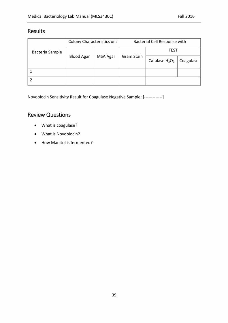
Results
Bacteria Sample
Colony Characteristics on: Bacterial Cell Response with
Blood Agar MSA Agar Gram Stain
TEST
Catalase H2O2 Coagulase
1
2
Novobiocin Sensitivity Result for Coagulase Negative Sample: [-------------]
Review Questions
What is coagulase?
What is Novobiocin?
How Manitol is fermented?

Medical Bacteriology Lab Manual (MLS3430C) Fall 2016
40
Week 7 & 8: Enteric Gram-Negative Rodes
(Enterobacteriaceae)
Morphology and Identification
Typical Organisms
The Enterobacteriaceae are short gram-negative rods. Typical morphology is seen in growth
on solid media in vitro, but morphology is highly variable in clinical specimens.
Culture
E coli and most of the other enteric bacteria form circular, convex, smooth colonies with
distinct edges. Enterobacter colonies are similar but somewhat more mucoid. Klebsiella
colonies are large and very mucoid and tend to coalesce with prolonged incubation. The
salmonellae and shigellae produce colonies similar to E coli but do not ferment lactose. Some
strains of E coli produce hemolysis on blood agar.
Growth Characteristics
Carbohydrate fermentation patterns and the activity of amino acid decarboxylases and other
enzymes are used in biochemical differentiation. Some tests, such as the production of indole
from tryptophan, are commonly used in rapid identification systems, but others, such as the
Voges-Proskauer reaction (production of acetylmethylcarbinol from dextrose), are used less
often. Culture on “differential” media that contain special dyes and carbohydrates (eg, eosin-
methylene blue [EMB], MacConkey, or deoxycholate medium) distinguishes lactose-
fermenting (colored) from non–lactose-fermenting colonies (nonpigmented) and may allow
rapid presumptive identification of enteric bacteria. Many complex media have been devised
to help in identification of the enteric bacteria. One such medium is triple sugar iron (TSI) agar,
which is often used to help differentiate salmonellae and shigellae from other enteric gram-
negative rods in stool cultures.
Escherichia
E coli typically produce positive test results for indole, lysine decarboxylase, and mannitol
fermentation and produces gas from glucose (TSI is A/A +Ve gas). An isolate from urine can

Medical Bacteriology Lab Manual (MLS3430C) Fall 2016
41
be quickly identified as E coli by its hemolysis on blood agar, typical colonial morphology with
an iridescent “sheen” on differential media such as EMB agar, and a positive spot indole test
result.
Shigella
Shigellae are nonmotile and usually do not ferment lactose but do ferment other
carbohydrates, producing acid but not gas (TSI is K/A, -Ve gas). They do not produce H2S. The
four Shigella species are closely related to E coli. Many share common antigens with one
another and with other enteric bacteria.
Salmonella
Salmonellae are motile rods that characteristically ferment glucose and mannose without
producing gas but do not ferment lactose or sucrose. Most salmonellae produce H2S. They
are often pathogenic for humans or animals when ingested.
Methyl red test positive
Voges-Proskauer test negative
Citrate positive (growth on Simmon's citrate agar)
Lysine decarboxylase positive
Urease negative
Indole production negative
Laboratory diagnostic techniques
Escherichia coli:
o Gram-negative rod
o Oxidase negative
o E. coli is a lactose fermenter: colonies with iridescent green sheen on EMB
Salmonella enterica Subsp. typhi
o Organisms can be isolated from blood, bone marrow, urine, and tissue biopsy from
the rose spots if present.

Medical Bacteriology Lab Manual (MLS3430C) Fall 2016
42
Salmonella enterica Subspecies Other Than typhi (S. enteritidis, S. typhimurium):
o Culture on Hektoen agar, H2S production
Shigella Species:
o Isolation from stool during illness and culture on selective media
Materials
Gram Stain
MacConkey & Kligler Iron Agar
Reagents for [Indole, Methyl Red, Voges-Proskauer, Citrate] tests
Procedure
1. Inoculate simulated fecal sample on MacConkey & Kligler Iron Agar (see page 86) then
Gram Stain (see page 61)
2. Do IMViC [Indole, Methyl Red, Voges-Proskauer, Citrate] test (see page 73, 75, 77 & 79)
Results
Comparison Between E. coli Salmonella
Media / Reaction Expected Result Expected Result
Indole + -
Methyl Red + +
Voges-Proskauer - -
Citrate Utilization - +
Colony Color on MacConkey Agar Pink Colorless
Ferment Lactose + -
Review questions
How could you differentiate between E. coli & Salmonella? State the principle behind the
fact.
Medical Bacteriology Lab Manual (MLS3430C) Fall 2016
43
Week 9: Vibrios, Campylobacters, Helicobacter & Associated
Bacteria
VIBRIO CHOLERAE
Morphology and Identification
Typical Organisms
Vibrios are among the most common bacteria in surface waters worldwide. They are curved
aerobic rods and are motile, possessing a polar flagellum
Culture
V cholerae produces convex, smooth, round colonies that are opaque and granular in
transmitted light. V cholera and most other vibrios grow well at 37°C on many kinds of media,
including defined media containing mineral salts and asparagine as sources of carbon and
nitrogen. V cholera grows well on thiosulfate-citrate-bile-sucrose (TCBS) agar, a media
selective for vibrios, on which it produces yellow colonies (sucrose fermented). Vibrios are
oxidase positive, which differentiates them from enteric gram-negative bacteria.
Characteristically, vibrios grow at a very high pH (8.5–9.5) and are rapidly killed by acid.
Cultures containing fermentable carbohydrates therefore quickly become sterile. V. cholerae
on Kligler’s iron agar (KIA), which contains glucose and lactose, is similar to those of
nonlactose-fermenting Enterobacteriaceae (K/A, no gas, no H2S)
CAMPYLOBACTER
Morphology and Identification
Typical Organisms
C jejuni and the other campylobacters are gram-negative rods with comma, S, or “gull wing”
shapes. They are motile, with a single polar flagellum, and do not form spores.
Growth Characteristics
C jejuni and the other campylobacters pathogenic for humans are positive for both oxidase
and catalase. Campylobacters do not oxidize or ferment carbohydrates. Gram-stained smears

Medical Bacteriology Lab Manual (MLS3430C) Fall 2016
44
show typical morphology. Nitrate reduction, hydrogen sulfide production, hippurate tests,
and antimicrobial susceptibilities can be used for further identification of species.
HELICOBACTER PYLORI
Morphology and Identification
Typical Organisms
H pylori has many characteristics in common with campylobacters. It has multiple flagella at
one pole and is actively motile.
Culture
H pylori grows in 3–6 days when incubated at 37°C in a microaerophilic environment, as for C
jejuni. The media for primary isolation include Skirrow’s medium with vancomycin, polymyxin
B, and trimethoprim, chocolate medium, and other selective media with antibiotics (eg,
vancomycin, nalidixic acid, amphotericin). The colonies are translucent and 1–2 mm in
diameter.
Growth Characteristics
H pylori is oxidase positive and catalase positive, has a characteristic morphology, is motile,
and is a strong producer of urease.
Laboratory diagnostic techniques
Vibrio cholera:
o Culture stool on TCBS
o Oxidase positive
Campylobacter jejuni:
o Culture on Campylobacter or Skirrow agar at 42°C
Helicobacter pylori:
o Biopsy with culture; histology with Giemsa or silver stain
o Breath test: 13C-urea swallowed; ammonia+13C-C02 exhaled
o Serology
Medical Bacteriology Lab Manual (MLS3430C) Fall 2016
45
Materials
MacConkey Agar
Kligler Iron Agar
Thiosulfate-citrate-bile-sucrose (TCBS) Agar
Gram Stain
Reagents for Indole, Methyl Red, Voges-Proskauer, Citrate, Catalase, Urease & Oxidase
tests
Procedure
1. Inoculate simulated fecal sample on MacConkey thiosulfate-citrate-bile-sucrose (TCBS)
& Kligler Iron Agar (see page 86) then Gram Stain (see page 61)
2. Do IMViC [Indole, Methyl Red, Voges-Proskauer, Citrate] test (see page 73, 75, 77 & 79)
3. Do Catalase, Oxidase & Urease test (see page 91, 97 & 99)
Results
Serological test results if applicable: [---------]
Result of Vibrio culture on TCBS: [Describe the Colonies]
Review questions
Differentiate between Vibrio cholera & Helicobacter pylori on the basis of morphology and
biochemical reactions.
Medical Bacteriology Lab Manual (MLS3430C) Fall 2016
46
Week 10: Pseudomonads & Anaerobic Bacteria
THE PSEUDOMONAD GROUP Pseudomonas aeruginosa
Morphology and Identification
Typical Organisms
P aeruginosa is motile and rod shaped. It is gram negative and occurs as single bacteria, in
pairs, and occasionally in short chains.
Culture
P aeruginosa is an obligate aerobe that grows readily on many types of culture media,
sometimes producing a sweet or grapelike or corn taco–like odor. Some strains hemolyze
blood. P aeruginosa forms smooth round colonies with a fluorescent greenish color. It often
produces the nonfluorescent bluish pigment pyocyanin, which diff uses into the agar. Other
Pseudomonas species do not produce pyocyanin.
Biochemical reactions
Catalase-positive
Oxidase-positive
Nitrate reduction-positive
Indole test-negative
Methyl red test-negative
Vp test-negative
Citrate test-positive
Urease test-negative
Laboratory diagnostic techniques
Pseudomonas aeruginosa:
o Gram stain and culture; it produces pyocyanin, pyoverdin

Medical Bacteriology Lab Manual (MLS3430C) Fall 2016
47
Materials
MacConkey Agar
Gram Stain
Reagents for Indole, Methyl Red, Voges-Proskauer, Citrate, Catalase, Urease & Oxidase
tests
Procedure
1. Inoculate samples on MacConkey Agar, then Gram Stain (see page 61)
2. Do IMViC [Indole, Methyl Red, Voges-Proskauer, Citrate] test (see page 73, 75, 77 & 79)
3. Do Catalase, Oxidase & Urease test (see page 91, 97 & 99)
Results
The characteristics of colonies will be described completely: [-------------------]
Review questions
What is the characteristic color of colonies of Pseudomonas aeruginosa? And what is the
source of this color?

Medical Bacteriology Lab Manual (MLS3430C) Fall 2016
48
Week 11: Haemophilus, Bordetelia & Legioneliae Haemophilus
genus
THE HAEMOPHILUS SPECIES
Morphology and Identification
This is a group of small, gram-negative, pleomorphic bacteria that require enriched media,
usually containing blood or its derivatives, for isolation. Haemophilus influenzae type b is an
important human pathogen.
Haemophilus influenza
Culture
On chocolate agar, flat, grayish brown colonies are present after 24 hours of incubation.
IsoVitaleX in media enhances growth. H influenzae does not grow on sheep blood agar except
around colonies of staphylococci (“satellite phenomenon”).
THE BORDETELLAE
Morphology and Identification
Typical Organisms
The organisms are minute gram-negative coccobacilli. With toluidine blue stain, bipolar
metachromatic granules can be demonstrated. A capsule is present.
Culture
Primary isolation of B pertussis requires enriched media. Bordet-Gengou medium (potato-
blood-glycerol agar).
Growth Characteristics
The organism is a strict aerobe and it is oxidase and catalase positive but nitrate, citrate, and
urea negative, the results of which are useful for differentiating among the other species of
bordetellae
Medical Bacteriology Lab Manual (MLS3430C) Fall 2016
49
LEGIONELLA PNEUMOPHILA
Morphology and Identification
Typical Organisms
Legionellae are fastidious, aerobic gram-negative bacteria. They often stain poorly by Gram’s
method and are not seen in stains of clinical specimens.
Culture
Legionellae can be grown on complex media such as buffered charcoal yeast extract agar with
α-ketoglutarate and iron (BCYE) at a pH of 6.9, temperature of 35°C, and 90% humidity.
Antibiotics can be added to make the medium selective for Legionella species. Legionellae
grow slowly; visible colonies are usually present after 3 days of incubation. Colonies that
appear after overnight incubation are not Legionella species. Colonies are round or fl at with
entire edges. They vary in color from colorless to iridescent pink or blue and are translucent
or speckled.
Growth Characteristics
The legionellae are catalase positive. L pneumophila is oxidase positive; the other legionellae
are variable in oxidase activity. L pneumophila hydrolyzes hippurate; the other legionellae do
not. Most legionellae produce gelatinase and β-lactamase; L micdadei produces neither
gelatinase nor β-lactamase.
Laboratory diagnostic techniques
Haemophilus influenza:
o Blood or CSF culture on chocolate agar
o PCR
o Antigen detection of capsule (latex particle agglutination)
Bordetella pertussis:
o Fastidious/delicate: Regan-Lowe or Bordet-Gengou media; either direct cough plates
or nasopharyngeal cultures
Medical Bacteriology Lab Manual (MLS3430C) Fall 2016
50
o Difficult to culture from the middle of paroxysmal stage on
o Direct immunotluorescence (DFA) on nasopharyngeal smear
o PCR and serologic tests available
Legionella pneumophila:
o Diagnosis: DFA (direct fluorescent antibody) on biopsy, (+) by Dieterle silver stain
o Antigen urine test for one serogroup only
o Fourfold increase in antibody
Materials
Blood, Chocolate & MacConkey Agar
Gram & capsule Stain
Reagents for Citrate, Gelatinase, Catalase, Urease & Oxidase tests
Procedure
1. Inoculate samples on Blood, Chocolate & MacConkey Agar, then Gram (see page 61) &
capsule Stain (see page 63)
2. Do Citrate, Gelatinase, Catalase, Oxidase & Urease test (see page 79, 91, 97, 99 & 101)
Results
Media / Stain /Reaction Bacterial Response
Blood Agar
Chocolate Agar
MacConkey Agar
Gram Stain
Capsule Stain
Citrate
Gelatinase
Catalase
Urease
Oxidase
Medical Bacteriology Lab Manual (MLS3430C) Fall 2016
51
Review questions
Can you culture Bordetella pertussis on Blood Agar?
o If yes, how?
o If no, why?

Medical Bacteriology Lab Manual (MLS3430C) Fall 2016
52
Week 12: Brucelia, Yersinia, Franciselia & Pasteurelia Brucella
genus
THE BRUCELLAE
Morphology and Identification
The brucellae are obligate parasites of animals and humans and are characteristically located
intracellularly. They are relatively inactive metabolically. Brucella melitensis typically infects
goats; Brucella suis, swine; Brucella abortus, cattle; and Brucella canis, dogs. Other species
are found only in animals.
Typical Organisms
The appearance in young cultures varies from cocci to rods 1.2 μm in length, with short
coccobacillary forms predominating. They are gram negative but often stain irregularly, and
they are aerobic, nonmotile, and nonspore forming.
Culture
Small, convex, smooth colonies appear on enriched media in 2–5 days.
Growth Characteristics
Fresh specimens from animal or human sources are usually inoculated on trypticase-soy agar
or blood culture media. Whereas B abortus requires 5–10% CO2 for growth, the other three
species grow in air. Brucellae use carbohydrates but produce neither acid nor gas in amounts
sufficient for classification. Catalase and oxidase are produced by the four species that infect
humans. Hydrogen sulfide is produced by many strains, and nitrates are reduced to nitrites.
FRANCISELLA TULARENSIS
Morphology and Identification
Typical Organisms
F tularensis is a small, gram-negative coccobacillus. It is rarely seen in smears of tissue.
Medical Bacteriology Lab Manual (MLS3430C) Fall 2016
53
Specimens
Blood is taken for serologic tests. The organism may be recovered in culture from lymph node
aspirates, bone marrow, peripheral blood, deep tissue, and ulcer biopsies.
Culture
Growth requires enriched media containing cysteine. In the past, glucose-cysteine blood agar
was preferred, but F tularensis grows on commercially available hemin containing media such
as chocolate agar, modified Thayer-Martin agar, and buffered charcoal yeast extract (BCYE)
agar used to grow Legionella species. Media should be incubated in CO2 at 35–37°C for 2–5
days.
Biochemical/Test Reactions .
Oxidase: Negative .
Catalase: Weak positive .
Urease: Negative
YERSINIA PESTIS
Morphology and Identification
Morphology and Identification
Y pestis is a gram-negative rod that exhibits striking bipolar staining with special stains such
as Wright, Giemsa, Wayson, or methylene blue. It is nonmotile. It grows as a facultative
anaerobe on many bacteriologic media. Growth is more rapid in media containing blood or
tissue fluids and fastest at 30°C. In cultures on blood agar at 37°C, colonies may be very small
at 24 hours. A virulent inoculum, derived from infected tissue, produces gray and viscous
colonies, but after passage in the laboratory, the colonies become irregular and rough. The
organism has little biochemical activity, and this is somewhat variable.
Y pestis produces nonlactose-fermenting colonies on MacConkey agar, and it grows better at
25°C than at 37°C. The organism is catalase positive; indole, oxidase, urease negative; and
nonmotile. The last two reactions are useful in differentiating Y pestis from other pathogenic
yersiniae.

Medical Bacteriology Lab Manual (MLS3430C) Fall 2016
54
PASTEURELLA
Morphology and Identification
Pasteurella species are primarily animal pathogens, but they can produce a range of human
diseases. Pasteurellae are nonmotile gram-negative coccobacilli with a bipolar appearance on
stained smears. They are aerobes or facultative anaerobes that grow readily on ordinary
bacteriologic media at 37°C. They are all oxidase positive and catalase positive but diverge in
other biochemical reactions.
Laboratory diagnostic techniques
Brucella:
o Culture is hazardous.
o Serum agglutination test, fourfold increase in titer
o Antibodies against Brucella > 1 : 160 considered positive
Francisella tularensis:
o Serodiagnosis; culture is hazardous.
o DFA
Yersinia pestis:
o Clinical specimens and cultures are hazardous.
o Serodiagnosis or direct immunotluorescence
o "Safety pin'' staining
Pasteurella:
o Rarely cultured because routine prophylaxis is common
Materials
BloodAgar
Chocolate Agar
MacConkey Agar
Gram Stain
Reagents for Indole, Methyl Red, Voges-Proskauer, Citrate Catalase, Urease & Oxidase
tests

Medical Bacteriology Lab Manual (MLS3430C) Fall 2016
55
Procedure
1. Inoculate samples on Blood, Chocolate & MacConkey Agar, then Gram Stain (see page
61)
2. Do IMViC [Indole, Methyl Red, Voges-Proskauer, Citrate] test (see page 73, 75, 77 & 79)
3. Do Catalase, Oxidase & Urease test (see page 91, 97 & 99)
Results
Serological test results if applicable: [---------]
Review questions
Why cultures of Brucella, Francisella tularensis & Yersinia pestis are considered hazardous?
Medical Bacteriology Lab Manual (MLS3430C) Fall 2016
56
Week 13: The Neisseriae & Unusual Bacterial Pathogens
Neisseriae genus & Mycobacteria
The Neisseriae
Morphology and Identification
Typical Organisms
The typical Neisseria is a gram-negative, nonmotile diplococcus, approximately 0.8 μm in
diameter. Individual cocci are kidney shaped; when the organisms occur in pairs, the flat or
concave sides are adjacent.
Culture
In 48 hours on enriched media (eg, modified Thayer-Martin, Martin-Lewis, GC-Lect, and New
York City), gonococci and meningococci form convex, glistening, elevated, mucoid colonies 1–
5 mm in diameter. Colonies are transparent or opaque, nonpigmented, and nonhemolytic.
Neisseria flavescens, Neisseria cinerea, Neisseria subflava, and Neisseria lactamica may have
yellow pigmentation. Neisseria sicca produces opaque, brittle, wrinkled colonies. Moraxella
catarrhalis produces nonpigmented or pinkish gray opaque colonies.
Growth Characteristics
The neisseriae grow best under aerobic conditions, but some grow in an anaerobic
environment. They have complex growth requirements. Most neisseriae oxidize
carbohydrates, producing acid but not gas, and their carbohydrate patterns are a means of
distinguishing them. The neisseriae produce oxidase and give positive oxidase reactions; the
oxidase test is a key test for identifying them. When bacteria are spotted on a filter paper
soaked with tetramethylparaphenylenediamine hydrochloride (oxidase), the neisseriae
rapidly turn dark purple.

Medical Bacteriology Lab Manual (MLS3430C) Fall 2016
57
Mycobacteria
Morphology and Identification
The mycobacteria are rod-shaped, aerobic bacteria that do not form spores. Although they
do not stain readily, after being stained, they resist decolorization by acid or alcohol and are
therefore called “acid-fast” bacilli.
Typical Organisms
In tissue, tubercle bacilli are thin, straight rods measuring about 0.4 × 3 μm. On artificial media,
coccoid and fi lamentous forms are seen with variable morphology from one species to
another. The Ziehl-Neelsen technique of staining is used for identification of acid-fast bacteria.
Culture
Semisynthetic Agar Media
These media (eg, Middlebrook 7H10 and 7H11) contain defined salts, vitamins, cofactors,
oleic acid, albumin, catalase, and glycerol; the 7H11 medium also contains casein hydrolysate.
The albumin neutralizes the toxic and inhibitory effects of fatty acids in the specimen or
medium.
Inspissated Egg Media
These media (eg, Löwenstein- Jensen) contain defined salts, glycerol, and complex organic
substances (eg, fresh eggs or egg yolks, potato flour, and other ingredients in various
combinations). Malachite green is included to inhibit other bacteria. Small inocula in
specimens from patients will grow on these media in 3–6 weeks.
Broth media
Broth media (eg, Middlebrook 7H9 and 7H12) support the proliferation of small inocula.
Ordinarily, mycobacteria grow in clumps or masses because of the hydrophobic character of
the cell surface. If tweens (water-soluble esters of fatty acids) are added, they wet the surface
and thus permit dispersed growth in liquid media. Growth is often more rapid than on
complex media.

Medical Bacteriology Lab Manual (MLS3430C) Fall 2016
58
Growth Characteristics
Mycobacteria are obligate aerobes and derive energy from the oxidation of many simple
carbon compounds. Increased CO2 tension enhances growth. Biochemical activities are not
characteristic, and the growth rate is much slower than that of most bacteria.
Laboratory diagnostic techniques
Neisseria meningitides:
o Ferments maltose
o Presumptive diagnosis by Gram stain of petechiae or CSF
o LATEX agglutination test b/c capsular polysaccharides
o PCR
Neisseria gonorrhoeae:
o Intracellular gram-negative diplococci in PMNs from urethral smear from
symptomatic male are suggestive of N. gonorrhoeae.
o Commonly: diagnosis by genetic probes with amplification
o Culture (when done) on Thayer-Martin medium
o Oxidase-positive colonies
o Maltose not fermented
o No capsule
Mycobacterium tuberculosis:
o Microscopy of sputum: screen with auramine-rhodamine stain (fluorescent apple-
green); no antibody involved; very sensitive; if positive, confirm with acid fast stain
o PPD skin test (Mantoux): measure zone of induration at 48-72 hours; positive if:
5 mm in HIV+ or anyone with recent TB exposure; AIDS patients have reduced
ability to mount skin test.
10 mm in high-risk population: IV drug abusers, people living in poverty, or
immigrants from high TB area
15 mm in low-risk population
o Positive skin test indicates only exposure but not necessarily active disease.
o Quantiferon-TB Gold Test: measures interferon-gamma production when leukocytes
exposed to TB antigens
Medical Bacteriology Lab Manual (MLS3430C) Fall 2016
59
o Slow-growing (3-6 weeks) colonies on Lowenstein-Jensen medium (faster new systems)
o Organisms produce niacin and are catalase-negative (68°C).
o No serodiagnosis
Materials
Blood Agar
Chocolate Agar
MacConkey Agar
Gram Stain
Acid-Fast Stain
Reagents for Oxidase test
Procedure
1. Inoculate samples on Blood, Chocolate & MacConkey Agar, then Gram (see page 61) &
Acid-Fast Stain (see page 64)
2. Do Oxidase test (see page 97)
Results
Record complete colony morphology of Neisseria on different Agar Plates
Record Oxidase result for your diplococci sample
Review questions
State reasons behind inability to culture Mycobacterium tuberculosis in routine work.

Medical Bacteriology Lab Manual (MLS3430C) Fall 2016
60
Week 14: The Analytical Profile Index (API)
API is a method commonly used to identify a wide range of microorganisms. APIs consist of a
number of plastic strips, each of which has about 20 miniature compartments containing
biochemical reagents. Almost all cultivatable bacterial groups and more than 550 different
species can be identified using the results of these API tests. These identification systems have
extensive databases of microbial biochemical reactions. The numerical clusters derived from
these tests identify different strains at selected levels of overall similarity (usually >80% at the
species level) on the basis of the frequency with which they share traits. (see page 106)
Medical Bacteriology Lab Manual (MLS3430C) Fall 2016
61
A / Staining Techniques
Staining Technique 1. Gram’s Stain Procedure:
1. A heat-fixed smear is flooded with a basic purple dye, usually crystal violet. Because the
purple stain imparts its color to all cells, it is referred to as a primary stain.
2. After 1 minute, the crystal violet is drained off and washed with distilled water. The smear
is then covered with Gram's iodine, a mordant or helper. When the iodine is washed off,
both gram-positive and gram-negative bacteria appear dark violet or purple.
3. Next, the slide is washed with alcohol (95% ethanol) or an alcohol-acetone solution. This
solution is a decolorizing agent. When the procedure is carried out, the slide is held at an
angle and 95% ethanol is poured until the draining solution no longer has a purple tint.
4. The alcohol is now rinsed off with distilled water and the slide is then stained with safranin,
a basic red dye known as the counter stain. The staining process is allowed to be carried
out for 2-3 minutes. The smear is washed again, heat dried and examined microscopically.

Medical Bacteriology Lab Manual (MLS3430C) Fall 2016
62
Staining Technique 2. Endospore Stain Procedure
1. Aseptically transfer one species of bacterium with an inoculating loop to each of the
respective slides, air dry (or use a slide warmer), and heat-fix.
2. Place the slide to be stained on a hot plate or boiling water bath equipped with a staining
loop or rack. Cover the smear with paper toweling that has been cut the same size as the
microscope slide.
3. Soak the paper with the malachite green staining solution. Gently heat on the hot plate
(just until the stain steams) for 5 to 6 minutes after the malachite green solution begins
to steam. Replace the malachite green solution as it evaporates so that the paper remains
saturated during heating.
Do not allow the slide to become dry.
4. Remove the paper using forceps, allow the slide to cool, and rinse the slide with water for
30 seconds.
5. Counterstain with safranin for 60 to 90 seconds.
6. Rinse the slide with water for 30 seconds.
7. Blot dry with bibulous paper and examine under oil immersion. A coverslip is not
necessary. The spores, both endospores and free spores, stain green; vegetative cells stain
red.
Medical Bacteriology Lab Manual (MLS3430C) Fall 2016
63
Staining Technique 3. Capsule Stain Procedure:
1. With a wax pencil, label the left hand corner of a clean glass slide with the name of the
bacterium that will be stained.
2. Aseptically transfer a loopful of culture with an inoculating loop to the slide. Allow the slide
to air dry.
[Do not heat-fix!] Heat-fixing can cause the bacterial cells to shrink and give a false
appearance to the capsule.
3.Place the slide on a staining rack. Flood the slide with crystal violet and let stand for 4 to 7
minutes.
4. Rinse the slide thoroughly with 20% copper sulfate.
5. Blot dry with bibulous paper.
6. Examine under oil immersion (a cover slip is not necessary). Capsules appear as faint halos
around dark cells.

Medical Bacteriology Lab Manual (MLS3430C) Fall 2016
64
Staining Technique 4. Acid Fast Stain Procedure:
1. Prepare a smear consisting of a mixture of E. coli and M. phlei.
2. Allow the smear to air dry and then heat-fix.
3. Place the slide on a hot plate that is within a chemical hood (with the exhaust fan on), and
cover the smear with a piece of paper towelling that has been cut to the same size as the
microscope slide. Saturate the paper with Ziehl’s carbolfuchsin. Heat for 3 to 5 minutes.
Do not allow the slide to dry out, and avoid excess flooding! Also, prevent boiling
by adjusting the hot plate to a proper temperature.
A boiling water bath with a staining rack or loop held 1 to 2 inches above the water surface
also works well. (Instead of using a hot plate to heatdrive the carbolfuchsin into the bacteria,
an alternate procedure is to cover the heat-fixed slide with a piece of paper towel. Soak the
towel with the carbolfuchsin and heat, well above a Bunsen burner flame).
4. Remove the slide, let it cool, and rinse with water for 30 seconds.
5. Decolorize by adding acid-alcohol drop by drop until the slide remains only slightly pink.
This requires 10 to 30 seconds and must be done carefully.
6. Rinse with water for 5 seconds.
7. Counterstain with alkaline methylene blue for about 2 minutes.
8. Rinse with water for 30 seconds.
9. Blot dry with bibulous paper.
10. There is no need to place a coverslip on the stained smear. Examine the slide under oil
immersion and record your results in the report for this exercise. Acid-fast organisms stain
red; the background and other organisms stain blue or brown.
11. Examine the prepared slide of Mycobacterium phlei.
Medical Bacteriology Lab Manual (MLS3430C) Fall 2016
65
B / Metabolic Activities of Bacteria
Background
Microbial metabolic processes are complex, but they permit the microbiologist to distinguish
among microorganisms grown in culture. Bacteria, especially, are identified by inoculating
pure, isolated colonies into media that contain one or more specific biochemical. The
biochemical reactions that take place in the culture can then be determined by relatively
simple indicator reagents, included in the medium or added to the culture later.
Some bacteria ferment simple carbohydrates, producing acidic, alcoholic, or gaseous end
products. Many different species are distinguished on the basis of the carbohydrates they do
or do not utilise, as well as by the nature of end products formed during fermentation. Still
others break down more complex carbohydrates, such as starch. (see page 106)
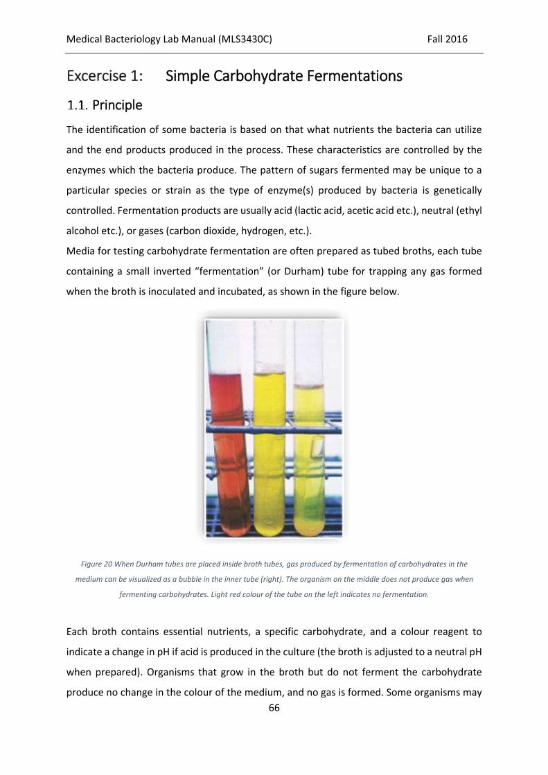
Medical Bacteriology Lab Manual (MLS3430C) Fall 2016
66
Simple Carbohydrate Fermentations
Principle
The identification of some bacteria is based on that what nutrients the bacteria can utilize
and the end products produced in the process. These characteristics are controlled by the
enzymes which the bacteria produce. The pattern of sugars fermented may be unique to a
particular species or strain as the type of enzyme(s) produced by bacteria is genetically
controlled. Fermentation products are usually acid (lactic acid, acetic acid etc.), neutral (ethyl
alcohol etc.), or gases (carbon dioxide, hydrogen, etc.).
Media for testing carbohydrate fermentation are often prepared as tubed broths, each tube
containing a small inverted “fermentation” (or Durham) tube for trapping any gas formed
when the broth is inoculated and incubated, as shown in the figure below.
Figure 20 When Durham tubes are placed inside broth tubes, gas produced by fermentation of carbohydrates in the
medium can be visualized as a bubble in the inner tube (right). The organism on the middle does not produce gas when
fermenting carbohydrates. Light red colour of the tube on the left indicates no fermentation.
Each broth contains essential nutrients, a specific carbohydrate, and a colour reagent to
indicate a change in pH if acid is produced in the culture (the broth is adjusted to a neutral pH
when prepared). Organisms that grow in the broth but do not ferment the carbohydrate
produce no change in the colour of the medium, and no gas is formed. Some organisms may

Medical Bacteriology Lab Manual (MLS3430C) Fall 2016
67
produce acid products in fermenting the sugar, but no gas, whereas others may form both
acid and gas. In some cases, organisms that do not ferment the carbohydrate use the protein
nutrients in the broth, thereby producing alkaline end products, a result that is also evidenced
by a change in indicator colour.
Objective
To distinguish bacterial species on the basis of simple carbohydrate fermentation
Materials
Tubed phenol red glucose broth with Durham tubes
Tubed phenol red lactose broth with Durham tubes
Tubed phenol red sucrose broth with Durham tubes
Slant cultures of Escherichia coli, Serratia marcescens, Pseudomonas aeruginosa, and
Proteus vulgaris
Procedure
1. Inoculate growth from each of the four cultures into separate tubes of each of the three
carbohydrate broths. Be certain no bubbles are inside the Durham tubes before
inoculation.
2. Label each of the 12 inoculated tubes with the name of the carbohydrate it contains and
the name of the bacterial culture.
3. Incubate at 35°C for 24 hours.
Results
Record your results in the following table. Use these symbols to indicate specific changes
observed in the broths.
A = acid production
K = alkaline colour change
N = neutral (no change in colour)
G = gas formation

Medical Bacteriology Lab Manual (MLS3430C) Fall 2016
68
Name of Organism Glucose Lactose Sucrose
Review Questions
What is the color of phenol red at an acid pH?
What is the function of a Durham tube?

Medical Bacteriology Lab Manual (MLS3430C) Fall 2016
69
Starch Hydrolysis
Principle
Some microorganisms split apart (hydrolyse) large organic molecules and then use the
component parts in further metabolic processes. Starch is a polysaccharide that is hydrolysed
by some bacteria. When iodine is added to the intact starch molecule a blue-coloured
complex forms. If starch is hydrolysed by bacterial enzymes, however, it is broken down to
simple sugars (glucose and maltose) that do not complex with iodine, and no colour reaction
is seen. The medium for this test is a nutrient agar containing starch, prepared in a petri plate.
The organism to be tested is streaked on the plate. When the culture has grown, the plate is
flooded with Gram’s iodine solution. The medium turns blue in all areas where the starch
remains intact. The areas of medium surrounding organisms that have hydrolysed the starch
remain clear and colourless.
Objective
To distinguish bacterial species on the basis of starch hydrolysis
Materials
Starch agar plates
Slant cultures of Escherichia coli, Pseudomonas aeruginosa, and Bacillus subtilis
Gram’s iodine solution
Procedure
1. Take one starch plate, invert it, and with your marking pencil mark three triangular
compartments on the back of the dish.
2. Inoculate one section of the agar with E. coli, using back-and-forth streaking; another
section with B. subtilis; and the third with P. aeruginosa.
3. Label each section of the plate on the back of the dish with the name of the organism
streaked in that area.
4. Incubate 24 to 48 hours at 35°C.
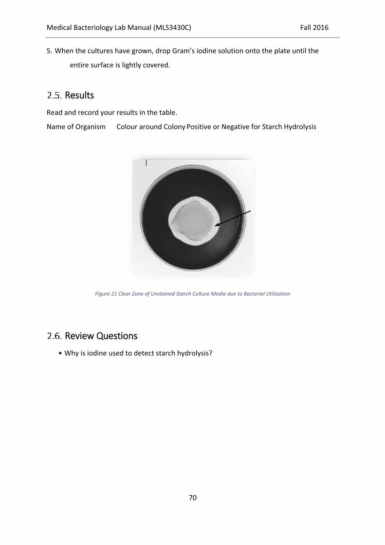
Medical Bacteriology Lab Manual (MLS3430C) Fall 2016
70
5. When the cultures have grown, drop Gram’s iodine solution onto the plate until the
entire surface is lightly covered.
Results
Read and record your results in the table.
Name of Organism Colour around Colony Positive or Negative for Starch Hydrolysis
Figure 21 Clear Zone of Unstained Starch Culture Media due to Bacterial Utilization
Review Questions
• Why is iodine used to detect starch hydrolysis?

Medical Bacteriology Lab Manual (MLS3430C) Fall 2016
71
Oxidation-Fermentation (OF) test
Principle
Fermentative organisms utilize the carbohydrate in both the open and sealed tubes as shown
by a change in colour of the medium from green to yellow. Oxidative organisms are able to
utilize the carbohydrate only in the open tube.
Objective
The test is used to differentiate those organisms that oxidize carbohydrate (aerobic utilization)
from those organisms that ferment carbohydrate (anaerobic utilization).
Materials
Slant culture of Pseudomonas aeruginosa
Slant culture of Escherichia coli
Tryptone or peptone agar medium containing carbohydrate (glucose or other
carbohydrate) and the indicator is bromothymol blue.
Procedure
1. Inoculate the test organism to the bottom of two tubes of sterile O-F medium (always use
a heavy inoculum).
2. Cover the inoculated medium of one tube with a 10 mm deep layer of sterile paraffin oil or
molten wax.
3. Incubate the tubes at 35-37°C for 14 days.
4. Examine daily for carbohydrate utilization.
Results
Open tube Sealed
tube
Interpretation Example

Medical Bacteriology Lab Manual (MLS3430C) Fall 2016
72
Yellow Green Oxidative organism
Yellow Yellow Fermentative organism
Green/Blue Green No utilization of
carbohydrates
Review Questions
• Name the indicator used in this experiment.
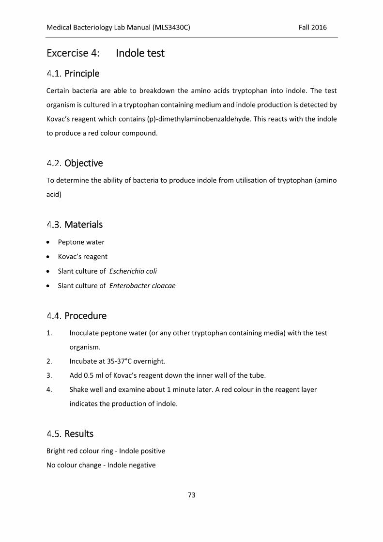
Medical Bacteriology Lab Manual (MLS3430C) Fall 2016
73
Indole test
Principle
Certain bacteria are able to breakdown the amino acids tryptophan into indole. The test
organism is cultured in a tryptophan containing medium and indole production is detected by
Kovac’s reagent which contains (p)-dimethylaminobenzaldehyde. This reacts with the indole
to produce a red colour compound.
Objective
To determine the ability of bacteria to produce indole from utilisation of tryptophan (amino
acid)
Materials
Peptone water
Kovac’s reagent
Slant culture of Escherichia coli
Slant culture of Enterobacter cloacae
Procedure
1. Inoculate peptone water (or any other tryptophan containing media) with the test
organism.
2. Incubate at 35-37°C overnight.
3. Add 0.5 ml of Kovac’s reagent down the inner wall of the tube.
4. Shake well and examine about 1 minute later. A red colour in the reagent layer
indicates the production of indole.
Results
Bright red colour ring - Indole positive
No colour change - Indole negative
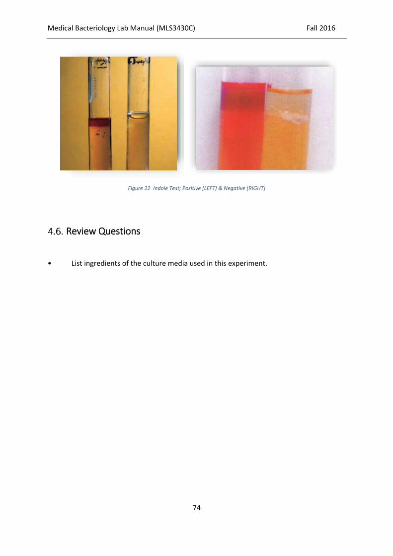
Medical Bacteriology Lab Manual (MLS3430C) Fall 2016
74
Figure 22 Indole Test; Positive [LEFT] & Negative [RIGHT]
Review Questions
• List ingredients of the culture media used in this experiment.

Medical Bacteriology Lab Manual (MLS3430C) Fall 2016
75
Methyl Red (MR) test
Principle
The test detects the ability of some organisms to produce sufficient acidity from the glucose
fermentation, after prolonged incubation, overcoming the pH buffering system of the
medium.
Objective
Determine whether tested organism is able to produce as much acid as buffering system
disturbing amount can do
Materials
Glucose phosphate broth
Methyl Red solution
Slant culture of Escherichia coli
Slant culture of Enterobacter cloacae
Procedure
1. Inoculate a colony of the test organism in 2 ml of sterile glucose phosphate broth.
2. Incubate at 35-37°C overnight.
3. Add 5 drops of Methyl Red solution.
4. Mix and read immediately.
5. Appearance of a red colour at the surface indicates a positive reaction.
Results
Bright red colour - Methyl Red positive
Orange / Yellow colour - Methyl Red negative
Medical Bacteriology Lab Manual (MLS3430C) Fall 2016
76
Figure 23 Methyl Red Test; Positive [LEFT] & Negative [RIGHT]
Review Questions
• Why did you shake the MR culture?

Medical Bacteriology Lab Manual (MLS3430C) Fall 2016
77
Voges-Proskauer (VP) test
Principle
Some organisms produce acetoin (acetylmethylcarbinol), a neutral-reacting end product, as
the main end product of the glucose fermentation. Acetoin is oxidized into diacetyl in the
presence of atmospheric oxygen and alkaline condition which forms a pink compound with
the creatine.
Objective
To identify organisms that mainly produce Acetoin from the fermentation of Glucose
Materials
Glucose phosphate peptone water
α-naphthol solution
40% KOH aqueous solution
Slant culture of Klebsiella pneumoniae
Slant culture of Escherichia coli
Procedure
1. Inoculate the test organism in to 2 ml of sterile glucose phosphate peptone water.
2. Incubate at 35-37°C for 48 hours.
3. Add 0.6 ml of α-naphthol solution followed by 0.2 ml of 40% KOH aqueous solution.
4. Shake well for maximum aeration.
5. Remove the bottle cap and leave for 1 hour at room temperature.
6. A pink – red colour indicates a positive reaction.
Results
Pink-red colour -VP positive
No pink-red colour -VP negative

Medical Bacteriology Lab Manual (MLS3430C) Fall 2016
78
Figure 24 Voges Proskauer Test
Review Questions
• Why did you shake the VP culture?

Medical Bacteriology Lab Manual (MLS3430C) Fall 2016
79
Citrate test
Principle
The test detects the ability of an organism to utilize citrate as the sole carbon and energy
source and ammonia as sole source of the nitrogen.
Objective
To determine whether the tested organism can utilize citrate as sole carbon source or not
Materials
Koser’s citrate medium
Or Simmons’ citrate medium
Slant culture of Klebsiella pneumoniae sub species aerogenes
Slant culture of Escherichia coli
Procedure
1. Make a light suspension of the organism in sterile water or saline.
2. Inoculate Koser’s or Simmons’ citrate medium using a sterile straight wire.
3. Incubate at 35-37°C for up to 7 days, checking daily for growth.
4. In Koser’s medium look for the turbidity; in Simmons’ medium look for a colour change.
Results
Koser’s Citrate
*Medium becomes turbid – Citrate positive

Medical Bacteriology Lab Manual (MLS3430C) Fall 2016
80
*No turbidity - Citrate negative
Simmon citrate
*Blue colour and growth on the streak line - Citrate positive
*No colour change and no growth - Citrate negative
Figure 25 Positive Citrate Utilization Test [RIGHT] & Negative [LEFT]
Review Questions
• What is the culture media used for Citrate test & what is the significant ingredient?

Medical Bacteriology Lab Manual (MLS3430C) Fall 2016
81
Figure 26 IMViC [Indole, Methyl Red, Voges-Proskauer, Citrate] set of reactions used to differentiate between E. coli with
reaction pattern of [+, +, -, -] & Enterobacter aerogenes with [-, -, +, +]

Medical Bacteriology Lab Manual (MLS3430C) Fall 2016
82
Production of Indole and Hydrogen Sulphide,
and Motility
Principle
Indole is a by-product of the metabolic breakdown of the amino acid tryptophan used by
some microorganisms. The presence of indole in a culture grown in a medium containing
tryptophan can be readily demonstrated by adding Kovac’s reagent to the culture. If indole is
present, it combines with the reagent to produce a brilliant red colour. If it is not present,
there will be no colour except that of the reagent itself. This test is of great value in the battery
used to identify enteric bacteria.
Hydrogen sulfide is produced when amino acids containing sulfur are metabolized by
microorganisms. If the medium contains metallic ions, such as lead, bismuth, or iron (in
addition to an appropriate amino acid), the hydrogen sulfide formed during growth combines
with the metallic ions to form a metal sulfide that blackens the medium. The most convenient
medium for testing for indole and/or hydrogen sulfide production is SIM medium (SIM is an
acronym for sulfide, indole, and motility). This is a tubed semisolid agar that can also be used
to demonstrate bacterial motility. It is inoculated by stabbing the wire loop (or preferably a
straight wire inoculating needle) straight down the middle of the agar to about one-fourth
the depth of the medium and withdrawing the wire along the same path.

Medical Bacteriology Lab Manual (MLS3430C) Fall 2016
83
Figure 27 Hydrogen Sulfide Production and/or Motility in SIM Media
Objective
To observe how a single medium can be used to test for three distinguishing features of
bacterial growth

Medical Bacteriology Lab Manual (MLS3430C) Fall 2016
84
Materials
Tubes of SIM medium
Xylene
Kovac’s reagent
5-ml pipettes
Bulb or other pipette aspiration device
Slant cultures of Escherichia coli, Proteus vulgaris, and Klebsiella pneumoniae
Broth cultures of Escherichia coli, Proteus vulgaris, and Klebsiella pneumoniae
Procedure
1. Inoculate growth from each of the three slant cultures into separate tubes of SIM
medium. Stab the inoculating wire straight down through the agar for a distance of
about one-fourth of its depth. Quickly withdraw the wire along the same path (do not
move it around in the agar).
2. Incubate the tubes at 35°C for 24 hours.
3. Examine the tubes for evidence of hydrogen sulphide production (browning or
blackening of the medium). Record results.
4. Examine the tubes for evidence of motility of the organism. A motile species grows away
from the line of stab into the surrounding agar. Lines of growth, or even general
turbidity, can be seen throughout the tube. The growth of a nonmotile organism is
restricted to the path of the stab. Record your observations.
5. Set up a hanging-drop or wet-mount preparation of each broth culture to confirm
results observed in SIM medium for motility.
6. Perform the Kovac test for indole.
a. Using a pipette bulb or other aspiration device, pipette 0.5 ml of xylene into the SIM
tube (it will layer over the top surface of the agar).
b. Pipette 0.5 ml of Kovac’s reagent in the same way as you did the xylene and add it to
the SIM tube.
c. Observe the colour of the xylene layer, and record.

Medical Bacteriology Lab Manual (MLS3430C) Fall 2016
85
Results
Record your observations and results in the table.
Name of Organism Sulphide Indole Motility
SIM
Medium
Hanging
Drop
Figure 28 Reactions in Sulfide Indole Motility medium [SIM] RIGHT negative for all, MIDDLE is positive for all whilst LEFT is
positive for Motility and Indole, but negative for Sulfide
Review Questions
Name one indole-positive organism.
How is indole produced in SIM medium? How is it detected?
How is hydrogen sulphide demonstrated in this medium?
Name two methods for determining bacterial motility.
Why is it essential to have pure cultures for biochemical tests?
Could a pH-sensitive colour indicator be used to reveal the presence of a contaminant
in a fluid that should be sterile? Explain.

Medical Bacteriology Lab Manual (MLS3430C) Fall 2016
86
Kligler Iron Agar (KIA)
Principle
There is an indicator (phenol red) which will change the colour of medium when acid is
produced. Ratio of lactose to glucose is 10:1 in the medium [TSI Triple Sugar Iron agar is similar
but sucrose is also added]. An alkalinity is produced in the slant due to the oxidative
deamination of the amino acids. The organism that has the ability to ferment glucose changes
the colour of the butt to yellow. But the acidity produced by glucose is not sufficient to
overcome the alkalinity produced by oxidative deamination of the amino acids in the slant
and change the colour of the slant. The organism that has the ability to ferment lactose
changes the colour of the butt as well as the slant.
Objective
KIA is used to look for the ability of an organism to ferment lactose and glucose and to produce
hydrogen sulphide. It is further used to check the production of gas while fermentation of
glucose and lactose.
Materials
KIA agar slants
Slant Culdtures of:
o Alcaligenes faecalis
o Escherichia coli
o Proteus vulgaris
o Pseudomonas aeruginosa
o Shigella flexneri
Procedure
1. Label each of the KIA agar slants with the name of the bacterium to be inoculated.
Use one of the tubes as a control. Place your name and date on each tube.
2. Using aseptic technique, streak the slant with the appropriate bacterium and then

Medical Bacteriology Lab Manual (MLS3430C) Fall 2016
87
3. stab the butt. Screw the caps on the tubes but do not tighten!
4. Incubate for only 18 to 24 hours at 35°C for changes in the butt and on the slant.
Tubes should be incubated and checked daily for up to seven days in order to
observe blackening.
5. Then
6. Examine all slant cultures for the colour of the slant and butt, and for the presence
or absence of blackening within the medium.
7. Record your results in the report
8. Using a straight wire inoculating needle, stab the butt of the tube and streak the
slant; the closure should not be tight
Results
9.5.1. Different patterns in KIA
1. Un-changed slant and butt: No fermentation of glucose or lactose. This is seen with most
strains of Acinetobacter.
2. Red slant with unchanged butt: Oxidative deamination of amino acids and no
fermentation of glucose or lactose. This is seen with most strains of Pseudomonas.
3. Red slant and yellow butt: Oxidative deamination of amino acids, fermentation of
glucose and no fermentation of lactose. This is seen with most strains of non-lactose
fermenting Enterobacteriaceae.
4. Yellow slant and yellow butt: Fermentation of lactose and glucose. This occurs with
Escherichia coli and other lactose fermenting enterobacteriaceae.
5. Cracks and bubbles in the medium: Gas production from glucose fermentation.
6. Blackening along the stab line or throughout the medium: Production of hydrogen
sulfide. Salmonella Typhi produces small amount of blackening whereas other Salmonella
species and few other species of enterobacteriaceae causes extensive blackening.

Medical Bacteriology Lab Manual (MLS3430C) Fall 2016
88
Figure 29 Kligler Iron Agar Reactions
Interpretation. (a) The tube on the left is an uninoculated control. Notice the red color. (b)
The second tube from the left has a yellow slant (acid), yellow butt (acid), gas production at
the bottom of the tube, and no H2S production. This would indicate a weak lactose fermenter.
(c) The third tube from the left has a red slant (alkaline), red butt (alkaline), and the black
indicates H2S production, but no gas. (d) The tube on the right has a red slant (alkaline), yellow
butt (acid), H2S production, but no gas production. This would indicate a nonlactose
fermenter.
Table 2 Reporting KIA Test
Review Questions
1. For what bacteria would you use the TSI test?
2. Why must TSI test observations be made between 18 to 24 hours after inoculation?
a b c d

Medical Bacteriology Lab Manual (MLS3430C) Fall 2016
89
3. Distinguish between an acid and alkaline slant.
4. What is the purpose of thiosulfate in the TSI agar?
5. What is meant by a saccharolytic bacterium? What reaction would it give in a TSI tube?
6. Why is there more lactose and sucrose in TSI agar than glucose?
7. What is the pH indicator in TSI agar?
Figure 30 KIA reactions.

Medical Bacteriology Lab Manual (MLS3430C) Fall 2016
90
C / Activities of Bacterial Enzymes
Background
Enzymes are the most important chemical mediators of every living cell’s activities. These
organic substances catalyse, or promote, the uptake and use of raw materials needed for
synthesis of cellular components or for energy. Enzymes are also involved in the breakdown
of unneeded substances or of metabolic side products that must be eliminated from the cell
and returned to the environment. As catalysts, enzymes promote changes only in very specific
substances or substrates, as they are often called. Thus, in the previous exercise, the changes
produced in simple carbohydrates and in starch substrates were brought about by different,
specific enzymes.
Since enzymes appear to be limited to particular substrates, it follows that each bacterial cell
must possess a large battery of different enzymes, each mediating a different metabolic
process. They are identified in terms of the type of change produced in the substrate. In
naming them, the suffix -ase is usually added to the name of the substrate affected. Thus,
urease is an enzyme that degrades urea, gelatinase breaks down gelatin (a protein),
penicillinase inactivates penicillin, and so on.
In this exercise, we shall see how many bacterial enzymes are demonstrated and how their
recognition in bacterial cultures leads to identification of species.

Medical Bacteriology Lab Manual (MLS3430C) Fall 2016
91
Activity of Catalase
Principle
Many bacteria produce the enzyme catalase, which breaks down hydrogen peroxide,
liberating oxygen. The simple test for catalase can be very useful in distinguishing between
organism groups. The hydrogen peroxide can be added directly to a slant culture or to bacteria
smeared on a clean glass slide. The test should not be performed with organisms growing on
a blood containing medium because catalase is found in red blood cells.
Objective
To observe bacterial catalase activity
Materials
3% hydrogen peroxide
Capillary pipettes
Pipette bulb or other aspiration device
Nutrient agar slant cultures of Staphylococcus epidermidis and Enterococcus faecalis
Clean glass slides
China-marking pencil or marking pen
Procedure
1. Divide a clean glass slide into two sections with your marking pen or pencil.
2. With a sterilized and cooled inoculating loop, pick up a small amount of the
Staphylococcus culture from the nutrient agar slant. Smear the culture directly onto
the left-hand side of the slide. The smear should be about the size of a pea.
3. Sterilize the loop again and smear a small amount of the Enterococcus culture on the
right-hand side of the slide.
4. With the capillary pipette, place one drop of hydrogen peroxide over each smear. Be
careful not to run the drops together.
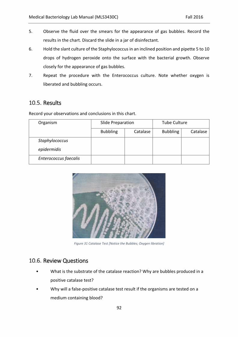
Medical Bacteriology Lab Manual (MLS3430C) Fall 2016
92
5. Observe the fluid over the smears for the appearance of gas bubbles. Record the
results in the chart. Discard the slide in a jar of disinfectant.
6. Hold the slant culture of the Staphylococcus in an inclined position and pipette 5 to 10
drops of hydrogen peroxide onto the surface with the bacterial growth. Observe
closely for the appearance of gas bubbles.
7. Repeat the procedure with the Enterococcus culture. Note whether oxygen is
liberated and bubbling occurs.
Results
Record your observations and conclusions in this chart.
Organism Slide Preparation Tube Culture
Bubbling Catalase Bubbling Catalase
Staphylococcus
epidermidis
Enterococcus faecalis
Figure 31 Catalase Test [Notice the Bubbles; Oxygen libration]
Review Questions
• What is the substrate of the catalase reaction? Why are bubbles produced in a
positive catalase test?
• Why will a false-positive catalase test result if the organisms are tested on a
medium containing blood?

Medical Bacteriology Lab Manual (MLS3430C) Fall 2016
93

Medical Bacteriology Lab Manual (MLS3430C) Fall 2016
94
Activity of Coagulase
Principle
Coagulase is an enzyme mainly found in Staphylococcus aureus. Therefore, this test will help
to distinguish Staphylococcus aureus from other commonly isolated Staphylococci.
Two forms of coagulase exist. One is bound to the cell and other one is excreted from the cell
as an enzyme. Bound coagulase is known as “clumping factor” which acts directly on the
fibrinogen in plasma and convert it to fibrin to form a coagulum. When the coagulase is
released as an enzyme, also called “free coagulase”, reacts with the coagulase reacting factor
(CRF) in plasma to form thrombin that then acts on fibrinogen in the plasma to form a fibrin
clot.
Fibrinogen Fibrin
EDTA blood is used. Citrated blood may give false positive result.
Objective
To implement coagulase activity in distinguishing between pathogenic (coagulase +ve) and
non-pathogenic (coagulase -ve) staphylococci
Materials
Plasma
Physiological saline.
Broth culture of Staphylococcus aureus
Broth culture of Staphylococcus epidermidis
Procedure
11.4.1. Slide method
1. Place two separate drops of physiological saline on a clean slide.
Medical Bacteriology Lab Manual (MLS3430C) Fall 2016
95
2. Emulsify a colony and make two thick suspensions.
3. If there is no auto agglutination add a drop of plasma to one of the suspensions and
mix gently.
4. Look for clumping within 10 seconds in the plasma added suspension.
5. If auto agglutination is present, this slide test cannot be read.
11.4.2. Tube test
11.4.2.1. Method 1
1. Prepare 1:10 diluted plasma in physiological saline.
2. Add 0.5 ml of diluted plasma in a tube.
3. Add 0.1 ml of an 18-24hour broth culture of the organisms.
4. Mix gently and incubate at 35-37°C.
5. Examine for formation of a coagulum after 1 hour; if no coagulum, examine at 2 and
6 hours.
6. Negative tubes should be left at room temperature overnight and re-examined.
7. Use negative and positive controls.
11.4.2.2. Method 2
1. Mix 0.5 ml undiluted plasma with an equal volume of an 18-24hour broth culture.
Incubate at 37oC for 4 hours.
2. Examine after 1 and 4 hours for coagulum.
3. Negative tubes should be left at room temperature overnight and then re-examine.
Results
Formation of coagulum - Positive test
No coagulum - Negative test

Medical Bacteriology Lab Manual (MLS3430C) Fall 2016
96
Review Questions
• What is coagulase?
• How does the enzyme coagulase function?
Figure 32 Coagulase Positive [LEFT] & Negative [RIGHT]

Medical Bacteriology Lab Manual (MLS3430C) Fall 2016
97
Activity of Oxidase
Principle
This test indicates the presence of enzyme cytochrome oxidase. When the organism is
oxidase-producing, the phenylenediamine in the reagent will be oxidized to a deep purple
colour.
Objective
To examine whether the sample bacteria contains the respiratory enzyme cytochrome c
oxidase or not
Materials
Slant cultures of Escherichia coli & Pseudomonas aeruginosa
Freshly prepared tetramethyl-p-phenylenediamine dihydrochloride (oxidase) reagent to
make 10 ml
Tetramethyl-p-phenylenediamine dihydrochloride 0.1 g
Distilled water 10 ml
Dissolve the chemical in the water. The reagent is not stable. It is therefore best prepared
immediately before use.
Procedure
1. Place a piece of filter paper in a clean Petri dish and add 2-3 drops of freshly prepared
oxidase reagent.
2. Using a piece of sterile stick or glass rod (not an oxidized wire loop), remove a colony of
the test organism and smear it on the filter paper.
3. Look for the development of a blue-purple colour within a few seconds.

Medical Bacteriology Lab Manual (MLS3430C) Fall 2016
98
4. Same can be done using a sterile cotton bud impregnated with the oxidase solution.
Results
Record your observations within 10 seconds.
Figure 33 Oxidase Positive [RIGHT] & Negative [LEFT]
Review Questions
1. What metabolic property characterizes bacteria that possess oxidase activity?
2. What is the importance of cytochrome oxidase to bacteria that possess it?
3. Do anaerobic bacteria require oxidase? Explain your answer.
4. What is the function of the test reagent in the oxidase test?
5. The oxidase test is used to differentiate among which groups of bacteria?
6. Why should nichrome or other iron-containing inoculating devices not be used in the
oxidase test?
7. Are there limitations to the oxidase test?

Medical Bacteriology Lab Manual (MLS3430C) Fall 2016
99
Activity of Urease
Principle
The test detects the ability of an organism to produce enzyme urease. The test organism is
cultured in a medium which contains urea and the indicator phenol red. If the organism
produces urease, the enzyme will breakdown the urea to ammonia and carbon-dioxide. The
medium becomes alkaline when ammonia is released which is indicated by the red-pink
colour.
Objective
To observe the activity of urease and to distinguish bacteria that produce it from those that
do not
Materials
Tubes of urea broth or agar
Slant cultures of Escherichia coli and Proteus vulgaris
Procedure
1. Inoculate a tube of urea broth or agar with E. coli, and another with the Proteus
culture.
2. Incubate the tubes at 35°C for 24 hours.
Results
Record your observations.
Name of
Organism
Colour of Urea Medium Urease
Before
Culture
After
Culture
Positive Negative

Medical Bacteriology Lab Manual (MLS3430C) Fall 2016
100
Figure 34 Urease Positive [LEFT] & Negative [RIGHT]
Review Questions
• What is a catalyst?
• Define an enzyme and a substrate. What is the value of enzyme tests in diagnostic
microbiology?
• What happens to urea in the presence of urease?

Medical Bacteriology Lab Manual (MLS3430C) Fall 2016
101
Activity of Gelatinase
Principle
Gelatin is a simple protein. When in solution, it liquefies at warm temperatures above 25°C.
At room temperature or below it becomes solid. When bacteria that produce the enzyme
gelatinase are grown in a gelatin medium, the enzyme breaks up the gelatin molecule and the
medium cannot solidify even at cold temperatures. An alternative method for detecting
gelatinase production is the use of X-ray film that is coated with a green gelatin emulsion.
Organisms that produce gelatinase remove the emulsion from the strip.
Objective
To observe the usefulness of a gelatinase test in distinguishing between bacterial species
Materials
Tubes of nutrient gelatin medium
1.25 inch strips of exposed, undeveloped X-ray film or gelatin strip
Tubes containing 0.5 ml sterile saline
Slant cultures of Serratia marcescens and Providencia stuartii
Procedure
1. Inoculate each of the two cultures into a separate tube of gelatin, stabbing the
inoculating wire straight down through the solid column of medium.
2. Incubate the inoculated tubes and one uninoculated tube of gelatin medium at 35°C.
3. Inoculate each of the two cultures into a separate tube of 0.5 ml saline. The
suspension should be very turbid.
4. Insert a strip of the X-ray or gelatin film into each saline suspension.
5. Incubate the tubes at 35°C. Observe at 1, 2, 3, 4, and 24 hours for removal of the
gelatin emulsion from the strip with subsequent appearance of the transparent strip
support.
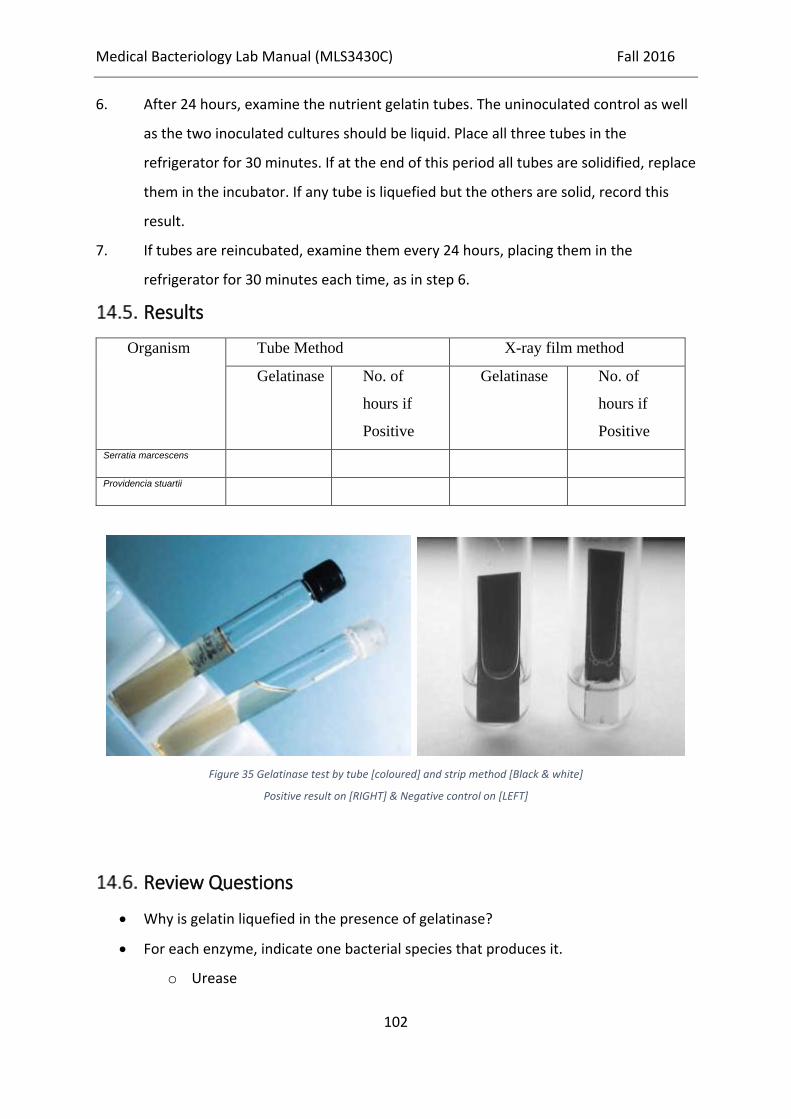
Medical Bacteriology Lab Manual (MLS3430C) Fall 2016
102
6. After 24 hours, examine the nutrient gelatin tubes. The uninoculated control as well
as the two inoculated cultures should be liquid. Place all three tubes in the
refrigerator for 30 minutes. If at the end of this period all tubes are solidified, replace
them in the incubator. If any tube is liquefied but the others are solid, record this
result.
7. If tubes are reincubated, examine them every 24 hours, placing them in the
refrigerator for 30 minutes each time, as in step 6.
Results
Organism Tube Method X-ray film method
Gelatinase No. of
hours if
Positive
Gelatinase No. of
hours if
Positive
Serratia marcescens
Providencia stuartii
Figure 35 Gelatinase test by tube [coloured] and strip method [Black & white]
Positive result on [RIGHT] & Negative control on [LEFT]
Review Questions
Why is gelatin liquefied in the presence of gelatinase?
For each enzyme, indicate one bacterial species that produces it.
o Urease

Medical Bacteriology Lab Manual (MLS3430C) Fall 2016
103
o Catalase
o Gelatinase

Medical Bacteriology Lab Manual (MLS3430C) Fall 2016
104
Phenylalaninedeaminase test / Phenylpyruvic
acid (PPA) test
Principle
The test is based on the ability of organism to breakdown phenylalanine by oxidative
deamination to produce phenylpyruvic acid. Phenylpyruvic acid is detected by adding FeCl3
which produces a green colour on the surface of the culture.
Phenylpyruvic Acid + Ferric Chloride Green Complex
Objectices
Examining whether the sample bacteria is able to breakdown phenylalanine by oxidative
deamination to produce phenylpyruvic acid or not.
Materials
o Phenylalanine agar Slants
o 10% aqueous solution of FeCl3
o Slant culture of Proteus vulgaris
o Slant culture of Klebsiella pneumoniae sub species pneumoniae
Procedure
1. Inoculate the slope of phenylalanine agar with the test organism.
2. Incubate at 35-37°C overnight.
3. Add 0.2 ml of freshly prepared 10% aqueous solution of FeCl3 to the culture.
4. Allow the reagent to run down the slope.
5. Watch immediately.
6. A positive reaction is indicated by a dark green colour which fades quickly.
Results
Green colour (within 5 minutes) - Positive test
No green colour - Negative test

Medical Bacteriology Lab Manual (MLS3430C) Fall 2016
105
Figure 36 Phenylalanine Deamination.
(LEFT) Uninoculated control. (MIDDLE) Phenylalanine negative. (RIGHT) Phenylalanine positive.
Review Questions
1. What is the purpose of the ferric chloride in the phenylalanine deamination test?
2. When would you use the phenylalanine deamination test?
3. Name some bacteria that can deaminate phenylalanine.
4. Describe the process of deamination.
Medical Bacteriology Lab Manual (MLS3430C) Fall 2016
106
API & AST tests + Lab Automation
Background
The biochemical tests performed in the preceding sections are representative of standard
methods for bacterial identification. In some instances, it is possible to identify a bacterium
correctly by using only a few tests, but more often an extensive biochemical “profile” is
needed. Because it is expensive and time consuming to make and keep a wide variety of
culture media on hand, many microbiology laboratories now use multimedia identification
kits. These are commercially available and are especially useful for identifying the common
enteric bacteria. The use of such kits is customarily referred to as an application of “rapid
methods,” even though they must be incubated overnight, as usual, before results can be
read. Some of them, indeed, are rapid to inoculate, while others permit complete
identification within 24 hours.
Principle
The API System (bioMérieux Inc.) is a kit for rapid identification of bacteria. This system
provides, in a single strip, a series of 20 microtubules (miniature test tubes) of dehydrated
media that are rehydrated with a saline suspension of the bacterium to be identified. The
tests included in the strip determine whether the organism ferments glucose, mannitol,
inositol, sorbitol, rhamnose, saccharose, melibiose, and amygdalin; produces indole and H2S;
splits urea; breaks down the amino acids tryptophan (same mechanism as phenylalanine),
lysine, ornithine, and arginine; produces gelatinase; forms acetylmethylcarbinol from glucose
(VP test); and splits the compound o-nitrophenyl-β-D-galactopyranoside (ONPG). The enzyme
that acts on ONPG, called β-galactosidase, also is responsible for lactose fermentation. Some
bacteria, however, are unable to transport lactose into their cells for breakdown, although
they possess β-galactosidase. In lactose broth, therefore, such bacteria fail to display acid
production, or do so only after a delay of days or weeks. By contrast, in ONPG medium their
β-galactosidase splits the substrate in a matter of hours, producing a bright yellow end
product. Thus, ONPG can be used for the rapid demonstration of an organism’s ability to
ferment lactose.

Medical Bacteriology Lab Manual (MLS3430C) Fall 2016
107
Objective
To observe the biochemical properties of bacteria grown in a multimedia system for rapid
identification
Materials
20E & 20NE API System (bioMérieux Inc.) kits for rapid identification of bacteria
Saline suspension of Escherichia coli
Saline suspension of Proteus vulgaris
Procedure
• Inoculated two API strips with Escherichia coli and Proteus vulgaris
• incubated for 24 hours
Results
After incubation of API strips, record the results observed for each organism in the blocks
provided under the following diagram.
Figure 37 Reactions in API 20E.

Medical Bacteriology Lab Manual (MLS3430C) Fall 2016
108
Review Questions
• What is the significant advantage of API strips
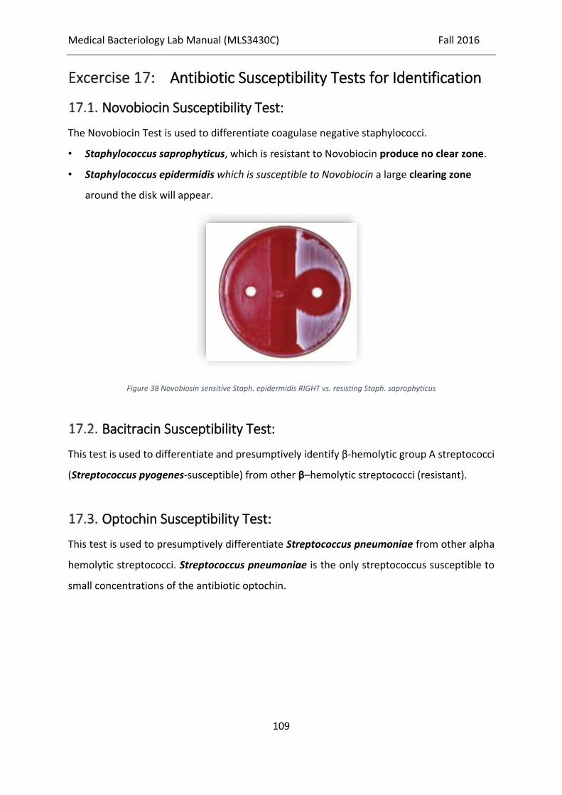
Medical Bacteriology Lab Manual (MLS3430C) Fall 2016
109
Antibiotic Susceptibility Tests for Identification
Novobiocin Susceptibility Test:
The Novobiocin Test is used to differentiate coagulase negative staphylococci.
• Staphylococcus saprophyticus, which is resistant to Novobiocin produce no clear zone.
• Staphylococcus epidermidis which is susceptible to Novobiocin a large clearing zone
around the disk will appear.
Figure 38 Novobiosin sensitive Staph. epidermidis RIGHT vs. resisting Staph. saprophyticus
Bacitracin Susceptibility Test:
This test is used to differentiate and presumptively identify β-hemolytic group A streptococci
(Streptococcus pyogenes-susceptible) from other β–hemolytic streptococci (resistant).
Optochin Susceptibility Test:
This test is used to presumptively differentiate Streptococcus pneumoniae from other alpha
hemolytic streptococci. Streptococcus pneumoniae is the only streptococcus susceptible to
small concentrations of the antibiotic optochin.

Medical Bacteriology Lab Manual (MLS3430C) Fall 2016
110
Some Culture Media Ingredients:
Blood Agar (pH 7.3)
Infusion from beef heart ............................. 500.0 g
Tryptose...................................................... 10.0 g
Sodium chloride.......................................... 5.0 g
Agar ............................................................ 15.0 g
Distilled water ............................................ 1,000.0 ml
Note: Dissolve the above ingredients and autoclave. Cool the sterile blood agar base to 45° to
50°C and aseptically add 50 ml of sterile, defibrinated blood. Mix thoroughly and then
dispense into plates while a liquid. Blood agar base for use in making blood agar also can be
purchased. A combination of hemoglobin and a commercial nutrient supplement can be used
in place of defibrinated blood.
Brain-Heart Infusion Agar (pH 7.4)
Calf brains, infusion from........................... 200.0 g
Beef hearts, infusion from .......................... 250.0 g
Proteose peptone......................................... 10.0 g
Dextrose...................................................... 2.0 g
Sodium chloride.......................................... 5.0 g
Disodium phosphate ................................... 2.5 g
Agar ............................................................ 15.0 g
Distilled water ............................................ 1,000.0 ml

Medical Bacteriology Lab Manual (MLS3430C) Fall 2016
111
Kligler Iron Agar (pH 7.4)
Beef extract................................................. 3.0 g
Yeast extract ............................................... 3.0 g
Peptone ....................................................... 15.0 g
Proteose peptone......................................... 5.0 g
Lactose........................................................ 10.0 g
Dextrose...................................................... 1.0 g
Ferrous sulfate ............................................ 0.2 g
Sodium chloride.......................................... 5.0 g
Sodium thiosulfate...................................... 0.3 g
Agar ............................................................ 12.0 g
Phenol red................................................... 0.024 g
Distilled water ............................................ 1,000.0 ml
Mannitol Salt Agar (pH 7.4)
Beef extract ............................................... 1.0 g
Peptone...................................................... 10.0 g
Sodium chloride ........................................ 75.0 g
D–mannitol................................................ 10.0 g
Agar........................................................... 15.0 g
Phenol red ................................................. 0.025 g
Distilled water ........................................... 1,000.0 ml

Medical Bacteriology Lab Manual (MLS3430C) Fall 2016
112
MacConkey’s Agar (pH 7.1)
Bacto peptone ............................................. 17.0 g
Proteose peptone......................................... 3.0 g
Lactose........................................................ 10.0 g
Bile salts mixture ........................................ 1.5 g
Sodium chloride ........................................ 5.0 g
Agar........................................................... 13.5 g
Neutral red................................................. 0.03 g
Crystal violet ............................................. 0.001 g
Distilled water ........................................... 1,000.0 ml
MR-VP Broth (pH 6.9)
Peptone...................................................... 7.0 g
Dextrose .................................................... 5.0 g
Potassium phosphate ................................. 5.0 g
Distilled water ........................................... 1,000.0 ml
Mueller-Hinton Agar (pH 7.4)
Beef, infusion ............................................ 300.0 g
Casamino acids ......................................... 17.5 g
Starch ........................................................ 1.5 g
Agar........................................................... 17.0 g
Distilled water ........................................... 1,000.0 ml
Nutrient Agar (pH 7.0)
Peptone...................................................... 5.0 g
Beef extract ............................................... 3.0 g
Agar........................................................... 15.0 g
Distilled water ........................................... 1,000.0 ml
Note: Autoclave at 121 lb pressure for 15 minutes.

Medical Bacteriology Lab Manual (MLS3430C) Fall 2016
113
Starch Agar (pH 7.5)
Beef extract ............................................... 3.0 g
Soluble starch ............................................ 10.0 g
Agar........................................................... 12.0 g
Distilled water ...........................................1,000.0 ml
Thioglycollate Broth (pH 7.1)
Peptone...................................................... 15.0 g
Yeast extract.............................................. 5.0 g
Dextrose .................................................... 5.0 g
L-cystine.................................................... 0.75 g
Thioglycollic acid ..................................... 0.5 g
Agar........................................................... 0.75 g
Sodium chloride ........................................ 2.5 g
Resazurin................................................... 0.001 g
Distilled water ........................................... 1,000.0 ml
Triple Sugar Iron Agar (pH 7.4)
Beef extract ............................................... 3.0 g
Yeast extract.............................................. 3.0 g
Peptone...................................................... 15.0 g
Peptose-peptone ........................................ 5.0 g
Lactose ...................................................... 10.0 g
Saccharose................................................. 10.0 g
Dextrose .................................................... 1.0 g
Ferrous sulfate........................................... 0.2 g
Sodium chloride ........................................ 5.0 g
Sodium thiosulfate .................................... 0.3 g
Phenol red ................................................. 0.024 g
Agar........................................................... 12.0 g
Distilled water ........................................... 1,000.0 ml