medpix medical image database cow - case of the week case contributor: clark brixey affiliation:...
TRANSCRIPT

MedPix Medical Image Database
COW - Case of the WeekCase Contributor: clark brixeyAffiliation: National Capital Consortium

MedPix No: 10096 - HistoryPt Demographics: Age = 0 y.o. Gender = 34 year old male involved in an MVA where he reports slamming into ceiling during the crash.
Downloaded by (-1)

MedPix No: 10096 - EXAM & LABSSevere pain lower mid back. No neurologic findings

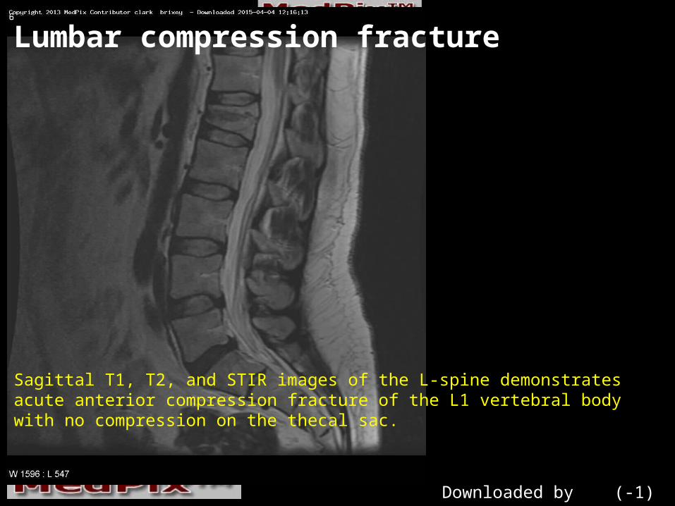
Lumbar compression fracture
Lateral radiograph of the L-spine demonstrates anterior wedge deformity of the L1 vertebral body.
Downloaded by (-1)

Lumbar compression fracture
Sagittal T1, T2, and STIR images of the L-spine demonstrates acute anterior compression fracture of the L1 vertebral body with no compression on the thecal sac.
Downloaded by (-1)
Lumbar compression fracture
Sagittal T1, T2, and STIR images of the L-spine demonstrates acute anterior compression fracture of the L1 vertebral body with no compression on the thecal sac.
Downloaded by (-1)

Lumbar compression fracture
Sagittal T1, T2, and STIR images of the L-spine demonstrates acute anterior compression fracture of the L1 vertebral body with no compression on the thecal sac.
Downloaded by (-1)

FINDINGSThe anteroposterior radiograph reveals bulking of the lateral cortices of the vertebral body close to the involved end plate, together with a decrease in the height of the body. In lateral-flexion injuries, compression forces may result in a wedge-shaped deformity of the vertebral body. In subtle cases, a clue to the diagnosis may be seen in a localized bulge of the paraspinal line secondary to hemorrhage and edema. However, it should be kept in mind that this finding may also be seen in pathologic fractures secondary to skeletal mets to the spine. On the lateral view, a simple vertebral compression fracture can be seen by a decrease in the height of the anterior part of the body, while the height of the posterior part and posterior cortex is maintained.

DIFFERENTIAL DIAGNOSISWhat is your Differential Diagnosis?Traumatic fracture, acute or chronic- Pathologic fracture-

Diagnosis: Lumbar compression fractureDx Confirmed by: Image findings and history

DISCUSSIONThis case nicely demonstrates the findings of compression fracture in an otherwise healthy man. Given the history, the diagnosis could be made from the plain film, but neurosurgery requested the MR to rule out any compression on the thecal sac/cord. Some would prefer CT over MRI to visualize canal and foraminal encroachment with greater visualization of bony detail.
