melanocytoma of the ciliary body misdiagnosed as iridodialysis · pdf filecase report open...
TRANSCRIPT

© 2014 Kim and Lee. This work is published by Dove Medical Press Limited, and licensed under Creative Commons Attribution – Non Commercial (unported, v3.0) License. The full terms of the License are available at http://creativecommons.org/licenses/by-nc/3.0/. Non-commercial uses of the work are permitted without any further
permission from Dove Medical Press Limited, provided the work is properly attributed. Permissions beyond the scope of the License are administered by Dove Medical Press Limited. Information on how to request permission may be found at: http://www.dovepress.com/permissions.php
Clinical Ophthalmology 2014:8 1051–1053
Clinical Ophthalmology Dovepress
submit your manuscript | www.dovepress.com
Dovepress 1051
C a s e R e p O Rt
open access to scientific and medical research
Open access Full text article
http://dx.doi.org/10.2147/OPTH.S63328
Journal name: Clinical OphthalmologyJournal Designation: Case ReportYear: 2014Volume: 8Running head verso: Kim and LeeRunning head recto: Melanocytoma of the ciliary body misdiagnosed as iridodialysisDOI: http://dx.doi.org/10.2147/OPTH.S63328
Melanocytoma of the ciliary body misdiagnosed as iridodialysis
Moosang Kimseung-Jun LeeDepartment of Ophthalmology, school of Medicine, Kangwon National University, Chuncheon, Republic of Korea
Abstract: A 62-year-old female presented to our institution with dimness of vision in her right
eye. On examination, her best corrected visual acuity was 20/100 in the right eye. The intraocular
pressures were 14 mmHg in both eyes. Slit-lamp examination revealed nuclear sclerotic cataracts
bilaterally and iridodialysis in her right eye. Seven days after the first visit, cataract surgery was
performed without any complications. One year later, she presented to our institution with acute
visual loss and ocular pain in the right eye. Best corrected visual acuity of the right eye was
light perception and the intraocular pressure was 44 mmHg. Slit-lamp examination revealed a
ciliary body mass with widespread pigment dispersion in the anterior segment. Due to no use-
ful vision and uncontrolled pain, enucleation of the right eye was performed. Histopathologic
examination revealed a melanocytoma of the ciliary body.
Keywords: ciliary body, iridodialysis, melanocytoma
IntroductionAlthough melanocytomas are typically located in the optic nerve head, they can also be
found anywhere along the uveal tract, including the iris, choroid, and ciliary body.1–3
Melanocytoma is a variant of melanocytic nevi, and its occurrence in the uveal tract
is relatively uncommon. In this paper, we report a case of melanocytoma of the ciliary
body misdiagnosed as iridodialysis.
Case reportA 62-year-old female presented to our institution with dimness of vision in the right eye.
On examination, her best corrected visual acuity was 20/100 in the right eye and 20/50
in the left eye. Intraocular pressures were 14 mmHg in both eyes. Slit-lamp examination
revealed nuclear sclerotic cataracts bilaterally and a small iridodialysis in her right eye.
The iridodialysis was within 20 degrees in the 11 o’clock direction, and the iris and its
surrounding structures were otherwise unremarkable. However, she had no history of
ocular trauma. The pupil was grossly concentric and almost fully dilated after instil-
lation of mydriatics, even though the finding was consistent with the iridodialysis.
Fundus examination was unremarkable in both eyes. We considered these findings in
the iris as a small asymptomatic dialysis that did not require any specific treatment.
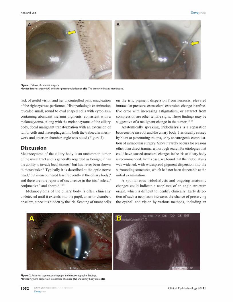
The patient wanted surgery for her right eye cataract. Seven days after presentation,
the cataract surgery was performed without any complications (Figure 1).
One year later, the patient presented to our institution with acute visual loss and
ocular pain in her right eye. Best corrected visual acuity of the right eye was light
perception and the intraocular pressure was 44 mmHg. Slit-lamp examination and
ultrasonography revealed an iridodialysis of nearly 40 degrees and a ciliary body mass
with widespread pigment dispersion in the anterior segment (Figure 2). Due to her
Correspondence: seung-Jun LeeBaengnyeong-ro 156, Chuncheon-si, Gangwon-Do Kangwon National University Hospital 200-722, Republic of Korea tel +823 3258 2014Fax +82 2966 7340email [email protected]
Clinical Ophthalmology 2014:8submit your manuscript | www.dovepress.com
Dovepress
Dovepress
1052
Kim and Lee
Figure 1 Views of cataract surgery.Notes: Before surgery (A) and after phacoemulsification (B). the arrow indicates iridodialysis.
A B
Figure 2 Anterior segment photograph and ultrasonographic findings.Notes: pigment dispersion in anterior chamber (A) and ciliary body mass (B).
lack of useful vision and her uncontrolled pain, enucleation
of the right eye was performed. Histopathologic examination
revealed small, round to oval shaped cells with cytoplasm
containing abundant melanin pigments, consistent with a
melanocytoma. Along with the melanocytoma of the ciliary
body, focal malignant transformation with an extension of
tumor cells and macrophages into both the trabecular mesh-
work and anterior chamber angle was noted (Figure 3).
DiscussionMelanocytoma of the ciliary body is an uncommon tumor
of the uveal tract and is generally regarded as benign; it has
the ability to invade local tissues,4 but has never been shown
to metastasize.5 Typically it is described at the optic nerve
head,1 but is encountered less frequently at the ciliary body,6
and there are rare reports of occurrence in the iris,7 sclera,8
conjunctiva,9 and choroid.10,11
Melanocytoma of the ciliary body is often clinically
undetected until it extends into the pupil, anterior chamber,
or sclera, since it is hidden by the iris. Seeding of tumor cells
on the iris, pigment dispersion from necrosis, elevated
intraocular pressure, extrascleral extension, change in refrac-
tive error with increasing astigmatism, or cataract from
compression are other telltale signs. These findings may be
suggestive of a malignant change in the tumor.12–14
Anatomically speaking, iridodialysis is a separation
between the iris root and the ciliary body. It is usually caused
by blunt or penetrating trauma, or by an iatrogenic complica-
tion of intraocular surgery. Since it rarely occurs for reasons
other than direct trauma, a thorough search for etiologies that
could have caused structural changes in the iris or ciliary body
is recommended. In this case, we found that the iridodialysis
was widened, with widespread pigment dispersion into the
surrounding structures, which had not been detectable at the
initial examination.
A spontaneous iridodialysis and ongoing anatomic
changes could indicate a neoplasm of an angle structure
origin, which is difficult to identify clinically. Early detec-
tion of such a neoplasm increases the chance of preserving
the eyeball and vision by various methods, including an

Clinical Ophthalmology
Publish your work in this journal
Submit your manuscript here: http://www.dovepress.com/clinical-ophthalmology-journal
Clinical Ophthalmology is an international, peer-reviewed journal covering all subspecialties within ophthalmology. Key topics include: Optometry; Visual science; Pharmacology and drug therapy in eye diseases; Basic Sciences; Primary and Secondary eye care; Patient Safety and Quality of Care Improvements. This journal is indexed on
PubMed Central and CAS, and is the official journal of The Society of Clinical Ophthalmology (SCO). The manuscript management system is completely online and includes a very quick and fair peer-review system, which is all easy to use. Visit http://www.dovepress.com/testimonials.php to read real quotes from published authors.
Dovepress
Clinical Ophthalmology 2014:8 submit your manuscript | www.dovepress.com
Dovepress
Dovepress
1053
Melanocytoma of the ciliary body misdiagnosed as iridodialysis
en bloc resection.15 If a spontaneous iridodialysis with no
plausible explanation is encountered, an early and thor-
ough search should be done in the anterior chamber angle,
including gonioscopy or anterior segment optical coherence
tomography. In summary, melanocytoma of the ciliary body
is a rare tumor, is clinically hard to detect, and can invade
chamber angle structures and appear as a pigmented mass
at the iris root.
ConclusionThis is a case of melanocytoma of the ciliary body mis-
diagnosed as iridodialysis. A physician should always keep
in mind that a careful evaluation of the anterior chamber
angle (eg, gonioscopy, anterior segment optical coherence
tomography) is the most important step for making an accu-
rate diagnosis.
DisclosureThe authors report no conflicts of interest in this work.
References1. Demirci H, Mashayekhi A, Shields CL, Eagle RC Jr, Shields JA. Iris
melanocytoma: clinical features and natural course in 47 cases. Am J Ophthalmol. 2005;139(3):468–475.
2. Brownstein S, Dorey MW, Mathew B, Little JM, Lindley JI. Melanocy-toma of the choroid: atypical presentation and review of the literature. Can J Ophthalmol. 2002;37(4):247–252.
3. Shields JA, Shields CL, Eagle RC Jr. Melanocytoma (hyperpigmented magnocellular nevus) of the uveal tract: the 34th G. Victor Simpson lecture. Retina. 2007;27(6):730–739.
4. Zimmerman LE. Melanocytes, melanocytic nevi and melanocytomas. Invest Ophthalmol. 1965;4:11–41.
5. Raichand M, Peyman GA, Juarez CP, Seetner AA, Sugar J, Goldberg MF. Resection of uveal melanocytoma: clinicopathological correlation. Br J Ophthalmol. 1983;67(4):236–243.
6. Shammas HJ, Minckler DS, Hulquist R, et al. Melanocytoma of the ciliary body. Ann Ophthalmol. 1981;13:1381–1383.
7. Shields JA, Augsburger JJ, Bernardino V Jr, Eller AW, Kulczycki E. Melanocytoma of the ciliary body and iris. Am J Ophthalmol. 1980; 89(5):632–635.
8. Lee JS, Smith RE, Minckler DS. Scleral melanocytoma. Ophthalmol-ogy. 1982;89(2):178–182.
9. Verdaguer J, Valenzuela H, Strozzi L. Melanocytoma of the conjunctiva. Arch Ophthalmol. 1974;91(5):363–366.
10. Shields JA, Font RL. Melanocytoma of the choroid clinically simulating a malignant melanoma. Arch Ophthalmol. 1972;87:396–400.
11. Jürgens I, Roca G, Sedó S, Pujol O, Berniell JA, Quintana M. Pre-sumed melanocytoma of the macula. Arch Ophthalmol. 1994;112(3): 305–306.
12. Bhorade AM, Edward DP, Goldstein DA. Ciliary body melanocytoma with anterior segment pigment dispersion and elevated intraocular pressure. J Glaucoma. 1999;8(2):129–133.
13. Cialdini AP, Sahel JA, Jalkh AE, Weiter JJ, Zakka K, Albert DM. Malignant transformation of an iris melanocytoma. A case report. Graefes Arch Clin Exp Ophthalmol. 1989;227(4):348–354.
14. Radcliffe NM, Finger PT. Eye cancer related glaucoma: current con-cepts. Surv Ophthalmol. 2009;54(1):47–73.
15. Weinstein GW, Quayle WH. An en bloc technique for the resection of anterior uveal tumors. Am J Ophthalmol. 1979;88(3 Pt 1):519–523.
Figure 3 Light microscopy views.Notes: (A) a pigmented mass in the ciliary body (40×), (B) involvement of trabecular meshwork by the tumor cells (100×), and (C) round to oval bland cytologic features of tumor cells (400×). they show prominent nucleoli (arrows) and marked cytoplasmic melanin pigments.
A X40 BX100 C X400