melanoma overview 2009 frances collichio associate professor, university of north carolina, chapel...
Post on 19-Dec-2015
220 views
TRANSCRIPT
Melanoma Overview 2009Frances Collichio
Associate Professor, University of North Carolina, Chapel Hill
Disclosure: OncoVex
Novartis
What is Melanoma?
• A Cancer of Melanocytes
• All Melanomas are malignant
• Melanocytes?– Cells that Make Pigment
• Melanoma therefore can start from any pigmented cell.
Extracutaneous Melanomas,
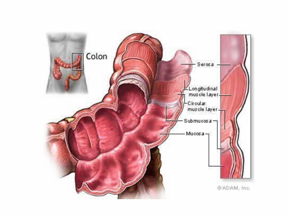
• Mucosal– GI tract, Head and Neck, vagina– Older pts– Poorer px– Disporportionally non-white

Ocular
• UvealCiliary body/Choroidal
Poor prognosisIris
Better px
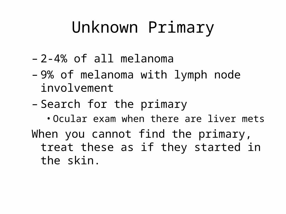
Unknown Primary
– 2-4% of all melanoma– 9% of melanoma with lymph node
involvement– Search for the primary
• Ocular exam when there are liver mets
When you cannot find the primary, treat these as if they started in the skin.

““Silent”Silent” NationalNational EpidemicEpidemic
0
2
4
6
8
10
12
14
16
18
1935 1950 1980 1985 1987 2000 2005 2010
• The incidence per year is rising faster than any other cancer!
Rate
/100,0
00
1:1500 1:600 1:250 1:150 1:135 1:75 1:65 1:60
estimated Lifetime RiskLifetime Risk
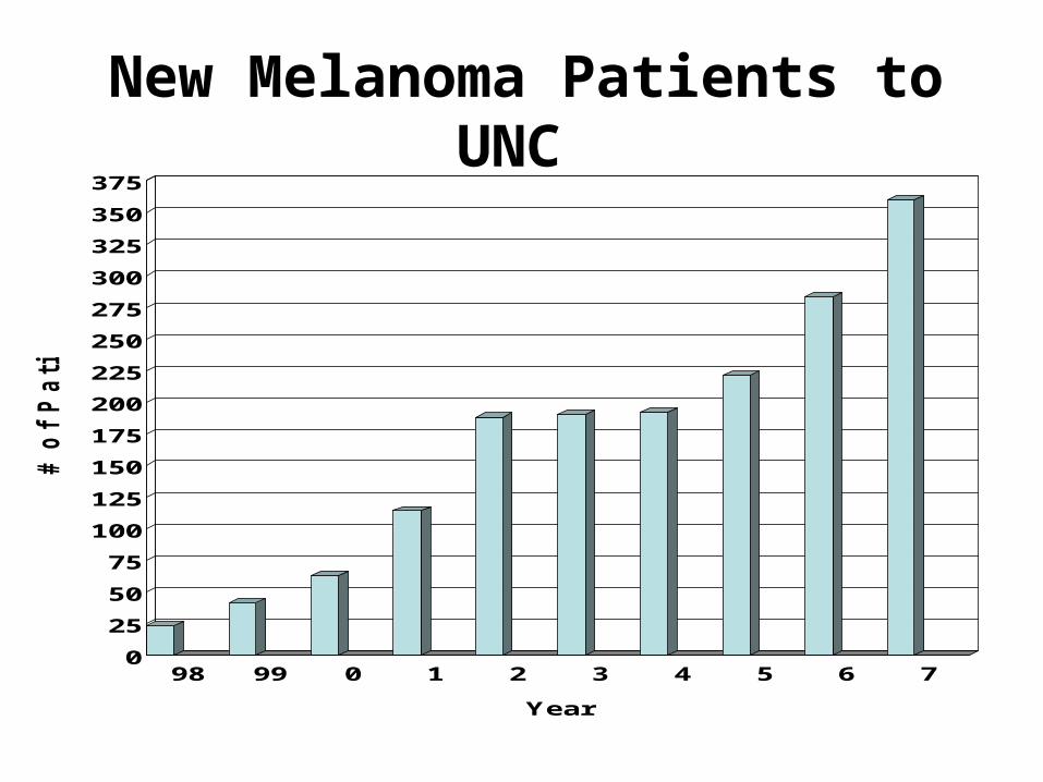
New Melanoma Patients to UNC
0
25
50
75
100
125
150
175
200
225
250
275
300
325
350
375
# o
f P
ati
en
ts
98 99 0 1 2 3 4 5 6 7
Year

2007 Estimated US Cancer Cases*
*Excludes basal and squamous cell skin cancers and in situ carcinomas except urinary bladder.Source: American Cancer Society, 2007.
Men766,860
Women678,060
•26% Breast
•15% Lung & bronchus
•11% Colon & rectum
•6% Uterine corpus
• 4% Non-Hodgkin lymphoma
•4% Melanoma of skin
• 4% Thyroid
• 3% Ovary
• 3% Kidney
•3% Leukemia
•21% All Other Sites
Prostate 29%
Lung & bronchus 15%
Colon & rectum 10%
Urinary bladder 7%
Non-Hodgkin4% lymphoma
Melanoma of skin 4%
Kidney 4%
Leukemia 3%
Oral cavity 3%
Pancreas 2%
All Other Sites 19%
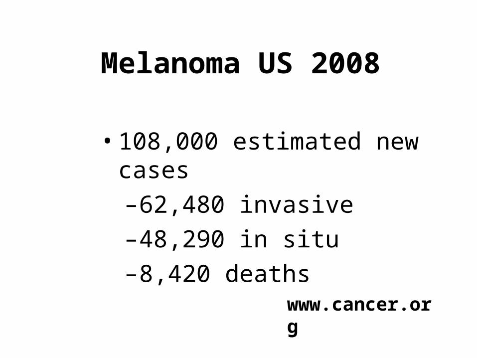
Melanoma US 2008
• 108,000 estimated new cases
–62,480 invasive
–48,290 in situ
–8,420 deaths
www.cancer.org
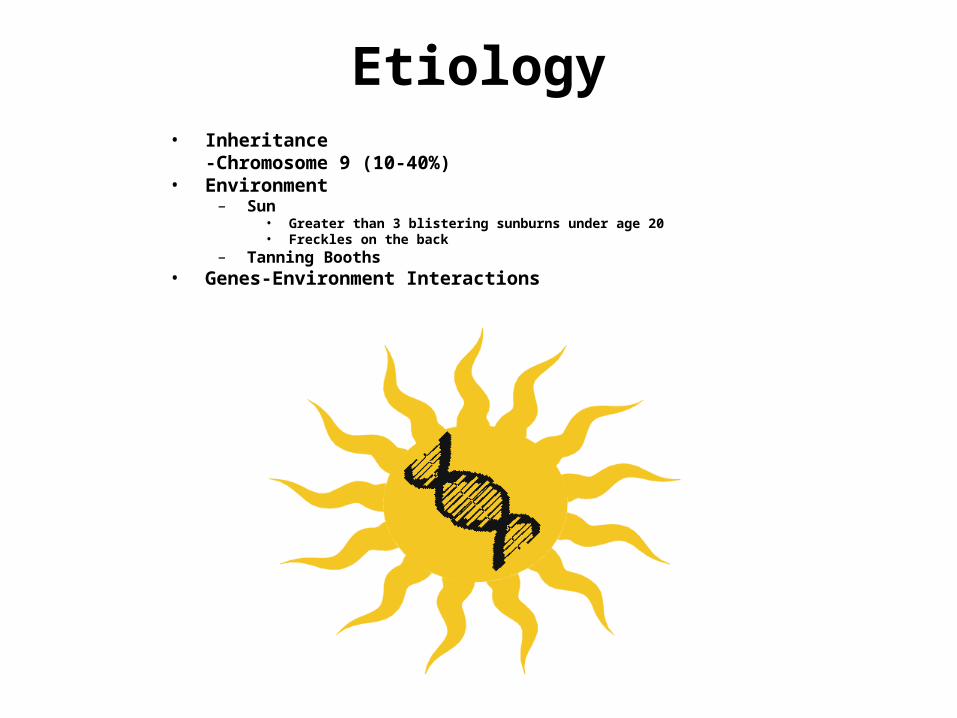
Etiology• Inheritance
-Chromosome 9 (10-40%)• Environment
– Sun• Greater than 3 blistering sunburns under age 20• Freckles on the back
– Tanning Booths• Genes-Environment Interactions

Melanoma
A—AsymmetryB—BorderC—ColorD—DiameterE—Evolution: A changing mole

Types of Melanoma

Nodular Amelanotic Melanoma
Invasive Melanoma
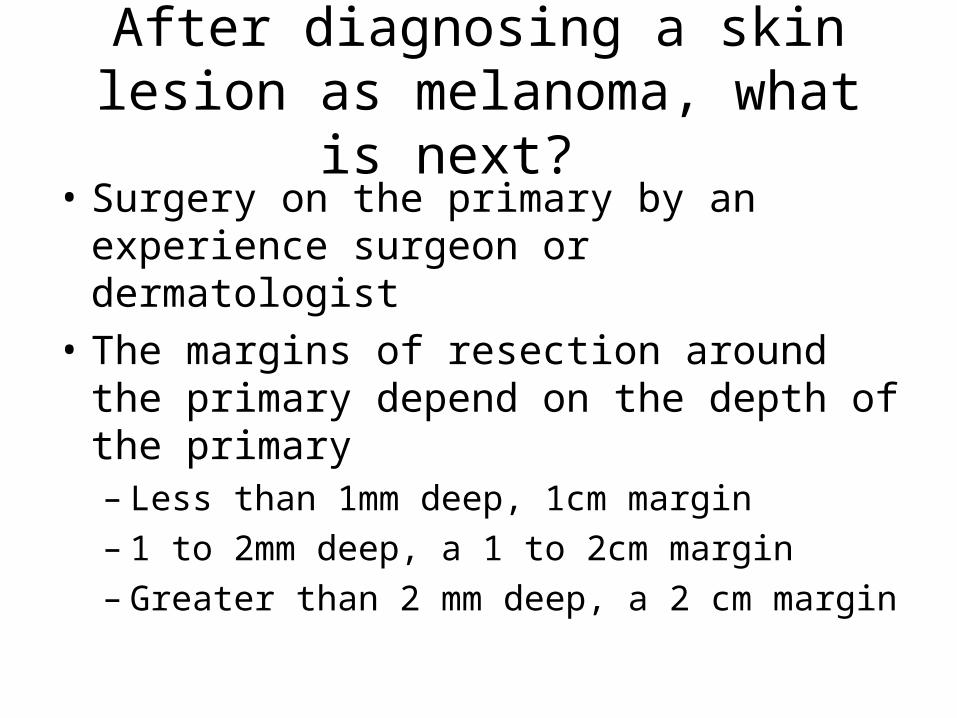
After diagnosing a skin lesion as melanoma, what is next?
• Surgery on the primary by an experience surgeon or dermatologist
• The margins of resection around the primary depend on the depth of the primary– Less than 1mm deep, 1cm margin– 1 to 2mm deep, a 1 to 2cm margin– Greater than 2 mm deep, a 2 cm margin
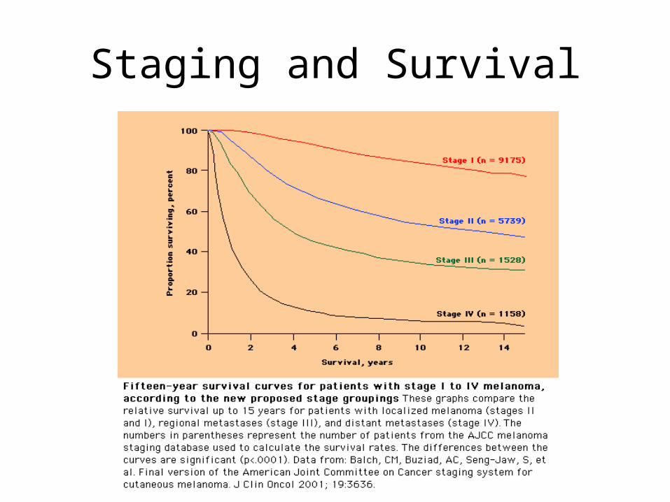
Staging and Survival
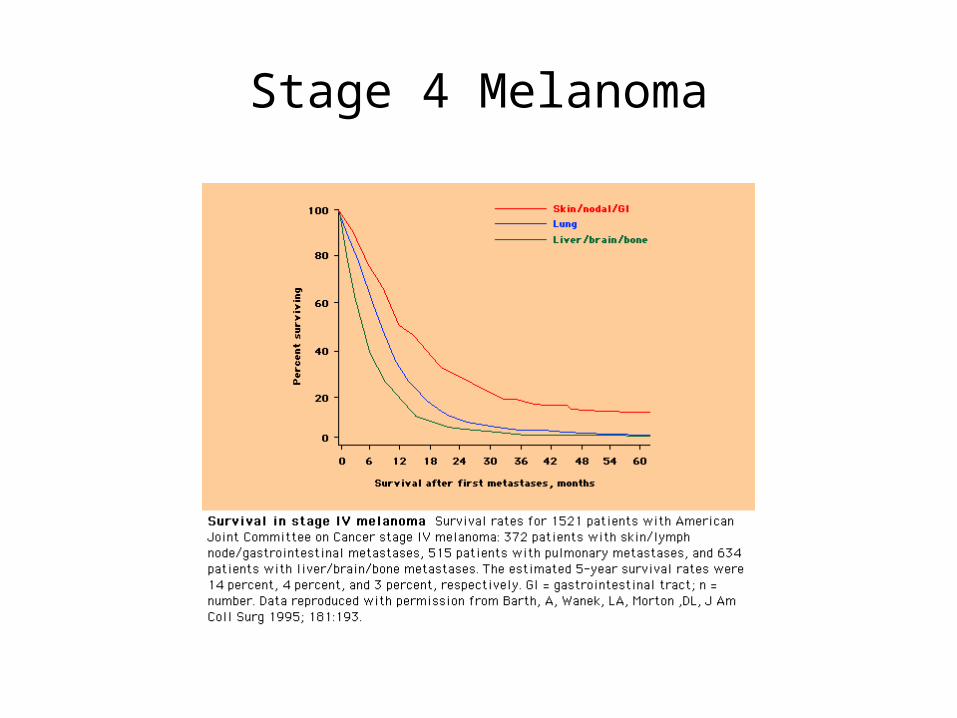
Stage 4 Melanoma

Staging, 2002 AJCC • T
–Breslow depth–Clark level---used only in T1 melanomas–Ulceration
• N–N1: Metastasis to one node–N2: Metastasis to two or three nodes–N3: Mets to 4 or more, or matted nodes, or in transit disease, or satellites (tumor w/in 5cm of the primary). –Micromets are defined by sentinel node. Macro mets are clinically detectable.
• M –M1a-skin, subcutaneous tissue or distant lymph nodes–M1b-lung–M1c-other sites
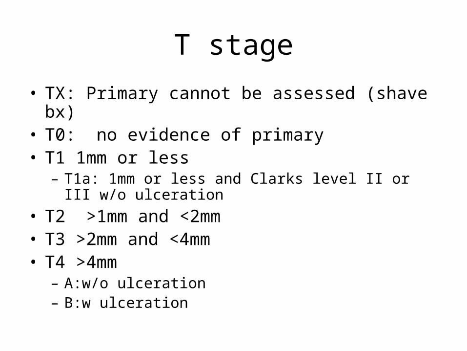
T stage
• TX: Primary cannot be assessed (shave bx)• T0: no evidence of primary• T1 1mm or less
– T1a: 1mm or less and Clarks level II or III w/o ulceration
• T2 >1mm and <2mm• T3 >2mm and <4mm• T4 >4mm
– A:w/o ulceration– B:w ulceration

Nodal Stage
• N– N1: Metastasis to one node
• N1a:micromet• N1b: Macromet
– N2: Metastasis to two or three nodes• N2a: Micromet• N2b: Macromet• N2c:in-transit met(s)/satellite(s) without metastatic lymph
nodes
– N3: Mets to 4 or more, or matted nodes, or in transit disease, or satellites (tumor w/in 5cm of the primary).
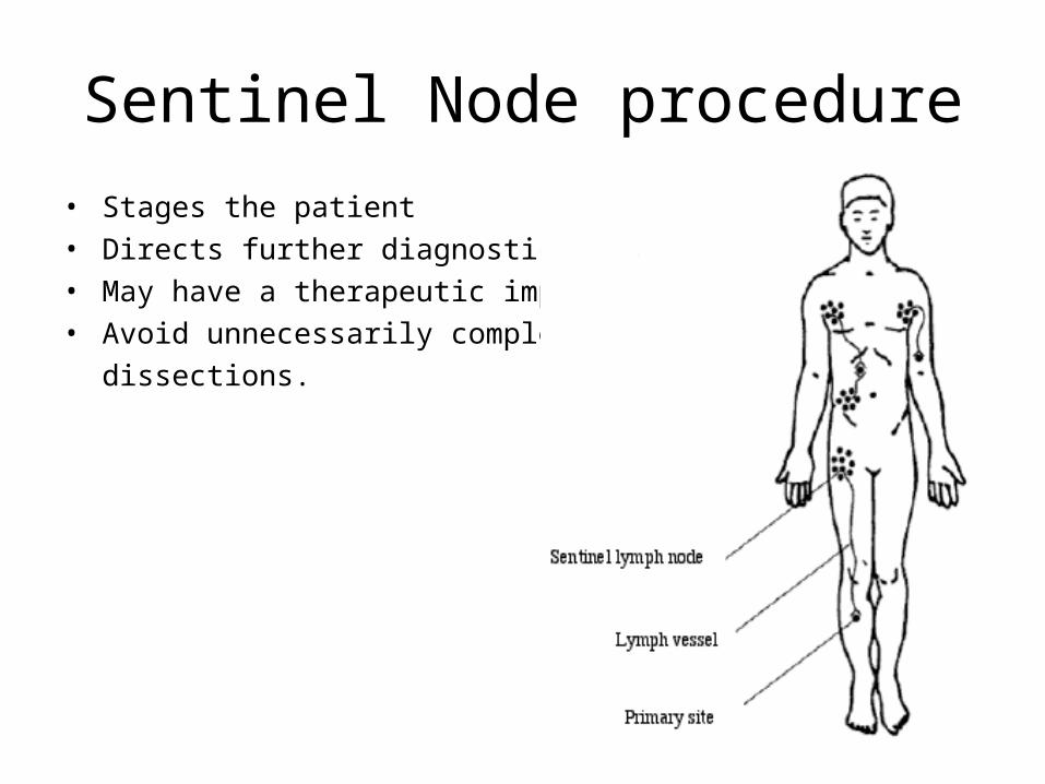
Sentinel Node procedure
• Stages the patient• Directs further diagnostic studies• May have a therapeutic impact• Avoid unnecessarily complex
dissections.

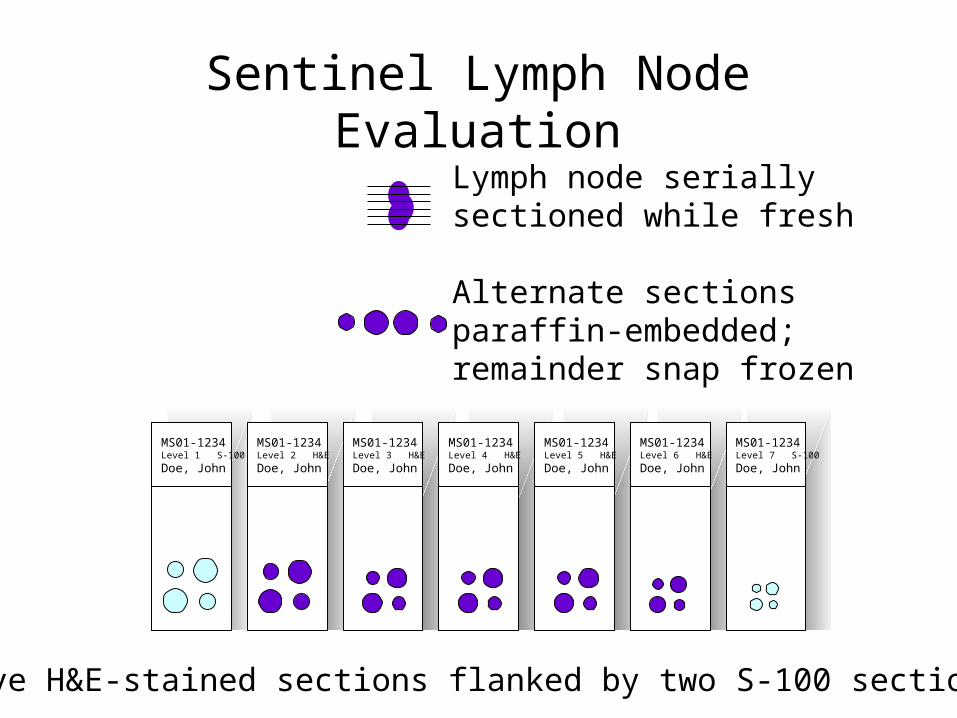
Sentinel Lymph Node Evaluation
MS01-1234Level 1 S-100
Doe, John
Lymph node serially sectioned while fresh
Alternate sections paraffin-embedded;remainder snap frozen
MS01-1234Level 2 H&E
Doe, John
MS01-1234Level 3 H&E
Doe, John
MS01-1234Level 4 H&E
Doe, John
MS01-1234Level 5 H&E
Doe, John
MS01-1234Level 6 H&E
Doe, John
MS01-1234Level 7 S-100
Doe, John
Five H&E-stained sections flanked by two S-100 sections

Indications for the Sentinel Lymph Node Procedure
• Thin melanomas (< 1 mm)• Consider for ulcerated primaries or Clarks IV, V
• Intermediate thickness melanomas (1-4 mm)– Risk: 18-20%
• Thick melanomas (>4 mm)– Risk: 40%+ nodal, 15-20% systemic
Patients with Postive Lymph Nodes
• Have completion Lymph node surgery.

In Transit Melanoma

After treating the primary and completing lymph node surgery,
what is next?

Now
• You know the T stage
• You know the N stage
• You base the extent of additional studies on those two facts.

Additional Studies?
• Stage I to IIA: (up to a 4mm thick with no ulceration) No additional Studies
• Stage IIB, IIC: Additional Studies as clinically indicated (CT, PET, MRI)
• Stage III: Baseline studies for staging or symptoms---Chest x-ray, CT + PET, MRI brain
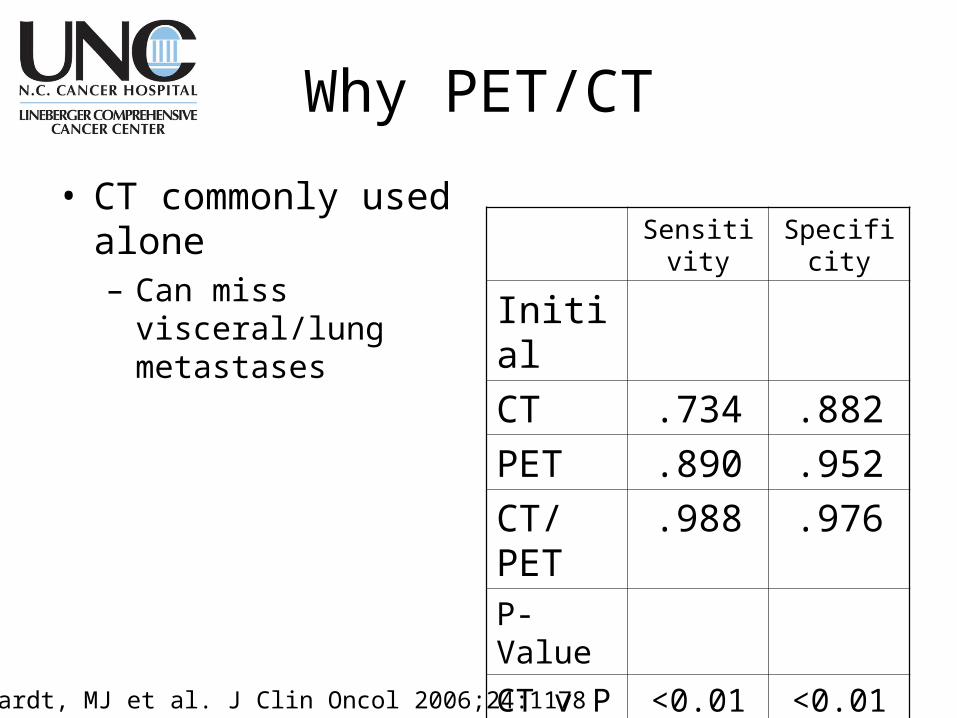
Why PET/CT
• CT commonly used alone– Can miss visceral/lung
metastases
Sensitivity Specificity
Initial
CT .734 .882
PET .890 .952
CT/PET
.988 .976
P-Value
CT v P <0.01 <0.01
CT v C/P <.00001 <.00001
P v C/P <0.01 NSReinhardt, MJ et al. J Clin Oncol 2006;24:1178

Treatment of Stage IIb (T4), IIc, and III Melanoma when all of the visible tumor has
been removed• Observation
• Clinical Trial
• Or Interferon alpha
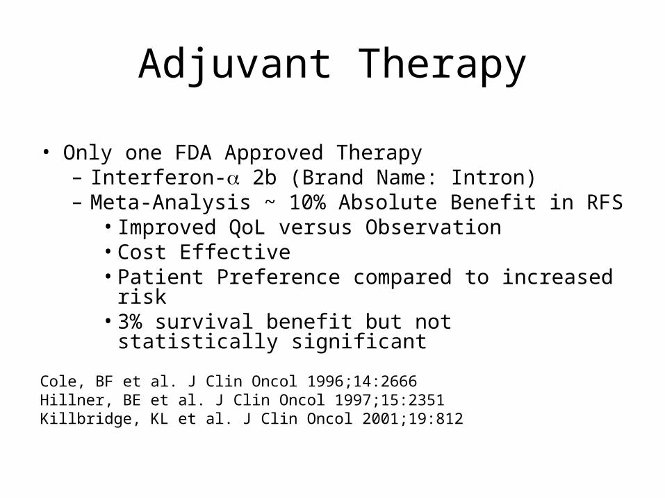
Adjuvant Therapy
• Only one FDA Approved Therapy– Interferon- 2b (Brand Name: Intron)– Meta-Analysis ~ 10% Absolute Benefit in RFS
• Improved QoL versus Observation• Cost Effective• Patient Preference compared to increased risk• 3% survival benefit but not statistically significant
Cole, BF et al. J Clin Oncol 1996;14:2666Hillner, BE et al. J Clin Oncol 1997;15:2351Killbridge, KL et al. J Clin Oncol 2001;19:812


Interferon Regimen
• IFN 2b– 20 Million Units/m2/Day IV
– (15MU often new starting point)
• 5 Days per week for 4 weeks
– 10 Million Units/m2/Day Subcutaneous• 3 times per week for 11 months
Side Effects:– Flu-like Sx, Hepatic Transaminitis, Fatigue,
HA, Nausea, Wt Loss, Myelosuppression, Depression

Adjuvant Radiation Therapy
• Consider RT to the nodal basin when there are multiple nodes involved or extranodal extension
(category IIb evidence NCCN)
Stage IV
• Median survival is 9 months and less than 5% probability of survival beyond 5 years.
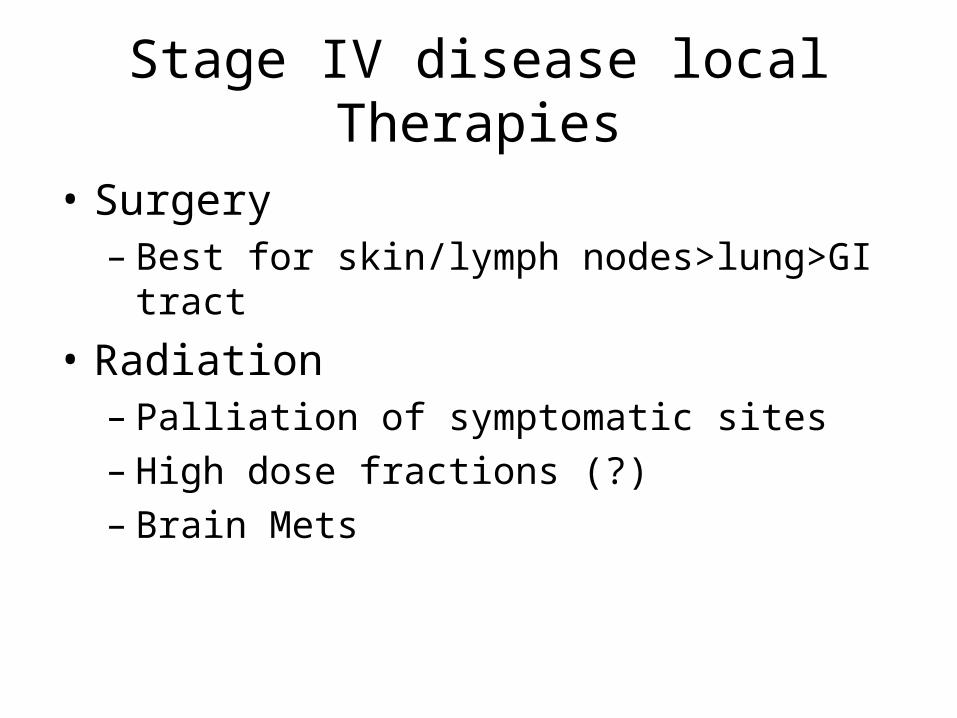
Stage IV disease local Therapies
• Surgery– Best for skin/lymph nodes>lung>GI tract
• Radiation– Palliation of symptomatic sites– High dose fractions (?)– Brain Mets

Systemic Treatments
• Where is this going– Genetic Footprint
• What Categories of treatment do we see?– Immune-therapy sensitive– Chemotherapy– Targetted Therapy

MAPK and PI3K/AKT pathwaysCell Membrane---------------------------------------------------------
SHC PI3K→AKTRAS PTEN ↓BRAFMEK ½ CCND1MAPK3 ↓
MAPK1 CDK4/6
↓ ↓ ↓ VEG CFOS ELK Proliferation Proliferation ↓
Cell cycle progression

MAPK and PI3K/AKT pathways
• Ras/Ras PI3/AKTCutaneous Not chronically sun exposed Cutaneous, Chronically sun exposed
Mucosal, Acral
Proliferative pathway Antiapototic pathway
CDKN2A gene mutations
CDK4 amplifcation in acral and mucosal

KIT
• Receptor tyrosine kinase• Critical regulator of growth of melanocytes.
• Dysfunctional KIT pigmentary defects. • Expression is lost in nevi• Amplification in chronically sun exposed
melanoma• Amplification in KIT exons 11, 13, 17 and
18 may correlate with Rx response
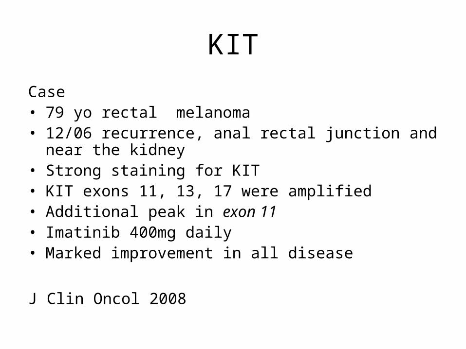
KIT
Case• 79 yo rectal melanoma• 12/06 recurrence, anal rectal junction and near the
kidney• Strong staining for KIT• KIT exons 11, 13, 17 were amplified• Additional peak in exon 11• Imatinib 400mg daily• Marked improvement in all disease
J Clin Oncol 2008

Imatinib Study
• For patients with KIT amplification or mutation
• Gleevac 400mg PO daily
Immunotherapy
• IL2
• Vaccines
• Other Modulators of the immune system– CTLA 4– New agents
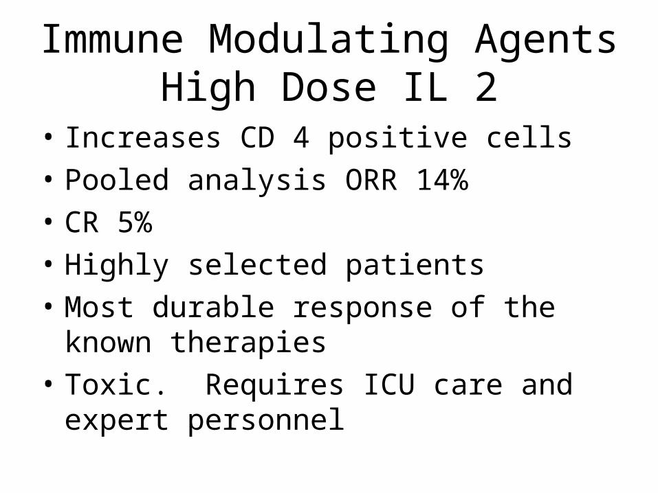
Immune Modulating AgentsHigh Dose IL 2
• Increases CD 4 positive cells
• Pooled analysis ORR 14%
• CR 5%
• Highly selected patients
• Most durable response of the known therapies
• Toxic. Requires ICU care and expert personnel
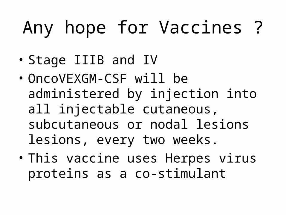
Any hope for Vaccines ?
• Stage IIIB and IV
• OncoVEXGM-CSF will be administered by injection into all injectable cutaneous, subcutaneous or nodal lesions lesions, every two weeks.
• This vaccine uses Herpes virus proteins as a co-stimulant
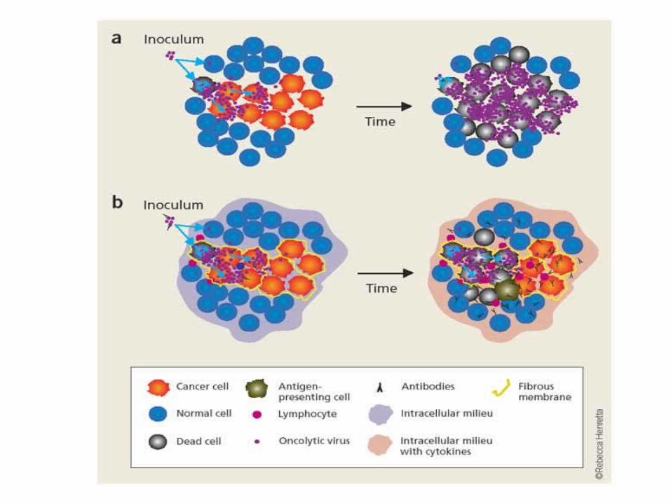

After 3 Injections

Resolution of un-injected disease in the lung. Injection sites in the knee/thigh
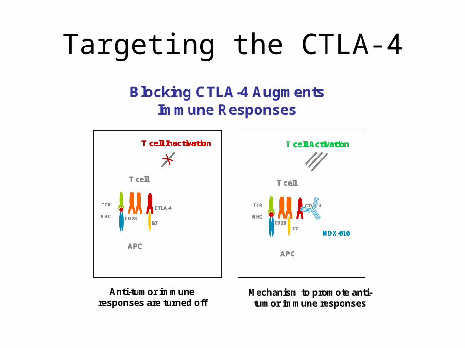
Targeting the CTLA-4
MHC
TCR
T cell
APC
CD28B7
CTLA-4
MDX-010
T cell Activation
MHC
TCR
T cell
APC
CD28
CTLA-4
T cell Inactivation
B7
MHC
TCR
T cell
APC
CD28B7
CTLA-4
MDX-010
T cell Activation
MHC
TCR
T cell
APC
CD28B7
CTLA-4
MDX-010
T cell ActivationT cell Activation
MHC
TCR
T cell
APC
CD28
CTLA-4
T cell Inactivation
B7
CTLA-4CTLA-4
T cell Inactivation
B7
T cell InactivationT cell Inactivation
B7B7
Blocking CTLA-4 Augments Immune Responses
Anti-tumor immune responses are turned off
Mechanism to promote anti-tumor immune responses

CTLA -4 Blockade: Clinical Development
• Fully human monoclonal antibodies
• Ipilimumab (MDX-010)—IgG
• Ticilimumab (CP-675,206)--IgG

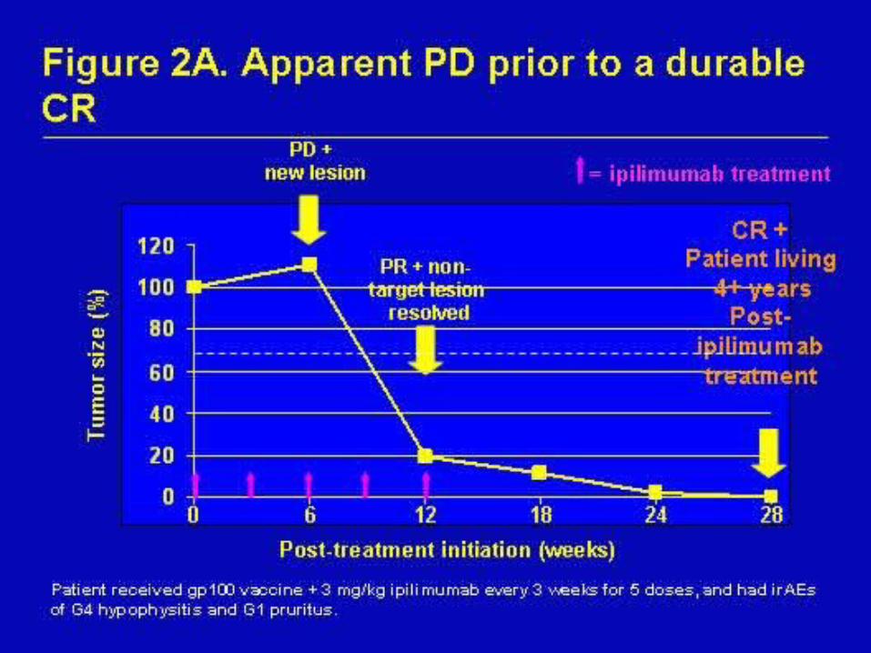
Durable Complete Response in Metastatic Melanoma
• Complete resolution of 2 subcutaneous nodules, 31 lung metastases and 0.5 cm brain metastasis. Patient previously failed chemotherapy and surgery.
• Response ongoing at 40+ months
Sources: Phan et al. PNAS. 2003 Jul;100(14):8372-8377 and Medarex unpublished data
Duration as of 1/10/05.

Immune Breakthrough Events
• Colitis
• Hepatitis
• Vitiligo
• Any autoimmune
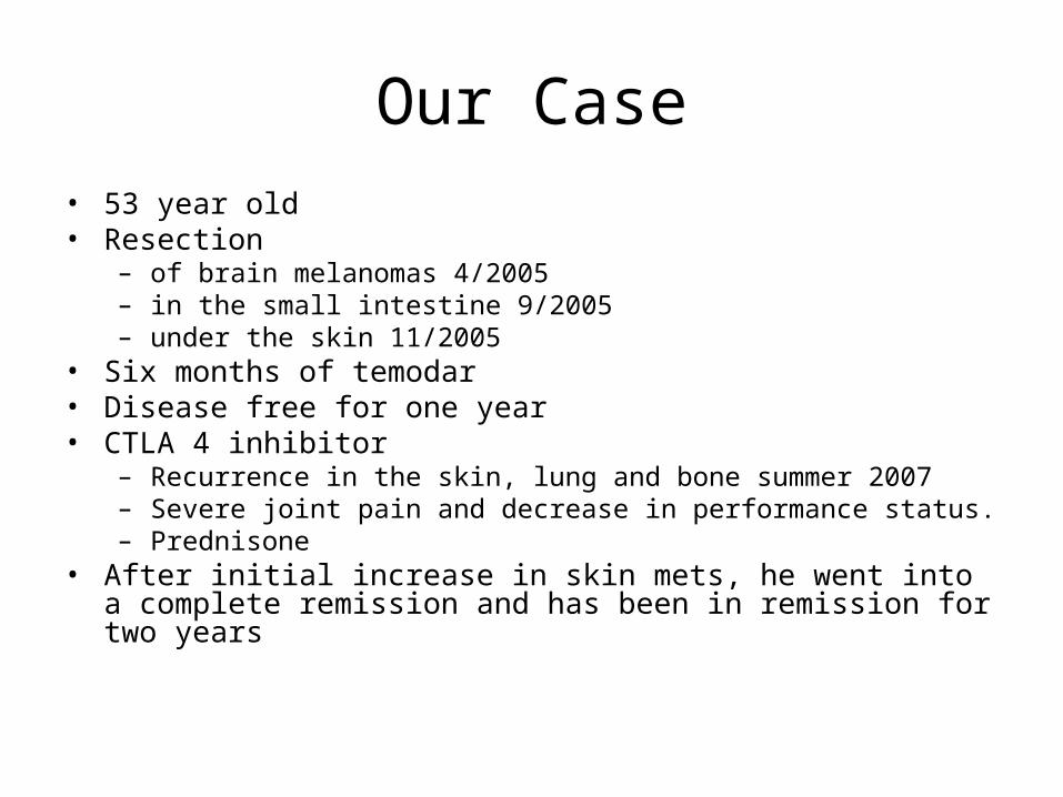
Our Case• 53 year old • Resection
– of brain melanomas 4/2005– in the small intestine 9/2005– under the skin 11/2005
• Six months of temodar• Disease free for one year• CTLA 4 inhibitor
– Recurrence in the skin, lung and bone summer 2007– Severe joint pain and decrease in performance status. – Prednisone
• After initial increase in skin mets, he went into a complete remission and has been in remission for two years
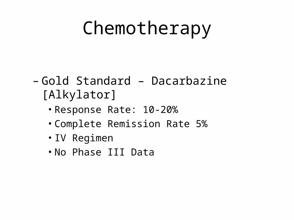
Chemotherapy
– Gold Standard – Dacarbazine [Alkylator]• Response Rate: 10-20%• Complete Remission Rate 5%• IV Regimen• No Phase III Data

Chemotherapy
• Temozolomide [Temodar™]– Oral agent converts to active metabolite of
dacarbazine– Penetrates CNS– FDA Approved for Brain Tumors only– Middleton et al: Phase III trial showing no
difference from dacarbazine
Middleton MR, Grob JJ, et al. J Clin Oncol 2000;18:158

Temodar TargetsTransmethylation of the O-6 site of guanine in DNA leads to
profound cytotoxicity
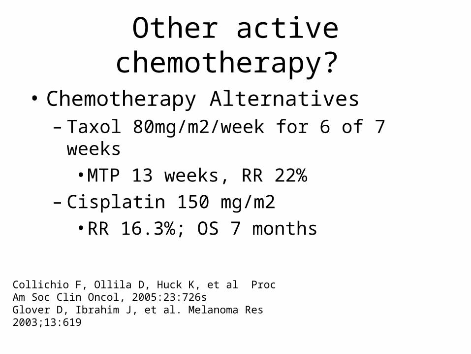
Other active chemotherapy?
• Chemotherapy Alternatives– Taxol 80mg/m2/week for 6 of 7 weeks
• MTP 13 weeks, RR 22%– Cisplatin 150 mg/m2
• RR 16.3%; OS 7 months
Collichio F, Ollila D, Huck K, et al Proc Am Soc Clin Oncol, 2005:23:726sGlover D, Ibrahim J, et al. Melanoma Res 2003;13:619

SYMMETRY• All patients received paclitaxel and ½ got
Elescomol
• First Line
• Measurable disease
• LDH less than 2.5 times normal
• No brain mets


What were the results of Symmetry
• Better response with Elesclomol and paclitaxel than paclitaxel alone
• Better time to disease progression
• But more deaths with the combination and the research is currently on hold
• UNC was number 2 in the USA for accrual

Conclusions
• Epidemiology– Sun– Tanning Booths
• Epidemic
• Types of Melanoma– Mucosal, Ocular, Cutaneous
• Recognition
Conclusion
• Surgery– With appropriate margins
• Sentinel Lymph node– For most cases except very thin– Positive nodes are followed by a completion
dissection
• Tests to do after knowing the T stage and nodal stage
• Reviewed Adjuvant Interferon Rx
Conclusions
• “Genetic footprint is the future”• Currently
– Targetted– Immune – Chemotherapy
• Research at UNC-KIT amplified or mutated-Vaccine Study of Oncolytic Virus