melanoma skin - queen's university belfast · care of patients with malignant melanoma of skin...
TRANSCRIPT

N. Ireland Cancer RegistryCentre for Clinical and Population SciencesMulhouse BuildingGrosvenor RoadBelfast BT12 6BJ T: (44) 028 9063 2573F: (44) 028 9024 8017E: [email protected]: www.qub.ac.uk/nicr CDS N110730
Care of Patients with Malignant Melanoma of Skin in Northern Ireland 2006
2006 Melanom
a Skin
Care of Patients with Malignant Melanoma of Skin in Northern Ireland 2006
Edited by Ann Smith and Anna Gavin
This report should be cited as “Smith A, & Gavin A. Care of Patients with Malignant Melanoma of Skin in Northern Ireland 2006.
N. Ireland Cancer Registry, 2008”
We welcome your comments:An evaluation form is available at www.qub.ac.uk which may be completed electronically or printed and posted.


N. IrelandCancer Registrypage 1
Melanoma 2006
Foreword 2
Acknowledgements 3
SECTION I – Introduction, Background and Methods 4
SECTION II – Results 9
Conclusions 37
SECTION III – Summary 38
References 41
Appendix A – Cases and deaths from malignant melanoma 1984 – 2005 42
Appendix B – Campbell Report: Recommendations 46
Appendix C – Staging of malignant melanoma 47

N. IrelandCancer Registry
Melanoma 2006
page 2
Foreword
This report describes the characteristics of patients diagnosed in N. Ireland with malignant melanoma of skin and their care. This process is supported by local clinicians and the recommendations will be taken forward through the NICaN tumour specific group on melanoma.
We are on a journey of continuous improvement in cancer care and this report provides valuable information which will be essential in helping us to track our progress and highlight areas where change is needed. This second report in a new series highlights the importance of the Cancer Registry as a valuable public health tool. I look forward to future reports.
Dr Michael McBride
Chief Medical Officer

N. IrelandCancer Registrypage 3
Melanoma 2006
Acknowledgements
I am grateful to the clinicians who helped with determining the data items to collect, their interpretation and final presentation.
The N. Ireland Cancer Registry is funded by the Department of Health, Social Services & Public Safety Northern Ireland (DHSSPSNI) and this project was possible thanks to grants from the four Health and Social Services Boards and G.A.I.N. (Guideline and Audit Implementation Network) previously known as the Regional Multiprofessional Audit Group (RMAG).
The quality of data in this project is a result of the work of the Registry Tumour Verification Officers especially Bernadette Anderson and Jackie Kelly who meticulously extracted detailed information from clinical records for analysis and presentation in this report. The analysis of data was undertaken by Dr Ann Smith. A special word of gratitude to the Medical Records staff of all the hospitals in Northern Ireland who have facilitated the Registry in this work.
The work of the N. Ireland Cancer Registry including the production of this report is the result of the work of the Registry team. I wish also to record my thanks to the Management Group and Council of the Registry who guide that work.
A Gavin Director, NICR
2008

N. IrelandCancer Registry
Melanoma 2006
page 4
SECTION I – Introduction, Background and Methods
This report is the second in a new series of Cancer Audit reports examining in detail the pathway of care for cancer patients in Northern Ireland. It is the first such report on Malignant Melanoma.
BACKGROUND – Malignant Melanoma
Melanoma is a malignant tumour of melanocytes which are found predominantly in skin. It is one of the rarer types of skin cancer but causes the majority of skin cancer related deaths1. Despite many years of intensive laboratory and clinical research, the sole effective cure currently is surgical resection of the primary tumour before it achieves a thickness greater than 1 mm2.
Around 160,000 new cases of melanoma are diagnosed worldwide each year3. The pattern of malignant melanoma in a population varies; in some populations more males develop it than females, but generally in a Caucasian population more females than males develop the condition. This is the case in Northern Ireland, Scotland, England and Wales where there is a record of higher levels in females. Melanoma is more common in Caucasian populations living in sunny climates than other groups4. According to a WHO Report about 48,000 melanoma related deaths occur worldwide annually5.
In Caucasian populations the recognised risk factors for the condition are fair skin, a large number of moles, sunbathing and the use of sunbeds. There is a reported trend in increasing melanoma due to increasing frequency of holidays in the sun.
There are four main types of melanoma:
(a) Superficial Spreading Melanoma that spreads superficially, is by far the most common type. (About 70 % of all cases).
(b) Lentigo Maligna usually appears as a flat or mildly elevated mottled tan, brown or dark brown skin discolouration. This type arises on chronically sun-exposed, damaged skin.
(c) Nodular Melanoma is the most aggressive of the melanomas, and is found in 10 to 15 percent of cases. It is usually invasive at the time it is first diagnosed.
(d) Acral Lentiginous Melanoma, spreads superficially before penetrating the skin more deeply. Unlike the other forms of melanoma it usually appears as a black or brown discoloration under the nails or on the soles of the feet or palms of the hands.
Others include the very rare:
Spindle Cell Melanoma – this is characterised by the epitheloid cell (spindle cell shape) naevus, and is most common in elderly male patients, but women are at risk as well.
Desmoplastic Melanoma – A rare form of melanoma with nonpigmented lesions, mostly found on head and neck.
People with melanoma will usually be treated, and in many cases cured, by having surgery to remove the lesion. If necessary, secondary surgery, and plastic surgery, such as skin grafts are carried out. The thickness of the melanoma and how deeply it has invaded the layers of the skin determines the extent of the surgery. Lymph nodes near the tumour may be removed because cancer can spread through the lymphatic system to other parts of the body. For more advanced disease, chemotherapy agents, such as Dacarbazine are utilised, or high doses of Interferon and Temozolomide may sometimes be used in specific cases.
Similarly, biological therapy/immunotherapy using substances called cytokines can be utilised. Radiation
N. IrelandCancer Registrypage 5
Melanoma 2006
therapy may be used to help control melanoma that has spread to the brain, bones, and other parts of the body. It may shrink the tumour and relieve symptoms6.
A trial was instigated in 2000 for malignant melanoma (the Middleton trial7) in an effort to gain curative therapy for malignant melanoma. Patients with stages III and IV are being randomly assigned to receive Temozolomide or Lomeguatrib, or a combination of these.
The incidence of invasive malignant melanomas in the UK has doubled over the past 20 years8 and it now claims about 1,700 lives a year in the UK.
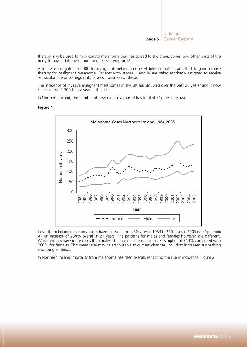
In Northern Ireland, the number of new cases diagnosed has trebled9 (Figure 1 below).
Figure 1
In Northern Ireland melanoma cases have increased from 80 cases in 1984 to 230 cases in 2005 (see Appendix A), an increase of 288% overall in 21 years. The patterns for males and females however, are different. While females have more cases than males, the rate of increase for males is higher at 340% compared with 260% for females. This overall rise may be attributable to cultural changes, including increased sunbathing and using sunbeds.
In Northern Ireland, mortality from melanoma has risen overall, reflecting the rise in incidence (Figure 2).

N. IrelandCancer Registry
Melanoma 2006
page 6
Figure 2
The number of deaths in males from melanoma rose from 26 in the year 1984 to 42 in year 2005, an increase of 162%. Deaths from melanoma in females has remained stable with no significant change throughout this time period.
Melanoma occurs more commonly in affluent populations. Socioeconomic analysis for Northern Ireland10 show that there were statistically significant higher rates in EASRs (European Age Standardised Rates) of malignant melanoma for new cases with increasing affluence (p<0.01). Males living in the most affluent areas had an EASR of 13.9 compared with 6.8 per 100,000 for males living in the most deprived areas. Similarly, females living in the most affluent areas had higher EASR incidence of malignant melanoma than those living in the most deprived areas (18.3 vs 8.4 per 100,000 respectively). Mortality rates from malignant melanoma were considerably lower than incidence, with no statistically significant patterns detectable with deprivation for either males, females or both combined.
The Campbell report of 199611 set the standards required in Northern Ireland for the care of cancer patients. (Summary in Appendix B.)
The proposed management of patients with cancer was outlined, proposing multidisciplinary, multiprofessional specialist cancer teams and the setting up of a major cancer centre at the City Hospital and Cancer Units for each Health Board in Northern Ireland. While there was no special recommendation for the control of malignant melanoma or other skin cancers, the Department produced strategies for the control of malignant melanoma and other skin cancers in 199912 and a Working Group produced a report on Skin Cancer in 200413. Both had a section on cancer treatment. The most recent document recommends the formation of a multidisciplinary melanoma audit team at each of the Cancer Units to review cases with a view to directing patients along agreed pathways of treatment.

N. IrelandCancer Registrypage 7
Melanoma 2006
Guidelines
Guidelines have been produced for the management of melanomas14, by a multidisciplinary working party related to the British Association of Dermatologists. These recommendations include the procedure for biopsy and excision of suspected melanomas, the histopathology reporting in detail and the recording of patient and family histories. Rapid referral, diagnosis and treatment are the key to successful management.
Targets include the patient being seen by a clinician specialising in the area, within two weeks of a •referral letter from the G.P.
Treatment of the primary lesion depends on the type (e.g. whether in situ or not) of melanoma and •the depth. Margins for excision range from 0.5 cm for lesions of less than 0.75 mm depth to 3 cm for lesions of greater than 4 mm depth.
Imaging investigations are not considered necessary for stages I and IIA patients, but patients with stage •IIB and over should have chest X-rays, ultrasound, CT scans, full blood counts and liver function tests.
Ideally patients with stage IIB or over should be managed in a Cancer Centre by a multidisciplinary team. •If clinical trails are available then these patients should be considered for their suitability to participate.
If there is any evidence of lymph node spread then the lymph node region should be excised radically. •Patients with further spread should be referred to oncologists, but current therapies are mostly palliative, •apart from further surgical resections.
All patients with invasive melanoma should be followed up every 3 months for 3 years. Where the •melanoma thickness was less than 1 mm the patient may be discharged; others should be followed up for a further two years at 6 monthly intervals.
At risk individuals should be encouraged to practice sun care and examine their moles on a regular •basis.
The National Institute for Health and Clinical Excellence (NICE) have also issued guidelines for health care management for people with skin cancer15. With respect to melanomas, they recommend the involvement of specialist skin cancer multidisciplinary teams, attached to Cancer Centres. Nationally agreed protocols should be adhered to and appropriate follow up care agreed. Because of the steady rise in incidence of melanomas, information on skin cancers should be collected and studied, in adequately funded cancer registries.
N. IrelandCancer Registry
Melanoma 2006
page 8
Study Aim
The aim of the report is to review the process of care for malignant melanoma patients diagnosed in Northern Ireland during 2006. This is the first population based audit on this condition here.
Study Methods
Data collection
Registry tumour verification officers (TVOs) collected data by reviewing clinical notes on patients diagnosed in the calendar year 2006 with malignant melanoma (ICD C43). The data included presenting symptoms, co-existing illnesses, investigations, treatment, staging, onward referral and survival. The data are not incidences because sufficient information was not available on all cases, but should reflect the patterns of occurrence of malignant melanoma and its treatment in Northern Ireland.
Data were entered into an electronic proforma, devised with the help of local clinicians; copy available at www.qub.ac.uk/nicr.
Exclusions
Patients were excluded if they resided outside Northern Ireland, if their records lacked sufficient information, or if the diagnosis was not primary melanoma diagnosed in 2006.
Method of data analysis
The data was imported into EXCEL for cleaning, sorting and validation and then analysis was carried out in SPSS v1516. Comparisons between groups were tested by ANOVA.

N. IrelandCancer Registrypage 9
Melanoma 2006
SECTION II – Results
Whilst 254 patients were registered with invasive malignant melanoma in Northern Ireland in 2006, this report has recorded details of 248 cases which have sufficient information to be included. Of these 145 were females and 103 were males. In addition to the 254 patients with invasive malignant melanoma there were, in 2006, 85 female and 63 male in situ melanomas which are not included in this audit.
Study patients
Patients Number of patients (%)
Total patients 254
Exclusions – lack of information 6
Total males 103 (41.5%)
Total females 145 (58.5%)
Total 248 (100%)
Average age at diagnosis – male 61 years
Average age at diagnosis – female 57 years
Melanoma is more common in females than males (3:2).•Males diagnosed with malignant melanoma are on average slightly older than females (p<0.001). •Nine patients (3.6%) were aged less than 25 years, 76 (30.6%) were aged between 25 and 50 years •and 163 (65.7%) were aged over 50 years.
Pattern with Deprivation
Deprivation quintile Number of patients (%)
Quintile 5 (most deprived) 35 (14.1%)
Quintile 4 32 (12.9%)
Quintile 3 57 (23.0%)
Quintile 2 49 (19.8%)
Quintile 1 (least deprived) 75 (30.2%)
Total 248 (100%)
Note: In the general population, if the disease is not linked with deprivation then it would be expected that 20% of all cases of disease would fall in each quintile.
In this group, more melanomas than expected occurred in the less deprived, more affluent, sectors of •the population, with more than twice as many in the least deprived quintile (Q1) compared with the most deprived (Q5). This is a significant finding (p<0.001).

N. IrelandCancer Registry
Melanoma 2006
page 10
Family History
Number of patients (% total)
Family history of melanoma 7 (2.8%)
Family history of skin cancer 5 (2.0%)
Family history of other cancer 39 (15.7%)
No family history of cancer 29 (11.7%)
Family history not recorded 174 (70.2%)
Note: Patients could record more than one family history, of more than one cancer.
Family history was poorly recorded.•A positive family history of malignant melanoma was recorded in 7 cases and indicates that this is not •a major risk factor (2.8%) in this group. This does not concur with research carried out in the USA that has tried to quantify the risk posed by a family history of melanoma. Clinical records of family members were searched and they found higher levels (17%) had a family history of melanoma, when searching records rather than that reported by asking the patient17.
Source of referral to specialist care
Source of referral Number of patients (%)
G.P. 208 (83.9%)
Dermatology clinics 17 (6.8%)
Physician 9 (3.6%)
A&E/self referral 1 (0.4%)
Private sector 2 (0.8%)
Not recorded 9 (3.6%)
Other 2 (0.8%)
Total 248 (100%)
Note: “Physician” included in-patient referrals for patients admitted to hospital for other conditions, “Other” includes referrals from podiatry and nurse practitioners
Most patients (84%) were referred by their G.P., while 7% were already under review by Dermatology •clinics that they had been attending for other skin conditions. About 8% were referred from other sources.
One hundred and thirty one patients (52.8%) were urgent referrals, whilst 15 (6.0%) were recorded as •semi-urgent referrals.
Patients presenting within their own Health Board
Board of residence Number of patients (% of patients in that board) presenting within own Board
NHSSB 39 (69.0%)
EHSSB 104 (97.8%)
SHSSB 48 (85.2%)
WHSSB 28 (92.9%)
Most patients presented in their own Health Board of residence, however, this was less so for the •Northern Board residents, many of whom presented to the Eastern Board.
N. IrelandCancer Registrypage 11
Melanoma 2006
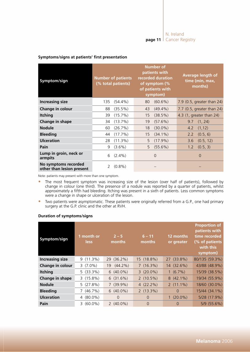
Symptoms/signs at patients’ first presentation
Symptom/signNumber of patients (% total patients)
Number of patients with
recorded duration of symptom (% of patients with
symptom)
Average length of time (min, max,
months)
Increasing size 135 (54.4%) 80 (60.6%) 7.9 (0.5, greater than 24)
Change in colour 88 (35.5%) 43 (49.4%) 7.7 (0.5, greater than 24)
Itching 39 (15.7%) 15 (38.5%) 4.3 (1, greater than 24)
Change in shape 34 (13.7%) 19 (57.6%) 9.7 (1, 24)
Nodule 60 (26.7%) 18 (30.0%) 4.2 (1,12)
Bleeding 44 (17.7%) 15 (34.1%) 2.2 (0.5, 6)
Ulceration 28 (11.3%) 5 (17.9%) 3.6 (0.5, 12)
Pain 9 (3.6%) 5 (55.6%) 1.2 (0.5, 3)
Lump in groin, neck or armpits 6 (2.4%) 0 0
No symptoms recorded other than lesion present 2 (0.8%) – –
Note: patients may present with more than one symptom.
The most frequent symptom was increasing size of the lesion (over half of patients), followed by •change in colour (one third). The presence of a nodule was reported by a quarter of patients, whilst approximately a fifth had bleeding. Itching was present in a sixth of patients. Less common symptoms were a change in shape or ulceration of the lesion.
Two patients were asymptomatic. These patients were originally referred from a G.P., one had primary •surgery at the G.P. clinic and the other at RVH.
Duration of symptoms/signs
Symptom/sign1 month or
less2 – 5
months6 – 11
months12 months or greater
Proportion of patients with time recorded (% of patients
with this symptom)
Increasing size 9 (11.3%) 29 (36.2%) 15 (18.8%) 27 (33.8%) 80/135 (59.3%)
Change in colour 3 (7.0%) 19 (44.2%) 7 (16.3%) 14 (32.6%) 43/88 (48.9%)
Itching 5 (33.3%) 6 (40.0%) 3 (20.0%) 1 (6.7%) 15/39 (38.5%)
Change in shape 3 (15.8%) 6 (31.6%) 2 (10.5%) 8 (42.1%) 19/34 (55.9%)
Nodule 5 (27.8%) 7 (39.9%) 4 (22.2%) 2 (11.1%) 18/60 (30.0%)
Bleeding 7 (46.7%) 6 (40.0%) 2 (13.3%) 0 15/44 (34.1%)
Ulceration 4 (80.0%) 0 0 1 (20.0%) 5/28 (17.9%)
Pain 3 (60.0%) 2 (40.0%) 0 0 5/9 (55.6%)

N. IrelandCancer Registry
Melanoma 2006
page 12
Duration of symptoms was moderately well recorded.•Bleeding, ulceration and pain, although not the most common symptoms, tended to be endured by the •patient for less time than changes in size, shape or colour.
The majority (n=18) of the 27 patients who had noticed increasing size of their lesion for over 12 •months had stages I – II disease at diagnosis (Breslow thickness 0.2 mm to 3.5 mm, with 10 of these with Breslow depth greater than 1 mm). One patient had a IIIB staged lesion (Breslow thickness 9.4 mm). All others (n=8) had insufficient information for staging, but had Breslow thicknesses of between 0.2 mm and 3.5 mm, with three of these having thicknesses of greater than 1 mm.
All patients who noticed change of lesion colour over at least 12 months (n=14), and with recorded •staging of the lesion had stages I – II. One of these patients with stage IB, Breslow depth 1.15 mm, died in the first year after diagnosis of malignant melanoma.
All patients who had noticed a change of shape for at least 12 months (n=8), and with recorded staging •of the lesion had final stages I – II (Breslow depth 0.4 mm to 1.4 mm). All patients in this category were alive at 1 year after diagnosis.
Duration of any symptoms by gender
1 month or less
2 – 5 months
6 – 11 months
12 months or more
Not recorded
No symptoms
Male 21 (20.4%) 22 (21.4%) 9 (8.7%) 7 (7.0%) 43 (42.2%) 1 (0.7%)
Female 18 (12.4%) 53 (36.6%) 24 (16.7%) 46 (31.7%) 3 (2.1%) 1 (1.0%)
All 39 (15.7%) 75 (30.2%) 33 (13.3%) 53 (21.4%) 46 (18.5%) 2 (0.8%)
Note: these are symptoms, not cases, as patients had more than one symptom. Two patients, one male, one female had no symptoms recorded.
Recording of symptom duration was better for women than men.•Of those recorded, women tended to have symptoms longer than men. •
Co-morbidities – Existing illness
Co-morbidity Number of patients (%)
Chronic Obstructive Pulmonary Disease 5 (2.1%)
Dementia 3 (1.2%)
Cerebrovascular disease 10 (4.1%)
Psychiatric condition 3 (1.2%)
Previous Basal Cell Carcinoma 21 (8.7%)
Previous melanoma 3 (1.2%)
Previous naevus 35 (14.5%)
Ischaemic heart disease 21 (8.7%)
Epilepsy/neurological disease 3 (1.2%)
Other malignancies 18 (7.4%)
No other morbidity reported 123 (50.8%)
Note: patients may have had more than one co-morbidity.
Melanoma patients had few co-morbidities overall compared with other cancer patients (see previous •Audit Reports from NICR).
Three patients had a previous melanoma on a completely different site. •Fourteen percent had previous naevus (non-malignant) recorded.•Of the “Other malignancies”, 5 were breast cancer. •

N. IrelandCancer Registrypage 13
Melanoma 2006
Site of malignant melanoma
SiteMales N (%)
Females N (%)
Number of patients N (%)
C43.1 (Eyelid) 1 (1.0%) 1 (0.7%) 2 (0.8%)
C43.2 (Ear and aural canal) 6 (5.8%) 2 (1.4%) 8 (3.2%)
C43.3 (Face) 15 (14.6%) 19 (13.1%) 34 (13.7%)
C43.4 (Scalp and neck) 7 (5.8%) 1 (0.7%) 8 (3.2%)
C43.5 ((Main torso) 41 (39.8%) 17 (11.7%) 58 (23.4%)
C43.6 (Upper limb, shoulder) 21 (20.4%) 41 (28.3%) 62 (25.0%)
C43.7 (lower Limb, hip) 12 (11.7%) 64 (43.4%) 76 (30.6%)
ALL SITES 103 145 248
All patients had site of melanoma recorded. •The most frequent site for melanoma in male was the main torso, with more than two times as many •males presenting with melanoma on this part of the body than females.
The greatest difference between the sexes was for the lower limb, with over five times as many females •as males presenting with melanoma at this location. Males had almost twice the percentage of lesions on the head and neck. These differences are all highly significant (p<0.001).
These patterns are similar to previous melanoma results for Northern Ireland and reflect patterns of skin •sun exposure.

N. IrelandCancer Registry
Melanoma 2006
page 14
History of Sun ExposureFifty eight patients (24.0%) had significant sun exposure recorded in the notes.•Durations for living abroad were recorded of greater than 2 years for six patients and six months for •one patient.
Comments recorded were:•“Sunny holidays” (2). –“Multiple sunburn over the years”. –“Sunburn in the past”. –“Sunburn and sun beds” (2). –“Sun worshipper”. –“Sun exposure in youth”. –“Prolonged sun exposure” (2). –
Patient Pathways
This diagram illustrates the patient pathways from referral.
Patients either attended their G.P. first (84%), were already attending dermatology clinics (7%), or presented from diverse sources, such as private clinics or other hospital clinic (Others, 8%).
The majority of patients (69%) were referred directly to a dermatology clinic by a General Practitioner. Patients could be referred to an oncologist after these initial pathways. Three percent of patients had initial removal of their lesion at their G.P. clinic, and were then referred to secondary care.
Note: Patient pathways for ‘others’ are diverse, can include many sources. Some patients did not have the pathway recorded.
157 patients (63% of total) were referred to a plastic surgeon, 9% from G.P. directly. Ninety seven •patients (41%) had further surgery performed in plastic surgery.
Eighty nine patients (36% of total) were referred to oncology after these initial pathways. •

N. IrelandCancer Registrypage 15
Melanoma 2006
Number of hospitals attended
Of the 248 patients in the final dataset, 106 attended one hospital for treatment, 96 attended two •hospitals and 46 attended three hospitals.
Number of hospitals attended
One hospital
Note: Belvoir Park closed in March 2006, so numbers for the Belfast City Hospital and this are combined for this year. The figures are for the one hospital attended.

N. IrelandCancer Registry
Melanoma 2006
page 16
The greatest number of patients who went to one hospital only (n=27, 10.9% of all patients) were •referred to the Ulster Hospital, where there is a specialist plastic surgery unit.
Many patients who attended only one hospital (n=18, 7.3%) were referred to Craigavon Hospital, the •Royal Victoria Hospital (n=14, 5.6%) or went to private clinics (n=7, 2.8%).
Most patients who attended two hospitals and had attended another hospital initially, were referred •to the Ulster Hospital’s plastic surgery unit as their second hospital (n=36). Numbers attending two hospitals were 96 (38.7% of all patients).
Some patients attending just two hospitals were referred on to the Cancer Centre at Belfast City/Belvoir •Park (n=11, 4.4%).
Most patients who attended at least three hospitals (17 patients) were referred on to the Ulster Hospital, •after having attended two other hospitals.
Total new melanoma patients ever attending each hospital for treatment 2006
HospitalTotal numbers of
attendances (% of total patients)
Numbers attending this hospital if only one hospital
attended (n=106)
Altnagelvin 12 (4.8%) 4 (3.8%)
Roe Valley 2 (0.8%) 0 (0%)
Erne 8 (3.2%) 4 (3.8%)
Tyrone County 5 (2.0%) 1 (0.9%)
Antrim 17 (6.9%) 3 (2.8%)
Causeway 15 (6.0%) 8 (7.5%)
Mid Ulster 7 (2.8%) 3 (2.8%)
Whiteabbey 6 (2.4%) 1 (0.9%)
Belfast City/Belvoir Park 46 (18.5%) 10 (9.4%)
Lagan Valley 11 (4.4%) 3 (2.8%)
Royal Victoria 37 (14.9%) 14 (13.2%)
Downe 4 (1.6%) 2 (1.9%)
Ulster 138 (55.6%) 27 (25.5%)
Daisy Hill 13 (5.2%) 1 (0.9%)
Craigavon 41 (16.5%) 18 (17.0%)
South Tyrone 3 (1.2%) 0 (0%)
Private Clinics 17 (6.9%) 7 (6.6%)
Note: Patients could have attended more than one hospital, but percentages are of total patients (248).
56% of patients attended the Ulster Hospital, at some stage of their treatment.•4.8% of patients had some or all of their treatment in the private sector.•76.5% of patients were seen at a cancer unit at some stage of their treatment.•

N. IrelandCancer Registrypage 17
Melanoma 2006
Hospital/Clinic of diagnosis/primary surgery and secondary surgery
HospitalPrimary surgery for diagnosis.
Number of patients (%)Location of secondary surgery.
Number of patients (%)
Altnagelvin 9 (3.6%) 4 (1.6%)
Roe Valley 2 (0.8%) 0
Erne 7 (2.8%) 4 (1.6%)
Tyrone County 3 (1.2%) 2 (0.8%)
Mid Ulster 5 (2.0%) 1 (0.4%)
Moyle 1 (0.4%) 0
Causeway 10 (4.1%) 9 (3.6%)
Coleraine 1 (0.4%) 1 (0.4%)
Antrim 6 (2.4%) 3 (1.2%)
Whiteabbey 2 (0.8%) 2 (0.8%)
Ards 5 (2.0%) 2 (0.8%)
Downe 4 (1.6%) 2 (0.8%)
Bangor 3 (1.2%) 2 (0.8%)
Belfast City/Belvoir Park 26 (10.6%) 11 (4.4%)
Lagan Valley 5 (2.0%) 3 (1.2%)
Royal Victoria 23 (9.3%) 19 (7.7%)
Ulster 56 (22.6%) 100 (40.3%)
Daisy Hill 3 (1.2%) 0
Armagh Community 1 (0.4%) 0
Craigavon 25 (10.2%) 19 (7.8%)
Banbridge 2 (0.8%) 2 (0.8%)
South Tyrone 5 (2.0%) 1 (0.4%)
G.P. clinics 32 (13.2%) 0 (0%)
Private Clinics 12 (4.8%) 12 (4.8%)
No further surgery 49 (19.8%)
Total 248 (100 %) 248 (100%)
Thirteen percent of patients had their lesions removed by G.P.s, who have minor surgery clinics, and who •carried out punch biopsies or simple excision of lesions. These were usually diagnostic procedures.
The Ulster Hospital carried out the highest rate (40%) of the secondary surgery for wider excisions after •histology indicated malignant melanoma.
Others were referred to several different places for secondary surgery, including Craigavon Hospital •dermatology clinic (7.8%), the Royal Victoria Hospital (7.7%) or the Cancer Centre at City/Belvoir Hospitals (4.4%).
N. IrelandCancer Registry
Melanoma 2006
page 18
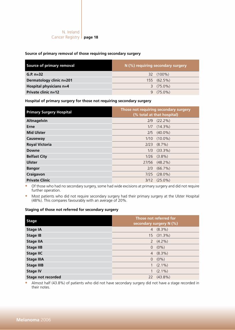
Source of primary removal of those requiring secondary surgery
Source of primary removal N (%) requiring secondary surgery
G.P. n=32 32 (100%)
Dermatology clinic n=201 155 (62.5%)
Hospital physicians n=4 3 (75.0%)
Private clinic n=12 9 (75.0%)
Hospital of primary surgery for those not requiring secondary surgery
Primary Surgery Hospital Those not requiring secondary surgery
(% total at that hospital)
Altnagelvin 2/9 (22.2%)
Erne 1/7 (14.3%)
Mid Ulster 2/5 (40.0%)
Causeway 1/10 (10.0%)
Royal Victoria 2/23 (8.7%)
Downe 1/3 (33.3%)
Belfast City 1/26 (3.8%)
Ulster 27/56 (48.2%)
Bangor 2/3 (66.7%)
Craigavon 7/25 (28.0%)
Private Clinic 3/12 (25.0%)
Of those who had no secondary surgery, some had wide excisions at primary surgery and did not require •further operation.
Most patients who did not require secondary surgery had their primary surgery at the Ulster Hospital •(48%). This compares favourably with an average of 20%.
Staging of those not referred for secondary surgery
Stage Those not referred for
secondary surgery N (%)
Stage IA 4 (8.3%)
Stage IB 15 (31.3%)
Stage IIA 2 (4.2%)
Stage IIB 0 (0%)
Stage IIC 4 (8.3%)
Stage IIIA 0 (0%)
Stage IIIB 1 (2.1%)
Stage IV 1 (2.1%)
Stage not recorded 22 (43.8%)
Almost half (43.8%) of patients who did not have secondary surgery did not have a stage recorded in •their notes.

N. IrelandCancer Registrypage 19
Melanoma 2006
Investigations
Frequency of investigations carried out on patients with malignant melanoma by hospital of primary diagnostic surgery
Hospital of primary diagnostic surgery
CT scans Number of patients (%)
PET scans Number of patients (%)
Ultrasound Number of patients (%)
Erne 4/7 (57.1%) 0/7 (0%) 0/7 (0%)
Altnagelvin 3/9 (33.3%) 4/9 (44.4%) 0/9 (0%)
Roe Valley 1/2 (50.0%) 0/2 (0%) 1/2 (50%)
Tyrone County 1/3 (33.3%) 1/3 (33.3%) 1/3 (33.3%)
Antrim 1/6 (16.7%) 1/6 (16.7%) 0/6 (0%)
Moyle 1/1 (100%) 0/1 (0%) 0/1 (0%)
Causeway 1/10 (10.0%) 2/10 (20.0%) 2/10 (20.0%)
Coleraine 1/1 (100%) 0/1 (0%) 0/1 (0%)
Mid Ulster 0/5 (0%) 0/5 (0%) 1/5 (20.0%)
Whiteabbey 0/2 (0%) 0/2 (0%) 0/2 (0%)
Ards 1/5 (20.0%) 2/5 (40.0%) 0 (0%)
Bangor 0/3 (0%) 0/3 (0%) 0 (0%)
Belfast City/Belvoir Park 3/26 (11.5%) 1/26 (3.8%) 2/8 (25%)
Armagh Community 0/1 (0%) 0/1 (0%) 0/1 (0%)
Downe 0/3 (0%) 1/3 (33.3%) 1/3 (33.3%)
Royal Victoria 4/23 (17.4%) 5/23 (21.7%) 0/23 (0%)
Ulster 22/56 (39.3%) 6/56 (10.7%) 4/56 (7.1%)
Lagan Valley 0/5 (0%) 0/5 (0%) 0/5 (0%)
Craigavon 4/25 (16.0%) 3/25 (12.0%) 4/25 (16.0%)
Banbridge 2/2 (100%) 1/2 (50.0%) 0 (0%)
Daisy Hill 0/4 (0%) 0/4 (0%) 0/4 (0%)
South Tyrone 0/5 (0%) 1/5 (20.0%) 0/5 (0%)
G.P. clinics 5/32 (15.6%) 3/32 (9.4%) 0/32 (0%)
Private Clinics 3/12 (25.0%) 1/12 (8.3%) 1/12 (8.3%)
Total of all patients 57 (23.0%) 32 (12.9%) 17 (6.9%)
The percentage of patients having various investigations varied by hospital of primary diagnosis. These •investigations may have been ordered subsequent to primary diagnosis.
Four patients had MRI scans, they had primary surgery at a G.P. clinic, Altnagelvin Hospital, Ulster •Hospital or the Ulster Independent Clinic.
Of the 33 patients who had lesions of stage IIB or greater, 30 (90.9%) had Full Blood Counts carried •out, 28 (84.8%) had Liver Function Tests carried out and 27 (81.8%) had both investigations carried out. This means that most patients had these investigations, in accordance with the recommendations given in the guidelines.

N. IrelandCancer Registry
Melanoma 2006
page 20
Imaging scans by Health Board of residence
Board of residence Number of patients (% patients residing in each Board)
CT scan PET scan Ultrasound
EHSSB 22 (20.8%) 10 (9.4%) 7 (6.6%)
NHSSB 13 (19.6%) 10 (17.9%) 3 (5.4%)
SHSSB 12 (24.8%) 6 (10.9%) 6 (10.9%)
WHSSB 10 (36.0%) 6 (21.4%) 1 (3.6%)
Total 57 (23.0%) 32 (12.9%) 17 (6.9%)
There was no significant difference in the rate of PET/CT/Ultrasound scans, carried out on patients, by •area of residence.
Those who resided in the WHSSB had an apparent higher percentage of CT scans but this figure is not •significantly different (p= 0.39) compared with Northern Ireland as a whole.
Similarly, when all scans were totalled there were no significant differences between all types of scans •carried out for each Board, compared with Northern Ireland as a whole.
Histopathology
Note: percentages are of gender.
All cases had histopathology.•The most common type (51.2% of total patients) was superficial spreading followed by nodular (26.9%) •and lentigo maligna (13.2%).
The difference between males and females presenting with superficial spreading melanoma was not •significant.
The ages of spindle cell patients were 52, 58 and 87 years.•Nineteen patients (23.3%) with nodular melanoma were aged less than 50 years, whilst 47 (28.8%) •were older than 50 years. The difference is not significant.

N. IrelandCancer Registrypage 21
Melanoma 2006
Staging
TNM staging
(See Appendix C for further information on staging.)
StageMale N (%)
Female N (%)
All patients N (%)
Without secondary
surgery N (%)
With secondary
surgery N (%)
IA 19 (19.4%) 30 (20.8%) 49 (20.2%) 4 (8.3%) 45 (23.2%)
IB 33 (33.7%) 46 (31.9%) 79 (32.2%) 15 (31.3%) 64 (33.0%)
IIA 8 (8.2%) 7 (4.9%) 15 (6.1%) 2 (4.2%) 13 (6.7%)
IIB 5 (5.1%) 8 (5.6%) 13 (5.3%) 0 (0%) 13 (6.7%)
IIC 5 (5.1%) 6 (4.2%) 11 (4.5%) 1 (2.1%) 10 (3.6%)
IIIA 0 (0%) 3 (2.1%) 3 (1.2%) 0 (0%) 3 (1.5%)
IIIB 1 (1.0%) 1 (0.7%) 2 (0.8%) 0 (0%) 2 (0.5%)
IV 1 (1.0%) 3 (2.1%) 4 (1.6%) 1 (0%) 3 (1.5%)
Insufficient data 31 (30.1%) 41 (28.3%) 72 (29.0%) 26 (53.1%) 46 (23.1%)
Total 103 (100%) 145 (100%) 248 (100%) 49 (100%) 199 (100%)
A total of 176 (71%) patients had sufficient information recorded for staging by the Registry staff who •examined the notes.
The most common stage at presentation was stage 1B malignancy (32.2%). Overall, 68.3% of patients •had stages I – II, whereas 3.6% had stages III and IV recorded.
There was no difference in stage by gender.•
Patients with insufficient data for staging
Area of residenceNumber of patients (% unstaged of total
patients in each area)
NHSSB 16 (27.9%)
EHSSB 24 (23.8%)
SHSSB 22 (39.2%)
WHSSB 10 (31.6%)
Northern Ireland 72 (29.0%)
Overall 29.0% had insufficient information in the notes to allow staging to be recorded by the Registry •staff. The figures for each Health Board were similar in this respect.
N. IrelandCancer Registry
Melanoma 2006
page 22
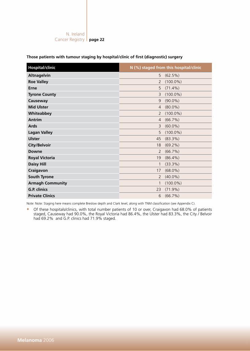
Those patients with tumour staging by hospital/clinic of first (diagnostic) surgery
Hospital/clinic N (%) staged from this hospital/clinic
Altnagelvin 5 (62.5%)
Roe Valley 2 (100.0%)
Erne 5 (71.4%)
Tyrone County 3 (100.0%)
Causeway 9 (90.0%)
Mid Ulster 4 (80.0%)
Whiteabbey 2 (100.0%)
Antrim 4 (66.7%)
Ards 3 (60.0%)
Lagan Valley 5 (100.0%)
Ulster 45 (83.3%)
City/Belvoir 18 (69.2%)
Downe 2 (66.7%)
Royal Victoria 19 (86.4%)
Daisy Hill 1 (33.3%)
Craigavon 17 (68.0%)
South Tyrone 2 (40.0%)
Armagh Community 1 (100.0%)
G.P. clinics 23 (71.9%)
Private Clinics 6 (66.7%)
Note: Note: Staging here means complete Breslow depth and Clark level, along with TNM classification (see Appendix C).
Of these hospitals/clinics, with total number patients of 10 or over, Craigavon had 68.0% of patients •staged, Causeway had 90.0%, the Royal Victoria had 86.4%, the Ulster had 83.3%, the City / Belvoir had 69.2% and G.P. clinics had 71.9% staged.

N. IrelandCancer Registrypage 23
Melanoma 2006
Symptoms at presentation by recorded TNM stage
SymptomTNM stage
IA IB IIA IIB IIC IIIA IIIB IV NR
Change in shape 10 15 3 1 1 4
Change in colour 23 24 7 5 2 2 2 23
Increasing size 27 42 10 10 8 2 2 2 32
Bleeding 2 8 4 7 7 1 3 12
Nodule 1 14 8 10 7 2 1 3 14
Ulceration 1 4 2 4 7 2 1 2 5
Itching 9 12 2 1 2 13
Pain 1 1 2 3 1 1
Lump in groin, neck or armpits 1 2 1 1 1
No recorded symptoms 1 1
Note: NR is not recorded. Patients may be recorded more than once.
Stage IV patients presented with increasing size, bleeding, nodule or ulceration. These symptoms were •however, also recorded commonly for earlier stage disease.
Diagnostic scans by TNM stage
ScanTNM stage
IA IB IIA IIB IIC IIIA IIIB IV NR Total
CT scan 3 21 7 3 7 2 2 4 8 57
PET scan 3 5 7 4 2 1 1 9 32
Ultrasound 1 3 2 4 1 2 1 3 17
Note: NR is not recorded.
Despite having scans, stage remained unrecorded in some patients.•

N. IrelandCancer Registry
Melanoma 2006
page 24
There are five Clark levels of invasion in melanoma18, defined as:Level I: Melanomas confined to the outermost layer of the skin, the epidermis. Also called “melanoma •in-situ.”
Level II: Penetration by melanomas into the second layer of the skin, the dermis. •Levels III – IV: Melanomas invade deeper through the dermis, but are still contained completely within •the skin.
Level V: Penetration of melanoma into the fat of the skin beneath the dermis, penetration into the third •layer of the skin, the subcutis.
Staging of melanoma: Example pT1a melanoma
(Breslow thickness 0.9 mm, invasion to Clark level III).
Clark level of lesion
Clark level Male Female All patients
II 7 (6.8%) 25 (17.4%) 32 (12.9%)
III 22 (21.4%) 28 (19.3%) 50 (20.2%)
IV 66 (64.1%) 79 (54.9%) 145 (58.5%)
V 4 (3.9%) 12 (8.3%) 16 (6.6%)
Not recorded 4 (3.9%) 1 (0.7%) 5 (2.0%)
Total 103 (100%) 145 (100%) 248 (100%)
Clark level IV was the most commonly recorded.•

N. IrelandCancer Registrypage 25
Melanoma 2006
Breslow depth of lesion
Breslow depth (mm) Male Female All patients
Less than 0.75 47 (45.6%) 62 (42.7%) 109 (44.0%)
0.75 – 1.50 19 (18.4%) 36 (24.8%) 55 (22.2%)
1.51 – 3.00 15 (14.6%) 26 (17.9%) 41 (16.5%)
3.01 or greater 21 (20.4%) 20 (13.8%) 41 (16.5%)
Not recorded 1 (1.0%) 1 (0.7%) 2 (0.8%)
Total 103 (100%) 145 (100%) 248 (100%)
Breslow depth less than 0.75 mm was the most commonly recorded.•There was no variation in Breslow depth/Clark level by gender.•Lesions on the torso were the same average Breslow depth as those elsewhere, with the same average •depths of 1.84 mm (range 0.2 mm, 6.5 mm) compared with 1.84 mm (range 0.03 mm, 9.6 mm) on other sites.
When the deprivation levels were categorised into two groups, 1 – 3 and 4 – 5, there was no detectable •variation in Breslow depth or Clark level with deprivation.
Clark level of lesion and Breslow depth by broad age category
Clark level n (%) patients with age less
than or equal to 50 yearsn (%) patients with age greater than 50 years
II 11 (12.9%) 21 (12.9%)
III 25 (29.4%) 25 (15.3%)
IV 47 (55.3%) 98 (60.1%)
V 0 (0%) 16 (9.8%)
NR 2 (2.4%) 3 (1.8%)
Total 85 (100%) 163 (100%) Note: NR is not recorded.
Breslow depth (mm)n (%) patients with age less
than or equal to 50 yearsn (%) patients with age greater than 50 years
less than 0.75 32 (37.6%) 77 (47.2%)
0.75 – 1.50 27 (31.8%) 28 (17.2%)
1.51 – 3.00 14 (16.4%) 27 (16.6%)
3.01+ 10 (11.8%) 31 (19.0%)
NR 2 (2.4%) 0 (0%)
Total 85 (100%) 163 (100%)
Note: NR is not recorded.
All patients with the deeper level V Clark lesions were over 50 years.•

N. IrelandCancer Registry
Melanoma 2006
page 26
Involvement of pathologist in TNM staging, Breslow depth and Clark staging, where these have been recorded
Regional pathologist involved
N (%)
Regional pathologist not involved
N (%)
Involvement of regional pathologist
not recorded N (%)
TNM stage recorded 93 (52.0%) 61 (34.1%) 25 (14.0%)
Breslow depth recorded 125 (50.4%) 79 (32.1%) 33 (13.4%)
Clark level recorded 124 (51.0%) 77 (31.7%) 32 (13.2%)
In cases where the regional pathologist was involved, TNM and Breslow depth were more likely to be •recorded.
Hospital/clinicNumber of patients with lateral margins
free (%)
Number of patients with deep margins
free (%)
Number of patients with regional
pathologist involved (%)
Altnagelvin 7/9 (77.8%) 7 (77.8%) 0 (0%)
Roe Valley 2/2 (100%) 2 (100%) 1 (50.0%)
Erne 6/7 (85.7%) 6 (85.7%) 2 (33.3%)
Tyrone County 2/3 (66.7%) 2 (66.7%) 1 (33.3%)
Mid Ulster 4/5 (80.0%) 4 (80.0%) 1 (20.0%)
Moyle 1/1 (100%) 1 (100%) 1 (100%)
Causeway 10/10 (100%) 8 (80.0%) 2 (20.0%)
Coleraine 1/1 (100%) 1 (100%) 0 (0%)
Antrim 4/6 (66.7%) 3 (50.0%) 3 (50.0%)
Whiteabbey 2/2 (100%) 1 (50.0%) 1 (50.0%)
Ards 5/5 (100%) 5 (100%) 4 (80.0%)
Downe 3/3 (100%) 2 (66.7%) 0 (0%)
Bangor 3/3 (100%) 3 (100%) 2 (66.7%)
Belfast City/Belvoir Park 24/26 (92.3%) 25 (96.2%) 11 (42.3%)
Lagan Valley 5/5 (100%) 5 (100%) 3 (60.0%)
Royal Victoria 17/23 (73.9%) 21 (91.3%) 19 (82.6%)
Ulster 51/56 (91.1%) 51 (91.1%) 46 (82.1%)
Daisy Hill 4/4 (100%) 4 (100%) 3 (75.0%)
Armagh Community 1/1 (100%) 1 (100%) 0 (0%)
Craigavon 21/25 (84.0%) 19 (76.0%) 6 (24.0%)
Banbridge 2/2 (100%) 2 (100%) 0 (0%)
South Tyrone 5/5 (100%) 5 (100%) 2 (40.0%)
G.P. clinics 20/32 (62.5%) 22 (68.8%) 13 (40.6%)
Private Clinics 9/12 (75.0%) 10 (83.3%) 5 (41.7%)
Total (n=248) 209 (84.3%) 210 (84.7%) 126 (50.8%)
Note: other patients either did not have their margins free or this was not recorded in the notes.
The regional pathologist was involved in assessing about half of the lesions. •Most patients had their lateral margins and deep margins tumour free at primary surgery (84.3% and •84.7% respectively).

N. IrelandCancer Registrypage 27
Melanoma 2006
Patients with margins recorded as being not free at primary surgery plus involvement of regional pathologist
Hospital/clinic
Number of patients with lateral margins
not free (%)
Number of patients with deep margins
not free (%)
Number of affected lesions with regional pathologist involved
(%)
Roe Valley 1/2 (50%) 0 (0%) 0 (0%)
Erne 1/7 (14.3%) 1 (14.3%) 1 (50%)
Tyrone County 1/3 (33.3%) 1 (33.3%) 1 (50%)
Mid Ulster 0/5 (0%) 1 (0%) 0 (0%)
Antrim 1/6 (16.7%) 2 (33.3%) 1 (33.3%)
Belfast City/Belvoir Park 2/26 (7.7%) 1 (3.8%) 0 (0%)
Royal Victoria 2/23 (8.7%) 0 (0%) 2 (100%)
Ulster 2/56 (3.6%) 2 (3.6%) 2 (50%)
Craigavon 1/25 (4.0%) 0 (0%) 1 (100%)
G.P. clinics 8/32 (25.0%) 3 (9.4%) 4 (36.4%)
Private Clinics 1/12 (8.3%) 0 (0%) 0 (0%)
Total (n=248) 20 (8.1%) 11 (4.4%) 12 (38.7%)
Note: some patients did not have a record as to whether their margins were free or not.
The lateral margins were not free at surgery for 8.1% of patients. The deep margins were not free at •surgery for 4.4% of patients. These patients usually have wider excision later.
The regional pathologist was involved in assessing 12 (38.7%) of the 31 lesions where either the lateral •or deep margins were not free at surgery. Seven patients had both types of margins not free at surgery, five of which were assessed by the regional pathologist.
Eight patients out of a total of 20, whose lateral margins were not free, had primary surgery at G.P. •clinics.
Similarly, for those 11 patients whose deep margins were not clear, primary surgery was performed on •3 patients at G.P. surgeries.
Of the seven patients who had both margins not clear, 3 were treated at G.P. clinics.•
Numbers of surgeons carrying out secondary operations
Number of procedures per year Number of surgeons N (% of patients)
20+ 1 28 (14.1%)
15-20 3 52 (21.0%)
10-14 1 10 (4.0%)
5-9 5 35 (17.6%)
2-4 18 52 (26.1%)
Single 18 18 (9.0%)
Not recorded - 4 (1.6%)
Total 46 199
Note: Surgeons = Consultant in charge and includes dermatologists, general surgeons and plastic surgeons.
Including dermatology, general surgery and plastic surgery there were 59 listed operators for primary •diagnostic surgery on 248 patients.
There were 46 operators recorded for secondary surgery on 199 patients with 18 single operators and •36 operators performing procedures on less than 5 patients in that year.

N. IrelandCancer Registry
Melanoma 2006
page 28
Multidisciplinary Team Meetings
There was no formal melanoma Multidisciplinary Team (MDT) meetings in place at the time of this audit. Discussion at MDT was, however, recorded for 7 patients. For the hospital of presentation, these were: 3 at Craigavon Area Hospital, 1 at Altnagelvin Hospital, 1 at Causeway Hospital, 1 at Ards Hospital, and 1 at Lagan Valley Hospital. Secondary surgery on these patients were recorded as 2 being referred to the Ulster Hospital, 1 to the Royal Victoria Hospital, 1 to Altnagelvin, 1 to Causeway hospital and 1 to a Private Clinic. One patient did not have secondary surgery. MDTs were recorded for 4 patients who had plastic surgery.
The site of lesion was not a factor in whether the patients were discussed at an MDT meeting: two patients had lesions in each site category of torso, upper limb or face, whilst one patient had a lesion on the lower limb.
ProceduresThose referred to a plastic surgeon by hospital of 1st (diagnostic) surgery
Hospital/clinic of primary diagnostic surgery N (% referred to plastic surgery by hospital)
Altnagelvin 4 (50.0%)
Roe Valley 2 (100.0%)
Erne 2 (28.6%)
Tyrone County 1 (33.3%)
Mid Ulster 2 (40.0%)
Causeway 3 (30.0%)
Antrim 3 (50.0%)
Whiteabbey 1 (50.0%)
Ulster 56 (100.0%)
Downe 1 (33.3%)
Lagan Valley 2 (40.0%)
Belfast City/Belvoir 20 (76.9%)
Banbridge 1 (50%)
Ards 5 (100.0%)
Royal Victoria 14 (52.2%)
South Tyrone 4 (80.0%)
Craigavon 6 (24.0%)
Daisy Hill 2 (66.7%)
G.P. clinics 25 (71.9%)
Private clinics 3 (27.3%)
Total 157
A total of 157 patients (62.3%) were referred to a plastic surgeon and 97 (63%) of these had further •surgery.
Final stage was recorded for 115 (74%) of these referred patients (not shown). •The average wait to be seen by a plastic surgeon from referral, for those 144 patients who had •both dates recorded, was 24 days (median time 11 days). The minimum time was 0 days when a patient had plastic surgery the same day as referral and the maximum was 495 days. This patient was lost in the system because of address change. Of those 4 patients who waited for plastic surgery of more than 100 days, referral was to the Ulster Hospital. These patients were either non-attendees at clinics or had changed address.
Delays in treatment by plastic surgery were also noted due to waits for complete staging, which would •require scans, etc.

N. IrelandCancer Registrypage 29
Melanoma 2006
Tumour staging of 157 patients referred to a plastic surgeon, compared with secondary surgery
Stage of tumourNo secondary
surgery N (%)
Any secondary surgery N (%)
Total tumoursNumber (%) of
patients referred to plastic surgery
IA 4 (8.2%) 45 (45.9%) 49 (100%) 23 (46.9%)
IB 15 (19.0%) 64 (81.0%) 79 “ 53 (67.0%)
IIA 2 (13.3%) 13 (86.7%) 15 “ 12 (80.0%)
IIB 0 (0%) 13 (100%) 13 “ 12 (92.3%)
IIC 4 (36.4%) 7 (63.6%) 11 “ 9 (81.8%)
IIIA 0 (0%) 3 (100%) 3 “ 3 (100%)
IIIB 1 (50%) 1 (33.3%) 2 “ 1 (50.0%)
IV 1 (25.0%) 3 (75.0%) 4 “ 3 (75.0%)
Not Recorded 22 (30.6%) 50 (69.4%) 72 “ 41 (56.9%)
Total 49 (19.8%) 199 (80.2%) 248 (100%) 157 (63.3%)
There were no differences in the staging level between those referred to a plastic surgeon compared •with those who had secondary surgery.
Clark level of those referred to a plastic surgeon
Clark level (Total patients with this level)
Number of patients referred to plastic
surgeon (%)
Number of patients not referred to a plastic surgeon
(%)
Number (%) of patients with no record of being
referred to a plastic surgeon
II 17 (53.1%) 12 (37.5%) 3 (9.4%)
III 28 (56.0%) 14 (28.0%) 8 (16.0%)
IV 94 (64.8%) 32 (22.0%) 19 (13.1%)
V 14 (87.5%) 1 (6.3%) 1 (6.3%)
Not Recorded 4 80.0%) 0 (0%) 1 (20.0%)
Total 157 59 32
A higher percentage of those with Clark level IV and V were referred to a plastic surgeon than levels •II and III.
Four patients who were referred to a plastic surgeon had no record of Clark level in their notes.•

N. IrelandCancer Registry
Melanoma 2006
page 30
Breslow depth recorded for those referred to a plastic surgeon
Breslow depth (mm)Number (%) of
patients referred to a plastic surgeon
Number (%) of patients not referred to a plastic surgeon
Number (%) of patients with no record of being
referred to a plastic surgeon
less than 0.75 67 (61.5%) 26 (23.9%) 16 (14.7%)
0.75 – 1.50 35 (63.6%) 15 (27.3%) 5 (9.1%)
1.51 – 3.00 22 (53.7%) 11 (26.8%) 8 (19.5%)
3.01+ 32 (78.0%) 6 (14.6%) 3 (7.3%)
Not recorded 1 (50.0%) 1 (50.0%) 0 (0%)
Total 157 (63.3%) 59 (23.0%) 32 (12.9%)
Mean depth 2.20 mm 1.26 mm 0.97 mm
Min depth 0.03 mm 0.17 mm 0.25 mm
Max depth 15.60 mm 7.00 mm 5.00 mm
Note: These Breslow depth subsets allow comparison of our data with standard American Joint Committee on Cancer (AJCC) categories and with classification schemes used in other publications and institutions. The depth relates to the thickness of the tumour penetrating into the skin and can be related to the prognosis of the patient.
Only one patient who was referred to a plastic surgeon did not have their Breslow depth recorded. •Those with Breslow depth greater than 3 mm were more likely to be referred to a plastic surgeon •(78.0% of patients compared with 14.6% who were not referred).
The mean depth of lesion of those sent to a plastic surgeon (2.20 mm) was greater than that of those •patients not referred to a plastic surgeon (1.26 mm). Those with no record of referral to a plastic surgeon had mean depth of 0.97 mm.
The minimum depth of lesion was 0.03 mm of those referred to a plastic surgeon compared with 0.17 •mm for those not referred. Those with no record of referral had a minimum depth of 0.25 mm.
The maximum depth of lesion was greater for those referred to a plastic surgeon (15.60 mm) compared •with those who were not referred to a surgeon (7.00 mm). Those with no record of referral had a maximum depth of 5.0 mm
In general, those with deeper lesions were referred to a plastic surgeon, because they required more •radical or wider surgery.

N. IrelandCancer Registrypage 31
Melanoma 2006
Referral to plastic surgeon by site of lesion
SiteNumber referred
(%)Not referred Not recorded
C43.1 (Eyelid) 1 (50.0%) 1 (50.0%) 0 (0%)
C43.2 (Ear and aural canal) 7 (87.5%%) 0 (0%) 1 (12.5%)
C43.3 (Face) 24 (70.6%) 6 (17.6%) 4 (11.8%)
C43.4 (Scalp and neck) 5 (62.5%) 1 (12.5%) 2 (25.0%)
C43.5 ((Main torso) 33 (56.9%) 19 (32.8%) 6 (10.3%)
C43.6 (Upper limb, shoulder) 31 (50.0%) 20 (32.3%) 11 (16.1%)
C43.7 (lower Limb, hip) 56 (74.6%) 12 (16.0%) 8 (10.7%)
ALL SITES 157 (63.3%) 59 (24.4%) 32 (12.9%)
A higher percentage of patients were referred to a plastic surgeon if they had a lesion on their head •(average of 67.7%) or lower limb (74.6%) compared with those with lesions on their main torso or upper limb (average 53.1%). This is significantly different (p=0.02).
Referral to Oncology
Tumour staging of patients who were referred to an oncologist
TNM stage of tumour Total tumoursNumber of patients referred
to an oncologist
IA 49 2 (4.1%)
IB 79 28 (35.4%)
IIA 15 13 (86.7%)
IIB 13 9 (69.2%)
IIC 11 9 (81.8%)
IIIA 3 3 (100%)
IIIB 2 2 (100%)
IV 4 3 (75.0%)
NR 72 20 (27.8%)
Total 248 89 (35.9%)
Eighty nine patients were referred to an oncologist.•All patients of stages III and IV were referred to an oncologist, apart from one patient who declined.•Most patients with stage II were also referred to an oncologist•Stage I patients tended not to be referred.•Twenty patients referred to an oncologist did not have the stage recorded (27.8%).•

N. IrelandCancer Registry
Melanoma 2006
page 32
Clark level of patients referred to an oncologist
Clark levelNumber of patients (%) referred to an
oncologist
Number of patients (%) not referred to an
oncologist
Number of patients (%) with no record of being referred to an
oncologist
II 1 (3.1%) 17 (53.1%) 14 (43.8%)
III 15 (30.0%) 23 (46.0%) 12 (24.0%)
IV 63 (43.4%) 51 (35.2%) 31 (21.4%)
V 9 (56.3%) 5 (31.3%) 2 (12.5%)
NR 1 (20.0%) 2 (40.0%) 2 (40.0%)
Total 89 98 61
Referral to oncology was more likely for patients with Clark level IV and V than other levels.•
Breslow depth of patients referred to an oncologist
Breslow depth (mm)Number of patients (%) referred to an
oncologist
Number of patients (%) not referred to an
oncologist
Number of patients (%) with no record of being referred to an
oncologist
less than 0.75 32 (29.4%) 41 (37.6%) 36 (33.0%)
0.75 – 1.50 26 (50.0%) 23 (41.8%) 6 (10.9%)
1.51 – 3.00 13 (31.7%) 18 (43.9%) 10 (24.4%)
3.01+ 17 (41.5%) 15 (36.6%) 9 (22.0%)
NR 1 (50.0%) 1 (50.0%) 0 (0%)
Total 89 98 61
Mean depth 2.98 mm 1.25 mm 1.11 mm
Min depth 0.20 mm 0.03 mm 0.17 mm
Max depth 15.60 mm 8.50 mm 13.0 mm
Note: One patient referred to an oncologist did not have the Breslow depth recorded because of sample difficulties.
There was little variation in referral to oncology by Breslow depth.•

N. IrelandCancer Registrypage 33
Melanoma 2006
Treatment
Surgery, radiotherapy or chemotherapy actually carried out (not total referrals)
Treatment N of procedures (% of patients)
Secondary surgery and plastic surgery 216 (80.2%)
Chemotherapy 8 (3.2%)
Radiotherapy 5 (2.0%)
Follow up surgery was recorded for 80% of patients. Ninety seven (39.1%) of all patients had plastic •surgery. All patients (100%) had primary surgery.
For surgery, 32% were recorded as being curative while 1% were recorded as being palliative, with •67% not recorded.
Five patients received Radiotherapy (2%), and these patients had stage level IB, IIC, IV while two did not •have stage recorded. The patient with initial recorded stage IB returned with metastasis at 14 months after diagnosis.
Eight patients received chemotherapy, and these patients had stage levels IB, IIB, IIC (2 patients), and IV •(2 patients) while 2 patients did not have stage recorded.
Timelines
Timelines were examined in line with the current standards regarding waiting times. The two targets examined are that of the times between diagnosis and the date of the first treatment (secondary surgery) (31 days) and the date of referral to 1st treatment (62 days). Additionally, the time between referral and diagnosis was also examined.
Summary timelines
TimeReferral to
diagnosis. Number (%) of patients
Referral to 1st treatment Number
(%) of patients
Diagnosis to 1st treatment Number
(%) of patients
Diagnosis made by G.P. (zero time) 22 (8.9%) 78 (39.2%) 90 (45.2%)
1 day – 31 days 65 (26.2%) 9 (4.5%) 16 (8.0%)
32 days – 62 days 53 (21.4%) 25 (12.6%) 15 (7.5%)
63 days – 93 days 35 (14.1%) 18 (9.0%) 23 (11.6%)
94 days – 124 days 15 (6.0%) 15 (7.5%) 17 (8.5%)
125 days – 155 days 15 (6.0%) 16 (8.0%) 14 (7.0%)
More than 155 days 23 (9.3%) 20 (10.1%) 23 (11.6%)
Dates of referral to diagnosis or treatment not recorded 20 (8.1%) 18 (9.0%) 1 (0.5%)
Total 248 (100%) 199 (100%) 199 (100%)
Note: referral to first treatment was decided to be wider excision. The primary excision or biopsy was regarded as being diagnosis rather than treatment. Note: there may occasionally be clinical reasons for delay in secondary surgery.

N. IrelandCancer Registry
Melanoma 2006
page 34
Fifty three percent of patients had their treatment within the 31 day target time from diagnosis to first •treatment.
Fifty six percent of patients had their treatment recorded within the 62 day target time from referral. •Almost 12% of patients waited more than 155 days for treatment after diagnosis.•Just over 10% of patients waited over 155 days for treatment following referral.•Twenty three patients (9.3%) waited more than 155 days for their diagnosis from referral.•The mean delay between referral and diagnosis was 69 days. (Median 38 days). •
Note: Diagnosis was usually taken as first diagnosis incision.

N. IrelandCancer Registrypage 35
Melanoma 2006
Note: Diagnosis to 1st treatment was generally the time between 1st excision and the wider (or 2nd) excision.
Some patients did not have both dates for referral and diagnosis/or first treatment recorded.•
Information recorded in notes
Information Number of patients (%)
Diagnosis discussed with patient 216 (87.0%)
Treatment plan discussed with patient 222 (89.5%)
Sun care advice given 100 (40.3%)
Referred to oncology centre 89 (35.9%)
Psychosocial needs considered 27 (10.9%)
Clinical trial discussed with patient 5 (2.0%)
Clinical trial entry recorded in notes 2 (0.8%)
Multidisciplinary team meeting 7 (2.8%)
Management plan recorded 239 (98.4%)
Prognosis recorded 81 (32.7%)
Total patients 248
Note: Patients may be recorded more than once.
Most patients had a record of diagnosis being discussed with them (87.0%) and treatment plan was •recorded for 89.5%.
Sun care advice was recorded as given to 100 (40.3%) of patients.•Eighty nine (35.9%) were referred to an oncologist.•Records indicate that twenty seven patients (10.9%) had their psychosocial needs considered.•Of the clinical trials discussed, three were informed regarding the possibility of the Middleton trial, one •about an HDI trial and one DNA vaccine trial (in the USA).
Two patients were entered into a clinical trial.•

N. IrelandCancer Registry
Melanoma 2006
page 36
Follow up care details
After care Number of patients
G.P. general practice 225 (93.0%)
Review planned at dermatology, plastic surgery, general surgery or oncology 238 (96.0%)
Planned review not recorded 10 (4.0%)
Total patients 248
Note: These figures relate to information recorded anywhere in the patients’ notes, including the discharge letters to G.P.s.
After treatment for malignant melanoma the patients were generally referred back to their G.P. with •planned review with a specialist.
Of those ten who had no follow up review planned, 2 were dead at one year after diagnosis; 1 was •discharged by consultant who thought review unnecessary; 4 were private patients; 1 was considered to be too old and frail while one had emigrated and another had defaulted.
Patient OutcomesSurvival from melanoma in the first year after diagnosis is good (98.8%). Figures for five year survival •of this cohort are not currently available.
Due to the good survival and short time of follow up, no difference in survival by patient age, stage, •etc. could be detected.
Five out of ten of the study patients who died within 465 days of diagnosis, died as a direct result of •malignant melanoma, 3 males and 2 females.
Of the five patients who died of malignant melanoma:
Two were recorded as stage IV, one as stage IB, one as stage IIC, whilst one was not recorded. –The average Breslow depth was 2.56mm (min 0.3mm, max 6.5mm). –Four were recorded as having Clark level IV and one had Clark level III. –Four of these patients had radiotherapy treatment and two patients had chemotherapy. –Three patients died in the first year after diagnosis. –The average age of these was 62 years. –
N. IrelandCancer Registrypage 37
Melanoma 2006
ConclusionsThe number of cases of malignant melanoma is rising particularly in males, pointing to the need to •increase efforts to encourage care in the sun and avoidance of sunbeds.
Fifty one patients (21.1%) had significant symptoms for over a year, indicating the need to further •increase awareness among the population.
The recording of tumour stage is poor (73%) and should be improved.•Only 7 patients were discussed at a multidisciplinary team meeting (MDT). A Northern Ireland regional •melanoma MDT is about to commence and should lead to more standardised treatment.
In 2006 only 56% of patients were treated within the 62 day referral to treatment target and only 53% •of patients were treated within the 31 day diagnosis to treatment target. This, however, was before the waiting list initiative.
It was difficult to access information for patients treated outside of the NHS.•The service should actively review the number of operators (46) performing secondary surgery on •(199) melanoma patients with the aim of reducing the number of low volume operators (36 less than 5 operations, including 18 single operations). This would ensure an equitable, high quality service for all patients.

N. IrelandCancer Registry
Melanoma 2006
page 38
SECTION III – Summary
PatientsAlmost sixty percent of patients with malignant melanoma were female and their average age at •diagnosis was 4 years younger than males (57 years compared with 61 years).
Melanoma was more common in groups from affluent areas compared with groups from deprived •areas.
3.6% of patients were less than 25 years at diagnosis and a third (34%) were under 50 years.•The level of co-morbidities was low, but a previous naevus was reported in 14% of cases. •Three patients had a history of melanoma.•Only 2.8% of patients reported having a family member with melanoma. •24% had a significant sun exposure recorded in their notes. •
PresentationThe majority of patients were referred by their G.P. (84%). •53% were referred urgently.•Most patients presented within their Health Board of residence.•The most common symptoms were an increase in size (54%) and/or a change in colour of their lesion •(36%) while 18% had bleeding, 16% had itching and 27% the presence of a nodule.
Bleeding, ulceration and pain, although not the most common symptoms, tended to be endured by the •patient for less time than changes in size, shape or colour.
Some patients had symptoms for over a year. •Recording of symptom duration was better for women than men and of those recorded women tended •to have symptoms longer than men.
Melanoma was more common on the head and torso in males and legs in females, reflecting patterns •of sun exposure.
Referral PatternsGPs referred 4% of patients to general surgery, 69% to dermatology and 9% directly to plastic •surgery.
157 (63%) patients were then referred to plastic surgery.•
Hospital of TreatmentOf the 248 patients in the final dataset, 106 attended just one hospital for treatment, 96 attended just •two hospitals and 46 attended 3 hospitals.
56% of patients attended the Ulster Hospital at some stage of their treatment.•4.8% of patients had some or all of their treatment in the private sector.•76.5% of patients were seen at a cancer unit at some stage of their treatment.•
Primary Excision/Biopsy59 operators performed primary surgery on 248 patients.•13% of patients had primary surgery in GP clinics, 5% in a private clinic and the remaining 204 patients •had their primary surgery in one of 22 hospitals, with over two thirds 68.7% having surgery in a cancer unit, the majority, 22.6%, in the Ulster Hospital.
Most patients had their lateral margins and deep margins tumour free at primary surgery (84.3% and •84.7% respectively).
N. IrelandCancer Registrypage 39
Melanoma 2006
Of those who had their primary surgery at the Ulster Hospital (48%) did not require secondary surgery, •compared with the average of 20%.
The lateral margins were not free for 8.1% of lesions, with deep margins not free for 4.4% of lesions. •Of the 7 patients who had both lateral and deep margins not free, 3 were treated at GP clinics and the regional pathologist was involved in assessing 5 of these.
HistologyThe regional pathologist was involved in the assessment of over half the lesions.•All patients had a morphology diagnosis.•The types of melanoma were: superficial spreading melanoma (51%); nodular melanoma (27%); •while lentigo maligna was recorded for 13% of patients. Other morphologies present were malignant melanoma, not otherwise specified (5%); acral lentiginous (3%); spindle cell (1%); desmoplastic melanoma (0.4%).
Types of melanoma were similar in males and females.•
InvestigationsA total of 57 patients (23%) had CT scans. PET scans were performed on 32 patients (13%). Ultrasound •scans were performed on 17 patients (7%), 4 MRI scans were carried out.
The percentage of patients having scans varied by hospital of primary surgery, but was not significantly •different between Health Boards of the patient’s residence.
The use of scans was not related to stage, Breslow depth or Clark level.•
Multidisciplinary meetingsOnly 7 patients had a record of discussion at MDM, but there were no formal melanoma Multidisciplinary •Team (MDT) meetings in place at the time of this audit.
Staging, Breslow and ClarkA total of 176 patients (71%) had sufficient information for full staging of their tumours and most of •these (n=128, 53% of all patients) were stage IA and IB.
Where the regional pathologist was involved, almost all cases had Breslow depth and Clark level recorded •and 74% had TNM stage recorded. This was significantly higher than where the regional pathologist was not involved.
There was no variation in staging by Health Board of residence.•Stages I and II were recorded for 67% of patients and 4% had stages III and IV. The remainder (29%) •did not have stage recorded.
Almost half (43.8%) of patients who did not have secondary surgery did not have a stage recorded in •their notes.
Twenty patients referred to an oncologist did not have the stage recorded (27.8%).•There was no difference in stage, Breslow depth or Clark level, at presentation by gender.•Forty four percent of patients had lesions with Breslow depth of less than 0.75 mm; 22% had depths •of 0.75 mm – 1.50 mm; 17% had depths of 1.51 mm – 3.00 mm and 17% had depths of greater than 3.01 mm.
Clark Level IV was most commonly recorded (59%) as was Breslow less than 0.75 mm (44%).•Despite having scans, stage remained unrecorded in some patients.•There was no detectable variation in Breslow depth or Clark level with deprivation.•Stage IV patients presented with increasing size, bleeding, nodule or ulceration. These symptoms were •however also recorded commonly for earlier stage disease.
All patients with the deeper Clark Level V lesions were over 50 years of age.•

N. IrelandCancer Registry
Melanoma 2006
page 40
SurgeryAfter the initial surgery of diagnosis, secondary surgery was recorded for 199 (80.2%) of the patients. •This meant that 49 patients (19.8%) had no further surgery beyond the initial biopsy/excision. Including dermatology, general surgery and plastic surgery there were 59 listed operators for primary •diagnostic surgery on 248 patients. There were 46 operators recorded for secondary surgery on 199 patients with 18 single operators and •36 operators performing procedures on less than 5 patients in that year.Plastic surgery was performed on 97 patients out of 157 referred to a plastic surgeon.•A higher percentage of patients were referred to a plastic surgeon if they had a lesion on their head •(average of 67.7%) or lower limb (74.6%) compared with those with lesions on their main torso or upper limb (average 53.1%). This is significantly different (p=0.02). A higher percentage of those with Clark Level IV and V were referred to a plastic surgeon than •Levels II and/or III.In general, those with deeper lesions were referred to plastic surgery. Those with Breslow depth greater •than 3mm were more likely to attend plastic surgery (78.0% vs 14.6% not referred).
Other treatmentEighty nine patients (36%) were referred to an oncologist.•Chemotherapy was given to 3.3% of patients.•Radiotherapy was given to 2.0% of patients.•All patients of stages III and IV were referred to an oncologist, apart from one patient who declined.•Most patients with stage II were also referred to an oncologist.•Stage I patients tended not to be referred to oncology.•Referral to oncology was more likely for patients with Clark Level IV and V than other levels.•There was little variation in referral to oncology by Breslow depth.•
TimelinesThe 62 day referral to treatment target was met for 56% of patients.•The 31 day diagnosis to treatment target was met for 53% of patients.•Nine percent of patients waited more than 155 days for treatment after diagnosis.•
Information Recorded in the notesMost patients (87.0%) had a record of diagnosis discussion while (89.5%) had a treatment plan •recorded. Sun care advice was recorded as given to 100 patients (40.3% of total).•Records indicate that twenty seven patients (10.9%) had their psychosocial needs considered.•Of the clinical trials discussed, three were informed regarding the possibility of the Middleton trial, one •about an HDI trial and one DNA vaccine trial (in the USA).Two patients were entered into a clinical trial.•
Onward referralMost patients (93%) were referred back to their G.P. and (96%) had a review with a specialist planned •to assess the patient in the future. Four percent of patients had no review recorded.
OutcomesSurvival from melanoma is good (98.4% in the first year). •Ten of the cohort of 248 patients studied had died at 465 days after diagnosis but only five of these as •a direct result of their melanoma.
N. IrelandCancer Registrypage 41
Melanoma 2006
References
1. Malignant melanoma. Available at http://seer.cancer.gov/statfacts/htmn/melan.html accessed 20/01/08
2. Stat. facts Melanoma. Available at http://seer.cancer.gov/statfacts/html/melan.html accessed 11/12/07
3. Ries LAG, et al, eds. SEER Cancer Statistics Review, 1975 – 2005. Bethesda, MD: National Cancer Institute; 2007
4. Parkin D, Bray F, Ferlay J, Pisani P. “Global cancer statistics, 2002.”. CA Cancer J Clin 55 (2): 74 – 108
5. Lucas, R. Global Burden of Disease of Solar Ultraviolet Radiation, Environmental Burden of Disease Series, July 25, 2006; No. 13. News release, World Health Organization
6. Treating melanoma. Cancer research U.K. available at http://www.cancerhelp.org.uk/help/default.asp?page=3000 accessed 18/02/08
7. Middleton MR, Grob JJ, Aaronson N, et al. Randomized phase III study of temozolomide versus dacarbazine in the treatment of patients with advanced metastatic malignant melanoma. J Clin Oncol 2000; 18: 158–166. [Erratum in: J Clin Oncol 2000;18:2351]
8. Malignant Melanoma Factsheet. Cancer Research UK. Available at http://info.cancerresearchuk.org/images/pdfs/melanomafactsheet2005
9. Incidence and Mortality Figures. Available at www.qub.ac.uk/research-centres/nicr/data/online statistics/
10. Cancer in Northern Ireland: Comprehensive reports (2). Northern Ireland Cancer Registry. Available at www.qub.ac.uk/research-centres/nicr/
11. Campbell report. Cancer Services – Investing for the Future. Department of Health and Social Services, Northern Ireland 1996
12. A strategy for the prevention, diagnosis and treatment of malignant. melanoma and other skin cancers in Northern Ireland. 1999. Department of Health and Social Services, Northern Ireland. The Stationery Office Northern Ireland
13. Report on Skin Cancer. Regional Advisory Committee on Cancer. 2004. Available at http://www.dhsspsni.gov.uk/dhs-54107-skin-cancer.pdf
14. Roberts DL, Anstey AV, Barlow RJ et al. UK guidelines for the management of cutaneous melanoma. R J Dematol 2002; 146: 7 – 17
15. Improving outcomes for people with skin tumours including melanoma. 2006 National Institute for Health and Clinical Excellence. Available at http://www.nice.org.uk
16. SPSS v 15 SPSS Corporation, Chicago. 2007. hppt://www.spss.com
17. Weinstock MA, Brodsky GL. Bias in the Assessment of Family History of Melanoma and its Association with Dysplastic Nevi in a Case-Control Study – Markers of increased melanoma risk for affected persons and blood relatives. J Clin Epidemiol, 1998; 51(12): 1299 – 1303
18. Clark level of invasion. Medicine Net.com Available at www.medterms.com/script/main/art accesses May 2008

N. IrelandCancer Registry
Melanoma 2006
page 42
Appendices
Appendix A – Number of cases per year of malignant melanoma from 1984 to 2005
(EASR is the European Age Standardised Rate per 100,000 population, with upper and lower confidence intervals.)
Males
Year No of casesPercentage
of TotalCrude
Rate/100,000EASR
EASR EASR
Lower Upper
1984 28 0.7% 3.7 4.4 2.7 6.0
1985 27 0.6% 3.5 4.1 2.5 5.6
1986 35 0.9% 4.6 5.1 3.4 6.7
1987 36 0.9% 4.7 5.3 3.5 7.0
1988 36 0.9% 4.7 5.2 3.5 6.9
1989 37 0.9% 4.8 5.3 3.6 7.0
1990 34 0.8% 4.4 5.2 3.4 7.0
1991 39 0.9% 5.0 5.7 3.9 7.6
1992 62 1.4% 7.8 8.8 6.6 11.0
1993 58 1.4% 7.3 8.3 6.2 10.5
1994 68 1.6% 8.5 9.2 7.0 11.5
1995 68 1.7% 8.5 9.3 7.1 11.6
1996 70 1.7% 8.6 9.5 7.3 11.8
1997 66 1.6% 8.1 8.8 6.6 10.9
1998 61 1.4% 7.5 8.0 6.0 10.0
1999 69 1.6% 8.4 8.7 6.6 10.8
2000 75 1.8% 9.1 9.7 7.5 12.0
2001 88 2.0% 10.7 11.0 8.7 13.3
2002 101 2.2% 12.2 12.5 10.0 14.9
2003 83 1.8% 10.0 10.3 8.1 12.5
2004 107 2.3% 12.8 12.7 10.3 15.1
2005 99 2.1% 11.7 11.7 9.4 14.0
N. IrelandCancer Registrypage 43
Melanoma 2006
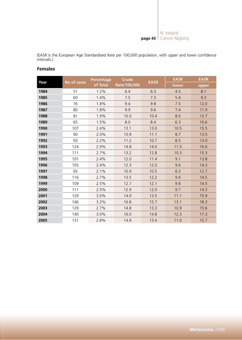
(EASR is the European Age Standardised Rate per 100,000 population, with upper and lower confidence intervals.)
Females
Year No of casesPercentage
of TotalCrude
Rate/100,000EASR
EASR EASR
lower upper
1984 51 1.2% 6.4 6.3 4.5 8.1
1985 60 1.4% 7.5 7.3 5.4 9.3
1986 76 1.8% 9.4 9.8 7.5 12.0
1987 80 1.8% 9.9 9.6 7.4 11.9
1988 81 1.9% 10.0 10.4 8.0 12.7
1989 65 1.5% 8.0 8.4 6.3 10.6
1990 107 2.4% 13.1 13.0 10.5 15.5
1991 90 2.0% 10.9 11.1 8.7 13.5
1992 93 2.2% 11.2 10.7 8.5 13.0
1993 124 2.9% 14.8 14.0 11.5 16.6
1994 111 2.7% 13.2 12.8 10.3 15.3
1995 101 2.4% 12.0 11.4 9.1 13.8
1996 105 2.4% 12.3 12.0 9.6 14.3
1997 93 2.1% 10.9 10.5 8.3 12.7
1998 116 2.7% 13.5 12.2 9.9 14.5
1999 109 2.5% 12.7 12.1 9.8 14.5
2000 111 2.5% 12.9 12.0 9.7 14.3
2001 129 3.0% 14.9 13.5 11.1 15.9
2002 146 3.2% 16.8 15.7 13.1 18.3
2003 129 2.7% 14.8 13.3 10.9 15.6
2004 140 3.0% 16.0 14.8 12.3 17.3
2005 131 2.8% 14.9 13.4 11.0 15.7

N. IrelandCancer Registry
Melanoma 2006
page 44
Deaths from malignant melanoma
(EASR is the European Age Standardised Rate per 100,000 population, with upper and lower confidence intervals.)
Male
YearNo of deaths
Percentage of Total
Crude Rate/100,000
EASREASR EASR
lower upper
1984 10 0.2% 1.3 1.5 0.6 2.4
1985 14 0.3% 1.8 2.1 1.0 3.2
1986 15 0.4% 2.0 2.2 1.1 3.3
1987 13 0.3% 1.7 1.8 0.8 2.7
1988 8 0.2% 1.0 1.3 0.4 2.1
1989 7 0.2% 0.9 1.0 0.2 1.7
1990 17 0.4% 2.2 2.7 1.4 4.0
1991 14 0.3% 1.8 1.9 0.9 3.0
1992 12 0.3% 1.5 1.7 0.7 2.7
1993 7 0.4% 0.9 1.0 0.2 1.7
1994 14 0.8% 1.7 2.0 0.9 3.1
1995 9 0.5% 1.1 1.2 0.4 2.0
1996 11 0.6% 1.4 1.6 0.7 2.6
1997 12 0.6% 1.5 1.6 0.7 2.5
1998 13 0.7% 1.6 1.7 0.8 2.6
1999 16 0.9% 2.0 2.0 1.0 3.0
2000 13 0.7% 1.6 1.8 0.8 2.7
2001 20 1.0% 2.4 2.5 1.4 3.6
2002 18 0.9% 2.2 2.4 1.3 3.5
2003 23 1.2% 2.8 2.8 1.6 4.0
2004 24 1.2% 2.9 2.9 1.7 4.0
2005 28 1.5% 3.3 3.2 2.0 4.4

N. IrelandCancer Registrypage 45
Melanoma 2006
(EASR is the European Age Standardised Rate per 100,000 population, with upper and lower confidence intervals.)
Female
YearNo of deaths
Percentage of Total
Crude Rate/100,000
EASREASR EASR
lower upper
1984 16 0.4% 2.0 2.1 1.0 3.1
1985 17 0.4% 2.1 2.1 1.1 3.2
1986 16 0.4% 2.0 1.8 0.9 2.7
1987 13 0.3% 1.6 1.3 0.6 2.0
1988 17 0.4% 2.1 2.1 1.0 3.1
1989 14 0.3% 1.7 1.6 0.7 2.4
1990 16 0.4% 2.0 1.8 0.9 2.7
1991 7 0.2% 0.8 0.7 0.2 1.2
1992 9 0.2% 1.1 0.8 0.2 1.4
1993 20 1.1% 2.5 2.6 1.5 3.8
1994 21 1.2% 2.6 3.0 1.7 4.3
1995 16 1.0% 2.0 2.2 1.1 3.3
1996 16 0.9% 2.0 2.1 1.1 3.2
1997 14 0.8% 1.7 1.8 0.8 2.7
1998 17 1.0% 2.1 2.2 1.1 3.2
1999 18 1.0% 2.2 2.4 1.3 3.5
2000 17 0.9% 2.1 2.1 1.1 3.1
2001 15 0.9% 1.8 1.8 0.9 2.8
2002 20 1.1% 2.4 2.5 1.4 3.6
2003 17 0.9% 2.0 2.1 1.1 3.1
2004 13 0.7% 1.6 1.5 0.6 2.3
2005 14 0.8% 1.7 1.6 0.8 2.5

N. IrelandCancer Registry
Melanoma 2006
page 46
Appendix B – Campbell report: Recommendations regarding Cancer Services in N. Ireland, 1996
1. The management of patients with cancer should be undertaken by appropriately trained, organ and disease specific medical specialists.
2. All patients with cancer should be managed by multidisciplinary, multiprofessional specialist cancer teams.
3. A Cancer Forum should be established involving all key interests in the delivery of cancer services.
4. Cancer Units should, in conjunction with local GPs and other providers, develop an effective communication strategy.
5. Northern Ireland should have one cancer centre, which in addition to its regional role, should act as a cancer Unit to its local catchment population of around half a million.
6. There should be four other Cancer Units, one in each Board Area, each serving a population of around quarter of a million.
7. Radiotherapy services, together with chemotherapy services, should be moved a soon as possible to the Belfast City Hospital and become and integral part of the regional Cancer Services.
8. Each Cancer Unit should develop a chemotherapy service. This service should be staffed by designated specialist nurses and pharmacists, and should be overseen by the non-surgical oncologist attached to the unit, with back-up from a haematologist.
9. There should be a minimum target of 13 consultants in non-surgical oncology for Northern Ireland by 2005.
10. Any new appointments of trained cancer specialists should be to Cancer Units or to the Cancer Centre.
11. Guidelines should be drawn up and agreed for the appropriate investigation and management of patients, presenting to non-Cancer Unit hospitals, who turn out to have cancer.
12. The Cancer Centre and Cancer Units should each develop a specialist multiprofessional palliative care team.
13 There should be a comprehensive review of palliative care service in Northern Ireland.
14. The Northern Ireland Cancer registry should be adequately resourced.
The above recommendations outlined the change that was necessary to improve cancer care.

N. IrelandCancer Registrypage 47
Melanoma 2006
Appendix C – Staging of Melanoma
Accurate staging is essential for the planning of appropriate treatment and for the comparison of the outcomes of such treatment (surgical and non-surgical). It is best achieved by a combination of techniques including physical examination, with careful inspection of the skin and regional lymph nodes. Adjuncts to staging such as CT scanning should be performed when clinically indicated.
Pathological staging adds significant information to this process. It involves histological examination of the surgically resected specimen including evaluation of the total number of regional nodes removed and the number containing metastatic tumour.
The TNM classification of Melanoma (6th Edition) is shown in table 1.
Determining the tumour size (T) factor
The T category is determined primarily by the thickness of the melanoma known as Breslow depth. The level of invasion (Clark Level) is used to subdivide T1 melanomas but not for T2 – T4. Melanoma ulceration (absence of an intact epidermis determined histopathologically) has a negative prognostic impact and is used to subdivide each T category.
Determining the (N) factor
Regional metastases include metastases to regional lymph nodes and also intralymphatic metastases which are either satellites around a primary melanoma or in-transit metastases between the primary melanoma and the regional nodes. When nodes are clinically and pathologically apparent this has a negative prognostic impact and this is used to futher subdivide the N categories.
Determining the (M) factor
Distant metastases are stage by the site or organ involved and also the presence of an elevated serum lactic dehydrogenase (LDH) has a negative prognostic impact and is used to further subdivide the M category.
Table 1: TNM classification of Melanoma
Tumour
Tumour Classification
TX Primary tumour cannot be assessed
Tis Melanoma in situ
T1
T1 Melanoma ≤ 1.0 mm with or without ulceration
T1a Melanoma ≤ 1.0 mm in thickness and Clark level II or II, no ulceration
T1b Melanoma ≤ 1.0 mm in thickness and Clark level IV or V, or with ulceration
T2
T2 Melanoma 1.01 – 2.0 mm in thickness with or without ulceration
T2a Melanoma 1.01 – 2.0 mm in thickness, no ulceration
T2b Melanoma 1.01 – 2.0 mm in thickness, with ulceration
T3
T3 Melanoma 2.01 – 4.0 mm in thickness with or without ulceration
T3a Melanoma 2.01 – 4.0 mm in thickness, no ulceration
T3b Melanoma 2.01 – 4.0 mm in thickness, with ulceration
T4
T4 Melanoma >4.0 mm in thickness with or without ulceration
T4a Melanoma >4.0 mm in thickness, no ulceration
T4b Melanoma >4.0 mm in thickness, with ulceration
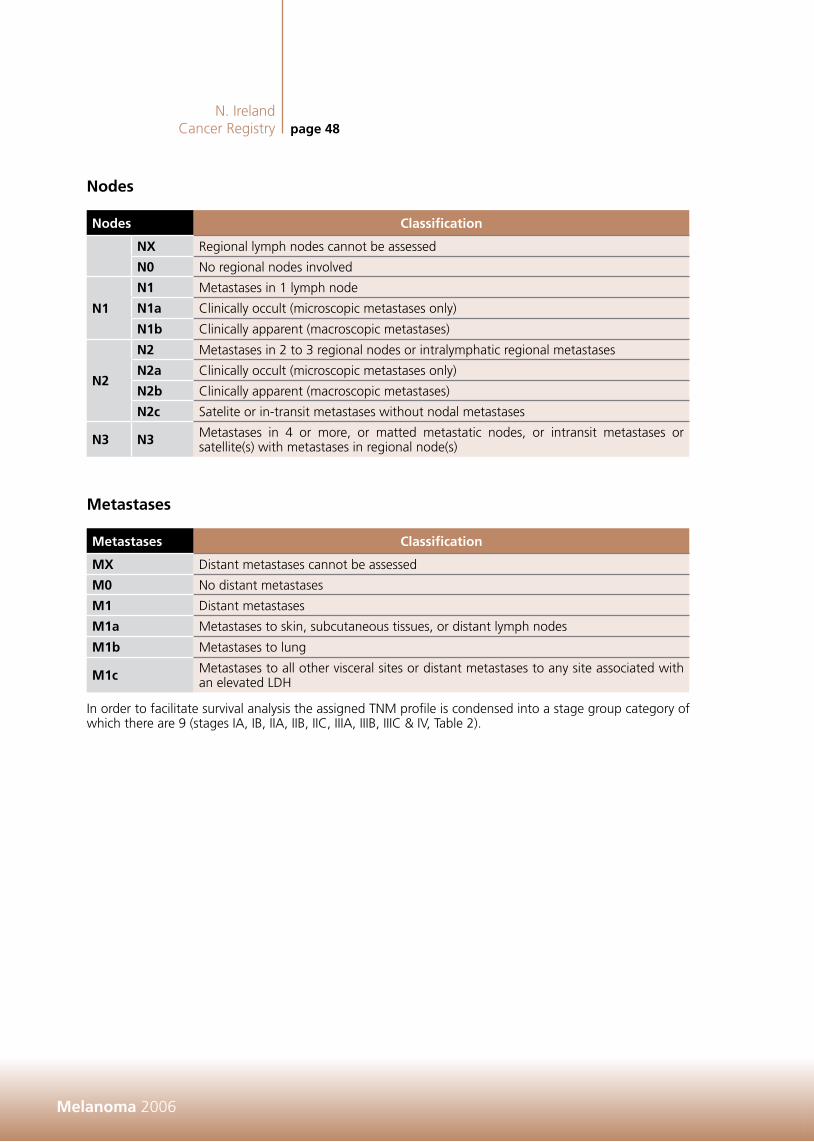
N. IrelandCancer Registry
Melanoma 2006
page 48
Nodes
Nodes Classification
NX Regional lymph nodes cannot be assessed
N0 No regional nodes involved
N1
N1 Metastases in 1 lymph node
N1a Clinically occult (microscopic metastases only)
N1b Clinically apparent (macroscopic metastases)
N2
N2 Metastases in 2 to 3 regional nodes or intralymphatic regional metastases
N2a Clinically occult (microscopic metastases only)
N2b Clinically apparent (macroscopic metastases)
N2c Satelite or in-transit metastases without nodal metastases
N3 N3 Metastases in 4 or more, or matted metastatic nodes, or intransit metastases or satellite(s) with metastases in regional node(s)
Metastases
Metastases Classification
MX Distant metastases cannot be assessed
M0 No distant metastases
M1 Distant metastases
M1a Metastases to skin, subcutaneous tissues, or distant lymph nodes
M1b Metastases to lung
M1c Metastases to all other visceral sites or distant metastases to any site associated with an elevated LDH
In order to facilitate survival analysis the assigned TNM profile is condensed into a stage group category of which there are 9 (stages IA, IB, IIA, IIB, IIC, IIIA, IIIB, IIIC & IV, Table 2).

N. IrelandCancer Registrypage 49
Melanoma 2006
Table 2: Stage Group Melanoma
Stage T N M
IA T1a N0 M0
IBT1b N0 M0
T2a N0 M0
IIAT2b N0 M0
T3a N0 M0
IIBT3b N0 M0
T4a N0 M0
IIC T4b N0 M0
IIIAT1 – 4a N1a M0
T1 – 4a N2a M0
IIIB
T1 – 4b N1a M0
T1 – 4b N2a M0
T1 – 4a N1b M0
T1 – 4a N2b M0
T1 – 4a/b N2c M0
IIIC
T1 – 4b N1b M0
T1 – 4b N2b M0
Ant T N3 M0
IV Any T Any N M1


N. Ireland Cancer RegistryCentre for Clinical and Population SciencesMulhouse BuildingGrosvenor RoadBelfast BT12 6BJ T: (44) 028 9063 2573F: (44) 028 9024 8017E: [email protected]: www.qub.ac.uk/nicr CDS N110730
Care of Patients with Malignant Melanoma of Skin in Northern Ireland 2006
2006 Melanom
a Skin