membrane and core periplasmic agrobacterium tumefaciens ...phenotype. crown gall tumors also produce...
TRANSCRIPT
Membrane and Core Periplasmic Agrobacterium tumefaciens VirulenceType IV Secretion System Components Localize to Multiple Sitesaround the Bacterial Perimeter during Lateral Attachment to PlantCells
Julieta Aguilar, Todd A. Cameron, John Zupan, and Patricia Zambryski
Department of Plant and Microbial Biology, University of California, Berkeley, Berkeley, California, USA
ABSTRACT Type IV secretion systems (T4SS) transfer DNA and/or proteins into recipient cells. Here we performed immunofluo-rescence deconvolution microscopy to localize the assembled T4SS by detection of its native components VirB1, VirB2, VirB4,VirB5, VirB7, VirB8, VirB9, VirB10, and VirB11 in the C58 nopaline strain of Agrobacterium tumefaciens, following inductionof virulence (vir) gene expression. These different proteins represent T4SS components spanning the inner membrane,periplasm, or outer membrane. Native VirB2, VirB5, VirB7, and VirB8 were also localized in the A. tumefaciens octopine strainA348. Quantitative analyses of the localization of all the above Vir proteins in nopaline and octopine strains revealed multiplefoci in single optical sections in over 80% and 70% of the bacterial cells, respectively. Green fluorescent protein (GFP)-VirB8expression following vir induction was used to monitor bacterial binding to live host plant cells; bacteria bind predominantlyalong their lengths, with few bacteria binding via their poles or subpoles. vir-induced attachment-defective bacteria or bacteriawithout the Ti plasmid do not bind to plant cells. These data support a model where multiple vir-T4SS around the perimeter ofthe bacterium maximize effective contact with the host to facilitate efficient transfer of DNA and protein substrates.
IMPORTANCE Transfer of DNA and/or proteins to host cells through multiprotein type IV secretion system (T4SS) complexes thatspan the bacterial cell envelope is critical to bacterial pathogenesis. Early reports suggested that T4SS components localized atthe cell poles. Now, higher-resolution deconvolution fluorescence microscopy reveals that all structural components of the Agro-bacterium tumefaciens vir-T4SS, as well as its transported protein substrates, localize to multiple foci around the cell perimeter.These results lead to a new model of A. tumefaciens attachment to a plant cell, where A. tumefaciens takes advantage of the mul-tiple vir-T4SS along its length to make intimate lateral contact with plant cells and thereby effectively transfer DNA and/or pro-teins through the vir-T4SS. The T4SS of A. tumefaciens is among the best-studied T4SS, and the majority of its components arehighly conserved in different pathogenic bacterial species. Thus, the results presented can be applied to a broad range of patho-gens that utilize T4SS.
Received 9 September 2011 Accepted 30 September 2011 Published 25 October 2011
Citation Aguilar J, Cameron TA, Zupan J, Zambryski P. 2011. Membrane and core periplasmic Agrobacterium tumefaciens virulence type IV secretion system componentslocalize to multiple sites around the bacterial perimeter during lateral attachment to plant cells. mBio 2(6):e00218-11. doi:10.1128/mBio.00218-11.
Editor Steven Lindow, University of California, Berkeley
Copyright © 2011 Aguilar et al. This is an open-access article distributed under the terms of the Creative Commons Attribution-Noncommercial-Share Alike 3.0 UnportedLicense, which permits unrestricted noncommercial use, distribution, and reproduction in any medium, provided the original author and source are credited.
Address correspondence to Patricia Zambryski, [email protected].
Type IV secretion systems (T4SS) are multiprotein complexesused by Gram-negative and Gram-positive bacteria for trans-
fer of DNA and/or proteins to other bacteria (1–3), plants (4),mammalian cells (5, 6), and yeast cells (7, 8). There are two majorclasses of T4SS. The first class comprises T4SS involved in conju-gative transfer of plasmid DNA between bacteria (1, 9). The sec-ond class is involved in pathogenesis by transferring effector pro-teins into eukaryotic host cells or the extracellular milieu. Humanpathogens such as Helicobacter pylori, Legionella pneumophila,Bordetella pertussis, and Rickettsia prowazekii require the T4SS fordisease (10).
The canonical model for T4SS is the virulence (vir)-induced T4SSof the plant pathogen Agrobacterium tumefaciens. vir-T4SS deliversboth DNA and proteins into plant cells, causing crown gall disease.Components of the vir-T4SS are encoded by the tumor-inducing
plasmid (pTi). The vir-T4SS transports pTi-encoded single-strandedDNA (T strand) and at least four Vir proteins, VirD2, VirE2, VirE3,and VirF, into host cells. Insertion of the T strand into plant genomicDNA and its subsequent expression lead to the overproduction ofT-DNA-encoded plant growth hormones, resulting in the tumorousphenotype. Crown gall tumors also produce opines, which are un-usual amino acid-derived compounds (11) specifically catabolized byagrobacteria as a source of carbon and nitrogen, thus providing aselective advantage for their growth in the rhizosphere (12, 13).Strains of A. tumefaciens can be differentiated on the basis of theunique opine produced by the tumor, and each type of opine is spe-cifically catabolized by the infecting strain (14). The two A. tumefa-ciens strains most extensively studied induce tumors that producenopaline or octopine opines. The vir-T4SS components from thesetwo strains are highly conserved (15).
RESEARCH ARTICLE
November/December 2011 Volume 2 Issue 6 e00218-11 ® mbio.asm.org 1
on February 27, 2020 by guest
http://mbio.asm
.org/D
ownloaded from
The vir-T4SS is composed of 11 VirB proteins (VirB1 to -11)and VirD4. All VirBs and VirD4 are essential for maximal DNAand protein transport. The vir-T4SS components can be placedinto three major groups. The first group consists of the T-piluscomponents and assembly factors VirB1 and VirB3. VirB2 is themajor T-pilus structural component, while the minor compo-nent, VirB5, is localized at the T-pilus tip (16). The N-terminalportion of VirB1 has homology to lytic transglycosylases andcleaves the peptidoglycan (17) to facilitate the assembly of thevir-T4SS in the periplasmic space. The C terminus of VirB1,VirB1*, is secreted to the cell surface (18, 19) and is required forT-pilus formation (20). VirB3 is also required for T-pilus assem-bly (21). The second group consists of VirB6 to VirB10, whichspan the inner and outer membranes and periplasm, forming thetranslocation channel (22). Cryo-electron microscopy and crystalstructures show that 14 copies each of VirB7, VirB9, and VirB10form a 1.05-MDa “core complex” that connects the cytosol to theouter surface of the bacterium with a central core diameter of 76 Å(23, 24). The arrangement of VirB6 and VirB8 with respect to thiscore is unknown, but both proteins interact with the T strand (25).The third group consists of VirB4, VirB11, and VirD4, which haveATPase homology and ATP-binding motifs (26–29) and may en-ergize assembly and/or substrate translocation. VirD4 is also thecoupling protein that brings the T strand and its associated pro-teins to the vir-T4SS (30–32).
To understand vir-T4SS function, it is critical to determine thelocalization pattern of the assembled vir-T4SS. Previously, weused deconvolution fluorescence microscopy to assess the local-ization of green fluorescent protein (GFP) fusions to structuralcomponents and substrates of the vir-T4SS. Due to the bulky na-ture of GFP, very few fusion proteins were functional; given thatmany T4SS proteins are multimeric or membrane spanning, GFPfusions are likely to interfere with complex assembly and function.Nevertheless, fusions of GFP to the cytoplasmic tails of VirD4 andVirB8 were functional, and these GFP fusion proteins localized ina helical/periodic pattern of multiple foci around the perimeter ofthe bacterial cell (33). However, as there are 12 vir-T4SS compo-nents, it is possible that there are subassemblies with differentlocalization patterns. Thus, it is critical to determine the localiza-tion pattern of the T4SS using probes to all vir-T4SS components.
Here, we performed immunofluorescence microscopy using an-tibodies to nine native Vir proteins residing in the inner mem-brane, periplasm, and outer membrane in the nopaline strain C58and detected similar patterns of multiple foci supporting our ini-tial findings. In addition, we extended our studies to the octopinestrain A348, and the localization of its vir-T4SS is identical to thatfound in the nopaline strain. Fluorescent labeling of the vir-T4SSallowed us to examine the orientation of bacteria during bindingto plant cells. The majority of bacteria bound laterally along theirlengths to plant cells, and multiple vir-T4SS foci continued to bepresent during this binding. These data support a model wheremultiple vir-T4SS around the bacterium maximize effective con-tact with the host to facilitate efficient transfer of DNA and proteinsubstrates.
RESULTSNopaline vir-T4SS membrane and periplasmic core structuralcomponents localize to multiple foci. Here we use immunofluo-rescence followed by deconvolution microscopy to detect ninedifferent structural components of the vir-T4SS. The use of anti-bodies allows localization of the native vir-T4SS. In our previouswork, GFP fusions to VirB8 and VirD4 and to vir-T4SS substrateproteins VirD2, VirE2, and VirF localized to multiple foci in ahelical/periodic pattern around the circumference of vir-inducedcells (33). Importantly, these fusion proteins did not interfere withtumor formation, and GFP-VirB8 rescued a virB8 deletion (33).In contrast, GFP fusions to VirB4, VirB6, VirB7, VirB9, VirB10,and VirB11 exhibited dominant negative effects on tumor forma-tion and did not form multiple foci, suggesting that the GFP fu-sion interfered with vir-T4SS complex assembly and/or function(33).
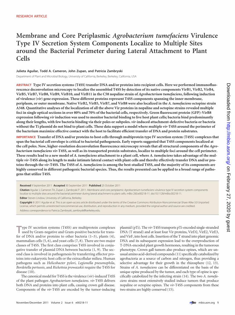
Immunofluorescent detection of nopaline strain vir-T4SScomponents VirB1, VirB2, VirB4, VirB5, VirB7, VirB8, VirB9,VirB10, and VirB11 revealed multiple foci (Fig. 1). Ten opticalsections were taken for each bacterial cell and then deconvolved toa three-dimensional (3D) image to better resolve the multiple foci(Fig. 1C). The images recapitulate those seen with GFP-VirB8fusions (33) and are suggestive of a periodic arrangement of foci.The numbers of foci in the panels in Fig. 1C vary between a min-imum of 10 and a maximum of 19.
FIG 1 Immunofluorescent detection of VirB proteins in vir-induced A. tumefaciens nopaline strain (C58). vir-induced A. tumefaciens C58 was probed withprimary antibodies to native VirB proteins (�-VirB1, �-VirB2, �-VirB4, �-VirB5, �-VirB7, �-VirB8, �-VirB9, �-VirB10, and �-VirB11) followed by fluorescentsecondary antibodies (left to right). (A and B) Bright-field images (A) corresponding to fluorescent panels (B). (C) Maximum-intensity z projections ofdeconvolved z stacks of a representative cell.
Aguilar et al.
2 ® mbio.asm.org November/December 2011 Volume 2 Issue 6 e00218-11
on February 27, 2020 by guest
http://mbio.asm
.org/D
ownloaded from
We next analyzed thousands of individual cells to provide aquantitative assessment of the frequency of multiple foci detectedby the nine different antibodies to native nopaline vir-T4SS com-ponents. As it is impractical to perform deconvolution micros-copy of stacked optical images of thousands of cells, we insteadcounted foci in a single plane of focus. We can easily detect mul-tiple foci, and we specifically assessed the frequency of cells exhib-iting 3 or more fluorescent foci (Fig. 1B). Over 80% of the cells had3 or more fluorescently labeled foci (Table 1). Most of the cells hadbetween 5 and 10 foci, and a very small percentage of cells had only3 or 4 foci. For example, the VirB8 antibody detected 3 or morefoci in 90% of the fluorescently labeled cells, reinforcing the re-sults seen with GFP-VirB8 in our earlier report (33). Antibodies tothe T-pilus components VirB2 and VirB5 revealed that 92% of thelabeled cells had 3 or more foci. In cells labeled with antibodies tothe energetic components VirB4 and VirB11, 83 to 85% of the cellscontained at least 3 foci. Antibodies to the core componentsVirB7, VirB9, and VirB10 showed that 85 to 91% of the labeledcells possessed 3 or more foci.
Each antibody labeled significantly more cells (P value, �0.05)of vir-induced cultures (�AS) than of noninduced cultures(�AS) (Table 2). The majority of antibodies labeled less than 7%of noninduced cells, signifying high specificity of the antibodies(Table 2). Antibodies to VirB1 and, to a lesser extent, antibodies toVirB7 were evidently less specific (Table 2). Nevertheless, anti-
bodies to VirB1 and VirB7 detected 3 or more foci more fre-quently in vir-induced cells than in noninduced cells, indicatingthat many of the foci observed in vir-induced cells were due tospecific antibody binding.
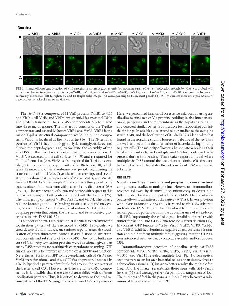
Octopine vir-T4SS structural components localize to multi-ple foci. The vir-T4SS components are highly conserved amongA. tumefaciens strains. For example, VirB8 shares 90% amino acididentity between nopaline and octopine strains (15). VirB8 is abitopic membrane protein (34) that accommodates GFP fusionsto its short cytoplasmic N-terminal tail without decreased func-tionality, as previously demonstrated in the nopaline strain (33).To test the localization of VirB8 in the octopine strain, we made aGFP fusion to the N terminus of octopine VirB8 and monitored itslocalization using deconvolution microscopy. Indeed, we detectedmultiple foci localized around the bacterial cell (Fig. 2), resem-bling the pattern of GFP-VirB8 fluorescence in the nopaline strain(33).
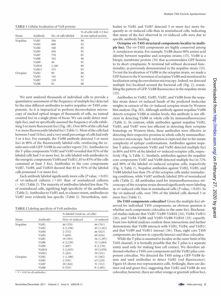
Antibodies to VirB2, VirB5, VirB7, and VirB8 from the nopa-line strain detect vir-induced bands of the predicted molecularweights in extracts of the vir-induced octopine strain by Westernblot analysis (Fig. 3D). Although antibody against nopaline VirB8detects octopine VirB8 at similar levels, this antibody is not effi-cient in detecting VirB8 in whole cells by immunofluorescence(Table 2). In contrast, while antibodies against nopaline VirB2,VirB5, and VirB7 were less efficient in detecting their octopinehomologs on Western blots, these antibodies were effective indetecting their respective proteins in whole cells by immunofluo-rescence microscopy. Such variation is expected due to the innatecomplexity of epitope conformations. Antibodies against nopa-line T-pilus components VirB2 and VirB5 detected multiple fociin 80% and 85% of the labeled vir-induced octopine cells, respec-tively (Fig. 3; Table 1). Similarly, antibodies against the nopalinecore components VirB7 and VirB8 detected multiple foci in 72%and 88% of the labeled vir-induced octopine cells, respectively(Fig. 3; Table 1). Nopaline antibodies against VirB2, VirB5, andVirB8 labeled less than 2% of the octopine cells under noninduc-ing conditions, while VirB7 antibody labeled 20% of noninducedcells (Table 2). All antibodies used for immunofluorescence mi-croscopy of the octopine strain showed significantly more labelingin vir-induced cells than in noninduced cells (P value, �0.05). Inthe vir-induced cells, over 70% of the labeled cells showed 3 ormore foci (Table 1).
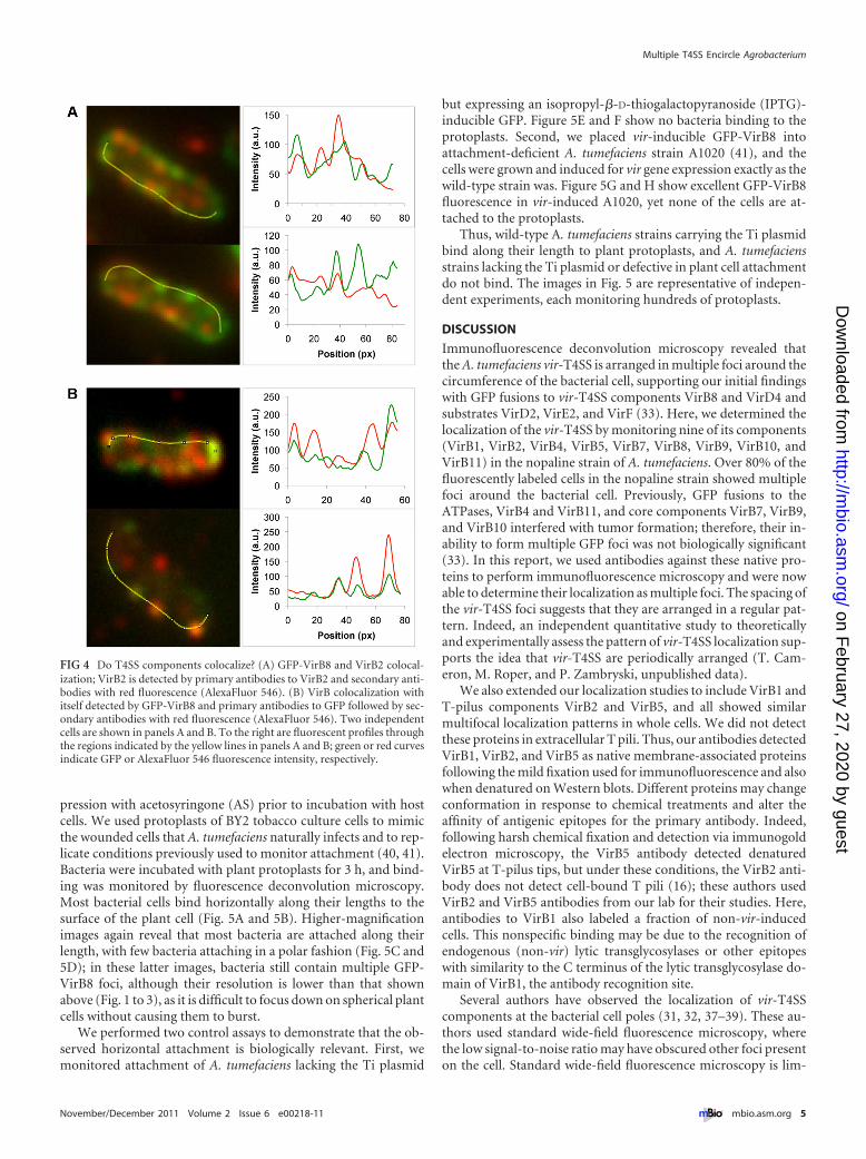
Do T4SS components colocalize? Given the multiple foci ob-served for individual T4SS components, an obvious question iswhether such components colocalize to the same foci. Biochemi-cal studies indicate that VirB7-VirB9-VirB10 (24), VirB4-VirB11(26), and VirB4-VirB8 and VirB5-VirB8-VirB10 (35) copurify.Yeast two-hybrid analyses confirm these interactions and furtherdemonstrate that VirB8 interacts with VirB1, VirB4, and VirB11and that VirB9 and VirB11 interact (36). Thus, eight core T4SScomponents are known to copurify/interact and thus colocalize.
While the T pilus is assumed to localize at the outer limit of theT4SS channel, it is formally possible that the T pilus is a separateentity used only for making host cell contact. We therefore ad-dressed whether a T4SS core component and the VirB2 pilus com-ponent colocalize. We detected the T4SS using a GFP-VirB8 fu-sion and used antibodies to detect VirB2 (red fluorescence).Figure 4A shows two representative cells. Strikingly, there are dis-tinct red and green foci, suggesting that VirB2 and VirB8 do notcolocalize; however, there are other orange or greenish yellow foci,
TABLE 1 Cellular localization of VirB proteins
Strain Antibody No. of cells labeled% of cells with �3 fociin one optical section
Nopaline VirB1 504 94VirB2 600 92VirB4 540 85VirB5 450 92VirB7 778 85VirB8 343 90VirB9 94 91VirB10 1,160 89VirB11 315 83
Octopine VirB2 90 80VirB5 145 85VirB7 159 72VirB8 83 88
TABLE 2 Labeling specificity of VirB antibodies
Strain Antibodya
% labeled (total no. of cells)
Non-vir induced vir induced
Nopaline VirB1 46 (570) 66 (761)VirB2 4 (1,194) 45 (1,341)VirB4 2 (752) 66 (823)VirB5 7 (609) 69 (654)VirB7 19 (593) 56 (1,397)VirB8 4 (2,239) 32 (1,064)VirB9 2 (487) 8 (1,170)VirB10 2 (2,055) 29 (3,970)VirB11 5 (265) 59 (531)
Octopine VirB2 1 (1,380) 15 (582)VirB5 2 (541) 63 (229)VirB7 20 (264) 51 (312)VirB8 0 (555) 8 (1,081)
a P � 0.05 for all antibodies.
Multiple T4SS Encircle Agrobacterium
November/December 2011 Volume 2 Issue 6 e00218-11 ® mbio.asm.org 3
on February 27, 2020 by guest
http://mbio.asm
.org/D
ownloaded from
suggesting partial colocalization. To the right of each fluorescentcell, we show quantification of the two fluorophores; these plotsreinforce the idea that some foci result from colocalized red andgreen fluorescence and that other foci emit predominantly red orgreen fluorescence. These data suggest that VirB2 and VirB8 onlysometimes colocalize.
As a control for the above, we monitored whether this approachcould accurately detect VirB8 colocalization with itself. We detectedVirB8 as a GFP fusion and with antibodies to VirB8 (red fluores-cence). The upper panel of Fig. 4B shows 3 possible colocalized fociand several foci that do not colocalize. The bottom panel and itstracing reveal excellent colocalization for 3 foci; however, the upperside of the same cell shows a clear red focus indicating no colocaliza-tion. Importantly, we expressed GFP-VirB8 in a virB8 deletion strain,so GFP fluorescence and VirB8 antibodies should detect the samemolecule, without any wild-type VirB8 present to compete for anti-body binding. We also tested for GFP-VirB8 colocalization usingantibodies to VirB8 (red fluorescence) and antibodies to GFP (far-redfluorescence) and obtained similar results that only some foci colo-calize (not shown). These data then lead to the unexpected conclu-
sion that combining GFP fusions and im-munofluorescence does not reliably resultin colocalized foci under these conditions.
Thus, the data testing for VirB2-VirB8and for VirB8-VirB8 colocalization aresimilar and do not allow us to concludethat colocalization occurs for all foci. Wesuggest two likely explanations. First,antibody detection is stochastic; follow-ing cell permeabilization and fixation, notall epitopes are in the correct conforma-tion for detection. Second, fixation ofcells may lead to unfolding of GFP andstochastic loss of GFP fluorescence.
The above data illustrate the short-comings of immunofluorescence in re-solving protein colocalization in the con-fines of bacterial cells. These results
potentially illuminate why a previous report utilizing immunolo-calization found several T4SS components localized to differentsites around the bacterial cell (37).
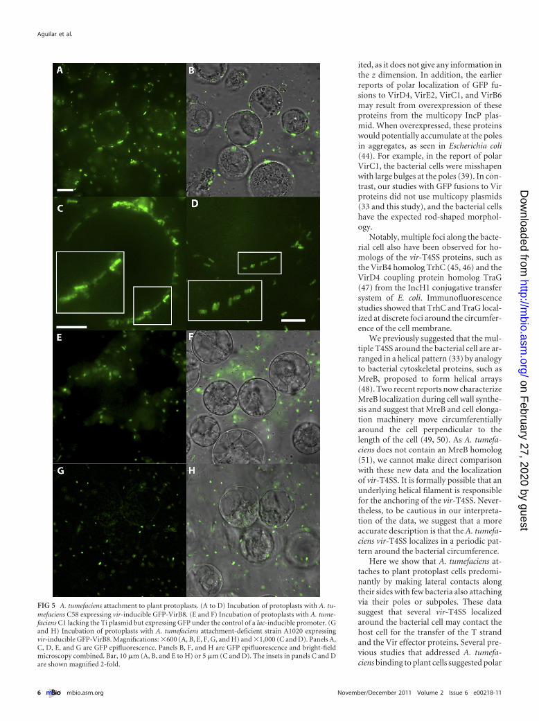
A. tumefaciens binds longitudinally to host cells. Attachmentbetween the host cell and A. tumefaciens is necessary for the trans-fer of the vir-T4SS substrates and the T-strand complex. Severalreports suggested that the vir-T4SS localizes to the cell poles (31,32, 37–39). Earlier reports of a few A. tumefaciens bacteria attach-ing to a single plant cell via their poles lent support for polarattachment (40, 41). Recent reviews continue to suggest polar at-tachment (42, 43) based on these earlier reports. However, ourpresent and previous findings (33) demonstrate that vir-T4SScomponents, including T pili, localize around the circumferenceof the cell, suggesting that there are multiple points of bacterialattachment to host cells. To test if attachment correlates with thelocalization of the vir-T4SS, we used GFP-VirB8 as a marker forthe localization of the vir-T4SS following incubation of A. tume-faciens with plant cells. A. tumefaciens carrying a vir-inducibleGFP-VirB8 plasmid in trans to pTi was induced for vir gene ex-
FIG 2 Localization of octopine VirB8 in a vir-induced A. tumefaciens octopine strain (A348). GFP-VirB8 in wild-type A. tumefaciens A348. (A) Wide-field image. (B) Maximum-intensity z projections ofdeconvolved z stacks of a representative cell. (C) Individual deconvolved slices from top to bottom ofthe cell in panel B.
FIG 3 Detection of VirB proteins in A. tumefaciens A348. Antibodies from the nopaline strain C58 were used to detect native VirB proteins in the octopine strainA348. (A) Bright-field images corresponding to fluorescent panels in panel B. (B and C) Immunofluorescence detection with primary antibodies to native A348VirB proteins (�-VirB2, �-VirB5, �-VirB7, and �-VirB8) followed by fluorescent secondary antibodies (left to right). (C) Maximum-intensity z projections ofdeconvolved z stacks of a representative cell. (D) A. tumefaciens bacteria from the nopaline and octopine strains were uninduced (�) or induced with 200 �M AS(�) for the expression of vir genes. AS-induced bands of the correct molecular weights were recognized by �-VirB2, �-VirB5, �-VirB7, and �-VirB8.
Aguilar et al.
4 ® mbio.asm.org November/December 2011 Volume 2 Issue 6 e00218-11
on February 27, 2020 by guest
http://mbio.asm
.org/D
ownloaded from
pression with acetosyringone (AS) prior to incubation with hostcells. We used protoplasts of BY2 tobacco culture cells to mimicthe wounded cells that A. tumefaciens naturally infects and to rep-licate conditions previously used to monitor attachment (40, 41).Bacteria were incubated with plant protoplasts for 3 h, and bind-ing was monitored by fluorescence deconvolution microscopy.Most bacterial cells bind horizontally along their lengths to thesurface of the plant cell (Fig. 5A and 5B). Higher-magnificationimages again reveal that most bacteria are attached along theirlength, with few bacteria attaching in a polar fashion (Fig. 5C and5D); in these latter images, bacteria still contain multiple GFP-VirB8 foci, although their resolution is lower than that shownabove (Fig. 1 to 3), as it is difficult to focus down on spherical plantcells without causing them to burst.
We performed two control assays to demonstrate that the ob-served horizontal attachment is biologically relevant. First, wemonitored attachment of A. tumefaciens lacking the Ti plasmid
but expressing an isopropyl-�-D-thiogalactopyranoside (IPTG)-inducible GFP. Figure 5E and F show no bacteria binding to theprotoplasts. Second, we placed vir-inducible GFP-VirB8 intoattachment-deficient A. tumefaciens strain A1020 (41), and thecells were grown and induced for vir gene expression exactly as thewild-type strain was. Figure 5G and H show excellent GFP-VirB8fluorescence in vir-induced A1020, yet none of the cells are at-tached to the protoplasts.
Thus, wild-type A. tumefaciens strains carrying the Ti plasmidbind along their length to plant protoplasts, and A. tumefaciensstrains lacking the Ti plasmid or defective in plant cell attachmentdo not bind. The images in Fig. 5 are representative of indepen-dent experiments, each monitoring hundreds of protoplasts.
DISCUSSION
Immunofluorescence deconvolution microscopy revealed thatthe A. tumefaciens vir-T4SS is arranged in multiple foci around thecircumference of the bacterial cell, supporting our initial findingswith GFP fusions to vir-T4SS components VirB8 and VirD4 andsubstrates VirD2, VirE2, and VirF (33). Here, we determined thelocalization of the vir-T4SS by monitoring nine of its components(VirB1, VirB2, VirB4, VirB5, VirB7, VirB8, VirB9, VirB10, andVirB11) in the nopaline strain of A. tumefaciens. Over 80% of thefluorescently labeled cells in the nopaline strain showed multiplefoci around the bacterial cell. Previously, GFP fusions to theATPases, VirB4 and VirB11, and core components VirB7, VirB9,and VirB10 interfered with tumor formation; therefore, their in-ability to form multiple GFP foci was not biologically significant(33). In this report, we used antibodies against these native pro-teins to perform immunofluorescence microscopy and were nowable to determine their localization as multiple foci. The spacing ofthe vir-T4SS foci suggests that they are arranged in a regular pat-tern. Indeed, an independent quantitative study to theoreticallyand experimentally assess the pattern of vir-T4SS localization sup-ports the idea that vir-T4SS are periodically arranged (T. Cam-eron, M. Roper, and P. Zambryski, unpublished data).
We also extended our localization studies to include VirB1 andT-pilus components VirB2 and VirB5, and all showed similarmultifocal localization patterns in whole cells. We did not detectthese proteins in extracellular T pili. Thus, our antibodies detectedVirB1, VirB2, and VirB5 as native membrane-associated proteinsfollowing the mild fixation used for immunofluorescence and alsowhen denatured on Western blots. Different proteins may changeconformation in response to chemical treatments and alter theaffinity of antigenic epitopes for the primary antibody. Indeed,following harsh chemical fixation and detection via immunogoldelectron microscopy, the VirB5 antibody detected denaturedVirB5 at T-pilus tips, but under these conditions, the VirB2 anti-body does not detect cell-bound T pili (16); these authors usedVirB2 and VirB5 antibodies from our lab for their studies. Here,antibodies to VirB1 also labeled a fraction of non-vir-inducedcells. This nonspecific binding may be due to the recognition ofendogenous (non-vir) lytic transglycosylases or other epitopeswith similarity to the C terminus of the lytic transglycosylase do-main of VirB1, the antibody recognition site.
Several authors have observed the localization of vir-T4SScomponents at the bacterial cell poles (31, 32, 37–39). These au-thors used standard wide-field fluorescence microscopy, wherethe low signal-to-noise ratio may have obscured other foci presenton the cell. Standard wide-field fluorescence microscopy is lim-
FIG 4 Do T4SS components colocalize? (A) GFP-VirB8 and VirB2 colocal-ization; VirB2 is detected by primary antibodies to VirB2 and secondary anti-bodies with red fluorescence (AlexaFluor 546). (B) VirB colocalization withitself detected by GFP-VirB8 and primary antibodies to GFP followed by sec-ondary antibodies with red fluorescence (AlexaFluor 546). Two independentcells are shown in panels A and B. To the right are fluorescent profiles throughthe regions indicated by the yellow lines in panels A and B; green or red curvesindicate GFP or AlexaFluor 546 fluorescence intensity, respectively.
Multiple T4SS Encircle Agrobacterium
November/December 2011 Volume 2 Issue 6 e00218-11 ® mbio.asm.org 5
on February 27, 2020 by guest
http://mbio.asm
.org/D
ownloaded from
ited, as it does not give any information inthe z dimension. In addition, the earlierreports of polar localization of GFP fu-sions to VirD4, VirE2, VirC1, and VirB6may result from overexpression of theseproteins from the multicopy IncP plas-mid. When overexpressed, these proteinswould potentially accumulate at the polesin aggregates, as seen in Escherichia coli(44). For example, in the report of polarVirC1, the bacterial cells were misshapenwith large bulges at the poles (39). In con-trast, our studies with GFP fusions to Virproteins did not use multicopy plasmids(33 and this study), and the bacterial cellshave the expected rod-shaped morphol-ogy.
Notably, multiple foci along the bacte-rial cell also have been observed for ho-mologs of the vir-T4SS proteins, such asthe VirB4 homolog TrhC (45, 46) and theVirD4 coupling protein homolog TraG(47) from the IncH1 conjugative transfersystem of E. coli. Immunofluorescencestudies showed that TrhC and TraG local-ized at discrete foci around the circumfer-ence of the cell membrane.
We previously suggested that the mul-tiple T4SS around the bacterial cell are ar-ranged in a helical pattern (33) by analogyto bacterial cytoskeletal proteins, such asMreB, proposed to form helical arrays(48). Two recent reports now characterizeMreB localization during cell wall synthe-sis and suggest that MreB and cell elonga-tion machinery move circumferentiallyaround the cell perpendicular to thelength of the cell (49, 50). As A. tumefa-ciens does not contain an MreB homolog(51), we cannot make direct comparisonwith these new data and the localizationof vir-T4SS. It is formally possible that anunderlying helical filament is responsiblefor the anchoring of the vir-T4SS. Never-theless, to be cautious in our interpreta-tion of the data, we suggest that a moreaccurate description is that the A. tumefa-ciens vir-T4SS localizes in a periodic pat-tern around the bacterial circumference.
Here we show that A. tumefaciens at-taches to plant protoplast cells predomi-nantly by making lateral contacts alongtheir sides with few bacteria also attachingvia their poles or subpoles. These datasuggest that several vir-T4SS localizedaround the bacterial cell may contact thehost cell for the transfer of the T strandand the Vir effector proteins. Several pre-vious studies that addressed A. tumefa-ciens binding to plant cells suggested polar
FIG 5 A. tumefaciens attachment to plant protoplasts. (A to D) Incubation of protoplasts with A. tu-mefaciens C58 expressing vir-inducible GFP-VirB8. (E and F) Incubation of protoplasts with A. tume-faciens C1 lacking the Ti plasmid but expressing GFP under the control of a lac-inducible promoter. (Gand H) Incubation of protoplasts with A. tumefaciens attachment-deficient strain A1020 expressingvir-inducible GFP-VirB8. Magnifications: �600 (A, B, E, F, G, and H) and �1,000 (C and D). Panels A,C, D, E, and G are GFP epifluorescence. Panels B, F, and H are GFP epifluorescence and bright-fieldmicroscopy combined. Bar, 10 �m (A, B, and E to H) or 5 �m (C and D). The insets in panels C and Dare shown magnified 2-fold.
Aguilar et al.
6 ® mbio.asm.org November/December 2011 Volume 2 Issue 6 e00218-11
on February 27, 2020 by guest
http://mbio.asm
.org/D
ownloaded from
attachment (40, 41). There are two significant differences betweenthe earlier reports and the data presented here. First, the earlyreports detected few bacteria binding to plant cells. In contrast, theuse of GFP facilitates the detection of numerous bacteria bindingto plant cells along their lengths. Second and more importantly,we used vir-induced A. tumefaciens to monitor attachment. vir-induced A. tumefaciens cells are likely primed and ready to attach.Under our conditions, agrobacteria are induced for 48 h at 19°C;this time frame results in maximal labeling of the T4SS (33). Wefurther suggest that A. tumefaciens attachment is likely a stepwisedynamic process. Pole- and subpole-attached A. tumefaciens ob-served here or previously may be in transition to become laterallyattached or in transition from detaching from the host cell follow-ing transfer of DNA and proteins.
The arrangement of multiple vir-T4SS around the bacterialcircumference should maximize effective contact between bacte-ria and host cells. Multiple vir-T4SS complexes and lateral attach-ment points may be required to facilitate efficient transfer ofT strands and Vir effector proteins into the host cell (Fig. 6). It iscurrently estimated that each induced bacterial cell produces 50T strands (39). To coat each T strand would require approxi-mately 30 molecules of VirE2 per kilobase of T-DNA (52, 53).Therefore, thousands of VirE2 molecules need to be delivered intothe host cytoplasm to prevent T-strand degradation. In additionto the numerous VirE2 molecules, VirE3 and VirF proteins alsomust be transported through the vir-T4SS. Thus, multiple vir-T4SS would be advantageous to the bacterial cell to expedite trans-port of the vast quantities of proteins and T strands as shown inthe model in Fig. 6.
MATERIALS AND METHODSBacterial strains and growth conditions. Wild-type A. tumefaciens strainC58 contains nopaline pTiC58, and strain A348 contains octopine pTiA6.Strain A1020 carries a Tn5 insertion at the chvB gene and is attachmentdeficient (41). For induction of the vir system, an overnight culture wasdiluted to an optical density at 600 nm (OD600) of 0.1 in minimal ABmedium, pH 5.5, and grown for 5 h at 19°C (18). Cultures were thenplated on AB agar plates with 200 �M acetosyringone (AS) for 2 days at19°C.
GFP was amplified using iProof DNA polymerase (Bio-Rad, Inc.) ac-cording to the manufacturer’s instructions. Primers incorporated an NdeIrestriction site at the 5= end of the coding sequence and an SpeI restrictionsite at the 3= end. The amplified product was digested with NdeI and SpeI
and ligated to similarly digested pSRKGm (54) to create pJZ210. Thisstrain was induced with 10 mM IPTG at 19°C for 48 h prior to incubationwith BY2 protoplasts.
Western analysis. Proteins from 108 cells were loaded into each lane.After protein separation, gels were transferred to Immobilon-P poly-vinylidene difluoride 0.45-�m membranes and analyzed by standardmethods for Western blotting.
Immunofluorescence microscopy. vir-induced cells were fixed in2.67% paraformaldehyde and 0.01% gluteraldehyde for 15 minutes atroom temperature. Cells were washed once in GTE (50 mM glucose,25 mM Tris, pH 8, 10 mM EDTA), pelleted, resuspended in GTE, andstored overnight at 4°C. Cells were washed in GTE and permeabilized bytreatment with 2 mg/ml of lysozyme and 5 mM EDTA for 45 min at roomtemperature. Primary antibodies were diluted in 1% bovine serum albu-min (BSA) in phosphate-buffered saline (PBS): anti-VirB1, anti-VirB2,anti-VirB4, anti-VirB5, and anti-VirB7 were diluted 1:50; anti-VirB8 wasdiluted 1:200; anti-VirB9 was diluted 1:50; anti-VirB10 and anti-VirB11were diluted 1:100; and chicken anti-GFP was diluted 1:100 (Aves Labs).Antibodies were added individually to each sample and incubated at37°C for 60 min. The cells were then washed twice with PBS containing0.05% Tween 20. The secondary antibodies were diluted in PBS:AlexaFluor 488 and AlexaFluor 546 goat anti-rabbit IgG were diluted1:250 and AlexaFluor 647 goat anti-chicken IgG was diluted 1:100. Sec-ondary antibodies were added as appropriate, and the samples were incu-bated at 37°C for 60 min. Cells were washed three times in PBS containing0.05% Tween 20 and once in PBS. Cells were resuspended in PBS andimaged with an Applied Precision Deltavision Spectris DV4 deconvolu-tion microscope as previously described (33). To assess colocalization,representative dually labeled cells were chosen from single wide-field im-ages for analysis. Fluorescence profiles were measured in ImageJ (http://rsbweb.nih.gov/ij) using the RGB Profiles Tool macro.
A. tumefaciens interaction with host cells. A. tumefaciens C58 con-taining GFP-VirB8 in trans to pTi was grown in AB minimal medium withappropriate antibiotics and induced with 200 �M AS for 48 hours asdescribed above. Bacteria were then scraped off plates and resuspended inAB medium. BY2 tobacco protoplasts were made by treating the culturecells with protoplast enzyme solution (1% cellulase, 0.01% pectolyase,and 0.4 M D-mannitol) at pH 5.5 for 150 minutes. BY2 protoplasts werecollected by centrifugation at 900 � g for 5 minutes. BY2 protoplasts werewashed twice with ice-cold 0.4 M mannitol and resuspended in protoplastmedium (66 mM calcium chloride, 7 mM sodium acetate, 247 mMD-mannitol, pH 5.8). A. tumefaciens was incubated with BY2 protoplastsfor 3 h at 19°C. Plant cells were imaged with an Applied Precision Delta-vision Spectris DV4 deconvolution microscope.
ACKNOWLEDGMENTS
We thank Steve Ruzin and Denise Schichnes of the College of NaturalResources Biological Imaging Facility for advice.
This work was supported by a predoctoral fellowship to J.A. (NIHgrant F31GM089088) and an NSF grant (MCB-0343566) to P.Z.
REFERENCES1. Llosa M, Gomis-Rüth FX, Coll M, de la Cruz F. 2002. Bacterial
conjugation: a two-step mechanism for DNA transport. Mol. Microbiol.45:1– 8.
2. Wallden K, Rivera-Calzada A, Waksman G. 2010. Type IV secretionsystems: versatility and diversity in function. Cell. Microbiol. 12:1203–1212.
3. Fronzes R, Christie PJ, Waksman G. 2009. The structural biology of typeIV secretion systems. Nat. Rev. Microbiol. 7:703–714.
4. Zupan J, Muth TR, Draper O, Zambryski P. 2000. The transfer of DNAfrom Agrobacterium tumefaciens into plants: a feast of fundamental in-sights. Plant J. 23:11–28.
5. Waters VL. 2001. Conjugation between bacterial and mammalian cells.Nat. Genet. 29:375–376.
6. Schröder G, Schuelein R, Quebatte M, Dehio C. 2011. Conjugative DNAtransfer into human cells by the VirB/VirD4 type IV secretion system of
FIG 6 Model for the localization of vir-T4SS and lateral attachment of A. tu-mefaciens to a plant cell. It may be advantageous for A. tumefaciens to assemblemultiple vir-T4SS and attach laterally to the host cell to facilitate efficienttransfer of the tens of T complexes and thousands of Vir-effector proteinstransported through the vir-T4SS.
Multiple T4SS Encircle Agrobacterium
November/December 2011 Volume 2 Issue 6 e00218-11 ® mbio.asm.org 7
on February 27, 2020 by guest
http://mbio.asm
.org/D
ownloaded from
the bacterial pathogen Bartonella henselae. Proc. Natl. Acad. Sci. U. S. A.108:14643–14648.
7. Bundock P, den Dulk-Ras A, Beijersbergen A, Hooykaas PJ. 1995.Trans-kingdom T-DNA transfer from Agrobacterium tumefaciens to Sac-charomyces cerevisiae. EMBO J. 14:3206 –3214.
8. Piers KL, Heath JD, Liang X, Stephens KM, Nester EW. 1996. Agrobac-terium tumefaciens-mediated transformation of yeast. Proc. Natl. Acad.Sci. U. S. A. 93:1613–1618.
9. Juhas M, Crook DW, Hood DW. 2008. Type IV secretion systems: toolsof bacterial horizontal gene transfer and virulence. Cell. Microbiol. 10:2377–2386.
10. Llosa M, Roy C, Dehio C. 2009. Bacterial type IV secretion systems inhuman disease. Mol. Microbiol. 73:141–151.
11. Dessaux Y, Petit A, Tempe J. 1993. Chemistry and biochemistry ofopines, chemical mediators of parasitism. Phytochemistry 34:31–38.
12. Gelvin SB, Thomashow MF, McPherson JC, Gordon MP, Nester EW.1982. Sizes and map positions of several plasmid-DNA-encoded tran-scripts in octopine-type crown gall tumors. Proc. Natl. Acad. Sci. U. S. A.79:76 – 80.
13. Ooms G, et al. 1982. T-DNA organization in homogeneous and hetero-geneous octopine-type crown gall tissues of Nicotiana tabacum. Cell 30:589 –597.
14. Montoya AL, Chilton MD, Gordon MP, Sciaky D, Nester EW. 1977.Octopine and nopaline metabolism in Agrobacterium tumefaciens andcrown gall tumor cells: role of plasmid genes. J. Bacteriol. 129:101–107.
15. Christie PJ, Atmakuri K, Krishnamoorthy V, Jakubowski S, Cascales E.2005. Biogenesis, architecture, and function of bacterial type IV secretionsystems. Annu. Rev. Microbiol. 59:451– 485.
16. Aly KA, Baron C. 2007. The VirB5 protein localizes to the T-pilus tips inAgrobacterium tumefaciens. Microbiology 153:3766 –3775.
17. Zahrl D, et al. 2005. Peptidoglycan degradation by specialized lytic trans-glycosylases associated with type III and type IV secretion systems. Micro-biology 151:3455–3467.
18. Llosa M, Zupan J, Baron C, Zambryski P. 2000. The N- and C-terminalportions of the Agrobacterium VirB1 protein independently enhance tu-morigenesis. J. Bacteriol. 182:3437–3445.
19. Baron C, Llosa M, Zhou S, Zambryski PC. 1997. VirB1, a component ofthe T-complex transfer machinery of Agrobacterium tumefaciens, is pro-cessed to a C-terminal secreted product, VirB1. J. Bacteriol. 179:1203–1210.
20. Zupan J, Hackworth CA, Aguilar J, Ward D, Zambryski P. 2007. VirB1*promotes T-pilus formation in the vir-type IV secretion system of Agro-bacterium tumefaciens. J. Bacteriol. 189:6551– 6563.
21. Jones AL, Shirasu K, Kado CI. 1994. The product of the virB4 gene ofAgrobacterium tumefaciens promotes accumulation of VirB3 protein.J. Bacteriol. 176:5255–5261.
22. Das A, Xie YH. 2000. The Agrobacterium T-DNA transport pore proteinsVirB8, VirB9, and VirB10 interact with one another. J. Bacteriol. 182:758 –763.
23. Chandran V, et al. 2009. Structure of the outer membrane complex of atype IV secretion system. Nature 462:1011–1015.
24. Fronzes R, et al. 2009. Structure of a type IV secretion system corecomplex. Science 323:266 –268.
25. Cascales E, Christie PJ. 2004. Definition of a bacterial type IV secretionpathway for a DNA substrate. Science 304:1170 –1173.
26. Atmakuri K, Cascales E, Christie PJ. 2004. Energetic components VirD4,VirB11 and VirB4 mediate early DNA transfer reactions required for bac-terial type IV secretion. Mol. Microbiol. 54:1199 –1211.
27. Gomis-Rüth FX, et al. 2001. The bacterial conjugation protein TrwBresembles ring helicases and F1-ATPase. Nature 409:637– 641.
28. Savvides SN, et al. 2003. VirB11 ATPases are dynamic hexamericassemblies: new insights into bacterial type IV secretion. EMBO J. 22:1969 –1980.
29. Yeo HJ, Savvides SN, Herr AB, Lanka E, Waksman G. 2000. Crystalstructure of the hexameric traffic ATPase of the Helicobacter pylori type IVsecretion system. Mol. Cell 6:1461–1472.
30. Cascales E, Atmakuri K, Liu Z, Binns AN, Christie PJ. 2005. Agrobac-terium tumefaciens oncogenic suppressors inhibit T-DNA and VirE2 pro-tein substrate binding to the VirD4 coupling protein. Mol. Microbiol.58:565–579.
31. Atmakuri K, Ding Z, Christie PJ. 2003. VirE2, a type IV secretion sub-strate, interacts with the VirD4 transfer protein at cell poles of Agrobacte-rium tumefaciens. Mol. Microbiol. 49:1699 –1713.
32. Kumar RB, Das A. 2002. Polar location and functional domains of theAgrobacterium tumefaciens DNA transfer protein VirD4. Mol. Microbiol.43:1523–1532.
33. Aguilar J, Zupan J, Cameron TA, Zambryski PC. 2010. Agrobacteriumtype IV secretion system and its substrates form helical arrays around thecircumference of virulence-induced cells. Proc. Natl. Acad. Sci. U. S. A.107:3758 –3763.
34. Thorstenson YR, Zambryski PC. 1994. The essential virulence proteinVirB8 localizes to the inner membrane of Agrobacterium tumefaciens.J. Bacteriol. 176:1711–1717.
35. Yuan Q, et al. 2005. Identification of the VirB4-VirB8-VirB5-VirB2 pilusassembly sequence of type IV secretion systems. J. Biol. Chem. 280:26349 –26359.
36. Ward DV, Draper O, Zupan JR, Zambryski PC. 2002. Peptide linkagemapping of the Agrobacterium tumefaciens vir-encoded type IV secretionsystem reveals protein subassemblies. Proc. Natl. Acad. Sci. U. S. A. 99:11493–11500.
37. Judd PK, Kumar RB, Das A. 2005. Spatial location and requirements forthe assembly of the Agrobacterium tumefaciens type IV secretion appara-tus. Proc. Natl. Acad. Sci. U. S. A. 102:11498 –11503.
38. Judd PK, Kumar RB, Das A. 2005. The type IV secretion apparatusprotein VirB6 of Agrobacterium tumefaciens localizes to a cell pole. Mol.Microbiol. 55:115–124.
39. Atmakuri K, Cascales E, Burton OT, Banta LM, Christie PJ. 2007.Agrobacterium ParA/MinD-like VirC1 spatially coordinates early conju-gative DNA transfer reactions. EMBO J. 26:2540 –2551.
40. Matthysse AG. 1987. Characterization of nonattaching mutants of Agro-bacterium tumefaciens. J. Bacteriol. 169:313–323.
41. Binns AN. 1991. Transformation of wall deficient cultured tobacco pro-toplasts by Agrobacterium tumefaciens. Plant Physiol. 96:498 –506.
42. Merritt PM, Danhorn T, Fuqua C. 2007. Motility and chemotaxis inAgrobacterium tumefaciens surface attachment and biofilm formation.J. Bacteriol. 189:8005– 8014.
43. Tomlinson AD, Fuqua C. 2009. Mechanisms and regulation of polarsurface attachment in Agrobacterium tumefaciens. Curr. Opin. Microbiol.12:708 –714.
44. Bardy SL, Maddock JR. 2007. Polar explorations: recent insights into thepolarity of bacterial proteins. Curr. Opin. Microbiol. 10:617– 623.
45. Gilmour MW, Taylor DE. 2004. A subassembly of R27-encoded transferproteins is dependent on TrhC nucleoside triphosphate-binding motifsfor function but not formation. J. Bacteriol. 186:1606 –1613.
46. Gilmour MW, Lawley TD, Rooker MM, Newnham PJ, Taylor DE. 2001.Cellular location and temperature-dependent assembly of IncHI1 plasmidR27-encoded TrhC-associated conjugative transfer protein complexes.Mol. Microbiol. 42:705–715.
47. Gunton JE, Gilmour MW, Alonso G, Taylor DE. 2005. Subcellularlocalization and functional domains of the coupling protein, TraG, fromIncHI1 plasmid R27. Microbiology 151:3549 –3561.
48. Shih YL, Rothfield L. 2006. The bacterial cytoskeleton. Microbiol. Mol.Biol. Rev. 70:729 –754.
49. Domínguez-Escobar J, et al. 2011. Processive movement of MreB-associated cell wall biosynthetic complexes in bacteria. Science 333:225–228.
50. Garner EC, et al. 2011. Coupled, circumferential motions of the cell wallsynthesis machinery and MreB filaments in B. subtilis. Science 333:222–225.
51. Daniel RA, Errington J. 2003. Control of cell morphogenesis in bacteria:two distinct ways to make a rod-shaped cell. Cell 113:767–776.
52. Zupan JR, Citovsky V, Zambryski P. 1996. Agrobacterium VirE2 proteinmediates nuclear uptake of single-stranded DNA in plant cells. Proc. Natl.Acad. Sci. U. S. A. 93:2392–2397.
53. Zambryski PC. 1992. Chronicles from the Agrobacterium-plant cell DNAtransfer story. Annu. Rev. Plant Physiol. Plant Mol. Biol. 43:465– 490.
54. Khan SR, Gaines J, Roop RM, II, Farrand SK. 2008. Broad-host-rangeexpression vectors with tightly regulated promoters and their use to ex-amine the influence of TraR and TraM expression on Ti plasmid quorumsensing. Appl. Environ. Microbiol. 74:5053–5062.
Aguilar et al.
8 ® mbio.asm.org November/December 2011 Volume 2 Issue 6 e00218-11
on February 27, 2020 by guest
http://mbio.asm
.org/D
ownloaded from