membrane proteomics of phagosomes suggests a …membrane proteomics of phagosomes suggests a...
TRANSCRIPT

Membrane proteomics of phagosomes suggestsa connection to autophagyWenqing Shuia, Leslie Sheub, Jun Liuc, Brian Smarta, Christopher J. Petzoldd, Tsung-yen Hsiehb, Austin Pitchera,Jay D. Keaslingd,e,f,1, and Carolyn R. Bertozzia,b,g,h,1
aDepartments of Chemistry, bMolecular and Cell Biology, and eChemical Engineering and Bioengineering, and hHoward Hughes Medical Institute, Universityof California, Berkeley, CA 94720; cBiological Products Division, Bayer HealthCare LLC, Berkeley, CA 94701; dPhysical Bioscience Division, and gMolecularFoundry, Lawrence Berkeley National Laboratory, Berkeley, CA 94720; and fJoint BioEnergy Institute, Emeryville, CA 94720
Contributed by Carolyn R. Bertozzi, September 17, 2008 (sent for review July 18, 2008)
Phagocytosis is the central process by which macrophage cellsinternalize and eliminate infectious microbes as well as apoptoticcells. During maturation, phagosomes containing engulfed parti-cles fuse with various endosomal compartments through theaction of regulatory molecules on the phagosomal membrane. Inthis study, we performed a proteomic analysis of the membranefraction from latex bead-containing (LBC) phagosomes isolatedfrom macrophages. The profile, which comprised 546 proteins,suggests diverse functions of the phagosome and potential con-nections to secretory processes, toll-like receptor signaling, andautophagy. Many identified proteins were not previously knownto reside in the phagosome. We characterized several proteins inLBC phagosomes that change in abundance on induction of auto-phagy, a process that has been previously implicated in the hostdefense against microbial pathogens. These observations suggestcrosstalk between autophagy and phagocytosis that may be rel-evant to the innate immune response of macrophages.
LC3 � phagocytosis
Phagosomes are specialized membrane-bound organelles gen-erated in phagocytic cells such as macrophages, neutrophils,
and dendritic cells. Their purpose is to internalize foreignparticles, microorganisms, or apoptotic cells, to mount an im-mune response, or to maintain tissue homeostasis (1). Thenascent phagosome undergoes a complex maturation processinvolving sequential fusion with endosomal compartments. Oncemature, the phagosome degrades its constituents and facilitatesantigen presentation in a highly controlled manner (2–4). Insightinto the biogenesis and immunity-related functions of the phago-some has come from analysis of its protein contents. Previousstudies have profiled the proteomes of latex bead-containing(LBC) phagosomes in cell lines from mice (5–7) and Drosophilasp. (8), as well as Dictyostelium discoideum (9) and Entamoebahistolytica (10). These elegant studies contribute to our under-standing of phagosome maturation (6, 9) and modulation bycytokines (7); however, because the entire contents of thephagosomes were sampled, abundant proteins, such as solublelysosomal hydrolases, might have obscured lower abundancemembrane-bound regulatory proteins or signaling factors.
To identify such lower abundance species, we enriched inte-gral and peripheral membrane proteins from macrophage LBCphagosomes by organelle sub-fractionation, before carrying outa large-scale proteomic analysis. The 546 proteins identified inour study included 49 membrane receptors, 64 transporterproteins, 107 regulators of vesicle and protein trafficking (in-cluding GTPases), and many components from cellular machin-eries other than phagosomes. One of the new proteins exclusivelyfound in our study, LC3-II, is considered a marker of autophagicactivity. Its presence in phagosomes suggests an unexploredlinkage between autophagy and phagocytosis. Apart from itshousekeeping role in maintaining cellular homeostasis, autoph-agy has been implicated in cancer, neurodegerative disorders,aging, and, more recently, immunity against intracellular mi-
crobes (11–13). We show here that LC3-II levels in phagosomesare modulated by autophagic activity, along with several otherproteins not previously associated with autophagy. These resultsunderscore the power of membrane-specific phagosomal pro-teomics for identifying new processes in which this organelle mayengage.
Results and DiscussionPhagosome Isolation and Membrane Fractionation. Fig. 1 illustratesour procedure for integrating sub-cellular fractionation tech-niques with the proteomic platform. We applied the method ofDesjardins and coworkers (14) for phagosome isolation. Briefly,latex beads were internalized into macrophages and the latexbead-containing (LBC) phagosomes were isolated by flotationon a sucrose gradient. Phagosomes isolated in this manner aredevoid of major contaminants (14, 15) and retain critical func-tional capabilities, such as sequential fusion with endocyticvesicles (16) and microbicidal activity (17). By using radio-labeling and proteomic analysis, Desjardins et al. (14) estimatethe potential contamination of LBC phagosomes to be below5%. The first proteomic study of LBC phagosomes, by using thismethod, identified �140 proteins. Not surprisingly, many of theobserved proteins were highly abundant lysosomal hydrolasesfrom the lumen of the vesicle.
To favor the recovery of integral or peripheral membraneproteins, which are at relatively low abundance, we lysed thephagosome pellet in sodium carbonate to release luminal proteins.The most loosely bound membrane-associated components werethen depleted by a second centrifugation. It should be noted thatseveral soluble proteins known to be transiently associated withphagosomal membranes participate in vesicle traffic and signaling(such as the Rab family) (18–20). In an effort to retain some ofthese functionally significant proteins, we refrained from extensivesubsequent washing of the membrane fraction. The resulting in-soluble pellet was re-suspended in concentrated detergent (4%SDS) to solubilize the residual proteins before separation bySDS/PAGE and identification by LC-MS/MS. Enrichment ofunique proteins in the purified phagosomal membrane fraction,compared to other fractions, was confirmed by SDS/PAGE [sup-porting information (SI) Fig. S1].
To verify the purity of isolated phagosomes and their mem-brane fractions, we probed for the presence of known cellularorganelle markers. GM-130 (Golgi-resident), Calnexin (ER-resident), and HSP-60 (mitochondria-resident) were detected
Author contributions: W.S. and C.R.B. designed research; W.S., L.S., J.L., and T.-y.H. per-formed research; W.S., J.L., B.S., C.J.P., and J.D.K. contributed new reagents/analytic tools;W.S., L.S., and A.P. analyzed data; and W.S., J.D.K., and C.R.B. wrote the paper.
The authors declare no conflict of interest.
1To whom correspondence may be addressed. E-mail: [email protected] [email protected].
This article contains supporting information online at www.pnas.org/cgi/content/full/0809218105/DCSupplemental.
© 2008 by The National Academy of Sciences of the USA
16952–16957 � PNAS � November 4, 2008 � vol. 105 � no. 44 www.pnas.org�cgi�doi�10.1073�pnas.0809218105
Dow
nloa
ded
by g
uest
on
May
27,
202
0

only in nonphagosomal fractions, whereas the lysosomal-associated membrane protein-1 (LAMP1) was clearly present inthe phagosomal fraction, as expected from the process ofphagosome–lysosome fusion (Fig. 2A). In the absence of latexbeads, LAMP1 was not observed in the corresponding fractionthat was obtained by centrifugation, thus confirming its associ-ation with phagosomal membranes.
We next evaluated the composition of the phagosomal mem-brane preparation with respect to membrane-associated versussoluble proteins. Western blot analysis demonstrated that threeknown membrane markers of endosomal compartments [earlyendosomal associated protein (EEA1), LAMP1, and transferrinreceptor (TfR)] were more abundantly represented in the mem-brane extract than in the soluble luminal fraction (Fig. 2B). All3 endosomal markers were also observed in other subcellularfractions that include endogenous endocytic vesicles. In contrastto membrane markers, a significant portion of the solublephagosomal protease cathepsin D (CatD) was released into thelumen fraction. However, this luminal protein was also observed
as a contaminant in our membrane fraction. Thus, although ourmethod enriches membrane-bound proteins considerably, themembrane fraction is not free of soluble contaminants.
Identification of Phagosomal Membrane-bound Proteins and Func-tional Categorization. Two types of mass spectrometers (Q-TOFand linear ion trap) were used to identify phagosomal mem-brane-bound proteins prepared in biological duplicates. The rawMS/MS data acquired from the two instruments were searchedby a single engine (Mascot), and a stringent set of filter criteriawas applied to select high-confidence protein IDs. A total of 546nonredundant IDs were generated in the 2 experiments by usingdifferent instruments. The details of protein identification arelisted in Dataset S1. Interestingly, when we compared ourdataset with that from the recent study conducted by Foster etal. (6) that identified 505 proteins from entire phagosomes of adifferent mouse macrophage cell line (without membrane frac-tionation), 318 IDs were exclusively found in our dataset, and 277IDs were unique hits revealed by Foster’s experiment (6) (Fig.3A). All of the 546 proteins identified in our analysis werecategorized into 14 major classes according to specific cellularprocesses or functions annotated in public databases (Fig. S2 andDataset S2). We also listed these proteins in order of relativeabundance, as indicated by their ID score and number ofidentified peptides (Dataset S3).
Dataset S4 highlights unique proteins identified in the previ-ously reported phagosomal proteome dataset, as well as the onewe acquired here, with a summary of their molecular weights(MW) and number of predicted transmembrane helices (TMHs)based on primary sequences (Figs. 3 B and C, respectively). Thehistogram of MWs shows an overall similar distribution profile,although our dataset contains slightly more IDs �40 kDa thanthe previously reported phagosomal proteomic dataset (6). Wefound 198 hits with �1 predicted TMH, which constitutes 36%of the entire phagosomal proteome in our analysis. By compar-ison, 27% of the phagosomal proteome reported by Foster et al.(6) was predicted to comprise transmembrane proteins, and aproteomic profile of Drosophila sp. phagosomes estimated thatvalue at 19.8% (8). Furthermore, proteins with more than 1TMH were represented at higher frequency in our uniquedataset compared to unique proteins in the previously reporteddatasets. Among our uniquely identified proteins are ion channeland solute carrier proteins, a class of predicted transmembraneproteins with unknown function, as well as members of the
Fig. 1. Macrophage subcellular fractionation of phagosomal membranes, and proteomic analyses.
Fig. 2. Immunoblot analysis of subcellular fractions. (A) Blot probed fororganelle markers. (B) Blot probed for membrane-bound endosomal markersand a lysosomal hydrolase. Fractions are denoted in Fig. 1. ‘‘No beads’’indicates a sample taken from the same position occupied by LBC phagosomesin the sucrose gradient, yet derived from macrophages without beads. Eachlane was loaded with 3 �g of total protein and equal loading was verified bysilver stain (see Fig. S1).
Shui et al. PNAS � November 4, 2008 � vol. 105 � no. 44 � 16953
CELL
BIO
LOG
Y
Dow
nloa
ded
by g
uest
on
May
27,
202
0
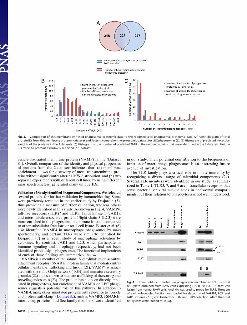
vesicle-associated membrane protein (VAMP) family (DatasetS4). Overall, comparison of the identity and physical propertiesof proteins from the 2 datasets indicates that: (a) membraneenrichment allows for discovery of more transmembrane pro-teins without significantly altering MW distribution, and (b) twoseparate experiments with different cell lines, by using differentmass spectrometers, generated many unique IDs.
Validation of Newly Identified Phagosomal Components. We selectedseveral proteins for further validation by immunoblotting. Somewere previously revealed in the earlier study by Desjardin (5),thus providing a measure of further validation, whereas otherswere newly identified in this study. As shown in Fig. 4, VAMP4,toll-like receptors (TLR)7 and TLR9, Janus kinase 1 (JAK1),and microtubule-associated protein 1/light chain 3 (LC3) weremore enriched in the phagosomal membrane fraction comparedto other subcellular fractions or total cell lysate. Foster et al. (6)also identified VAMP4 in macrophage phagosomes by massspectrometry, and certain TLRs were similarly identified byDesjardin (7) in a recent study of macrophage activation bycytokines. By contrast, JAK1 and LC3, which participate inimmune signaling and autophagy, respectively, had not beenidentified previously in phagosomes. The functional implicationsof each of these findings are summarized below.
VAMP4 is a member of the soluble N-ethylmaleimide-sensitiveattachment receptor (SNARE) protein family that mediates intra-cellular membrane trafficking and fusion (21). VAMP4 is associ-ated with the trans-Golgi network (TGN) and immature secretorygranules (22) and is known to mediate trafficking of the sorting andrecycling endosomes (23). The protein has not been directly impli-cated in phagocytosis, but enrichment of VAMP4 on LBC phago-somes suggests a potential role in this pathway. In addition toVAMP4, many other annotated proteins with relevance to ‘‘vesicleand protein trafficking’’ (Dataset S2), such as VAMP3, t-SNARE-interacting proteins, and Sec family members, were identified
in our study. Their potential contribution to the biogenesis orfunction of macrophage phagosomes is an interesting futureavenue of investigation.
The TLR family plays a critical role in innate immunity byrecognizing a diverse range of microbial components (24).Several TLR members were identified in our study, as summa-rized in Table 1. TLR3, 7, and 9 are intracellular receptors thatsense bacterial or viral nucleic acids in endosomal compart-ments, but their relation to phagocytosis is not well understood.
Fig. 3. Comparison of this membrane-enriched phagosomal proteomic data to the reported total phagosomal proteomic data. (A) Venn diagram of totalprotein IDs from this membrane proteomic dataset and Foster’s comprehensive proteomic dataset for LBC phagosomes (6). (B) Histogram of predicted molecularweights of the proteins in the 2 datasets. (C) Histogram of the number of predicted TMH in the unique proteins that were identified in the 2 datasets. UniqueIDs refers to proteins exclusively reported in 1 dataset.
Fig. 4. Immunoblots of proteins in phagosomal membranes. TCL� � totalcell lysate obtained from RAW cells expressing HA-TLR9; TCL� � total celllysate from normal RAW cells. Anti-HA was used to probe for TLR9. Three �gof each sub-cellular fraction was loaded for detection of VAMP4, LC3, andJAK1, whereas 7 �g was loaded for TLR7 and TLR9 detection. All of the totalcell lysates were loaded at 15 �g.
16954 � www.pnas.org�cgi�doi�10.1073�pnas.0809218105 Shui et al.
Dow
nloa
ded
by g
uest
on
May
27,
202
0
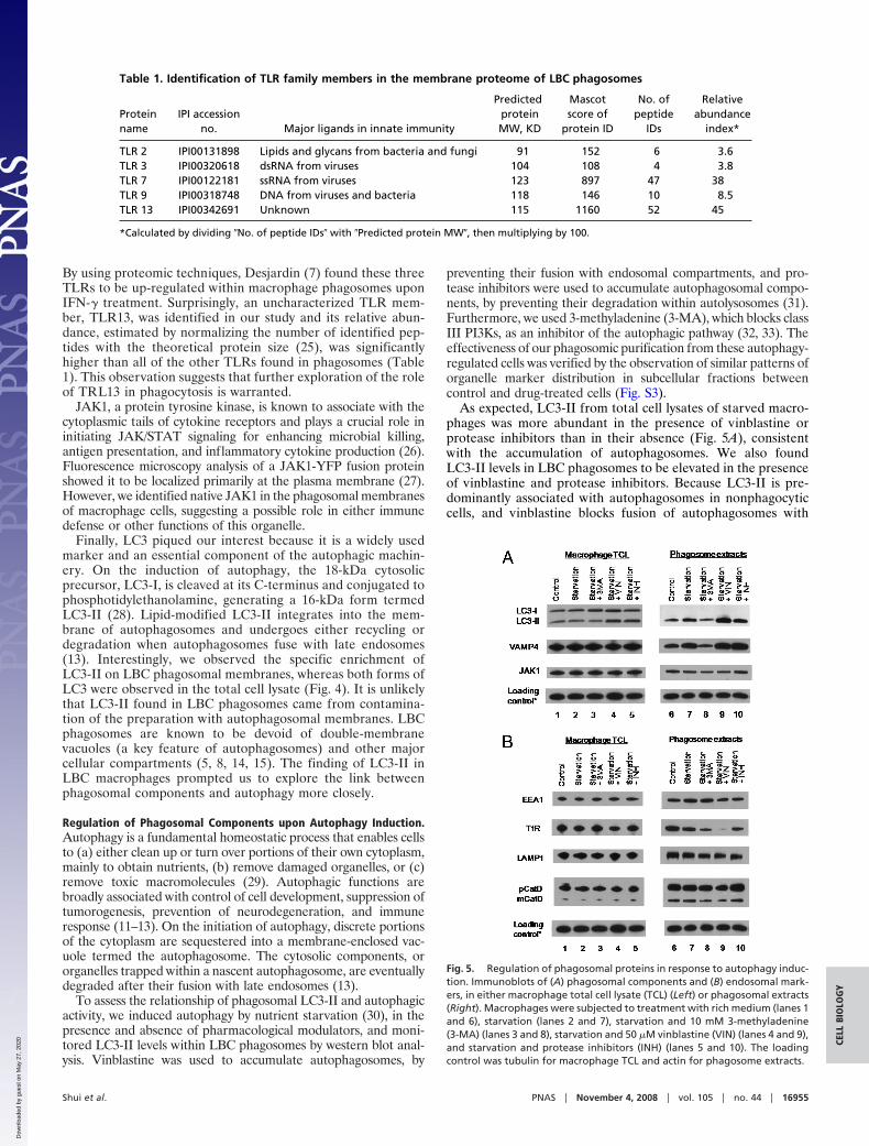
By using proteomic techniques, Desjardin (7) found these threeTLRs to be up-regulated within macrophage phagosomes uponIFN-� treatment. Surprisingly, an uncharacterized TLR mem-ber, TLR13, was identified in our study and its relative abun-dance, estimated by normalizing the number of identified pep-tides with the theoretical protein size (25), was significantlyhigher than all of the other TLRs found in phagosomes (Table1). This observation suggests that further exploration of the roleof TRL13 in phagocytosis is warranted.
JAK1, a protein tyrosine kinase, is known to associate with thecytoplasmic tails of cytokine receptors and plays a crucial role ininitiating JAK/STAT signaling for enhancing microbial killing,antigen presentation, and inflammatory cytokine production (26).Fluorescence microscopy analysis of a JAK1-YFP fusion proteinshowed it to be localized primarily at the plasma membrane (27).However, we identified native JAK1 in the phagosomal membranesof macrophage cells, suggesting a possible role in either immunedefense or other functions of this organelle.
Finally, LC3 piqued our interest because it is a widely usedmarker and an essential component of the autophagic machin-ery. On the induction of autophagy, the 18-kDa cytosolicprecursor, LC3-I, is cleaved at its C-terminus and conjugated tophosphotidylethanolamine, generating a 16-kDa form termedLC3-II (28). Lipid-modified LC3-II integrates into the mem-brane of autophagosomes and undergoes either recycling ordegradation when autophagosomes fuse with late endosomes(13). Interestingly, we observed the specific enrichment ofLC3-II on LBC phagosomal membranes, whereas both forms ofLC3 were observed in the total cell lysate (Fig. 4). It is unlikelythat LC3-II found in LBC phagosomes came from contamina-tion of the preparation with autophagosomal membranes. LBCphagosomes are known to be devoid of double-membranevacuoles (a key feature of autophagosomes) and other majorcellular compartments (5, 8, 14, 15). The finding of LC3-II inLBC macrophages prompted us to explore the link betweenphagosomal components and autophagy more closely.
Regulation of Phagosomal Components upon Autophagy Induction.Autophagy is a fundamental homeostatic process that enables cellsto (a) either clean up or turn over portions of their own cytoplasm,mainly to obtain nutrients, (b) remove damaged organelles, or (c)remove toxic macromolecules (29). Autophagic functions arebroadly associated with control of cell development, suppression oftumorogenesis, prevention of neurodegeneration, and immuneresponse (11–13). On the initiation of autophagy, discrete portionsof the cytoplasm are sequestered into a membrane-enclosed vac-uole termed the autophagosome. The cytosolic components, ororganelles trapped within a nascent autophagosome, are eventuallydegraded after their fusion with late endosomes (13).
To assess the relationship of phagosomal LC3-II and autophagicactivity, we induced autophagy by nutrient starvation (30), in thepresence and absence of pharmacological modulators, and moni-tored LC3-II levels within LBC phagosomes by western blot anal-ysis. Vinblastine was used to accumulate autophagosomes, by
preventing their fusion with endosomal compartments, and pro-tease inhibitors were used to accumulate autophagosomal compo-nents, by preventing their degradation within autolysosomes (31).Furthermore, we used 3-methyladenine (3-MA), which blocks classIII PI3Ks, as an inhibitor of the autophagic pathway (32, 33). Theeffectiveness of our phagosomic purification from these autophagy-regulated cells was verified by the observation of similar patterns oforganelle marker distribution in subcellular fractions betweencontrol and drug-treated cells (Fig. S3).
As expected, LC3-II from total cell lysates of starved macro-phages was more abundant in the presence of vinblastine orprotease inhibitors than in their absence (Fig. 5A), consistentwith the accumulation of autophagosomes. We also foundLC3-II levels in LBC phagosomes to be elevated in the presenceof vinblastine and protease inhibitors. Because LC3-II is pre-dominantly associated with autophagosomes in nonphagocyticcells, and vinblastine blocks fusion of autophagosomes with
Table 1. Identification of TLR family members in the membrane proteome of LBC phagosomes
Proteinname
IPI accessionno. Major ligands in innate immunity
PredictedproteinMW, KD
Mascotscore of
protein ID
No. ofpeptide
IDs
Relativeabundance
index*
TLR 2 IPI00131898 Lipids and glycans from bacteria and fungi 91 152 6 3.6TLR 3 IPI00320618 dsRNA from viruses 104 108 4 3.8TLR 7 IPI00122181 ssRNA from viruses 123 897 47 38TLR 9 IPI00318748 DNA from viruses and bacteria 118 146 10 8.5TLR 13 IPI00342691 Unknown 115 1160 52 45
*Calculated by dividing �No. of peptide IDs� with �Predicted protein MW�, then multiplying by 100.
Fig. 5. Regulation of phagosomal proteins in response to autophagy induc-tion. Immunoblots of (A) phagosomal components and (B) endosomal mark-ers, in either macrophage total cell lysate (TCL) (Left) or phagosomal extracts(Right). Macrophages were subjected to treatment with rich medium (lanes 1and 6), starvation (lanes 2 and 7), starvation and 10 mM 3-methyladenine(3-MA) (lanes 3 and 8), starvation and 50 �M vinblastine (VIN) (lanes 4 and 9),and starvation and protease inhibitors (INH) (lanes 5 and 10). The loadingcontrol was tubulin for macrophage TCL and actin for phagosome extracts.
Shui et al. PNAS � November 4, 2008 � vol. 105 � no. 44 � 16955
CELL
BIO
LOG
Y
Dow
nloa
ded
by g
uest
on
May
27,
202
0
endocytic compartments (28, 34), we suspect that LC3-II wasdirectly transferred from autophagosomes to LBC phagosomes.Consistent with this hypothesis, 3-MA treatment, which inhib-ited autophagosome formation, reduced LC3-II levels withinLBC phagosomes (Fig. 5A).
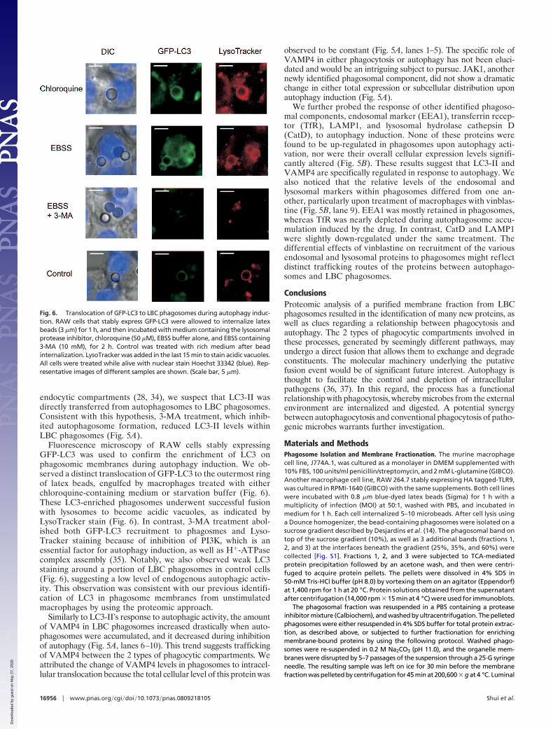
Fluorescence microscopy of RAW cells stably expressingGFP-LC3 was used to confirm the enrichment of LC3 onphagosomic membranes during autophagy induction. We ob-served a distinct translocation of GFP-LC3 to the outermost ringof latex beads, engulfed by macrophages treated with eitherchloroquine-containing medium or starvation buffer (Fig. 6).These LC3-enriched phagosomes underwent successful fusionwith lysosomes to become acidic vacuoles, as indicated byLysoTracker stain (Fig. 6). In contrast, 3-MA treatment abol-ished both GFP-LC3 recruitment to phagosmes and Lyso-Tracker staining because of inhibition of PI3K, which is anessential factor for autophagy induction, as well as H�-ATPasecomplex assembly (35). Notably, we also observed weak LC3staining around a portion of LBC phagosomes in control cells(Fig. 6), suggesting a low level of endogenous autophagic activ-ity. This observation was consistent with our previous identifi-cation of LC3 in phagosome membranes from unstimulatedmacrophages by using the proteomic approach.
Similarly to LC3-II’s response to autophagic activity, the amountof VAMP4 in LBC phagosomes increased drastically when auto-phagosomes were accumulated, and it decreased during inhibitionof autophagy (Fig. 5A, lanes 6–10). This trend suggests traffickingof VAMP4 between the 2 types of phagocytic compartments. Weattributed the change of VAMP4 levels in phagosomes to intracel-lular translocation because the total cellular level of this protein was
observed to be constant (Fig. 5A, lanes 1–5). The specific role ofVAMP4 in either phagocytosis or autophagy has not been eluci-dated and would be an intriguing subject to pursue. JAK1, anothernewly identified phagosomal component, did not show a dramaticchange in either total expression or subcellular distribution uponautophagy induction (Fig. 5A).
We further probed the response of other identified phagoso-mal components, endosomal marker (EEA1), transferrin recep-tor (TfR), LAMP1, and lysosomal hydrolase cathepsin D(CatD), to autophagy induction. None of these proteins werefound to be up-regulated in phagosomes upon autophagy acti-vation, nor were their overall cellular expression levels signifi-cantly altered (Fig. 5B). These results suggest that LC3-II andVAMP4 are specifically regulated in response to autophagy. Wealso noticed that the relative levels of the endosomal andlysosomal markers within phagosomes differed from one an-other, particularly upon treatment of macrophages with vinblas-tine (Fig. 5B, lane 9). EEA1 was mostly retained in phagosomes,whereas TfR was nearly depleted during autophagosome accu-mulation induced by the drug. In contrast, CatD and LAMP1were slightly down-regulated under the same treatment. Thedifferential effects of vinblastine on recruitment of the variousendosomal and lysosomal proteins to phagosomes might reflectdistinct trafficking routes of the proteins between autophago-somes and LBC phagosomes.
ConclusionsProteomic analysis of a purified membrane fraction from LBCphagosomes resulted in the identification of many new proteins, aswell as clues regarding a relationship between phagocytosis andautophagy. The 2 types of phagocytic compartments involved inthese processes, generated by seemingly different pathways, mayundergo a direct fusion that allows them to exchange and degradeconstituents. The molecular machinery underlying the putativefusion event would be of significant future interest. Autophagy isthought to facilitate the control and depletion of intracellularpathogens (36, 37). In this regard, the process has a functionalrelationship with phagocytosis, whereby microbes from the externalenvironment are internalized and digested. A potential synergybetween autophagocytosis and conventional phagocytosis of patho-genic microbes warrants further investigation.
Materials and MethodsPhagosome Isolation and Membrane Fractionation. The murine macrophagecell line, J774A.1, was cultured as a monolayer in DMEM supplemented with10% FBS, 100 units/ml penicillin/streptomycin, and 2 mM L-glutamine (GIBCO).Another macrophage cell line, RAW 264.7 stably expressing HA tagged-TLR9,was cultured in RPMI-1640 (GIBCO) with the same supplements. Both cell lineswere incubated with 0.8 �m blue-dyed latex beads (Sigma) for 1 h with amultiplicity of infection (MOI) at 50:1, washed with PBS, and incubated inmedium for 1 h. Each cell internalized 5–10 microbeads. After cell lysis usinga Dounce homogenizer, the bead-containing phagosomes were isolated on asucrose gradient described by Desjardins et al. (14). The phagosomal band ontop of the sucrose gradient (10%), as well as 3 additional bands (fractions 1,2, and 3) at the interfaces beneath the gradient (25%, 35%, and 60%) werecollected [Fig. S1]. Fractions 1, 2, and 3 were subjected to TCA-mediatedprotein precipitation followed by an acetone wash, and then were centri-fuged to acquire protein pellets. The pellets were dissolved in 4% SDS in50-mM Tris-HCl buffer (pH 8.0) by vortexing them on an agitator (Eppendorf)at 1,400 rpm for 1 h at 20 °C. Protein solutions obtained from the supernatantafter centrifugation (14,000 rpm � 15 min at 4 °C) were used for immunoblots.
The phagosomal fraction was resuspended in a PBS containing a proteaseinhibitor mixture (Calbiochem), and washed by ultracentrifugation. The pelletedphagosomes were either resuspended in 4% SDS buffer for total protein extrac-tion, as described above, or subjected to further fractionation for enrichingmembrane-bound proteins by using the following protocol. Washed phago-somes were re-suspended in 0.2 M Na2CO3 (pH 11.0), and the organelle mem-branes were disrupted by 5–7 passages of the suspension through a 25-G syringeneedle. The resulting sample was left on ice for 30 min before the membranefraction was pelleted by centrifugation for 45 min at 200,600 � g at 4 °C. Luminal
Fig. 6. Translocation of GFP-LC3 to LBC phagosomes during autophagy induc-tion. RAW cells that stably express GFP-LC3 were allowed to internalize latexbeads (3 �m) for 1 h, and then incubated with medium containing the lysosomalprotease inhibitor, chloroquine (50 �M), EBSS buffer alone, and EBSS containing3-MA (10 mM), for 2 h. Control was treated with rich medium after beadinternalization. LysoTracker was added in the last 15 min to stain acidic vacuoles.All cells were treated while alive with nuclear stain Hoechst 33342 (blue). Rep-resentative images of different samples are shown. (Scale bar, 5 �m).
16956 � www.pnas.org�cgi�doi�10.1073�pnas.0809218105 Shui et al.
Dow
nloa
ded
by g
uest
on
May
27,
202
0
soluble proteins from the supernatant were precipitated and re-dissolved simi-larly as the proteins in fraction B1–B3. The phagosomal membrane pellet wasresuspended in 4% SDS buffer for protein extraction as described above. Proteinconcentration was determined by the BCA assay (Piece).
SDS/PAGE and In-gel Digestion. Membrane-extracted proteins (35 �g) wereseparated by SDS/PAGE (4–12%, Bio-Rad). The entire Coomassie-stained gelwas cut into 23 consecutive bands, and the gel slices were subjected to in-geldigestion (38). Biological duplicates were prepared for analysis by using 2types of mass spectrometers.
Protein Identification by LC-MS/MS and Data Analysis. The in-gel digestion ofeach band was analyzed on either a Q-TOF (QSTAR�, Applied Biosystems) or alinear ion trap (LTQ, Thermo Inc.). LC-MS/MS data were searched against datain the International Protein Index (IPI) mouse database (v3.24 � 50,000entries) by using Mascot (v2.1, Matrix Science). Analytical details of LC-MS/MSand Mascot search parameters are provided in SI Materials and Methods. Eachprotein ID was assigned a major cellular function by GO-Getter (http://bmf2.colorado.edu/go-getter/help.psp) based on Genome Ontology (GO)terms. The number of transmembrane helices in each protein was predicted byusing the TMHMM program. (http://www.cbs.dtu.dk/services/TMHMM/).
Autophagy Induction by Starvation and Drug Treatment for Biochemical Study.J774 cells were washed with PBS and incubated in the starvation medium, Earle’sbalancedsalts solution (EBSS, Invitrogen), for2hat37 °C (31).Vinblastine (Sigma)was added to EBSS at 50 �M to accumulate autophagosomes (39). Alternatively,protease inhibitors, E64 (10 �M) and pepstatin A (2 �M), were added to EBSS topreventdegradationofautophagosomal components.3-Methyladenine (Sigma)was added to EBSS at 10 mM for autophagy inhibition (32). Cells were harvestedafter 2 h starvation, either in the presence, or in the absence, of a particular drug.
Subcellular fractionation from autophagy-induced J774 cells was also performedto acquire phagosomes in the same manner described earlier.
Autophagy Induction for Live Cell Imaging. A macrophage cell line stablyexpressing GFP-LC3 (kindly provided by Patrick Fitzgerald, St Jude’s Children’sResearch Hospital, Memphis, TN) was used here. Cells were seeded on NuncLab-Tek Chambered Coverglass microscopy slides (Fisher) 20 h before theimaging experiment. After incubation with latex beads (3.0 �m microspheres,Polysciences) for 1 h and extensive washes, the cells were treated with eitherchloroquine (50 �M) or 3-MA (10 mM) for 2 h. During the final 15 min,LysoTracker Red DND-99 (Molecular Probes) was added at 100 nM. Cells werewashed with PBS and treated with 15 �g/ml Hoechst 33342 (Molecular Probes)in PBS for 2 min. Finally, the live cells were rinsed and imaged by using a Zeiss200-M epi-fluorescent microscope. All images were deconvolved by using thenearest neighbor deconvolution algorithm from the instrument softwareSlidebook 4.2 (Intelligent Imaging Innovations).
Immunoblots. The total lysate from starved and drug-treated cells was ob-tained by sonicating cells in a 2% SDS, 50 mM Tris-HCl buffer (pH 8.0) withprotease inhibitor mixture (Calbiochem). After centrifugation, the superna-tant was collected, and the protein was quantified by the BCA assay. Proteinextracts were separated by SDS/PAGE and analyzed by western blots withrelevant antibodies.
ACKNOWLEDGMENTS. We thank Dr. Gregory Barton and Sarah Ewald for thekind gift of RAW cells that stably express the TLR9-HA fusion protein, Dr.Patrick Fitzgerald for the generous offer of LC3-GFP-expressing RAW cells, Dr.Hu Cang for assistance with proteomic data analysis, and Dr. Qing Zhong forhelpful discussions. This work was supported by a grant from the NationalInstitutes of Health (AI51622) and a grant from the Genomics:GTL program ofthe US Department of Energy (DE-AC02-05CH11231).
1. Stuart LM, Ezekowitz RA (2005) Phagocytosis: Elegant complexity. Immunity22(5):539–550.
2. Underhill DM, Ozinsky A (2002) Phagocytosis of microbes: complexity in action. AnnuRev Immunol 20:825–852.
3. Jutras I, Desjardins M (2005) Phagocytosis: at the crossroads of innate and adaptiveimmunity. Annu Rev Cell Dev Bi 21:511–527.
4. Desjardins M, Griffiths G (2003) Phagocytosis: Latex leads the way. Curr Opin Cell Biol15:498–503.
5. Garin J, et al. (2001) The phagosome proteome: Insight into phagosome functions.J Cell Biol 152:165–180.
6. Rogers LD, Foster LJ (2007) The dynamic phagosomal proteome and the contributionof the endoplasmic reticulum. Proc Natl Acad Sci USA 104:18520–18525.
7. Jutras I, et al. (2008) Modulation of the phagosome proteome by interferon-gamma.Mol Cell Proteomics 7:697–715.
8. Stuart LM, et al. (2007) A systems biology analysis of the Drosophila phagosome.Nature 445:95–101.
9. Gotthardt D, et al. (2006) Proteomics fingerprinting of phagosome maturation andevidence for the role of a Galpha during uptake. Mol Cell Proteomics 5:2228–2243.
10. Marion S, Laurent C, Guillen N (2005) Signalization and cytoskeleton activity throughmyosin IB during the early steps of phagocytosis in Entamoeba histolytica: A proteomicapproach. Cell Microbiol 7:1504–1518.
11. Mizushima N, Levine B, Cuervo AM, Klionsky DJ (2008) Autophagy fights diseasethrough cellular self-digestion. Nature 451:1069–1075.
12. Shintani T, Klionsky DJ (2004) Autophagy in health and disease: A double-edged sword.Science 306:990–995.
13. Levine B, Deretic V (2007) Unveiling the roles of autophagy in innate and adaptiveimmunity. Nat Rev Immunol 7:767–777.
14. Desjardins M, et al. (1994) Molecular characterization of phagosomes. J Biol Chem269:32194–32200.
15. Desjardins M, Huber LA, Parton RG, Griffiths G (1994) Biogenesis of phagolysosomesproceeds through a sequential series of interactions with the endocytic apparatus.J Cell Biol 124:677–688.
16. Desjardins M, Nzala NN, Corsini R, Rondeau C (1997) Maturation of phagosomes isaccompanied by changes in their fusion properties and size-selective acquisition ofsolute materials from endosomes. J Cell Sci 110:2303–2314.
17. Claus V, et al. (1998) Lysosomal enzyme trafficking between phagosomes, endosomes,and lysosomes in J774 macrophages. Enrichment of cathepsin H in early endosomes.J Biol Chem 273:9842–9851.
18. Scianimanico S, et al. (1999) Impaired recruitment of the small GTPase rab7 correlateswith the inhibition of phagosome maturation by Leishmania donovani promastigotes.Cell Microbiol 1:19–32.
19. Duclos S, et al. (2000) Rab5 regulates the kiss and run fusion between phagosomes andendosomes and the acquisition of phagosome leishmanicidal properties in RAW 264.7macrophages. J Cell Sci 113:3531–3541.
20. Grosshans BL, Ortiz D, Novick P (2006) Rabs and their effectors: Achieving specificity inmembrane traffic. Proc Natl Acad Sci USA 103:11821–11827.
21. Chen YA, Scheller RH (2001) SNARE-mediated membrane fusion. Nat Rev Mol Cell Biol2:98–106.
22. Steegmaier M, Klumperman J, Foletti DL, Yoo JS, Scheller RH (1999) Vesicle-associatedmembrane protein 4 is implicated in trans-Golgi network vesicle trafficking. Mol BiolCell 10:1957–1972.
23. Tran TH, Zeng Q, Hong W (2007) VAMP4 cycles from the cell surface to the trans-Golginetwork via sorting and recycling endosomes. J Cell Sci 120:1028–1041.
24. Barton GM (2007) Viral recognition by Toll-like receptors. Semin Immunol 19:33–40.25. Bagshaw RD, Mahuran DJ, Callahan JW (2005) A proteomic analysis of lysosomal
integral membrane proteins reveals the diverse composition of the organelle. Mol CellProteomics 4:133–143.
26. Schindler C, Levy DE, Decker T (2007) JAK-STAT signaling: From interferons to cyto-kines. J Biol Chem 282:20059–20063.
27. Behrmann I, et al. (2004) Janus kinase (Jak) subcellular localization revisited: Theexclusive membrane localization of endogenous Janus kinase 1 by cytokine receptorinteraction uncovers the Jak.receptor complex to be equivalent to a receptor tyrosinekinase. J Biol Chem 279:35486–35493.
28. Kabeya Y, et al. (2000) LC3, a mammalian homologue of yeast Apg8p, is localized inautophagosome membranes after processing. EMBO J 19:5720–5728.
29. Levine B, Klionsky DJ (2004) Development by self-digestion: Molecular mechanismsand biological functions of autophagy. Dev Cell 6:463–477.
30. Petiot A, Pattingre S, Arico S, Meley D, Codogno P (2002) Diversity of signaling controlsof macroautophagy in mammalian cells. Cell Struct Funct 27:431–441.
31. Harris J, et al. (2007) T helper 2 cytokines inhibit autophagic control of intracellularMycobacterium tuberculosis. Immunity 27:505–517.
32. Stroikin Y, Dalen H, Loof S, Terman A (2004) Inhibition of autophagy with 3-methy-ladenine results in impaired turnover of lysosomes and accumulation of lipofuscin-likematerial. Eur J Cell Biol 83:583–590.
33. Petiot A, Ogier-Denis E, Blommaart EF, Meijer AJ, Codogno P (2000) Distinct classes ofphosphatidylinositol 3�-kinases are involved in signaling pathways that control mac-roautophagy in HT-29 cells. J Biol Chem 275:992–998.
34. Kabeya Y, et al. (2004) LC3, GABARAP, and GATE16 localize to autophagosomalmembrane depending on form-II formation. J Cell Sci 117:2805–2812.
35. Fratti RA, Chua J, Vergne I, Deretic V (2003) Mycobacterium tuberculosis glycosylatedphosphatidylinositol causes phagosome maturation arrest. Proc Natl Acad Sci USA100:5437–5442.
36. Gutierrez MG, et al. (2004) Autophagy is a defense mechanism inhibiting BCG andMycobacterium tuberculosis survival in infected macrophages. Cell 119:753–766.
37. Alonso S, Pethe K, Russell DG, Purdy GE (2007) Lysosomal killing of Mycobacteriummediated by ubiquitin-derived peptides is enhanced by autophagy. Proc Natl Acad SciUSA 104:6031–6036.
38. Gu S, et al. (2003) Comprehensive proteomic profiling of the membrane constituentsof a Mycobacterium tuberculosis strain. Mol Cell Proteomics 2:1284–1296.
39. Rez G, et al. (1996) Time course of vinblastine-induced autophagocytosis and changesin the endoplasmic reticulum in murine pancreatic acinar cells: A morphometric andbiochemical study. Eur J Cell Biol 71:341–350.
Shui et al. PNAS � November 4, 2008 � vol. 105 � no. 44 � 16957
CELL
BIO
LOG
Y
Dow
nloa
ded
by g
uest
on
May
27,
202
0