membranes serve as allosteric activators of … serve as allosteric activators of phospholipase a 2,...
TRANSCRIPT
Membranes serve as allosteric activators ofphospholipase A2, enabling it to extract, bind,and hydrolyze phospholipid substratesVarnavas D. Mouchlisa,b,1, Denis Bucherb, J. Andrew McCammona,b,c,1, and Edward A. Dennisa,b,1
Departments of aPharmacology and bChemistry and Biochemistry, and cHoward Hughes Medical Institute, University of California, San Diego, La Jolla,CA 92093-0601
Contributed by J. Andrew McCammon, December 30, 2014 (sent for review December 9, 2014; reviewed by Jeffry D. Madura)
Defining the molecular details and consequences of the associa-tion of water-soluble proteins with membranes is fundamental tounderstanding protein–lipid interactions and membrane function-ing. Phospholipase A2 (PLA2) enzymes, which catalyze the hydro-lysis of phospholipid substrates that compose the membranebilayers, provide the ideal system for studying protein–lipid inter-actions. Our study focuses on understanding the catalytic cycleof two different human PLA2s: the cytosolic Group IVA cPLA2 andcalcium-independent Group VIA iPLA2. Computer-aided techniquesguided by deuterium exchange mass spectrometry data, were usedto create structural complexes of each enzyme with a single phos-pholipid substrate molecule, whereas the substrate extraction pro-cess was studied using steered molecular dynamics simulations.Molecular dynamic simulations of the enzyme–substrate–membranesystems revealed important information about the mechanisms bywhich these enzymes associate with the membrane and then ex-tract and bind their phospholipid substrate. Our data support thehypothesis that the membrane acts as an allosteric ligand that bindsat the allosteric site of the enzyme’s interfacial surface, shifting itsconformation from a closed (inactive) state in water to an open(active) state at the membrane interface.
GIVA cPLA2 | GVIA iPLA2 | PAPC | MD simulations | DXMS
During the last half century, much effort has been devotedto understanding how members of the phospholipase A2
(PLA2) enzyme superfamily interact with membranes. This takeson great importance because of the various biological functionsassociated with their ability to catalyze the hydrolysis of the esterbond at the sn-2 position of phospholipid molecules in mem-branes, liberating arachidonic acid (AA) and other polyunsaturatedfree fatty acids (PUFAs), as well as lysophospholipids (1). AAand other PUFAs are metabolic precursors for the biosynthesisof eicosanoids, including prostaglandins and leukotrienes, whichare implicated in many inflammatory conditions (2). Through theeicosanoid pathways, PLA2s have been implicated in many in-flammatory diseases, including asthma, arthritis, atherosclerosis,pain, and cancer (3–5). There are 16 groups and many subgroupsof PLA2, but they can be considered as six types: cytosolic (cPLA2),secreted (sPLA2), calcium-independent (iPLA2), platelet-activat-ing factor acetylhydrolase (PAF-AH), also known as lipoprotein-associated PLA2 (Lp-PLA2), lysosomal PLA2 (LPLA2), andadipose-PLA2 (AdPLA2) (1). Among them, the two cytosolicwater-soluble membrane-associated enzymes described in thisstudy are implicated in numerous inflammatory and metabolicdiseases (1, 6, 7). Thus, both enzymes are attractive targets forinhibitor development (8–10). However, there are no reportedstudies describing their interactions with a single phospholipidsubstrate molecule extracted from the membrane into the bindingpocket of these enzymes.The human Group IVA cytosolic PLA2 (GIVA cPLA2, cPLA2α)
was cloned and sequenced from U937 cells in 1990 (11). Thisenzyme contains 749 amino acids and has a molecular weight of85.2 kDa (12). Its X-ray crystal structure [Protein Data Bank
(PDB) ID 1CJY; Fig. S1A] revealed an N-terminal C2 domainand a C-terminal α/β hydrolase catalytic domain (13). The GIVAcPLA2 is regulated by intracellular calcium, which binds to acalcium binding site located on the C2 domain of the enzyme,resulting in the translocation of the enzyme to the surface of thephospholipid membrane (14). The enzyme is then able to accessits phospholipid substrate, and uses a catalytic dyad of Ser/Asp tocatalyze the hydrolysis (15). MAP kinase phosphorylation, as wellas the lipid mediators ceramide-1-phosphate and phosphatidyl-inositol 4,5-bis phosphate, were shown to further enhance theactivity of GIVA cPLA2 (16–18). GIVA cPLA2 exhibits highspecificity for phospholipids containing AA at the sn-2 position (19).The human Group VIA calcium-independent PLA2 (GVIA
iPLA2, iPLA2β) gene was found to express multiple splice var-iants (20). Among them, transcript variant 1 is the longer splicevariant, containing 806 amino acids, and has a molecular weightof 89.9 kDa. The X-ray crystal structure of the enzyme has notbeen solved yet, but sequence alignment studies revealed sevenankyrin (ANK) repeats, a linker region, and an α/β hydrolasecatalytic domain (20). The sequence of the human transcriptvariant 1 shows the insertion of an additional 54 amino acids,compared with the human transcript variant 2, which interruptsthe eighth ANK repeat (20, 21). ANK repeats are often involvedin protein–protein interactions regulating the activity of manyenzymes (22). The human enzyme is similar to the earlier dis-covered murine iPLA2 (23). GVIA iPLA2 also uses a catalyticdyad of Ser/Asp; its catalytic activity is enhanced by ATP anddoes not show specificity for the sn-2 fatty acid (24).
Significance
The catalytic mechanisms of interfacial enzymes acting directlyon the interfacial surface of the membrane are notoriouslydifficult to study experimentally with X-ray crystallographyand other biophysical methods. This scientific study is, to ourknowledge, the first to highlight similarities and differences inthe extraction and binding of a phospholipid molecule into thesubstrate binding pocket of two human phospholipases A2
(PLA2s): the cytosolic Group IVA cPLA2 and the calcium-inde-pendent Group VIA iPLA2. Molecular dynamics simulations,guided by deuterium exchange experiments, are used to showthat pathways to the active sites of these PLA2s are openedupon allosteric interaction with the membrane to facilitateentry of the substrate lipid. These enzymes are involved invarious diseases, and understanding their mechanisms will aidin the discovery of therapeutics.
Author contributions: V.D.M., D.B., J.A.M., and E.A.D. designed research; V.D.M. per-formed research; V.D.M., D.B., J.A.M., and E.A.D. analyzed data; and V.D.M., D.B.,J.A.M., and E.A.D. wrote the paper.
Reviewers included: J.D.M., Duquesne University.
The authors declare no conflict of interest.1To whom correspondence may be addressed. Email: [email protected], [email protected], or [email protected].
This article contains supporting information online at www.pnas.org/lookup/suppl/doi:10.1073/pnas.1424651112/-/DCSupplemental.
E516–E525 | PNAS | Published online January 26, 2015 www.pnas.org/cgi/doi/10.1073/pnas.1424651112
The PLA2 catalytic cycle can be described in four steps: asso-ciation of the enzyme with the membrane, extraction and bindingof a single phospholipid molecule from the membrane into thebinding pocket, hydrolysis, and product diffusion. The mostcritical step of the PLA2 catalytic cycle is the association of theenzyme with the membrane through its interfacial binding region,which interacts with the membrane by means of electrostatic,hydrophobic, and hydrogen-bonding (H-bonding) interactions.Understanding the mechanistic details of how PLA2s interact withmembranes to access their substrate is a very challenging bi-ological question that we have addressed in this study. Althoughseveral PLA2 X-ray crystal structures have been published in thepast, none of them contained cocrystallized natural phospholipidsubstrates or membranes. Hydrogen/deuterium exchange massspectrometry (DXMS) (25) has been successfully used in the pastto study PLA2–substrate, –inhibitor, and –membrane interactions(26, 27). In this study, computer modeling and simulations havebeen used to interpret previously published DXMS data (28–31),in an effort to understand mechanistic questions related to thestructural characteristics of GIVA cPLA2 and GVIA iPLA2. Inparticular, the extraction and binding of a single 1-palmitoyl-2-arachidonoyl-sn-glycero-3-phosphocholine (PAPC) phospholipidsubstrate into the binding pocket of these enzymes were studiedusing induced fit docking (IFD) (32) and steered molecular dy-namics (SMD) simulations (33). Long molecular dynamics (MD)simulations were carried out to the enzyme–PAPC complexesin the presence of 1-palmitoyl-2-oleoyl-sn-glycero-3-phosphocho-line (POPC) membrane patches using NAMD (33). The GIVAcPLA2 has a strong preference for AA in the sn-2 position of thephospholipid substrate, which also serves as an optimal substratefor GVIA iPLA2, although GVIA iPLA2 is more permissive as tothe fatty acid it hydrolyzes; thus for a direct comparison of the twoenzymes, we used PAPC as substrate, as is typically done for ki-netic comparisons. However, the most abundant and represen-tative phospholipid in membranes is POPC, so this phospholipidwas used for the membrane interaction studies. Our results pro-vide insight into the binding of both enzymes with the PAPCsubstrate molecule and the POPC membranes, suggesting that themembrane is an important factor that should be taken into con-sideration in future computer-aided design efforts on PLA2s.
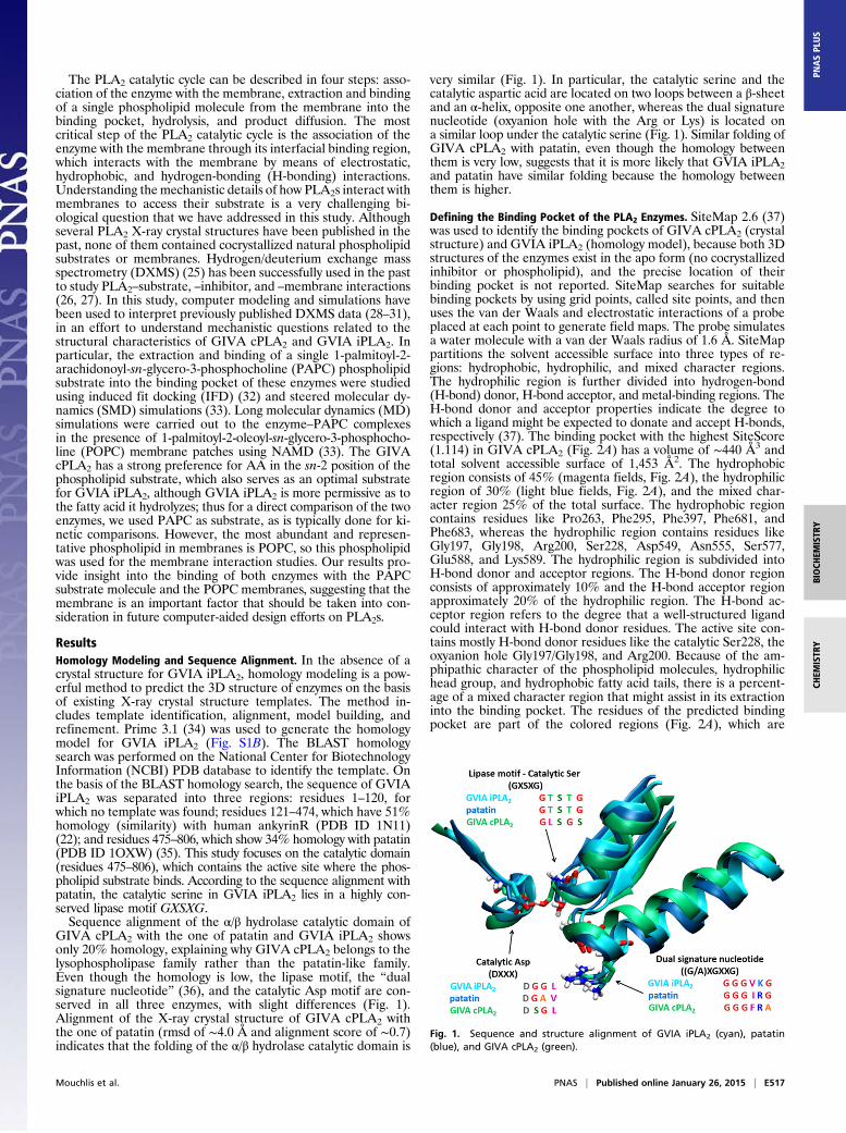
ResultsHomology Modeling and Sequence Alignment. In the absence of acrystal structure for GVIA iPLA2, homology modeling is a pow-erful method to predict the 3D structure of enzymes on the basisof existing X-ray crystal structure templates. The method in-cludes template identification, alignment, model building, andrefinement. Prime 3.1 (34) was used to generate the homologymodel for GVIA iPLA2 (Fig. S1B). The BLAST homologysearch was performed on the National Center for BiotechnologyInformation (NCBI) PDB database to identify the template. Onthe basis of the BLAST homology search, the sequence of GVIAiPLA2 was separated into three regions: residues 1–120, forwhich no template was found; residues 121–474, which have 51%homology (similarity) with human ankyrinR (PDB ID 1N11)(22); and residues 475–806, which show 34% homology with patatin(PDB ID 1OXW) (35). This study focuses on the catalytic domain(residues 475–806), which contains the active site where the phos-pholipid substrate binds. According to the sequence alignment withpatatin, the catalytic serine in GVIA iPLA2 lies in a highly con-served lipase motif GXSXG.Sequence alignment of the α/β hydrolase catalytic domain of
GIVA cPLA2 with the one of patatin and GVIA iPLA2 showsonly 20% homology, explaining why GIVA cPLA2 belongs to thelysophospholipase family rather than the patatin-like family.Even though the homology is low, the lipase motif, the “dualsignature nucleotide” (36), and the catalytic Asp motif are con-served in all three enzymes, with slight differences (Fig. 1).Alignment of the X-ray crystal structure of GIVA cPLA2 withthe one of patatin (rmsd of ∼4.0 Å and alignment score of ∼0.7)indicates that the folding of the α/β hydrolase catalytic domain is
very similar (Fig. 1). In particular, the catalytic serine and thecatalytic aspartic acid are located on two loops between a β-sheetand an α-helix, opposite one another, whereas the dual signaturenucleotide (oxyanion hole with the Arg or Lys) is located ona similar loop under the catalytic serine (Fig. 1). Similar folding ofGIVA cPLA2 with patatin, even though the homology betweenthem is very low, suggests that it is more likely that GVIA iPLA2and patatin have similar folding because the homology betweenthem is higher.
Defining the Binding Pocket of the PLA2 Enzymes. SiteMap 2.6 (37)was used to identify the binding pockets of GIVA cPLA2 (crystalstructure) and GVIA iPLA2 (homology model), because both 3Dstructures of the enzymes exist in the apo form (no cocrystallizedinhibitor or phospholipid), and the precise location of theirbinding pocket is not reported. SiteMap searches for suitablebinding pockets by using grid points, called site points, and thenuses the van der Waals and electrostatic interactions of a probeplaced at each point to generate field maps. The probe simulatesa water molecule with a van der Waals radius of 1.6 Å. SiteMappartitions the solvent accessible surface into three types of re-gions: hydrophobic, hydrophilic, and mixed character regions.The hydrophilic region is further divided into hydrogen-bond(H-bond) donor, H-bond acceptor, and metal-binding regions. TheH-bond donor and acceptor properties indicate the degree towhich a ligand might be expected to donate and accept H-bonds,respectively (37). The binding pocket with the highest SiteScore(1.114) in GIVA cPLA2 (Fig. 2A) has a volume of ∼440 Å3 andtotal solvent accessible surface of 1,453 Å2. The hydrophobicregion consists of 45% (magenta fields, Fig. 2A), the hydrophilicregion of 30% (light blue fields, Fig. 2A), and the mixed char-acter region 25% of the total surface. The hydrophobic regioncontains residues like Pro263, Phe295, Phe397, Phe681, andPhe683, whereas the hydrophilic region contains residues likeGly197, Gly198, Arg200, Ser228, Asp549, Asn555, Ser577,Glu588, and Lys589. The hydrophilic region is subdivided intoH-bond donor and acceptor regions. The H-bond donor regionconsists of approximately 10% and the H-bond acceptor regionapproximately 20% of the hydrophilic region. The H-bond ac-ceptor region refers to the degree that a well-structured ligandcould interact with H-bond donor residues. The active site con-tains mostly H-bond donor residues like the catalytic Ser228, theoxyanion hole Gly197/Gly198, and Arg200. Because of the am-phipathic character of the phospholipid molecules, hydrophilichead group, and hydrophobic fatty acid tails, there is a percent-age of a mixed character region that might assist in its extractioninto the binding pocket. The residues of the predicted bindingpocket are part of the colored regions (Fig. 2A), which are
Fig. 1. Sequence and structure alignment of GVIA iPLA2 (cyan), patatin(blue), and GIVA cPLA2 (green).
Mouchlis et al. PNAS | Published online January 26, 2015 | E517
BIOCH
EMISTR
YCH
EMISTR
YPN
ASPL
US
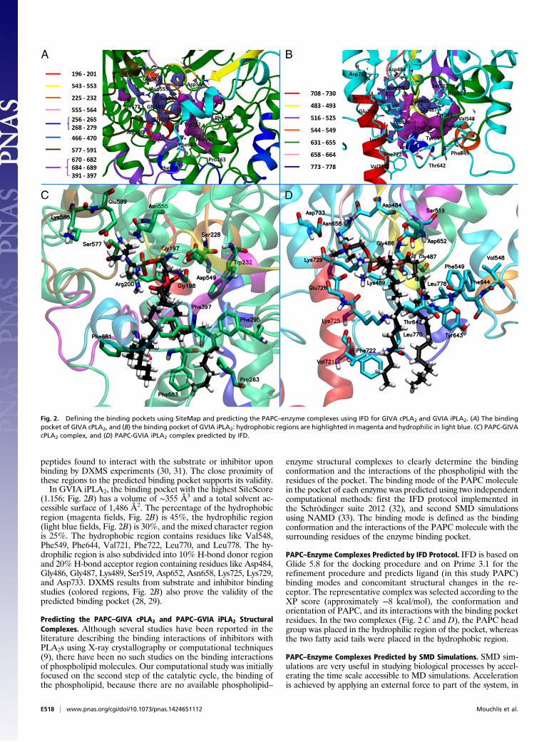
peptides found to interact with the substrate or inhibitor uponbinding by DXMS experiments (30, 31). The close proximity ofthese regions to the predicted binding pocket supports its validity.In GVIA iPLA2, the binding pocket with the highest SiteScore
(1.156; Fig. 2B) has a volume of ∼355 Å3 and a total solvent ac-cessible surface of 1,486 Å2. The percentage of the hydrophobicregion (magenta fields, Fig. 2B) is 45%, the hydrophilic region(light blue fields, Fig. 2B) is 30%, and the mixed character regionis 25%. The hydrophobic region contains residues like Val548,Phe549, Phe644, Val721, Phe722, Leu770, and Leu778. The hy-drophilic region is also subdivided into 10% H-bond donor regionand 20% H-bond acceptor region containing residues like Asp484,Gly486, Gly487, Lys489, Ser519, Asp652, Asn658, Lys725, Lys729,and Asp733. DXMS results from substrate and inhibitor bindingstudies (colored regions, Fig. 2B) also prove the validity of thepredicted binding pocket (28, 29).
Predicting the PAPC–GIVA cPLA2 and PAPC–GVIA iPLA2 StructuralComplexes. Although several studies have been reported in theliterature describing the binding interactions of inhibitors withPLA2s using X-ray crystallography or computational techniques(9), there have been no such studies on the binding interactionsof phospholipid molecules. Our computational study was initiallyfocused on the second step of the catalytic cycle, the binding ofthe phospholipid, because there are no available phospholipid–
enzyme structural complexes to clearly determine the bindingconformation and the interactions of the phospholipid with theresidues of the pocket. The binding mode of the PAPC moleculein the pocket of each enzyme was predicted using two independentcomputational methods: first the IFD protocol implemented inthe Schrödinger suite 2012 (32), and second SMD simulationsusing NAMD (33). The binding mode is defined as the bindingconformation and the interactions of the PAPC molecule with thesurrounding residues of the enzyme binding pocket.
PAPC–Enzyme Complexes Predicted by IFD Protocol. IFD is based onGlide 5.8 for the docking procedure and on Prime 3.1 for therefinement procedure and predicts ligand (in this study PAPC)binding modes and concomitant structural changes in the re-ceptor. The representative complex was selected according to theXP score (approximately −8 kcal/mol), the conformation andorientation of PAPC, and its interactions with the binding pocketresidues. In the two complexes (Fig. 2 C and D), the PAPC headgroup was placed in the hydrophilic region of the pocket, whereasthe two fatty acid tails were placed in the hydrophobic region.
PAPC–Enzyme Complexes Predicted by SMD Simulations. SMD sim-ulations are very useful in studying biological processes by accel-erating the time scale accessible to MD simulations. Accelerationis achieved by applying an external force to part of the system, in
Fig. 2. Defining the binding pockets using SiteMap and predicting the PAPC–enzyme complexes using IFD for GIVA cPLA2 and GVIA iPLA2. (A) The bindingpocket of GIVA cPLA2, and (B) the binding pocket of GVIA iPLA2: hydrophobic regions are highlighted in magenta and hydrophilic in light blue. (C) PAPC-GIVAcPLA2 complex, and (D) PAPC-GVIA iPLA2 complex predicted by IFD.
E518 | www.pnas.org/cgi/doi/10.1073/pnas.1424651112 Mouchlis et al.
this study on the substrate (PAPC molecule), to lead it along apredefined direction (33). Although this introduces a bias in thesystem, equilibrium thermodynamic quantities such as the freeenergy profile or potential of mean force along the reactioncoordinate can in principle be recovered from such study.Even though GIVA cPLA2 and GVIA iPLA2 are water-solu-
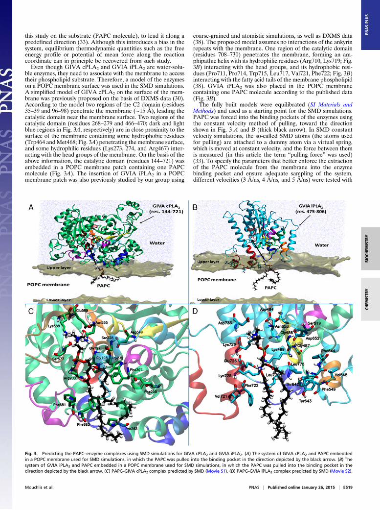
ble enzymes, they need to associate with the membrane to accesstheir phospholipid substrate. Therefore, a model of the enzymeson a POPC membrane surface was used in the SMD simulations.A simplified model of GIVA cPLA2 on the surface of the mem-brane was previously proposed on the basis of DXMS data (30).According to the model two regions of the C2 domain (residues35–39 and 96–98) penetrate the membrane (∼15 Å), leading thecatalytic domain near the membrane surface. Two regions of thecatalytic domain (residues 268–279 and 466–470; dark and lightblue regions in Fig. 3A, respectively) are in close proximity to thesurface of the membrane containing some hydrophobic residues(Trp464 andMet468; Fig. 3A) penetrating the membrane surface,and some hydrophilic residues (Lys273, 274, and Arg467) inter-acting with the head groups of the membrane. On the basis of theabove information, the catalytic domain (residues 144–721) wasembedded in a POPC membrane patch containing one PAPCmolecule (Fig. 3A). The insertion of GVIA iPLA2 in a POPCmembrane patch was also previously studied by our group using
coarse-grained and atomistic simulations, as well as DXMS data(38). The proposed model assumes no interactions of the ankyrinrepeats with the membrane. One region of the catalytic domain(residues 708–730) penetrates the membrane, forming an am-phipathic helix with its hydrophilic residues (Arg710, Lys719; Fig.3B) interacting with the head groups, and its hydrophobic resi-dues (Pro711, Pro714, Trp715, Leu717, Val721, Phe722; Fig. 3B)interacting with the fatty acid tails of the membrane phospholipid(38). GVIA iPLA2 was also placed in the POPC membranecontaining one PAPC molecule according to the published data(Fig. 3B).The fully built models were equilibrated (SI Materials and
Methods) and used as a starting point for the SMD simulations.PAPC was forced into the binding pockets of the enzymes usingthe constant velocity method of pulling, toward the directionshown in Fig. 3 A and B (thick black arrow). In SMD constantvelocity simulations, the so-called SMD atoms (the atoms usedfor pulling) are attached to a dummy atom via a virtual spring,which is moved at constant velocity, and the force between themis measured (in this article the term “pulling force” was used)(33). To specify the parameters that better enforce the extractionof the PAPC molecule from the membrane into the enzymebinding pocket and ensure adequate sampling of the system,different velocities (3 Å/ns, 4 Å/ns, and 5 Å/ns) were tested with
Fig. 3. Predicting the PAPC–enzyme complexes using SMD simulations for GIVA cPLA2 and GVIA iPLA2. (A) The system of GIVA cPLA2 and PAPC embeddedin a POPC membrane used for SMD simulations, in which the PAPC was pulled into the binding pocket in the direction depicted by the black arrow. (B) Thesystem of GVIA iPLA2 and PAPC embedded in a POPC membrane used for SMD simulations, in which the PAPC was pulled into the binding pocket in thedirection depicted by the black arrow. (C) PAPC–GIVA cPLA2 complex predicted by SMD (Movie S1). (D) PAPC–GVIA iPLA2 complex predicted by SMD (Movie S2).
Mouchlis et al. PNAS | Published online January 26, 2015 | E519
BIOCH
EMISTR
YCH
EMISTR
YPN
ASPL
US
a force constant (k) of 10 kcal/mol*Å2. For the preliminary SMDsimulations the GVIA iPLA2 was used. The position of PAPCduring the time of the SMD simulations was calculated for allthree simulations (Fig. S2 A–C). A constant velocity of 5 Å/nsindicated a smoother extraction of the PAPC molecule from themembrane into the enzyme pocket (Fig. S2C) and therefore waschosen for all of the SMD simulations in this study. The pre-dicted SMD PAPC–enzyme complexes (Fig. 3 C and D) revealeda binding mode similar to the one predicted by IFD (Fig. 2 C andD) for both GIVA cPLA2 and GVIA iPLA2.Two independent methods, the IFD protocol that incorporates
partial receptor flexibility and the SMD simulations allowing fullreceptor flexibility, resulted in similar binding modes for PAPC inboth enzymes, confirming the validity of the proposed complexes.
Binding Mode of PAPC in the Enzyme Binding Pocket. Even thoughthe two binding modes predicted by IFD and SMD are verysimilar, the H-bonds in the complex predicted by IFD are moreoptimized. On the other hand, SMD gives full flexibility to boththe PAPC and the enzyme, allowing them to relax and adopta more optimized conformation in contrast to IFD that allowspartial flexibility (backbone atoms fixed, side chains flexible) ofthe enzymes. The predicted orientation of the PAPC in thebinding pocket is very similar in both IFD (Fig. 2 C and D) andSMD (Fig. 3 C and D) complexes, even though the fatty acidchains adopt different conformations due to high flexibility (rmsdof 4 Å). In particular, the PAPC head group was placed near thehydrophilic regions of the pocket (light blue fields in Fig. 2 A andB), with the phosphate group forming an H-bond with Arg200 inGIVA cPLA2 and Lys489 in GVIA iPLA2, in both IFD and SMDcomplexes. Arg200 and Lys489 are part of the dual signaturenucleotide stabilizing the negatively charged phosphate group,not only through H-bonding but also through electrostatic inter-actions because they are positively charged. Another two resi-dues, Asn555 in GIVA cPLA2 and Asn658 in GVIA iPLA2, are inclose proximity with the phosphate group. The sn-2 carbonylgroup of PAPC is participating in H-bonding with the oxyanionhole, Gly197/Gly198 in GIVA cPLA2 and Gly486/Gly487 inGVIA iPLA2. The sn-2 carbonyl group of PAPC is located nearthe catalytic Ser288 in GIVA cPLA2 and Ser519 in GVIA iPLA2because it attacks the carbon atom of the PAPC sn-2 carbonylgroup, forming a serine-acyl intermediate during the hydrolysisstep of the catalytic cycle. The fatty acid tails of PAPC wereplaced in the hydrophobic regions of the binding pocket (magentafields in Fig. 2 A and B) of each enzyme in both IFD (Fig. 2 C andD) and SMD (Fig. 3C andD) complexes. In general, the fatty acidtails in GIVA cPLA2 interact with residues like Trp232, Pro263,Phe295, Phe397, Phe681, and Phe683 located on regions showingchanges in on-exchange rates in DXMS binding studies (coloredregions in Figs. 2 A and C and 3C). In GVIA iPLA2, the fatty acidtails interact with residues like Val548, Phe549, Phe643, Phe644,Val721, Phe722, Leu770, and Leu778 also located on regionsshowing decreases in on-exchange rates in DXMS studies (coloredregions in Figs. 2 B and D and 3D).
Extraction of PAPC from the Membrane into the Binding Pocket.SMD has been successfully used in the past to address substratebinding and transport in water channels (39, 40). In this study,SMD revealed interesting information about the extraction pro-cess of a single PAPC substrate molecule from the membrane intothe pocket of each enzyme and the associated role that variousresidues as well as the membrane play during the extraction forGIVA cPLA2 (Movie S1) and for GVIA iPLA2 (Movie S2).In the case of GIVA cPLA2, three regions are located near the
entrance of the pocket: residues 261–268, 405–415, and 670–686.These regions contain residues like Asn268, Ser408, Gln411,Asn682, and Gln684, which interact with the PAPC moleculethrough H-bonding (broken yellow lines in Movie S1) during itsextraction into the binding pocket (Movie S1). Arg200 is part ofthe dual signature nucleotide, and in the absence of the PAPCmolecule interacts with Ser680 and Glu418. When the PAPC
enters the active site approximately two-thirds of its length,Arg200 interacts with the phosphate group through H-bondingand electrostatic interactions, stabilizing its binding into thepocket of the enzyme. Glu418 is part of the lid (cyan region) thatexhibits flexibility, whereas the PAPC enters the binding pocket.After the complete accommodation of the PAPC into the pocket,Glu418 and Glu589 are located near to the choline group, stabi-lizing its binding through electrostatic interactions. Finally, thetwo fatty acid tails are accommodated in the hydrophobic re-gion of the pocket, with the sn-2 carbonyl group located nearthe oxyanion hole (Gly197/Gly198) and the four double bondsin close proximity with the aromatic residues Trp232, Phe295,Phe397, Phe681, and Phe683.The SMD simulation of GVIA iPLA2 also shed light on the
extraction of a single PAPC molecule into the enzyme pocket(Movie S2). Two regions (residues 720–730 and 640–648) seemto play a significant role on the PAPC extraction. In particular,region 640–648 contains Thr642 and Tyr643, which seem to serveas gate keepers controlling the volume of the binding pocket.While PAPC enters the pocket, Tyr643 interacts throughH-bonding with the phosphate group to help the extraction process.After PAPC reached Lys489, which is part of the dual signaturenucleotide, it participates in H-bonding and electrostatic inter-actions with the phosphate. The phosphate group flips into thepocket, where it is stabilized by Lys489. On region 720–730,another residue, Lys725 also plays an important role in the ex-traction process by interacting with the sn-1 carbonyl group.Lys725 is also in close proximity with the phosphate group andseems to participate in H-bonding and electrostatic interactions,assisting further the extraction of PAPC into the pocket. Thecholine group was seen to repel Lys729 away from the pocket,locating itself near the negatively charged residues Asp733 andAsp484, which stabilize its binding through electrostatic inter-actions. Likewise, the two fatty acid tails are accommodated inthe hydrophobic region of the pocket, with the sn-2 carbonylgroup located near the oxyanion hole (Gly486/Gly487), but theyare relatively exposed to the solvent.
MD Simulations of PAPC–Enzyme Complexes. MD simulations arean important computational tool widely used in understandingthe dynamic properties of biological macromolecules associatedwith their biological function (41). DXMS is also a powerful tech-nique used in understanding biological processes, including ligandbinding, membrane association, and conformational changes(27). This experimental technique can be effectively combinedwith MD simulations to better address questions related toconformational changes that occur on PLA2 enzymes when theyfunction on the surface of the membrane (28–31).The complexes of PAPC with GIVA cPLA2 (X-ray structure)
and GVIA iPLA2 (homology model) predicted by IFD wereinitially placed on the surface of a POPC membrane patch, andthe PAPC–enzyme–membrane system was subsequently placedin water (“explicit water model”) in the presence of 100 mM so-dium chloride. The two systems were equilibrated (SI Materialsand Methods) and then subjected to 300-ns MD simulations. Thecoordinates extracted from the SMD simulations were also firstequilibrated, to ensure stability of the complex, before conducting300 ns of MD simulations. The structures and conformationalflexibility observed in these two independent simulations were verysimilar, and therefore we will mainly discuss the trajectories of thesimulations initiated from the IFD complexes, with frequent ref-erences to the trajectories of the SMD complexes. To identify themost populated conformation, the frames of each trajectory wereclustered using the clustering plugin in VMD with an rmsd (allheavy-atom) cutoff of 2.0 Å (42). Among the five conformationclusters obtained, the most populated represented ∼70% of theweight and was chosen as the most representative structure for theenzyme–PAPC complex.Each simulation was performed in the NPT ensemble, and the
rmsd of the protein backbone atoms over the time of the simu-lation was calculated to ensure that each system had equilibrated.
E520 | www.pnas.org/cgi/doi/10.1073/pnas.1424651112 Mouchlis et al.
In the simulation of the GIVA cPLA2–PAPC IFD complex, thermsd for the enzyme backbone atoms was stabilized at ∼2 Å rel-ative to the starting structure (green curve in Fig. S3A), whereas inthe simulation of GIVA cPLA2-PAPC SMD the rmsd was stabi-lized at ∼2.5 Å relative to the starting structure (blue curve in Fig.S3A). In both simulations the proposed enzyme–PAPC finalcomplexes do not deviate much from each other, leading to a verysimilar enzyme conformation and PAPC binding mode (Fig. S3C;the structures are colored according to the curve color). All heavy-atom–based alignment of the enzyme final complex conformationsgave an rmsd of ∼2.7 Å, indicating that the two conformations arevery similar, whereas all heavy-atom–based alignment of thePAPC final complex conformations gave an rmsd of ∼1.7 Å, alsoshowing that the two conformations are very similar.The rmsd in the simulation of the GVIA iPLA2–PAPC IFD
complex was stabilized at ∼3.9 Å relative to the starting structure(cyan curve in Fig. S3B), whereas in the simulation of the GVIAiPLA2–PAPC SMD complex the rmsd was stabilized at ∼4 Å(blue curve in Fig. S3B). The fact that the conformation of theenzyme in simulations of GVIA iPLA2 was stabilized around anrmsd of ∼4 Å, which is ∼2 Å more than the ones in simulationsof GIVA cPLA2, might be either because the homology modelneeds more time to equilibrate and reach a potential energy localminimum or because the catalytic domain of GVIA iPLA2 ex-hibits higher flexibility than the one of GIVA cPLA2. Similarlyto the simulations of GIVA cPLA2–PAPC complexes, the oneson GVIA iPLA2–PAPC complexes led to a very similar proposedfinal complex in terms of enzyme conformation and PAPCbinding mode (Fig. S3D; the structures are colored according tothe curve color). Likewise, all heavy-atom–based alignment ofthe enzyme final complex conformations gave an rmsd of ∼2.5 Å,whereas all heavy-atom–based alignment of the PAPC finalcomplex conformations gave an rmsd of ∼2.4 Å.Two independent simulations of GIVA cPLA2 and GVIA
iPLA2—the first on the IFD and the second on the SMDenzyme–PAPC complex—yielded similar final conformations forthe enzyme and PAPC binding modes, supporting the validity ofthe proposed structural complexes.
PAPC Binding Mode After the MD Simulations. Although the com-plexes predicted by IFD and SMD provided important infor-mation about the binding mode of the PAPC in each enzymebinding pocket, the MD simulations revealed further conforma-tional adjustments of the enzyme-PAPC initial complexes, whichled to an optimized binding mode (Fig. 4 A and B and Movies S3and S4).The fatty acid tails of the PAPC in GIVA cPLA2 move into a
small hydrophobic cavity located near the main binding pocket(Fig. 4A). This small cavity contains two peptides, residues 268–279 (blue region in Fig. 4A) and 466–470 (light blue region inFig. 4A), which according to DXMS experiments exhibited adecrease in on-exchange rates in the presence of phospholipidvesicles (30). These amphipathic peptides are part of two helicesplaying a dual role because their exterior surface contains hy-drophobic and hydrophilic residues helping in the association ofthe GIVA cPLA2 catalytic domain with the membrane, and theirinterior surface contains hydrophobic and aromatic residues likeVal272, Tyr275, Val276, Phe291, Phe401, Leu405, and His466,which interact with fatty acid tails of PAPC (Fig. 4A). The deepchannel binding pocket of GIVA cPLA2 can accommodate thePAPC molecule in its entirety. Three main H-bonds were ob-served during the simulation: two between Arg200 and Asn555and the phosphate group, and one between the oxyanion hole(Gly197/Gly198) and the sn-2 carbonyl group. Fig. 4C shows theaverage number of H-bonds that the PAPC formed with thebinding pocket during the time of the simulation (green curve).These three H-bonds were maintained during the first 150 ns ofthe simulation, whereas one H-bond with Arg200 was mainlyformed during the next 100 ns, and finally two H-bonds withArg200 and Asn555 were mainly observed for the last 50 ns. Thecatalytic dyad of Ser228/Asp549 interacts with each other, and
with a water molecule that penetrates into the binding pocket(Movie S3).The MD simulations of GIVA cPLA2–PAPC complex re-
vealed interesting information about the dynamic nature of theenzyme binding pocket. Root mean square fluctuation (rmsf)data showed two loops controlling the entrance of the deepchannel binding pocket (regions A and B in Fig. S4A). These twoloops contain predominantly hydrophilic residues like Ser410,Gln411, Thr680, and Gln684 that exhibit high flexibility, withrmsf more than 4 Å (Fig. S4 B and C). The role of region B(residues 408–414) is dual, because it also constitutes one of thehinges that control the movement of the lid (residues 415–432),with the other hinge region C (residues 433–457) showing highflexibility too (rmsf > 4 Å in Fig. S4B). The Lid itself showsintermediate flexibility, with its exterior surface containing acidicresidues including Glu418 and Glu419, and its interior surfacecontaining hydrophobic residues like Met417, Leu421, andIle424 that interact with PAPC. Another flexible region near thebinding pocket is D (Fig. S4D). This region contains Ser577 andGlu589 that interact periodically with PAPC. Lys588 constantlyforms H-bonds with Glu418, which is located on the Lid, keepingthe interior hydrophobic surface of the Lid near to the PAPCmolecule (Fig. 4A and Movie S3). The information revealed bythe MD simulations helps us to better understand the role thatvarious peptide regions play in the binding of PAPC, includingregions A and D that showed decreases in on-exchange rates inour published DXMS experiments on GIVA cPLA2 (30, 31).In the case of GVIA iPLA2 regions A and B (Fig. S5A), which
were reported to exhibit a decrease in on-exchange rates inDXMS experiments in the presence of phospholipid vesicles (29),they showed high and intermediate flexibility, respectively. Thermsf values for region A (residues 700–719) were higher than 4 Å(Fig. S5B), and it was previously reported to play an importantrole in the association of the enzyme with the membrane (38).Region B (residues 720–730) exhibited rmsf values between1.5 Å and 4 Å (Fig. S5B), and it plays an important role in theextraction (see Discussion) and binding of the PAPC molecule. Forinstance, Val721 and Phe722 interact with the sn-1 fatty acid tail,whereas Lys729 participates in an H-bond either with the phosphategroup or the sn-1 carbonyl group of PAPC (Fig. 4B andMovie S4).Regions C, D, and E (Fig. S5A) showed also high flexibility(Fig. S5 C–E), containing peptides that exhibited decreases in on-exchange rates in our previously published DXMS experiments(28, 29). All of the above regions have an amphipathic characterbecause they act near to the membrane surface and are highlyflexible, controlling the entrance to the binding pocket (Fig. S5A).The binding pocket of GVIA iPLA2 does not seem as deep as
the one in GIVA cPLA2. When the GIVA cPLA2–PAPC com-plex was placed on the surface of the membrane patch containingPOPC, and during the MD simulations, the PAPC molecule didnot show any contact with the POPC molecules of the membranepatch, because it can be completely accommodated in the deepchannel binding pocket of the enzyme (Fig. S6A and Movie S5).On the contrary, placement of the GVIA iPLA2–PAPC complexon the surface of the membrane patch showed that the fatty acidtails of the PAPC molecule are in contact with the POPC mol-ecules of the membrane patch (Fig. S6B). During the MD sim-ulation of the GVIA iPLA2–PAPC complex, the sn-2 fatty acidtail of PAPC becomes separated from the sn-1 fatty acid tail andis accommodated in the hydrophobic region of the enzyme in-teracting with residues including Tyr541, Val548, Phe549, Ile523,Tyr643, and Phe644 (Fig. 4A and Movie S4). The simulation re-vealed a synergy between GVIA iPLA2 and the equilibratedPOPC membrane during the binding of the PAPC molecule,showing that the POPC molecules assist in the separation of thesn-2 chain (Movie S6). The same synergy was not observed duringthe simulation of the GIVA cPLA2–PAPC complex (Movie S5).Three H-bonds were observed for the first 150 ns of the simulation(Fig. 4C): two between Lys489 and Asn658 and the PAPC phos-phate group, and one between the oxyanion hole (Gly486/Gly487)and the sn-2 carbonyl group. For the last 150 ns of the simulation
Mouchlis et al. PNAS | Published online January 26, 2015 | E521
BIOCH
EMISTR
YCH
EMISTR
YPN
ASPL
US
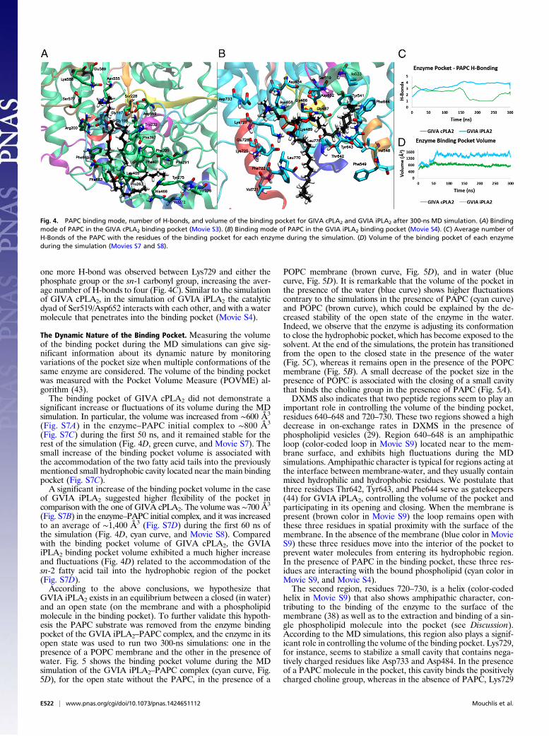
one more H-bond was observed between Lys729 and either thephosphate group or the sn-1 carbonyl group, increasing the aver-age number of H-bonds to four (Fig. 4C). Similar to the simulationof GIVA cPLA2, in the simulation of GVIA iPLA2 the catalyticdyad of Ser519/Asp652 interacts with each other, and with a watermolecule that penetrates into the binding pocket (Movie S4).
The Dynamic Nature of the Binding Pocket. Measuring the volumeof the binding pocket during the MD simulations can give sig-nificant information about its dynamic nature by monitoringvariations of the pocket size when multiple conformations of thesame enzyme are considered. The volume of the binding pocketwas measured with the Pocket Volume Measure (POVME) al-gorithm (43).The binding pocket of GIVA cPLA2 did not demonstrate a
significant increase or fluctuations of its volume during the MDsimulation. In particular, the volume was increased from ∼600 Å3
(Fig. S7A) in the enzyme–PAPC initial complex to ∼800 Å3
(Fig. S7C) during the first 50 ns, and it remained stable for therest of the simulation (Fig. 4D, green curve, and Movie S7). Thesmall increase of the binding pocket volume is associated withthe accommodation of the two fatty acid tails into the previouslymentioned small hydrophobic cavity located near the main bindingpocket (Fig. S7C).A significant increase of the binding pocket volume in the case
of GVIA iPLA2 suggested higher flexibility of the pocket incomparison with the one of GIVA cPLA2. The volume was ∼700 Å3
(Fig. S7B) in the enzyme–PAPC initial complex, and it was increasedto an average of ∼1,400 Å3 (Fig. S7D) during the first 60 ns ofthe simulation (Fig. 4D, cyan curve, and Movie S8). Comparedwith the binding pocket volume of GIVA cPLA2, the GVIAiPLA2 binding pocket volume exhibited a much higher increaseand fluctuations (Fig. 4D) related to the accommodation of thesn-2 fatty acid tail into the hydrophobic region of the pocket(Fig. S7D).According to the above conclusions, we hypothesize that
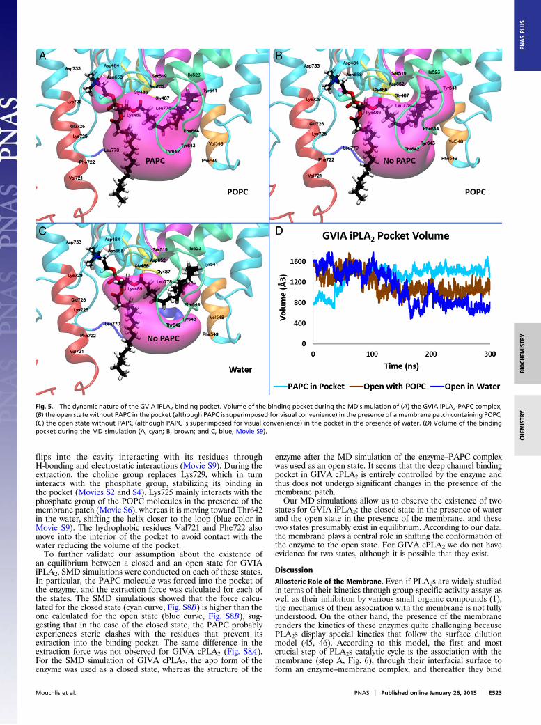
GVIA iPLA2 exists in an equilibrium between a closed (in water)and an open state (on the membrane and with a phospholipidmolecule in the binding pocket). To further validate this hypoth-esis the PAPC substrate was removed from the enzyme bindingpocket of the GVIA iPLA2–PAPC complex, and the enzyme in itsopen state was used to run two 300-ns simulations: one in thepresence of a POPC membrane and the other in the presence ofwater. Fig. 5 shows the binding pocket volume during the MDsimulation of the GVIA iPLA2–PAPC complex (cyan curve, Fig.5D), for the open state without the PAPC, in the presence of a
POPC membrane (brown curve, Fig. 5D), and in water (bluecurve, Fig. 5D). It is remarkable that the volume of the pocket inthe presence of the water (blue curve) shows higher fluctuationscontrary to the simulations in the presence of PAPC (cyan curve)and POPC (brown curve), which could be explained by the de-creased stability of the open state of the enzyme in the water.Indeed, we observe that the enzyme is adjusting its conformationto close the hydrophobic pocket, which has become exposed to thesolvent. At the end of the simulations, the protein has transitionedfrom the open to the closed state in the presence of the water(Fig. 5C), whereas it remains open in the presence of the POPCmembrane (Fig. 5B). A small decrease of the pocket size in thepresence of POPC is associated with the closing of a small cavitythat binds the choline group in the presence of PAPC (Fig. 5A).DXMS also indicates that two peptide regions seem to play an
important role in controlling the volume of the binding pocket,residues 640–648 and 720–730. These two regions showed a highdecrease in on-exchange rates in DXMS in the presence ofphospholipid vesicles (29). Region 640–648 is an amphipathicloop (color-coded loop in Movie S9) located near to the mem-brane surface, and exhibits high fluctuations during the MDsimulations. Amphipathic character is typical for regions acting atthe interface between membrane-water, and they usually containmixed hydrophilic and hydrophobic residues. We postulate thatthree residues Thr642, Tyr643, and Phe644 serve as gatekeepers(44) for GVIA iPLA2, controlling the volume of the pocket andparticipating in its opening and closing. When the membrane ispresent (brown color in Movie S9) the loop remains open withthese three residues in spatial proximity with the surface of themembrane. In the absence of the membrane (blue color in MovieS9) these three residues move into the interior of the pocket toprevent water molecules from entering its hydrophobic region.In the presence of PAPC in the binding pocket, these three res-idues are interacting with the bound phospholipid (cyan color inMovie S9, and Movie S4).The second region, residues 720–730, is a helix (color-coded
helix in Movie S9) that also shows amphipathic character, con-tributing to the binding of the enzyme to the surface of themembrane (38) as well as to the extraction and binding of a sin-gle phospholipid molecule into the pocket (see Discussion).According to the MD simulations, this region also plays a signif-icant role in controlling the volume of the binding pocket. Lys729,for instance, seems to stabilize a small cavity that contains nega-tively charged residues like Asp733 and Asp484. In the presenceof a PAPC molecule in the pocket, this cavity binds the positivelycharged choline group, whereas in the absence of PAPC, Lys729
Fig. 4. PAPC binding mode, number of H-bonds, and volume of the binding pocket for GIVA cPLA2 and GVIA iPLA2 after 300-ns MD simulation. (A) Bindingmode of PAPC in the GIVA cPLA2 binding pocket (Movie S3). (B) Binding mode of PAPC in the GVIA iPLA2 binding pocket (Movie S4). (C) Average number ofH-Bonds of the PAPC with the residues of the binding pocket for each enzyme during the simulation. (D) Volume of the binding pocket of each enzymeduring the simulation (Movies S7 and S8).
E522 | www.pnas.org/cgi/doi/10.1073/pnas.1424651112 Mouchlis et al.
flips into the cavity interacting with its residues throughH-bonding and electrostatic interactions (Movie S9). During theextraction, the choline group replaces Lys729, which in turninteracts with the phosphate group, stabilizing its binding inthe pocket (Movies S2 and S4). Lys725 mainly interacts with thephosphate group of the POPC molecules in the presence of themembrane patch (Movie S6), whereas it is moving toward Thr642in the water, shifting the helix closer to the loop (blue color inMovie S9). The hydrophobic residues Val721 and Phe722 alsomove into the interior of the pocket to avoid contact with thewater reducing the volume of the pocket.To further validate our assumption about the existence of
an equilibrium between a closed and an open state for GVIAiPLA2, SMD simulations were conducted on each of these states.In particular, the PAPC molecule was forced into the pocket ofthe enzyme, and the extraction force was calculated for each ofthe states. The SMD simulations showed that the force calcu-lated for the closed state (cyan curve, Fig. S8B) is higher than theone calculated for the open state (blue curve, Fig. S8B), sug-gesting that in the case of the closed state, the PAPC probablyexperiences steric clashes with the residues that prevent itsextraction into the binding pocket. The same difference in theextraction force was not observed for GIVA cPLA2 (Fig. S8A).For the SMD simulation of GIVA cPLA2, the apo form of theenzyme was used as a closed state, whereas the structure of the
enzyme after the MD simulation of the enzyme–PAPC complexwas used as an open state. It seems that the deep channel bindingpocket in GIVA cPLA2 is entirely controlled by the enzyme andthus does not undergo significant changes in the presence of themembrane patch.Our MD simulations allow us to observe the existence of two
states for GVIA iPLA2: the closed state in the presence of waterand the open state in the presence of the membrane, and thesetwo states presumably exist in equilibrium. According to our data,the membrane plays a central role in shifting the conformation ofthe enzyme to the open state. For GIVA cPLA2 we do not haveevidence for two states, although it is possible that they exist.
DiscussionAllosteric Role of the Membrane. Even if PLA2s are widely studiedin terms of their kinetics through group-specific activity assays aswell as their inhibition by various small organic compounds (1),the mechanics of their association with the membrane is not fullyunderstood. On the other hand, the presence of the membranerenders the kinetics of these enzymes quite challenging becausePLA2s display special kinetics that follow the surface dilutionmodel (45, 46). According to this model, the first and mostcrucial step of PLA2s catalytic cycle is the association with themembrane (step A, Fig. 6), through their interfacial surface toform an enzyme–membrane complex, and thereafter they bind
Fig. 5. The dynamic nature of the GVIA iPLA2 binding pocket. Volume of the binding pocket during the MD simulation of (A) the GVIA iPLA2-PAPC complex,(B) the open state without PAPC in the pocket (although PAPC is superimposed for visual convenience) in the presence of a membrane patch containing POPC,(C) the open state without PAPC (although PAPC is superimposed for visual convenience) in the pocket in the presence of water. (D) Volume of the bindingpocket during the MD simulation (A, cyan; B, brown; and C, blue; Movie S9).
Mouchlis et al. PNAS | Published online January 26, 2015 | E523
BIOCH
EMISTR
YCH
EMISTR
YPN
ASPL
US
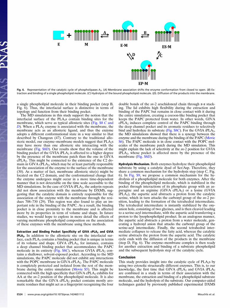
a single phospholipid molecule in their binding pocket (step B,Fig. 6). Thus, the interfacial surface is distinctive in terms oftopology and function from their binding pocket.The MD simulations in this study support the notion that the
interfacial surface of the PLA2s contain binding sites for themembrane, which serve as typical allosteric sites (Fig. S8 C andD). When a PLA2 enzyme is associated with the membrane, themembrane acts as an allosteric ligand, and thus the enzymeadopts a different conformational state in a way similar to thatdescribed by Changeux (47). Contrary to the traditional allo-steric model, our enzyme–membrane models suggest that PLA2smay have more than one allosteric site interacting with themembrane (Fig. S8D). Our results show that the volume of thebinding pocket of the GVIA iPLA2 is affected to a higher degreeby the presence of the membrane patch than the one in GIVAcPLA2. This might be connected to the existence of the C2 do-main in GIVA cPLA2, which may be at least partially responsiblefor the association of the enzyme to the surface of the membrane(30). As a matter of fact, membrane allosteric site(s) might belocated on the C2 domain, and the conformational change thatthe enzyme undergoes might occur in a more time-dependentmanner that is not detectable on the time scale accessible to theMD simulations. In the case of GVIA iPLA2, the ankyrin repeatsdid not show association with the membrane by DXMS, sug-gesting that the catalytic domain is entirely responsible for theassociation of the enzyme with the membrane through the resi-dues 708–730 (29). This region was also found to play an im-portant role in the binding of the PAPC. As a result, the bindingpocket is in close proximity to the membrane and is affectedmore by its properties in term of volume and shape. In futurestudies, we would hope to explore in more detail the effects ofvarying membrane phospholipid composition on the associationof these enzymes with the membrane using these methods.
Extraction and Binding Pocket Specificity of GIVA cPLA2 and GVIAiPLA2. In addition to the allosteric site on the interfacial sur-face, each PLA2 contains a binding pocket that is unique in termsof its volume and shape. GIVA cPLA2, for instance, containsa deep channel binding pocket that accommodates the PAPCmolecule in its entirety (Fig. S8C), whereas GVIA iPLA2 con-tains a shallower, solvent-exposed pocket. According to the MDsimulations, the PAPC molecule did not exhibit any interactionswith the POPC membrane in GIVA cPLA2. The PAPC moleculeis completely protected and isolated from the rest of the mem-brane during the entire simulation (Movie S5). This might beconnected with the high specificity that GIVA cPLA2 exhibits forAA at the sn-2 position of the phospholipid molecule. It is alsoremarkable that the GIVA cPLA2 pocket contains mostly aro-matic residues that might act as a fingerprint recognizing the four
double bonds of the sn-2 arachidonoyl chain through π-π stack-ing. The lid exhibits high flexibility during the extraction andbinding of the PAPC but remains in close contact with it duringthe entire simulation, creating a cocoon-like binding pocket thatkeeps the PAPC protected from water. In other words, GIVAcPLA2 induces complete control of the PAPC binding throughthe deep channel pocket and its aromatic residues to selectivelybind and hydrolyze its substrate (Fig. S8C). For the GVIA iPLA2,the MD simulations showed that there is a synergy between theenzyme and the membrane during the binding of the PAPC (MovieS6). The PAPC molecule is in close contact with the POPC mol-ecules of the membrane patch during the MD simulation. Thismight explain the lack of selectivity at the sn-2 position for GVIAiPLA2, whose pocket is affected more by the presence of themembrane (Fig. S8D).
Hydrolysis Mechanism. Both enzymes hydrolyze their phospholipidsubstrate by using a catalytic dyad of Ser/Asp. Therefore, theyshare a common mechanism for the hydrolysis step (step C, Fig.6). In Fig. S9, we propose a common mechanism for the hy-drolysis of a phospholipid molecule for both enzymes. After thebinding of the phospholipid molecule, which is stabilized in thepocket through interactions of its phosphate group with an as-paragine and an arginine (GIVA cPLA2) or a lysine (GVIAiPLA2), the aspartic acid abstracts a proton from the catalyticserine, which in turn attacks the carbonyl group of the sn-2 po-sition, leading to the formation of the tetrahedral intermediate.The tetrahedral intermediate is instantly stabilized by the oxy-anion hole, consisting of two glycines, and is then cleaved leadingto a serine-acyl intermediate, with the aspartic acid transferring aproton to the lysophospholipid product. In an analogous manner,the aspartic acid abstracts a proton from a water molecule, gen-erating a hydroxyl anion that attacks the carbonyl group of theserine-acyl intermediate. Finally, the second tetrahedral inter-mediate collapses to release the fatty acid, whereas the catalyticserine abstracts the proton from the aspartic acid. The catalyticcycle ends with the diffusion of the products in the membrane(step D, Fig. 6). The enzyme–membrane complex is then readyfor another extraction and binding of a substrate phospholipidand the subsequent hydrolysis step of the catalytic cycle.
ConclusionThis study provides insight into the catalytic cycle of PLA2s byusing two cytosolic structurally different enzymes. This is, to ourknowledge, the first time that GIVA cPLA2 and GVIA iPLA2are combined in a study in terms of their association with themembrane, the extraction and binding of a phospholipid substratemolecule, and the hydrolysis of the substrate. Our computer-aidedtechniques guided by previously published experimental DXMS
Fig. 6. Representation of the catalytic cycle of phospholipases A2. (A) Membrane association shifts the enzyme conformation from closed to open. (B) Ex-traction and binding of a single phospholipid molecule. (C) Hydrolysis of the bound phospholipid molecule. (D). Diffusion of the products into the membrane.
E524 | www.pnas.org/cgi/doi/10.1073/pnas.1424651112 Mouchlis et al.

results (28–31) revealed interesting information about the differ-ent role that the membrane plays for these two enzymes. We havealso found topological and functional differences in the bindingpockets of the two enzymes. All of the above information will beused to rationally design potent and selective inhibitors for thesetwo enzymes that might evolve to future therapeutic agents.
Materials and MethodsA detailed description of the computational methods used in this study isprovided in SI Materials and Methods. Briefly, the homology model ofGVIA iPLA2 was built using the Prime 3.1 protein structure suite programs(34). The X-ray crystal structure of GIVA cPLA2 (PDB ID 1CJY) was preparedusing the Protein Preparation Wizard module (48). The GIVA cPLA2 and
GVIA iPLA2 binding pockets were identified using SiteMap 2.6 (37). TheIFD protocol was used to generate the enzyme–PAPC complexes (32). The MDand SMD simulations were performed using NAMD 2.9 (33), and the results wereanalyzed using the VMD package (42) and the POVME algorithm (43).
ACKNOWLEDGMENTS. We thank Drs. Robert Konecny and Brain Fox fortheir help with the Schrödinger suite. This work was supported by NIH GrantGM20501 (to E.A.D.). Work in the J.A.M. group is supported in part by theNational Science Foundation (NSF), NIH, Howard Hughes Medical Institute,and NBCR (National Biomedical Computation Resource). Anton computertime was provided by the MMBioS through Grant P41GM103712-S1 fromthe NIH and the Pittsburgh Supercomputing Center. The Anton machine atPSC (Pittsburgh Supercomputing Center) was generously made available byD.E. Shaw Research. This work used the XSEDE (Extreme Science and Engineer-ing Discovery Environment), which is supported by NSF Grant ACI-1053575.
1. Dennis EA, Cao J, Hsu YH, Magrioti V, Kokotos G (2011) Phospholipase A2 enzymes:Physical structure, biological function, disease implication, chemical inhibition, andtherapeutic intervention. Chem Rev 111(10):6130–6185.
2. Buczynski MW, Dumlao DS, Dennis EA (2009) Thematic Review Series: Proteomics. Anintegrated omics analysis of eicosanoid biology. J Lipid Res 50(6):1015–1038.
3. Pniewska E, Pawliczak R (2013) The involvement of phospholipases A2 in asthma andchronic obstructive pulmonary disease. Mediators Inflamm 2013:793505.
4. Masuda S, et al. (2005) Various secretory phospholipase A2 enzymes are expressed inrheumatoid arthritis and augment prostaglandin production in cultured synovial cells.FEBS J 272(3):655–672.
5. Rosenson RS, Hurt-Camejo E (2012) Phospholipase A2 enzymes and the risk of ath-erosclerosis. Eur Heart J 33(23):2899–2909.
6. Nagase T, et al. (2002) A pivotal role of cytosolic phospholipase A(2) in bleomycin-induced pulmonary fibrosis. Nat Med 8(5):480–484.
7. Malhotra A, et al. (2009) Role of calcium-independent phospholipase A2 in thepathogenesis of Barth syndrome. Proc Natl Acad Sci USA 106(7):2337–2341.
8. Mouchlis VD, et al. (2012) Binding conformation of 2-oxoamide inhibitors to groupIVA cytosolic phospholipase A2 determined by molecular docking combined withmolecular dynamics. J Chem Inf Model 52(1):243–254.
9. Mouchlis VD, Barbayianni E, Mavromoustakos TM, Kokotos G (2011) The application ofrational design on phospholipase A(2) inhibitors. Curr Med Chem 18(17):2566–2582.
10. Magrioti V, et al. (2013) New potent and selective polyfluoroalkyl ketone inhibitors ofGVIA calcium-independent phospholipase A2. Bioorg Med Chem 21(18):5823–5829.
11. Kramer RM, Roberts EF, Manetta J, Putnam JE (1991) The Ca2(+)-sensitive cytosolicphospholipase A2 is a 100-kDa protein in human monoblast U937 cells. J Biol Chem266(8):5268–5272.
12. Sharp JD, et al. (1991) Molecular cloning and expression of human Ca(2+)-sensitivecytosolic phospholipase A2. J Biol Chem 266(23):14850–14853.
13. Dessen A, et al. (1999) Crystal structure of human cytosolic phospholipase A2 revealsa novel topology and catalytic mechanism. Cell 97(3):349–360.
14. Channon JY, Leslie CC (1990) A calcium-dependent mechanism for associating a sol-uble arachidonoyl-hydrolyzing phospholipase A2 with membrane in the macrophagecell line RAW 264.7. J Biol Chem 265(10):5409–5413.
15. Pickard RT, et al. (1996) Identification of essential residues for the catalytic function of85-kDa cytosolic phospholipase A2. Probing the role of histidine, aspartic acid, cys-teine, and arginine. J Biol Chem 271(32):19225–19231.
16. Lin LL, et al. (1993) cPLA2 is phosphorylated and activated by MAP kinase. Cell 72(2):269–278.
17. Nakamura H, Hirabayashi T, Shimizu M, Murayama T (2006) Ceramide-1-phosphateactivates cytosolic phospholipase A2α directly and by PKC pathway. Biochem Phar-macol 71(6):850–857.
18. Mosior M, Six DA, Dennis EA (1998) Group IV cytosolic phospholipase A2 binds withhigh affinity and specificity to phosphatidylinositol 4,5-bisphosphate resulting indramatic increases in activity. J Biol Chem 273(4):2184–2191.
19. Clark JD, et al. (1991) A novel arachidonic acid-selective cytosolic PLA2 contains a Ca(2+)-dependent translocation domain with homology to PKC and GAP. Cell 65(6):1043–1051.
20. Larsson Forsell PK, Kennedy BP, Claesson HE (1999) The human calcium-independentphospholipase A2 gene multiple enzymes with distinct properties from a single gene.Eur J Biochem 262(2):575–585.
21. Ma Z, Wang X, Nowatzke W, Ramanadham S, Turk J (1999) Human pancreatic isletsexpress mRNA species encoding two distinct catalytically active isoforms of group VIphospholipase A2 (iPLA2) that arise from an exon-skipping mechanism of alternativesplicing of the transcript from the iPLA2 gene on chromosome 22q13.1. J Biol Chem274(14):9607–9616.
22. Michaely P, Tomchick DR, Machius M, Anderson RG (2002) Crystal structure of a 12ANK repeat stack from human ankyrinR. EMBO J 21(23):6387–6396.
23. Balboa MA, Balsinde J, Jones SS, Dennis EA (1997) Identity between the Ca2+-inde-pendent phospholipase A2 enzymes from P388D1 macrophages and Chinese hamsterovary cells. J Biol Chem 272(13):8576–8580.
24. Lio YC, Dennis EA (1998) Interfacial activation, lysophospholipase and transacylase ac-tivity of group VI Ca2+-independent phospholipase A2. Biochim Biophys Acta 1392(2-3):320–332.
25. Percy AJ, Rey M, Burns KM, Schriemer DC (2012) Probing protein interactions with
hydrogen/deuterium exchange and mass spectrometry—a review. Anal Chim Acta
721:7–21.26. Harkewicz R, Dennis EA (2011) Applications of mass spectrometry to lipids and
membranes. Annu Rev Biochem 80:301–325.27. Cao J, Burke JE, Dennis EA (2013) Using hydrogen/deuterium exchange mass spec-
trometry to define the specific interactions of the phospholipase A2 superfamily with
lipid substrates, inhibitors, and membranes. J Biol Chem 288(3):1806–1813.28. Hsu Y-H, et al. (2013) Fluoroketone inhibition of Ca(2+)-independent phospholipase
A2 through binding pocket association defined by hydrogen/deuterium exchange and
molecular dynamics. J Am Chem Soc 135(4):1330–1337.29. Hsu YH, Burke JE, Li S, Woods VL, Jr, Dennis EA (2009) Localizing the membrane binding
region of Group VIA Ca2+-independent phospholipase A2 using peptide amide hydro-
gen/deuterium exchange mass spectrometry. J Biol Chem 284(35):23652–23661.30. Burke JE, et al. (2008) A phospholipid substrate molecule residing in the membrane
surface mediates opening of the lid region in group IVA cytosolic phospholipase A2.
J Biol Chem 283(45):31227–31236.31. Burke JE, et al. (2009) Location of inhibitors bound to group IVA phospholipase A2
determined by molecular dynamics and deuterium exchange mass spectrometry. J Am
Chem Soc 131(23):8083–8091.32. Suite 2012 (2012) Schrödinger Suite 2012 Induced Fit Docking Protocol; Glide Version
5.8, Schrödinger, LLC, New York, NY, 2012; Prime Version 3.1 (Schrödinger, LLC, New
York).33. Phillips JC, et al. (2005) Scalable molecular dynamics with NAMD. J Comput Chem
26(16):1781–1802.34. Suite 2012 (2012) Prime, Version 3.1 (Schrödinger, LLC, New York).35. Rydel TJ, et al. (2003) The crystal structure, mutagenesis, and activity studies reveal
that patatin is a lipid acyl hydrolase with a Ser-Asp catalytic dyad. Biochemistry 42(22):
6696–6708.36. Jenkins CM, et al. (2004) Identification, cloning, expression, and purification of three
novel human calcium-independent phospholipase A2 family members possessing tri-
acylglycerol lipase and acylglycerol transacylase activities. J Biol Chem 279(47):
48968–48975.37. Suite 2012 (2012) SiteMap, Version 2.6 (Schrödinger, LLC, New York, NY).38. Bucher D, Hsu YH, Mouchlis VD, Dennis EA, McCammon JA (2013) Insertion of the
Ca²⁺-independent phospholipase A₂ into a phospholipid bilayer via coarse-grained
and atomistic molecular dynamics simulations. PLOS Comput Biol 9(7):e1003156.39. Chen H, et al. (2007) Charge delocalization in proton channels, I: The aquaporin
channels and proton blockage. Biophys J 92(1):46–60.40. Wang Y, Schulten K, Tajkhorshid E (2005) What makes an aquaporin a glycerol
channel? A comparative study of AqpZ and GlpF. Structure 13(8):1107–1118.41. Karplus M, McCammon JA (2002) Molecular dynamics simulations of biomolecules.
Nat Struct Mol Biol 9(9):647–653.42. Humphrey W, Dalke A, Schulten K (1996) VMD: Visual molecular dynamics. J Mol
Graph 14(1):33–38, 27–28.43. Durrant JD, de Oliveira CA, McCammon JA (2011) POVME: An algorithm for mea-
suring binding-pocket volumes. J Mol Graph Model 29(5):773–776.44. Zuccotto F, Ardini E, Casale E, Angiolini M (2010) Through the “gatekeeper door”:
Exploiting the active kinase conformation. J Med Chem 53(7):2681–2694.45. Carman GM, Deems RA, Dennis EA (1995) Lipid signaling enzymes and surface di-
lution kinetics. J Biol Chem 270(32):18711–18714.46. Deems RA, Eaton BR, Dennis EA (1975) Kinetic analysis of phospholipase A2 activity
toward mixed micelles and its implications for the study of lipolytic enzymes. J Biol
Chem 250(23):9013–9020.47. Changeux J-P (2012) Allostery and the Monod-Wyman-Changeux model after 50
years. Annu Rev Biophys 41:103–133.48. Suite 2012 (2012) Schrödinger Suite 2012 Protein Preparation Wizard; Epik Version
2.3, Schrödinger, LLC, New York, NY, 2012; Impact Version 5.8, Schrödinger, LLC, New
York, NY, 2012; Prime Version 3.1 (Schrödinger, LLC, New York).
Mouchlis et al. PNAS | Published online January 26, 2015 | E525
BIOCH
EMISTR
YCH
EMISTR
YPN
ASPL
US