membranous nephropathy with crescent after hematopoietic
TRANSCRIPT
91
doi: 10.2169/internalmedicine.1251-18
Intern Med 58: 91-96, 2019
http://internmed.jp
【 CASE REPORT 】
Membranous Nephropathy with Crescent afterHematopoietic Cell Transplantation
Mineaki Kitamura 1,2, Satoshi Hisano 3, Yuka Kurobe 2, Shinichi Abe 2, Yuki Ota 2,
Yasushi Sawayama 4, Tadashi Uramatsu 2, Yoko Obata 2, Junya Fukuoka 5, Yasushi Miyazaki 4,
Hiroshi Mukae 6 and Tomoya Nishino 2
Abstract:A 44-year-old man who received allogenic hematopoietic stem cell transplantation after being diagnosed
with acute myeloid leukemia developed nephrosis when the dose of tacrolimus was tapered. A renal biopsy
showed the granular deposition of immunoglobulin G in the glomerular basement membrane and subepithe-
lial electron-dense deposits, crescent formation, C4d-positive staining of the peritubular capillary, and suben-
dothelial swelling, suggesting that the main pathological diagnosis was membranous nephropathy and that
chronic graft-versus-host disease played a role in the etiology of nephrosis. We herein report a case of mem-
branous nephropathy with various pathological findings. C4d deposition suggests complement activation and
the involvement of humoral factors.
Key words: graft-versus-host disease, membranous nephropathy, crescent, C4d positivity on peritubular
capillaries
(Intern Med 58: 91-96, 2019)(DOI: 10.2169/internalmedicine.1251-18)
Introduction
The number of allogenic hematopoietic cell transplants
for leukemia has recently increased. Consequently, the rec-
ognition of glomerular diseases associated with nephrotic
syndrome as a complication has also increased (1).
According to the pertinent literature, the incidence of
nephrotic syndrome in adult recipients ranges from 0.4% to
6% (2). Nephrotic syndrome has many causes; however, the
most critical cause of nephrotic syndrome in patients after
hematopoietic cell transplantation is graft-versus-host disease
(GVHD) (3). Although acute GVHD tends to cause throm-
botic microangiopathy (TMA), chronic GVHD tends to
cause nephrotic syndrome, especially membranous nephro-
pathy (MN) (2). There have been case series of MN after al-
logenic hematopoietic stem cell transplantation that focused
on IgG subclasses and the phospholipase A2 receptor
(PLA2R) to distinguish this form of MN from primary
MN (4-6). However, the characteristics of MN after hema-
topoietic stem cell transplantation remain unclear, since sev-
eral mechanisms are involved in chronic GVHD and the
pathological findings may vary among cases.
We herein report a case of MN with unusual renal patho-
logical findings after hematopoietic cell transplantation.
Case Report
A 44-year-old man diagnosed with acute myelogenous
leukemia (AML) was treated with idarubicin and cytarabine.
He was also administered intensification therapy with the
first-course drugs of mitoxantrone and cytarabine and
second-course drugs of daunorubicin and cytarabine. After-
ward, he underwent unrelated peripheral stem cell transplan-
1Division of Blood Purification, Nagasaki University Hospital, Japan, 2Department of Nephrology, Nagasaki University Hospital, Japan, 3Depart-
ment of Pathology, Faculty of Medicine, Fukuoka University, Japan, 4Department of Hematology, Nagasaki University Hospital, Japan, 5Depart-
ment of Pathology, Nagasaki University Graduate School of Biomedical Sciences, Japan and 6Department of Respiratory Medicine, Nagasaki
University Graduate School of Biomedical Sciences, Japan
Received: March 23, 2018; Accepted: June 10, 2018; Advance Publication by J-STAGE: August 24, 2018
Correspondence to Dr. Mineaki Kitamura, [email protected]
Intern Med 58: 91-96, 2019 DOI: 10.2169/internalmedicine.1251-18
92
Table. Laboratory Data on Admission.
Peripheral blood AST 25 U/L Urinalysis
WBC 10,200 μL ALT 22 U/L Gravity ≥1.050
RBC 5.63×10⁶ /μL ALP 310 U/L pH 6.5
Hb 17.5 g/dL LDH 22 U/L protein (4+)
Hct 51.4 % γGTP 26 U/L occult blood (2+)
Plt 259×103 /μL GLU 123 mg/dL glucose (+/-)
CRP 0.09 mg/dL Sediments
Blood chemistry Serological tests RBC 31~50 /HPF
Na 142 mmol/L anti nuclear Ab (-) WBC 1~2 /HPF
K 3.9 mmol/L CH50 52.1 /mL hyaline cast 5~10 /LPF
Cl 108 mmol/L C3 170.4 mg/dL dysmorphic RBC (+)
TP 5.2 g/dL C4 38.1 mg/dL
Alb 2.0 g/dL HBs antigen 0.1 COI Urinary chemistry
BUN 15 mg/dL anti HCV Ab 0.1 COI TP/Cr 23.02 g/gCr
Cr 0.96 mg/dL IgA 160 mg/dL NAG 158.5 U/L
UA 7.1 mg/dL IgG 650 mg/dL β2MG 4,600 μg/L
TC 379 mg/dL IgM 79 mg/dL selectivity index. 0.036
TG 322 mg/dL IgE 269.9 IU/mL
HDL-C 66 mg/dL MPO-ANCA (-)
LDL-C 250 mg/dL PR3-ANCA (-)
β2MG: β2 microglobulin, NAG: N-acetyl-β-D-glucosaminidase
tation, and there was no human leukocyte antigen (HLA)
(A, B, C, DR) mismatch with the unrelated donor. A condi-
tioning regimen of methotrexate and total-body irradiation
was instituted. GVHD prophylaxis initially consisted of
methotrexate but was later changed to tacrolimus monother-
apy. Even with reduced-dose tacrolimus, no symptoms re-
lated to GVHD were observed, and he was discharged from
the hospital four months after transplantation. The
tacrolimus dosage was decreased to 0.4 mg/day through 20
months post-transplantation.
Post-transplantation, the serum creatinine (Cr) and serum
albumin levels were normal (0.78 mg/dL and 4 g/dL, re-
spectively); however, at 21 months post-transplantation, the
patient noticed bilateral leg swelling. New laboratory data
showed that the serum Cr level remained normal but had in-
creased to 0.96 mg/dL, and the serum albumin level had de-
creased to 2 g/dL. The serum total protein level was 5 g/dL,
and the serum albumin level was 2 g/dL. The urinary pro-
tein/creatinine ratio was 23.02 g/gCr. Due to substantial pro-
teinuria, the patient received a nephrology consultation and
was transferred to the nephrology department, where a kid-
ney biopsy was performed at 23 months after transplanta-
tion. The urinary protein excretion was 10.5 g/day, and the
proteinuria selectivity index was 0.036. The number of red
blood cells in urinary sediment was 31-50 per high-power
field. The patient’s laboratory data upon admission are
shown in the table. The only complication the patient expe-
rienced was hyperlipidemia. The results of a serological
analysis for hepatitis B virus (HBV) and hepatitis C virus
(HCV) were negative. Myeloperoxidase anti-neutrophil cyto-
plasmic antibody (MPO-ANCA), proteinase (PR) 3-ANCA,
and antinuclear antibodies were also absent.
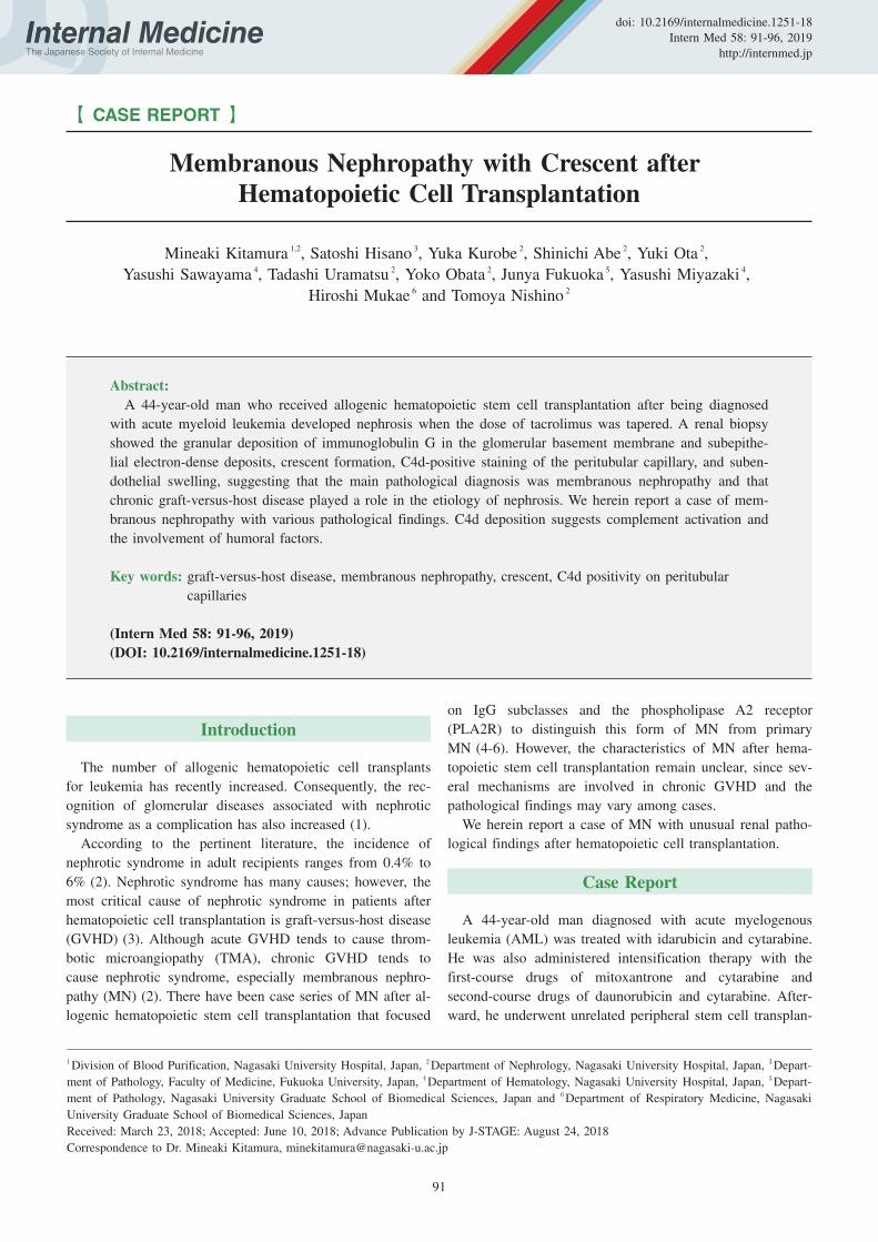
The renal biopsy sample contained 39 glomeruli, 2 of
which were globally sclerosed. One glomerulus showed seg-
mental endocapillary hypercellularity and extracapillary cel-
lular proliferation (Fig. 1A and B). The peripheral glomeru-
lar capillary walls were not thickened and showed no appar-
ent bubbling or spikes. In the two collapsing glomeruli, the
Bowman’s capsules were thickened with prominent intersti-
tial fibrosis surrounding them. The proportion of interstitial
fibrosis and tubular atrophy area in the cortex was 10%.
There was no intimal thickening in the interlobular arteries,
but partial nephrotoxicity of calcineurin inhibitors, such as
increasing arteriolar hyalinosis and small-vessel narrowing,
was observed. Typical TMA morphologies, such as fibrin
thrombi within glomeruli and mesangiolysis, were not pre-
sent.
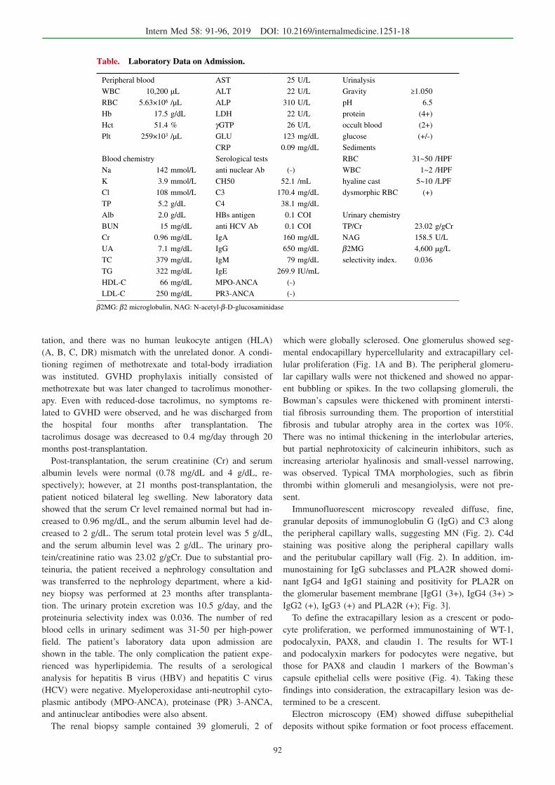
Immunofluorescent microscopy revealed diffuse, fine,
granular deposits of immunoglobulin G (IgG) and C3 along
the peripheral capillary walls, suggesting MN (Fig. 2). C4d
staining was positive along the peripheral capillary walls
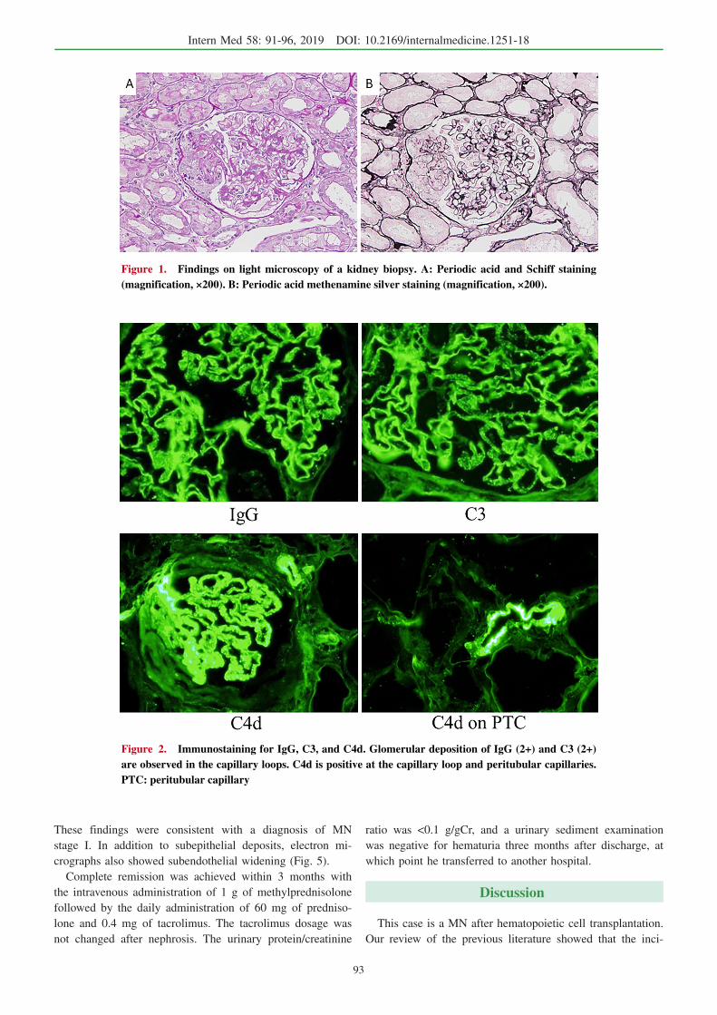
and the peritubular capillary wall (Fig. 2). In addition, im-
munostaining for IgG subclasses and PLA2R showed domi-
nant IgG4 and IgG1 staining and positivity for PLA2R on
the glomerular basement membrane [IgG1 (3+), IgG4 (3+) >
IgG2 (+), IgG3 (+) and PLA2R (+); Fig. 3].
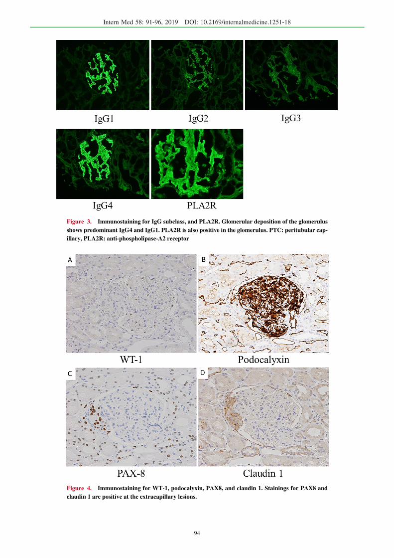
To define the extracapillary lesion as a crescent or podo-
cyte proliferation, we performed immunostaining of WT-1,
podocalyxin, PAX8, and claudin 1. The results for WT-1
and podocalyxin markers for podocytes were negative, but
those for PAX8 and claudin 1 markers of the Bowman’s
capsule epithelial cells were positive (Fig. 4). Taking these
findings into consideration, the extracapillary lesion was de-
termined to be a crescent.
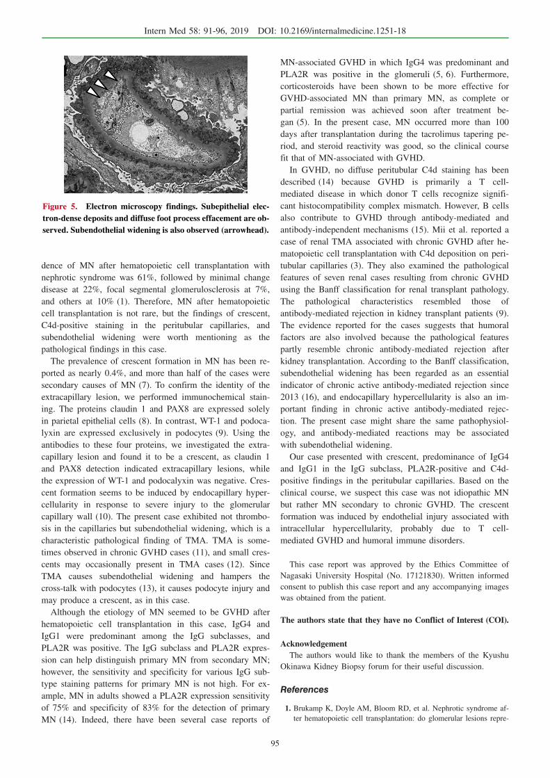
Electron microscopy (EM) showed diffuse subepithelial
deposits without spike formation or foot process effacement.
Intern Med 58: 91-96, 2019 DOI: 10.2169/internalmedicine.1251-18
93
Figure 1. Findings on light microscopy of a kidney biopsy. A: Periodic acid and Schiff staining (magnification, ×200). B: Periodic acid methenamine silver staining (magnification, ×200).
Figure 2. Immunostaining for IgG, C3, and C4d. Glomerular deposition of IgG (2+) and C3 (2+) are observed in the capillary loops. C4d is positive at the capillary loop and peritubular capillaries. PTC: peritubular capillary
These findings were consistent with a diagnosis of MN
stage I. In addition to subepithelial deposits, electron mi-
crographs also showed subendothelial widening (Fig. 5).
Complete remission was achieved within 3 months with
the intravenous administration of 1 g of methylprednisolone
followed by the daily administration of 60 mg of predniso-
lone and 0.4 mg of tacrolimus. The tacrolimus dosage was
not changed after nephrosis. The urinary protein/creatinine
ratio was <0.1 g/gCr, and a urinary sediment examination
was negative for hematuria three months after discharge, at
which point he transferred to another hospital.
Discussion
This case is a MN after hematopoietic cell transplantation.
Our review of the previous literature showed that the inci-
Intern Med 58: 91-96, 2019 DOI: 10.2169/internalmedicine.1251-18
94
Figure 3. Immunostaining for IgG subclass, and PLA2R. Glomerular deposition of the glomerulus shows predominant IgG4 and IgG1. PLA2R is also positive in the glomerulus. PTC: peritubular cap-illary, PLA2R: anti-phospholipase-A2 receptor
Figure 4. Immunostaining for WT-1, podocalyxin, PAX8, and claudin 1. Stainings for PAX8 and claudin 1 are positive at the extracapillary lesions.
Intern Med 58: 91-96, 2019 DOI: 10.2169/internalmedicine.1251-18
95
Figure 5. Electron microscopy findings. Subepithelial elec-tron-dense deposits and diffuse foot process effacement are ob-served. Subendothelial widening is also observed (arrowhead).
dence of MN after hematopoietic cell transplantation with
nephrotic syndrome was 61%, followed by minimal change
disease at 22%, focal segmental glomerulosclerosis at 7%,
and others at 10% (1). Therefore, MN after hematopoietic
cell transplantation is not rare, but the findings of crescent,
C4d-positive staining in the peritubular capillaries, and
subendothelial widening were worth mentioning as the
pathological findings in this case.
The prevalence of crescent formation in MN has been re-
ported as nearly 0.4%, and more than half of the cases were
secondary causes of MN (7). To confirm the identity of the
extracapillary lesion, we performed immunochemical stain-
ing. The proteins claudin 1 and PAX8 are expressed solely
in parietal epithelial cells (8). In contrast, WT-1 and podoca-
lyxin are expressed exclusively in podocytes (9). Using the
antibodies to these four proteins, we investigated the extra-
capillary lesion and found it to be a crescent, as claudin 1
and PAX8 detection indicated extracapillary lesions, while
the expression of WT-1 and podocalyxin was negative. Cres-
cent formation seems to be induced by endocapillary hyper-
cellularity in response to severe injury to the glomerular
capillary wall (10). The present case exhibited not thrombo-
sis in the capillaries but subendothelial widening, which is a
characteristic pathological finding of TMA. TMA is some-
times observed in chronic GVHD cases (11), and small cres-
cents may occasionally present in TMA cases (12). Since
TMA causes subendothelial widening and hampers the
cross-talk with podocytes (13), it causes podocyte injury and
may produce a crescent, as in this case.
Although the etiology of MN seemed to be GVHD after
hematopoietic cell transplantation in this case, IgG4 and
IgG1 were predominant among the IgG subclasses, and
PLA2R was positive. The IgG subclass and PLA2R expres-
sion can help distinguish primary MN from secondary MN;
however, the sensitivity and specificity for various IgG sub-
type staining patterns for primary MN is not high. For ex-
ample, MN in adults showed a PLA2R expression sensitivity
of 75% and specificity of 83% for the detection of primary
MN (14). Indeed, there have been several case reports of
MN-associated GVHD in which IgG4 was predominant and
PLA2R was positive in the glomeruli (5, 6). Furthermore,
corticosteroids have been shown to be more effective for
GVHD-associated MN than primary MN, as complete or
partial remission was achieved soon after treatment be-
gan (5). In the present case, MN occurred more than 100
days after transplantation during the tacrolimus tapering pe-
riod, and steroid reactivity was good, so the clinical course
fit that of MN-associated with GVHD.
In GVHD, no diffuse peritubular C4d staining has been
described (14) because GVHD is primarily a T cell-
mediated disease in which donor T cells recognize signifi-
cant histocompatibility complex mismatch. However, B cells
also contribute to GVHD through antibody-mediated and
antibody-independent mechanisms (15). Mii et al. reported a
case of renal TMA associated with chronic GVHD after he-
matopoietic cell transplantation with C4d deposition on peri-
tubular capillaries (3). They also examined the pathological
features of seven renal cases resulting from chronic GVHD
using the Banff classification for renal transplant pathology.
The pathological characteristics resembled those of
antibody-mediated rejection in kidney transplant patients (9).
The evidence reported for the cases suggests that humoral
factors are also involved because the pathological features
partly resemble chronic antibody-mediated rejection after
kidney transplantation. According to the Banff classification,
subendothelial widening has been regarded as an essential
indicator of chronic active antibody-mediated rejection since
2013 (16), and endocapillary hypercellularity is also an im-
portant finding in chronic active antibody-mediated rejec-
tion. The present case might share the same pathophysiol-
ogy, and antibody-mediated reactions may be associated
with subendothelial widening.
Our case presented with crescent, predominance of IgG4
and IgG1 in the IgG subclass, PLA2R-positive and C4d-
positive findings in the peritubular capillaries. Based on the
clinical course, we suspect this case was not idiopathic MN
but rather MN secondary to chronic GVHD. The crescent
formation was induced by endothelial injury associated with
intracellular hypercellularity, probably due to T cell-
mediated GVHD and humoral immune disorders.
This case report was approved by the Ethics Committee of
Nagasaki University Hospital (No. 17121830). Written informed
consent to publish this case report and any accompanying images
was obtained from the patient.
The authors state that they have no Conflict of Interest (COI).
AcknowledgementThe authors would like to thank the members of the Kyushu
Okinawa Kidney Biopsy forum for their useful discussion.
References
1. Brukamp K, Doyle AM, Bloom RD, et al. Nephrotic syndrome af-
ter hematopoietic cell transplantation: do glomerular lesions repre-

Intern Med 58: 91-96, 2019 DOI: 10.2169/internalmedicine.1251-18
96
sent renal graft-versus-host disease? Clin J Am Soc Nephrol 1:
685-694, 2006.
2. Hingorani S. Renal complications of hematopoietic-cell transplan-
tation. N Engl J Med 374: 2256-2267, 2016.
3. Mii A, Shimizu A, Utsumi K, et al. Renal thrombotic microan-
giopathy associated with chronic humoral graft versus host disease
after hematopoietic stem cell transplantation. Pathol Int 61: 34-41,
2011.
4. Hiramatsu R, Ubara Y, Imafuku A, et al. Clinicopathological
analysis of allogeneic hematopoietic stem cell transplantation-
related. Hum Pathol 50: 187-194, 2016.
5. Huang X, Qin W, Liu Z, et al. Detection of anti-PLA2R autoanti-
bodies and IgG subclasses in post-allogeneic hematopoietic stem
cell transplantation membranous nephropathy. Am J Med Sci 346:
32-37, 2013.
6. Byrne-Dugan CJ, Collins AB, Batal I, et al. Membranous nephro-
pathy as a manifestation of graft-versus-host disease: association
with HLA antigen typing, phospholipase A2 receptor, and C4d.
Am J Kidney Dis 64: 987-993, 2014.
7. Rodriguez EF, Nasr SH, Larsen CP, Sethi S, Fidler ME, Cornell
LD. Membranous nephropathy with crescents: a series of 19 cases.
Am J Kidney Dis 64: 66-73, 2014.
8. Ohse T, Vaughan MR, Chang AM, et al. De novo expression of
podocyte proteins in parietal epithelial cells during experimental
glomerular disease. Am J Physiol Renal Physiol 298: 702-711,
2010.
9. Palmer RE, Kotsianti A, Haber DA, et al. WT1 regulates the ex-
pression of the major glomerular podocyte membrane protein Po-
docalyxin. Curr Biol 11: 1805-1809, 2001.
10. Charles Jennette J. Rapidly progressive crescentic glomeru-
lonephritis. Kidney Int 63: 1164-1177, 2003.
11. Mii A, Shimizu A, Fujino T, et al. Renal thrombotic microan-
giopathy associated with chronic graft-versus-host disease after al-
logeneic hematopoietic stem cell transplantation. Pathol Int 61:
518-527, 2011.
12. Jennette JC, Silva FG, D’Agati VD, et al. Heptinstall’s pathology
of the kidney. Wolters Kluwer Health/Lippincott Williams &
Wilkins, Philadelphia, PA, 2015.
13. Dimke H, Maezawa Y, Quaggin SE. Crosstalk in glomerular injury
and repair. Curr Opin Nephrol Hypertens 6: 356-372, 2015.
14. Larsen CP, Messias NC, Walker PD, et al. Determination of pri-
mary versus secondary membranous glomerulopathy utilizing
phospholipase A2 receptor staining in renal biopsies. Mod Pathol
26: 709-715, 2013.
15. Jacobson CA, Ritz J. B-cell-directed therapy for chronic graft-
versus-host disease. Haematologica 95: 1811-1813, 2010.
16. Haas M, Sis B, Colvin RB, et al. Banff 2013 meeting report: in-
clusion of C4d-negative antibody-mediated rejection and antibody-
associated arterial lesions. Am J Transplant 14: 272-283, 2014.
The Internal Medicine is an Open Access journal distributed under the Creative
Commons Attribution-NonCommercial-NoDerivatives 4.0 International License. To
view the details of this license, please visit (https://creativecommons.org/licenses/
by-nc-nd/4.0/).
Ⓒ 2019 The Japanese Society of Internal Medicine
Intern Med 58: 91-96, 2019