membranous organelles -...
TRANSCRIPT
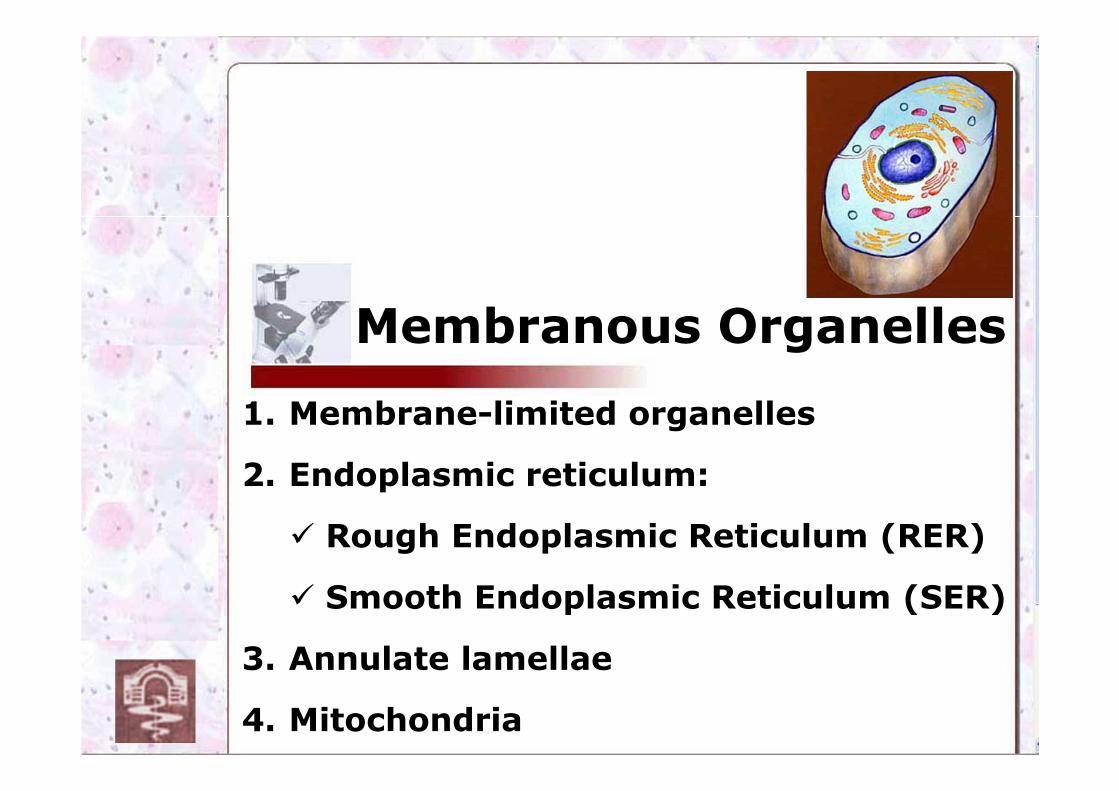
Membranous Organelles
1. Membrane-limited organelles
2. Endoplasmic reticulum:
� Rough Endoplasmic Reticulum (RER)
� Smooth Endoplasmic Reticulum (SER)
3. Annulate lamellae
4. Mitochondria
Prof. Dr. Nikolai Lazarov
2
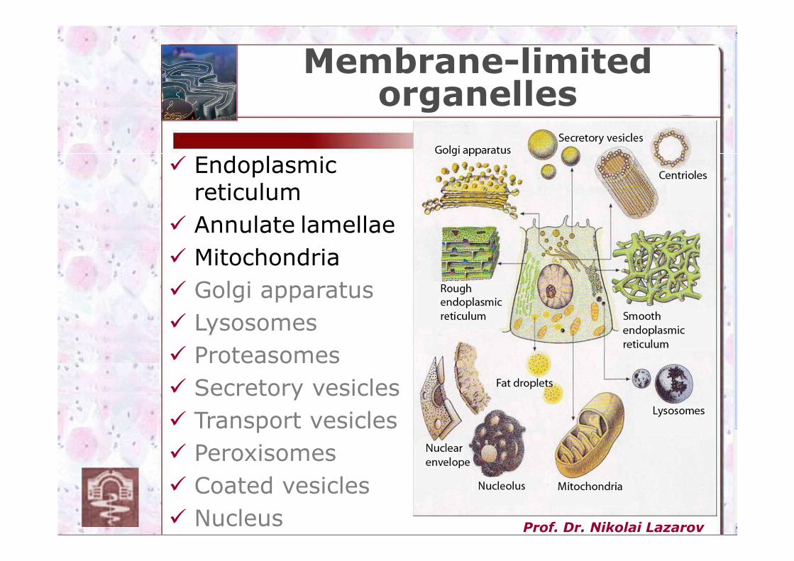
Membrane-limited organelles
� Endoplasmic reticulum
� Annulate lamellae
� Mitochondria
� Golgi apparatus
� Lysosomes
� Proteasomes
� Secretory vesicles
� Transport vesicles
� Peroxisomes
� Coated vesicles
� Nucleus
Prof. Dr. Nikolai Lazarov
3
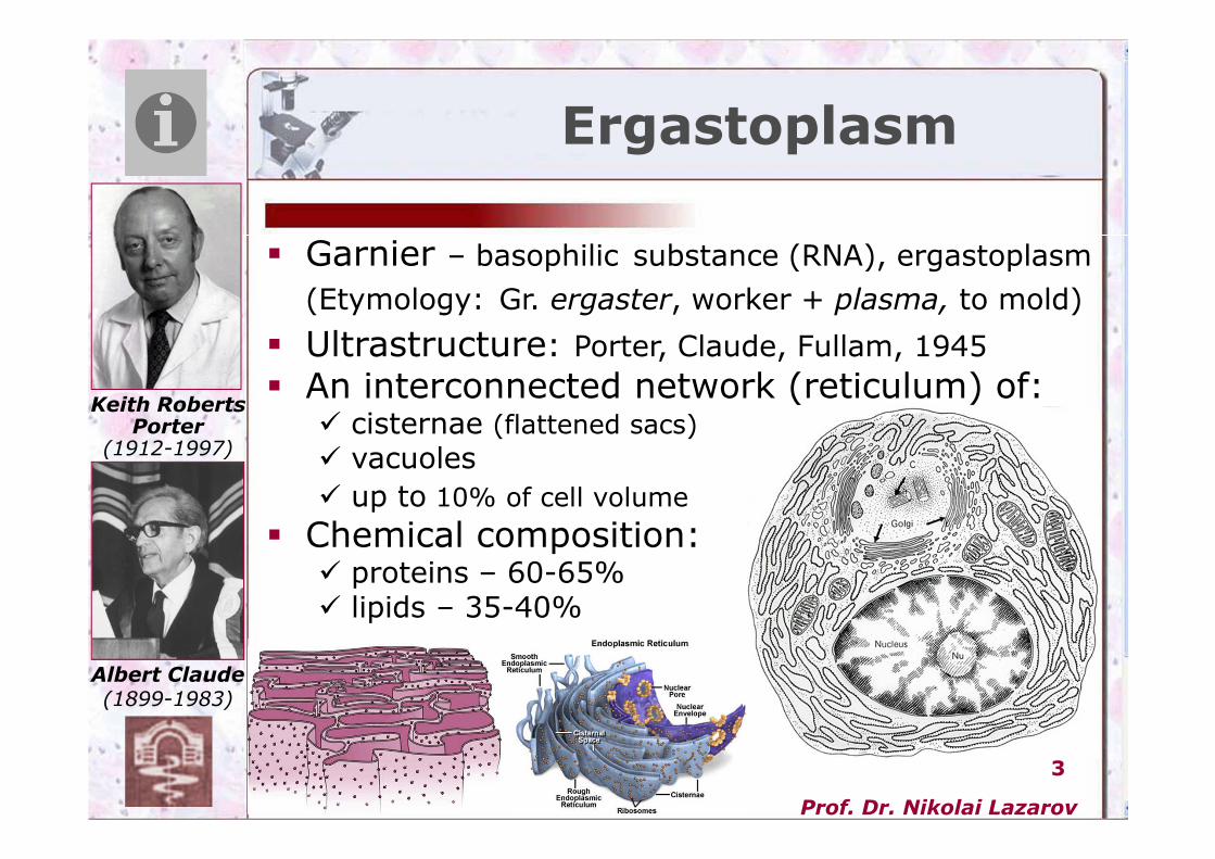
� Garnier – basophilic substance (RNA), ergastoplasm
(Etymology: Gr. ergaster, worker + plasma, to mold)
� Ultrastructure: Porter, Claude, Fullam, 1945
� An interconnected network (reticulum) of:� cisternae (flattened sacs)
� vacuoles
� up to 10% of cell volume
� Chemical composition:� proteins – 60-65%� lipids – 35-40%
Ergastoplasm
Keith Roberts
Porter
(1912-1997)
Albert Claude
(1899-1983)
Prof. Dr. Nikolai Lazarov
4
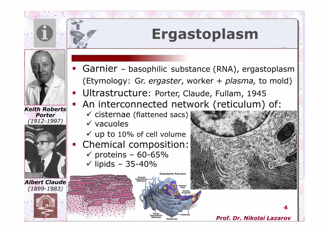
� Garnier – basophilic substance (RNA), ergastoplasm
(Etymology: Gr. ergaster, worker + plasma, to mold)
� Ultrastructure: Porter, Claude, Fullam, 1945
� An interconnected network (reticulum) of:� cisternae (flattened sacs)
� vacuoles
� up to 10% of cell volume
� Chemical composition:� proteins – 60-65%� lipids – 35-40%
Ergastoplasm
Keith Roberts
Porter
(1912-1997)
Albert Claude
(1899-1983)
Prof. Dr. Nikolai Lazarov
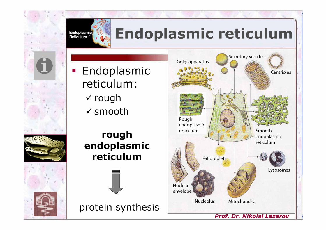
5protein synthesis
roughendoplasmicreticulum
� Endoplasmic reticulum:
� rough
�smooth
Endoplasmic reticulum
Prof. Dr. Nikolai Lazarov
6
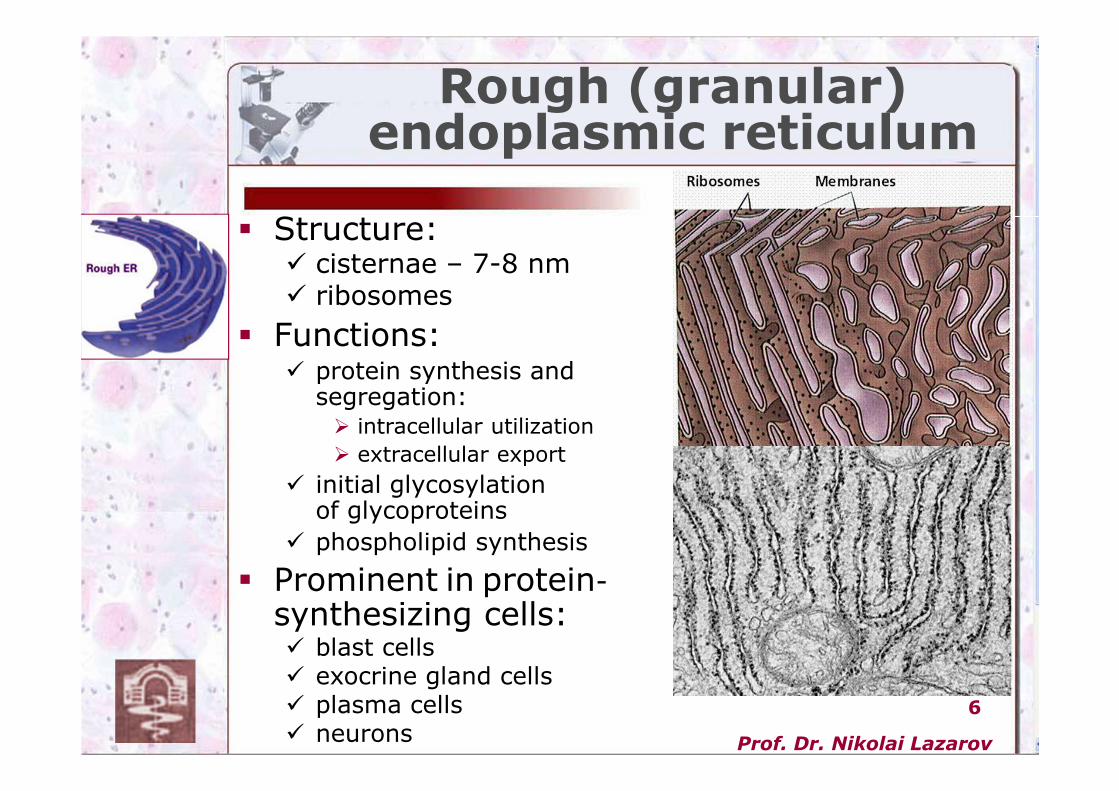
� Structure:� cisternae – 7-8 nm� ribosomes
� Functions:� protein synthesis and
segregation:� intracellular utilization
� extracellular export
� initial glycosylationof glycoproteins
� phospholipid synthesis
� Prominent in protein-
synthesizing cells:� blast cells� exocrine gland cells� plasma cells� neurons
Rough (granular) endoplasmic reticulum
Prof. Dr. Nikolai Lazarov
7
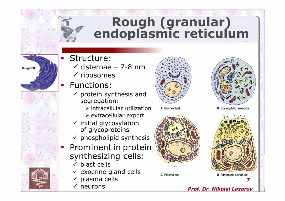
� Structure:� cisternae – 7-8 nm� ribosomes
� Functions:� protein synthesis and
segregation:� intracellular utilization
� extracellular export
� initial glycosylationof glycoproteins
� phospholipid synthesis
� Prominent in protein-
synthesizing cells:� blast cells� exocrine gland cells� plasma cells� neurons
Rough (granular) endoplasmic reticulum
Prof. Dr. Nikolai Lazarov
8
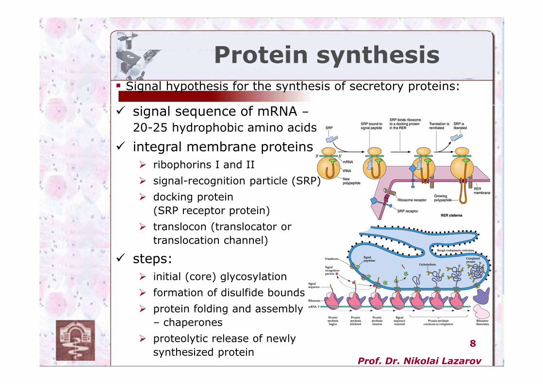
� signal sequence of mRNA –
20-25 hydrophobic amino acids
� integral membrane proteins
� ribophorins І and ІІ
� signal-recognition particle (SRP)
� docking protein
(SRP receptor protein)
� translocon (translocator or
translocation channel)
� steps:
� initial (core) glycosylation
� formation of disulfide bounds
� protein folding and assembly
– chaperones
� proteolytic release of newly
synthesized protein
Protein synthesis� Signal hypothesis for the synthesis of secretory proteins:
Prof. Dr. Nikolai Lazarov
9
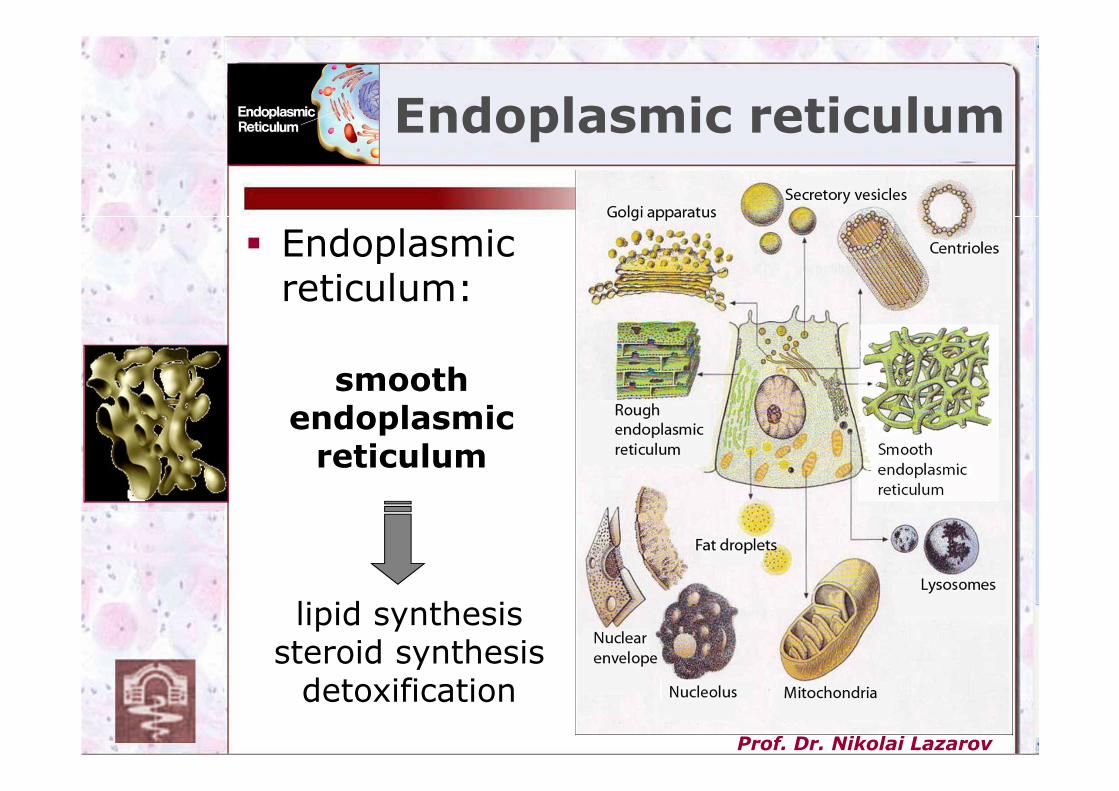
lipid synthesissteroid synthesis
detoxification
smoothendoplasmicreticulum
� Endoplasmic reticulum:
Endoplasmic reticulum
Prof. Dr. Nikolai Lazarov
10
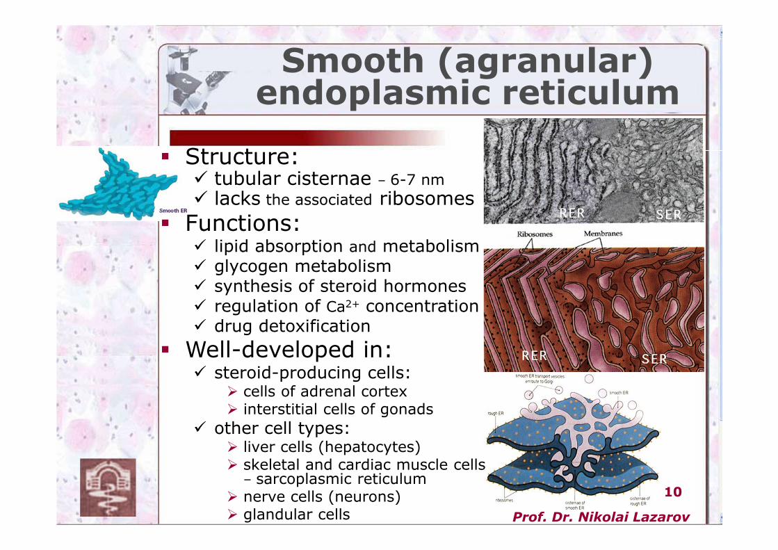
� Structure:� tubular cisternae – 6-7 nm
� lacks the associated ribosomes
� Functions:� lipid absorption and metabolism� glycogen metabolism� synthesis of steroid hormones� regulation of Ca2+ concentration� drug detoxification
� Well-developed in:� steroid-producing cells:
� cells of adrenal cortex� interstitial cells of gonads
� other cell types:� liver cells (hepatocytes)� skeletal and cardiac muscle cells
– sarcoplasmic reticulum� nerve cells (neurons)� glandular cells
Smooth (agranular) endoplasmic reticulum
Prof. Dr. Nikolai Lazarov
11
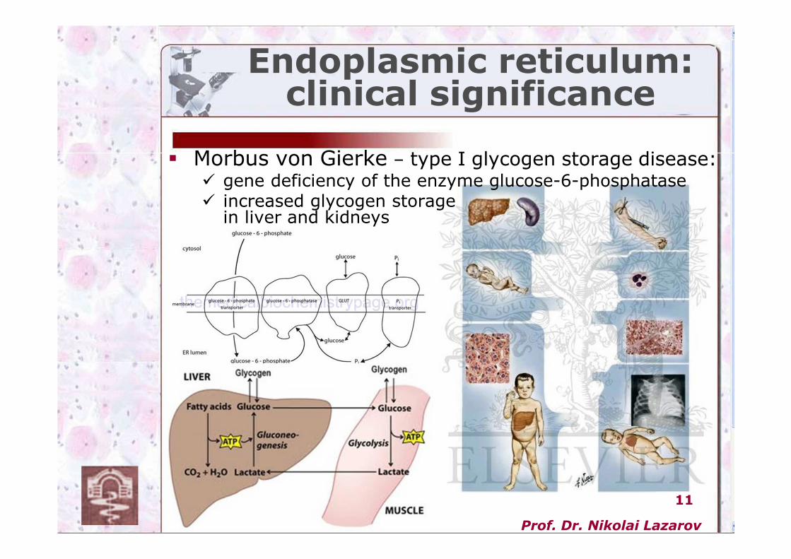
Endoplasmic reticulum:clinical significance
� Morbus von Gierke – type I glycogen storage disease:� gene deficiency of the enzyme glucose-6-phosphatase� increased glycogen storage
in liver and kidneys
Prof. Dr. Nikolai Lazarov
12
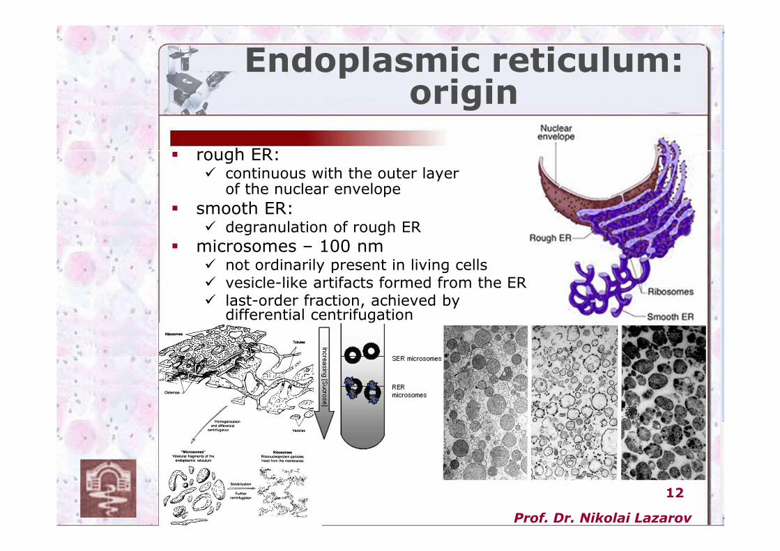
� rough ER:� continuous with the outer layer
of the nuclear envelope
� smooth ER:� degranulation of rough ER
� microsomes – 100 nm� not ordinarily present in living cells � vesicle-like artifacts formed from the ER� last-order fraction, achieved by
differential centrifugation
Endoplasmic reticulum:origin
Prof. Dr. Nikolai Lazarov
13
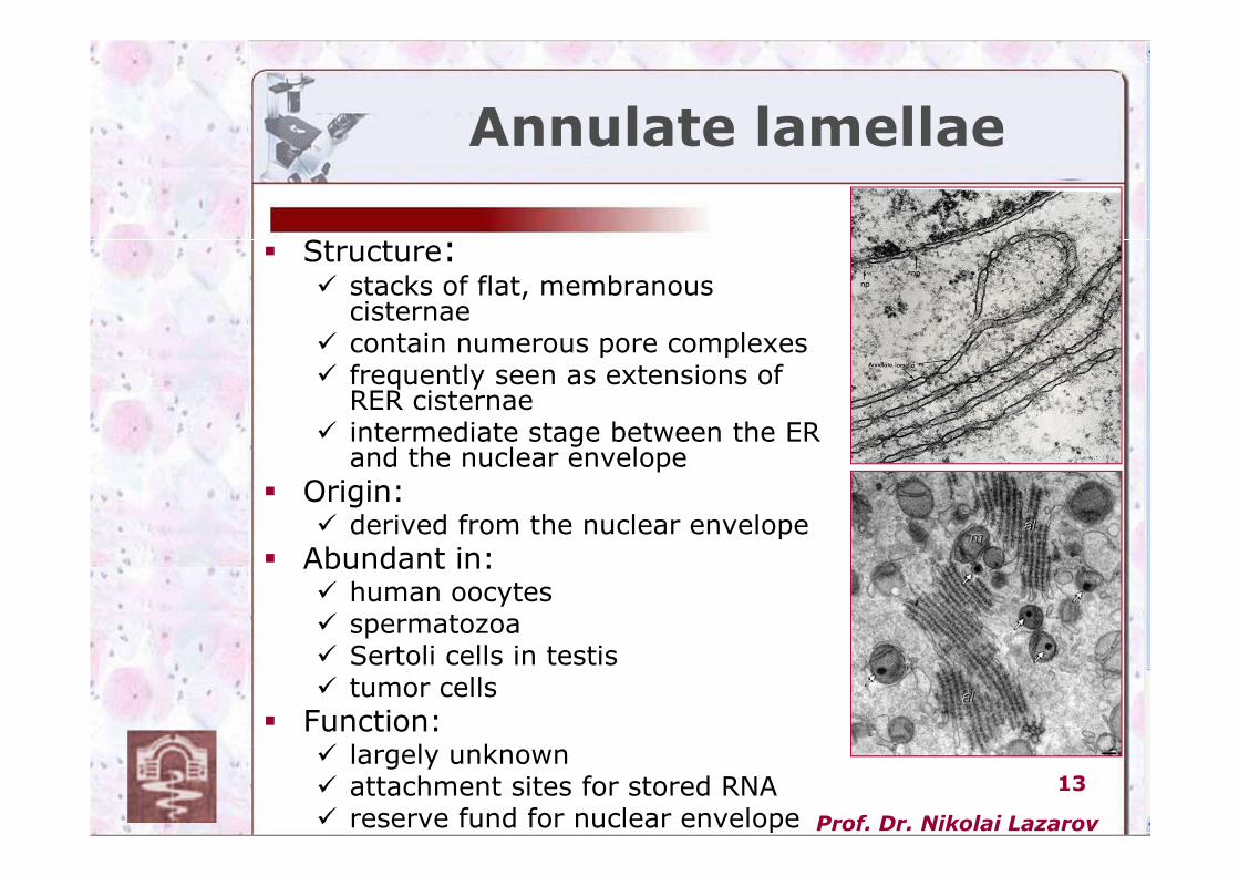
Annulate lamellae
� Structure:� stacks of flat, membranous
cisternae� contain numerous pore complexes � frequently seen as extensions of
RER cisternae � intermediate stage between the ER
and the nuclear envelope
� Origin:� derived from the nuclear envelope
� Abundant in:� human oocytes� spermatozoa� Sertoli cells in testis� tumor cells
� Function:� largely unknown� attachment sites for stored RNA� reserve fund for nuclear envelope
Prof. Dr. Nikolai Lazarov
14
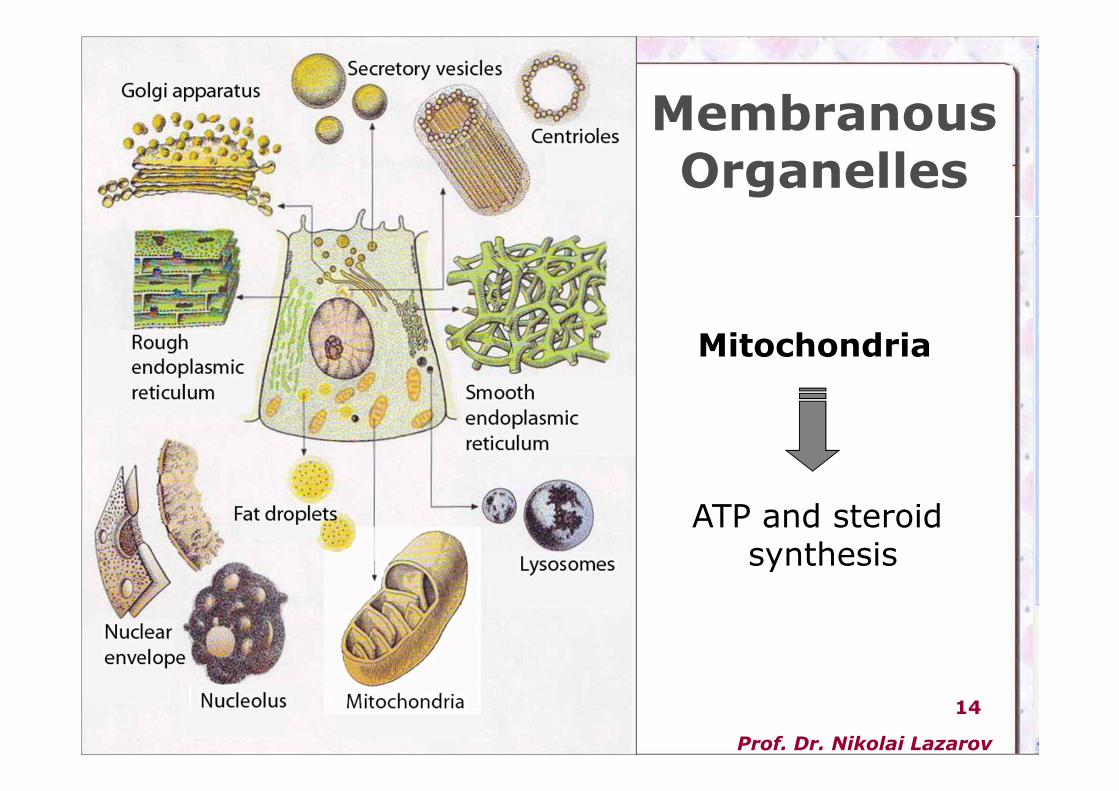
Membranous Organelles
ATP and steroidsynthesis
Mitochondria
Prof. Dr. Nikolai Lazarov
15
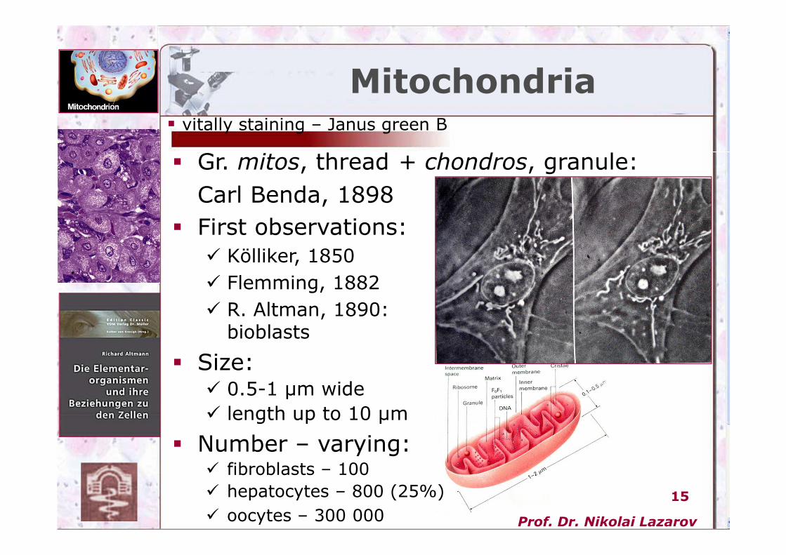
� Gr. mitos, thread + chondros, granule:
Carl Benda, 1898
� First observations:
� Kölliker, 1850
� Flemming, 1882
� R. Altman, 1890: bioblasts
� Size:� 0.5-1 µm wide
� length up to 10 µm
� Number – varying:� fibroblasts – 100
� hepatocytes – 800 (25%)
� oocytes – 300 000
Mitochondria� vitally staining – Janus green B
Prof. Dr. Nikolai Lazarov
16
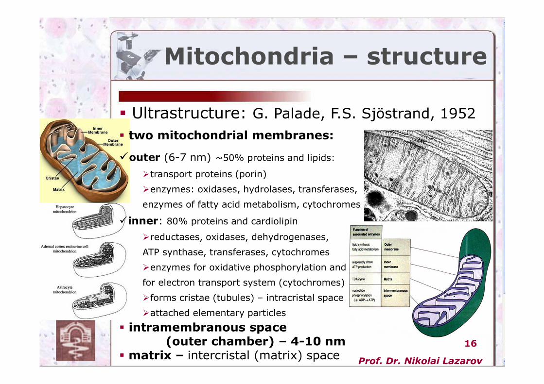
Mitochondria – structure
� Ultrastructure: G. Palade, F.S. Sjöstrand, 1952
� two mitochondrial membranes:
�outer (6-7 nm) ~50% proteins and lipids:
�transport proteins (porin)
�enzymes: oxidases, hydrolases, transferases,
enzymes of fatty acid metabolism, cytochromes
�inner: 80% proteins and cardiolipin
�reductases, oxidases, dehydrogenases,
ATP synthase, transferases, cytochromes
�enzymes for oxidative phosphorylation and
for electron transport system (cytochromes)
�forms cristae (tubules) – intracristal space
�attached elementary particles
� intramembranous space(outer chamber) – 4-10 nm
� matrix – intercristal (matrix) space
Prof. Dr. Nikolai Lazarov
17
Mitochondria – structure
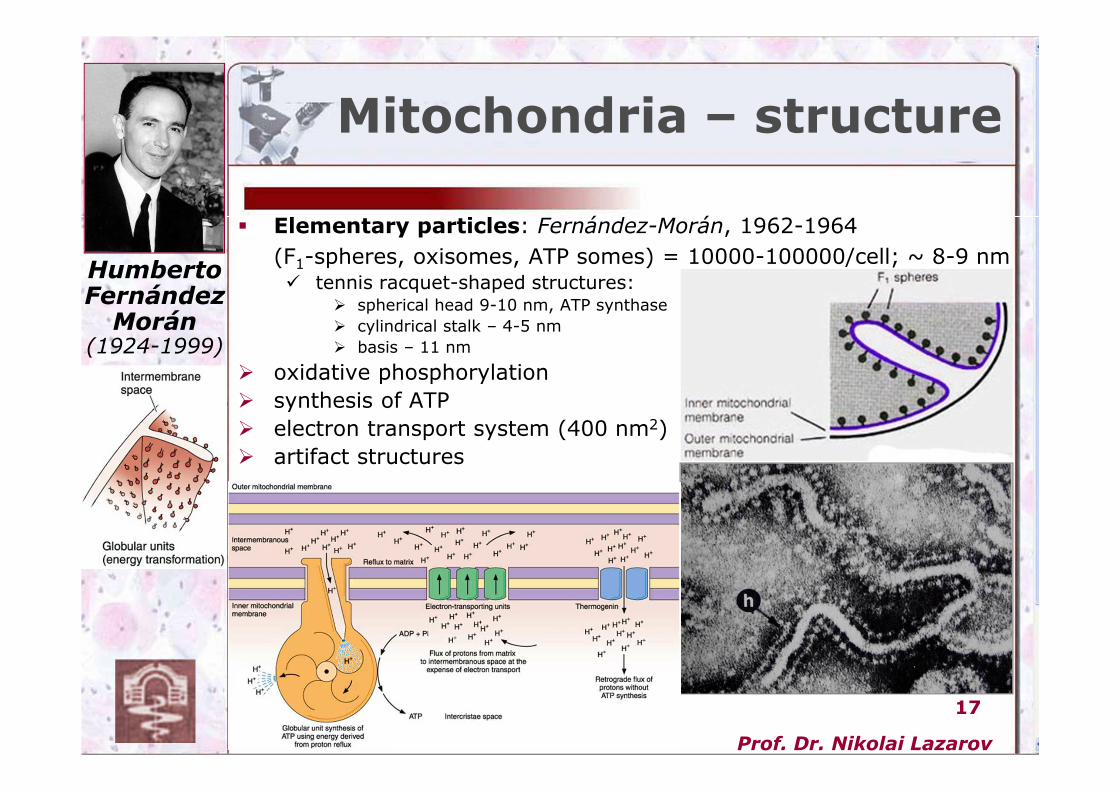
� Elementary particles: Fernández-Morán, 1962-1964
(F1-spheres, oxisomes, ATP somes) = 10000-100000/cell; ~ 8-9 nm� tennis racquet-shaped structures:
� spherical head 9-10 nm, ATP synthase
� cylindrical stalk – 4-5 nm
� basis – 11 nm
� oxidative phosphorylation
� synthesis of ATP
� electron transport system (400 nm2)
� artifact structures
Humberto
Fernández
Morán(1924-1999)
Prof. Dr. Nikolai Lazarov
18
Mitochondria –structure and function
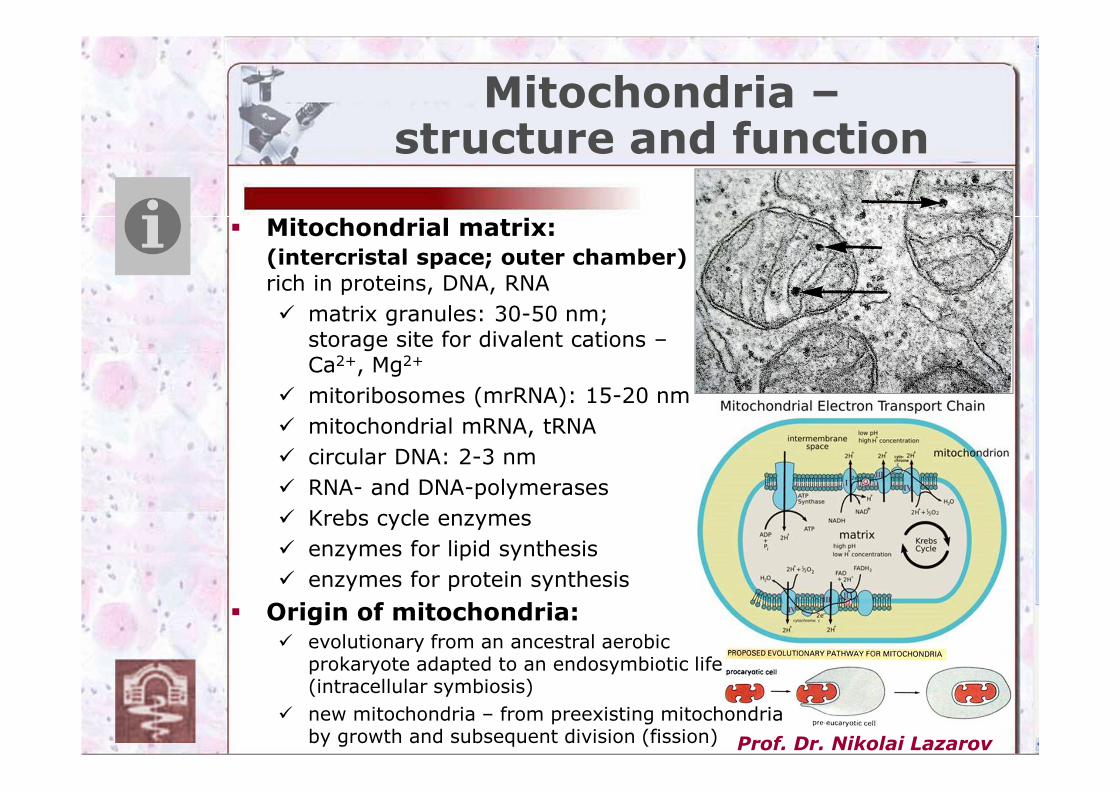
� Mitochondrial matrix: (intercristal space; outer chamber)
rich in proteins, DNA, RNA
� matrix granules: 30-50 nm; storage site for divalent cations –Ca2+, Mg2+
� mitoribosomes (mrRNA): 15-20 nm
� mitochondrial mRNA, tRNA
� circular DNA: 2-3 nm
� RNA- and DNA-polymerases
� Krebs cycle enzymes
� enzymes for lipid synthesis
� enzymes for protein synthesis
� Origin of mitochondria:� evolutionary from an ancestral aerobic
prokaryote adapted to an endosymbiotic life (intracellular symbiosis)
� new mitochondria – from preexisting mitochondria by growth and subsequent division (fission)
Prof. Dr. Nikolai Lazarov
19
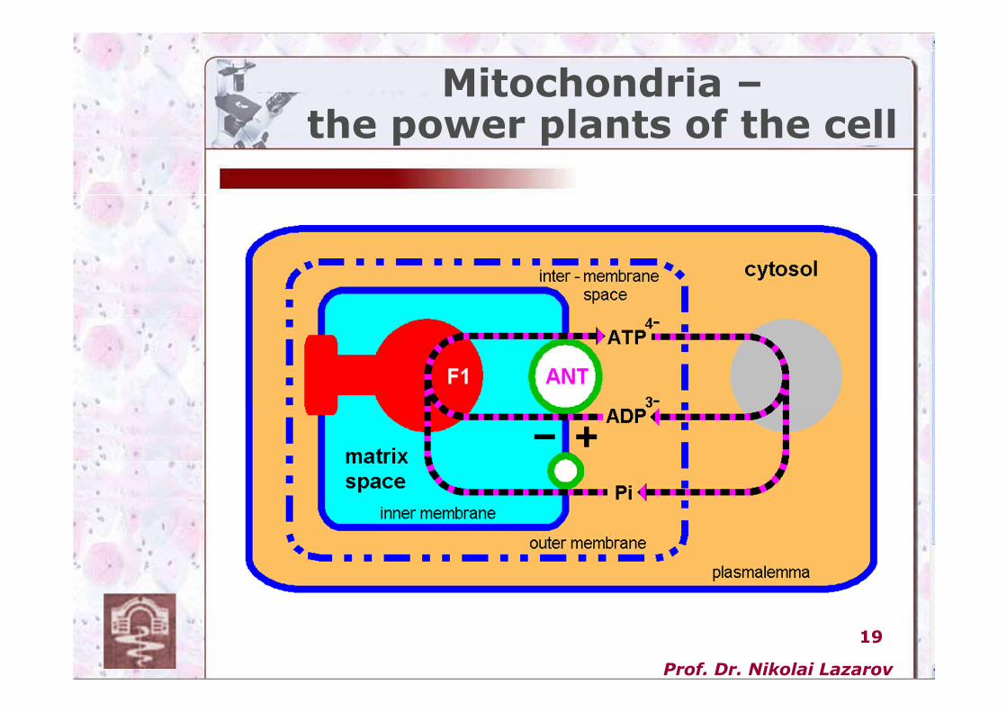
Mitochondria –the power plants of the cell
Prof. Dr. Nikolai Lazarov
20
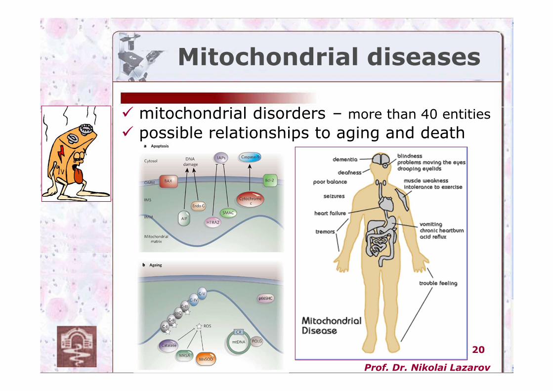
Mitochondrial diseases
� mitochondrial disorders – more than 40 entities
� possible relationships to aging and death
Prof. Dr. Nikolai Lazarov
21Thank you…
Mitochondria –“the cell’s brain”