mercury in dental amalgam and resin-based alternatives - world
TRANSCRIPT

Mercury in Dental Amalgamand Resin-Based Alternatives:A Comparative Health Risk EvaluationJ U N E 2 0 1 2
H E A L T H C A R E R E S E A R C H C O L L A B O R A T I V E
A U T H O R :
Serap Erdal, Ph.D.
I N C O L L A B O R A T I O N W I T H :
Peter Orris, M.D., M.P.H.
Health Care Without Harm
has initiated a research
collaborative coordinated
by faculty of the University
of Illinois at Chicago School
of Public Health, with
support from the Pioneer
Portfolio of the Robert Wood
Johnson Foundation, aimed
at stimulating collaborative
research around health and
safety improvements in
health care. The Research
Collaborative is designed to
increase the evidence base
concerning the impacts
of sustainable design,
construction, organization,
operations, and materials and
chemicals choices in the health
care sector on patient, worker
and environmental safety.
This paper is the tenth in
a series of papers in which
the Collaborative provides
research and analysis of factors
influencing patient, worker
and environmental safety and
sustainability in the healthcare
sector. The editors of this series
are Peter Orris, MD, MPH and
Susan Kaplan, JD.

TA B L E O F C O N T E N T S
EXECUTIVE SUMMARY ............................................................................................................................5
I. Introduction ..........................................................................................................................................8
II. Background ...........................................................................................................................................9
2.1 Chemical Composition ................................................................................................................................ 10
2.1.1 Dental Amalgam Composition ....................................................................................................... 10
2.1.2 Types and compositions of dental amalgam alternatives ................................................................ 10
2.1.2.1 Composite resins .................................................................................................................. 11
2.1.2.2 Glass ionomer (Glass polyalkenoate) cements ................................................................... 15
2.1.2.3 Resin-Modified Glass Ionomer Cement .............................................................................. 17
2.1.2.4 Compomers .......................................................................................................................... 18
2.1.2.5 Giomers ................................................................................................................................ 18
2.2 Environmental Behavior and Emissions ...................................................................................................... 19
2.2.1 Environmental Behavior and Emissions: Dental Amalgam ........................................................... 19
2.2.2 Environmental Behavior and Presence: Alternative Materials ..................................................... 20
III. Exposure Assessment ..........................................................................................................................25
3.1. Exposure Assessment: Dental Amalgam .................................................................................................... 25
3.1.1 Mercury Exposure Estimates related to Dental Amalgam in General Population and Children .. 25
3.1.2 Occupational Mercury Exposure Estimates ................................................................................... 26
3.2 Exposure Assessment: Alternative Materials .................................................................................... 27
3.2.1 Inhalation Exposure ........................................................................................................................ 27
3.2.2 Occupational Inhalation Average Daily Dose Estimates .............................................................. 30
3.2.3 Dermal Exposure ............................................................................................................................ 30
IV. Hazard Identification ..........................................................................................................................32
4.1 Hazard Identification: Human Health Effects of Dental Amalgam ........................................................... 32
subhead 3table title

4.2 Hazard Identification: Alternative Materials ............................................................................................. 35
4.2.1 Acute Toxicity Data (LD50, LC50) ............................................................................................................35
4.2.2 Cytotoxicity .................................................................................................................................... 35
4.2.3 Carcinogenicity ............................................................................................................................... 38
4.2.4 Estrogenicity .................................................................................................................................... 41
4.2.5 Allergic Reactions ........................................................................................................................... 41
V. Dose-Response Assessment .................................................................................................................43
5.1 Dose-Response Assessment: Dental Amalgam ............................................................................................ 43
5.2 Dose-Response Assessment: Alternative Materials .................................................................................... 44
Table 7. Summary of Available Toxicity Values for Constituents of Resin-based Alternative Materials ........ 44
VI. Discussion and Comparative Assessment .............................................................................................45
VII. Policy Recommendations .....................................................................................................................48
REFERENCES ............................................................................................................................................49
TABLES
Table 1. Typical composition of dental amalgam (Van Noort 2007) .......................................................................... 10
Table 2: Summary of chemicals used as constituents in dental composites (SCENIHR 2008, Powers and Wataha 2008) ....................................................................................................... 14
Table 3. Typical composition of a glass-ionomer cement powder (Combe and Grant 1992) .................................... 15
Table 4. Summary of constituents found in formulations of resin-based alternatives as compiled from product Material Safety Data Sheets (MSDSs) .................................................... 21
Table 5. Occupational Inhalation Average Daily Dose Estimates (mg/kg-day) ........................................................ 31
Table 6. Summary of Available Toxicity Values for Constituents of Dental Amalgam ............................................. 43

APPENDIX ................................................................................................................................................53
Table A-1. Chemical composition of dental resin composites commercially available in the U.S., as reported in MSDSs................................................................................... 53
Table A-2. Chemical composition of dental resin composite preparation and application materials commercially available in the U.S., as reported in MSDSs ......................................... 57
Table A-3. Chemical composition of glass ionomers commercially available in the U.S., as reported in MSDSs................................................................................... 61
Table A-4. Chemical composition of compomers commercially available in the U.S., as reported in MSDSs................................................................................... 62
Table A-5. Environmental fate and transport properties of constituents of resin-based alternative materials ............................................................................................... 63
Table A-6. Acute toxicity information for constituents of resin-based alternative materials (HSDB-NLM) ............................................................................................... 65
Table A-7. Acute toxicity information for methyl methacrylate (MMA) (HSDB-NLM) ........................................ 68

AcknowledgementsThe author wishes to thank the following reviewers of this document for their helpful comments and critique:
Fernando Bejarano
Carolyn Vickers
Dr. Graeme Munro-Hall
Dr. Lillian Lasaten Ebuen
In addition, the author thanks Drs. Jerome Bowman, Daniel Meyer, and Stuart Johnston for providing very useful critiques, and Julian Fisher for helping to coor-dinate some of these critical reviews. The author also extends her appreciation to Antony G. Milazzo, DDS for his contribution and practical guidance in relation to dental restoration.

5Mercury in Dental Amalgam and Resin-Based Alternatives: A Comparative Health Risk Evaluation
Current StatusUse of mercury in dental amalgam reconstruction for cavities has been debated by the scientific community due to well-documented adverse environmental and health implications of mercury. There has been consid-erable controversy concerning the health risks and ben-efits of utilizing mercury-containing amalgam. Neither epidemiologic studies nor consensus statements have identified evidence of harm to individuals due to their mercury amalgams. At the same time, the contribution of mercury dental amalgam use to the environmental mercury burden and its contribution to the neurotoxic damage of methyl mercury in children is well estab-lished. In 2005, the United Nations Environment Programme estimated that 362 tons of dental mercury are consumed annually worldwide.
The use of alternative products to replace mercury in dental amalgam is growing and in some areas has virtually replaced mercury in all its dental restorative uses. Specifically, Denmark, Sweden, and Norway have banned dental amalgam except when a specific excep-tion is requested for individual cases, and several other
countries (e.g., Canada, Italy, Australia) have taken steps to reduce amalgam use. Yet, the substitutes have not yet received systematic scrutiny as to their hazards.
Risk AssessmentThis report begins the process of risk assessment byevaluating the clinical, environmental, and occupa-tional exposures and the toxicity of the alternatives to mercury containing dental amalgam. It uses the four-step human health risk assessment approach used by U.S. federal agencies.
Basing itself on the primary literature, this four-step paradigm includes hazard identification, exposure assessment, toxicity assessment, and risk characteriza-tion. Material Safety Data Sheets were secured for the various composite, glass ionomer, and compomer formulations along with preparation and application formulations (etchants, primers, activators, coupling agents, adhesives, and bonding agents). Seventy-eight constituents were identified, organized, and summarized for the different formulations.
E X E C U T I V E S U M M A R Y
In 2005, the United Nations Environment Programme estimated
that 362 tons of dental mercury are consumed annually worldwide.

Mercury in Dental Amalgam and Resin-Based Alternatives: A Comparative Health Risk Evaluation6
Environmental Behavior of AlternatesThe environmental fate and transport property data revealed that constituents of resin-based restorative materials are complex in their environmental behavior, and while some are rapidly biodegradable, others are persistent.
Human ExposureDental professionals are exposed to components from resin-based restorative materials (including BPA) dur-ing routine practice. These exposures occur through inhalation and dermal absorption. No studies have been done estimating exposures to many of these com-ponents. Though, methacrylates, a class of chemicals used in several of these processes, has had three studies published estimating exposures to dental personnel. They ranged from an Average Daily Dose between 8E-08 and 6E-06 mg/ kg-d. to between 1E-03 to 4E-02 mg/kg-d.
Toxicity of AlternativesPeer reviewed studies of the acute toxicity, cytotoxicity, carcinogenicity, estrogenicity and sensitizing potential of these alternative materials were abstracted from the literature. Only 22 of the 78 constituents (i.e., 28%) were found to have any acute toxicity data. Primary attention has been paid to the methacrylates.
A majority of the methacrylates are skin-sensitizers, and these fillers used in resin formulations are respira-tory irritants. Furthermore, some of the monomers used have neurotoxic effects. With increasing clinical usage, case reports on hypersensitivity reactions to composites have emerged as well.
While no studies are available as to the short-lived Bisphenol A exposure in one of these processes, a number of studies provide evidence of cytotoxicity due to methacrylate monomer released. This release is, primarily, due to incomplete polymerization (i.e., the filling has not been allowed adequate setting time) and, partially, due to normal degradation in the oral environment.
Risk of AlternativesAlthough some in vitro studies have shown genotoxity, methacrylates are categorized by IARC as not classifi-able as to their carcinogenicity to humans (Group 3).
A summary of available toxicity values (RfD/RfC/CSF)for the constituents of dental amalgam and resin-based alternatives indicates that the inhalation Hazard Quotient (HQ), an indicator of non cancerous risk, varied from 4E-07 to 0.2. These estimates are signifi-cantly less than 1, indicating little or no risk, though it must be noted that risk for mixtures have not been assessed.
In sum, though data gaps continue to exist for the health effects of the alternatives to mercury amalgam, other than individual allergies to components of one or another composite, there is no current evidence of significant personal or environmental toxicity.

7Mercury in Dental Amalgam and Resin-Based Alternatives: A Comparative Health Risk Evaluation
Substitution of Alternatives for Mercury AmalgamBased on current evidence, therefore, the ultimate goal of a phase-out of virtually all usage of dental mercury is recommended. This phase-out must be planned and deliberate, assuring continued emphasis on adequate restorations to prevent continued tooth decay and the potential of malnutrition in economically impover-ished areas.
Such a phase-out, therefore, must take into account the practical availability of alternative materials, the equipment needed to utilize non-mercury alternatives, the training of dentists to utilize these alternatives, and the costs to the patient and society.
Based on this comparative review and the practical experience of countries and dentists that have essen-tially eliminated mercury amalgams, a virtual phase-out of dental amalgam, with exceptions provided for dif-ficult cases, is possible and advisable. Dental personnel handling these materials should take proper exposure control measures due to the demonstrated genotoxicity and allergenicity of some of these compounds. In con-clusion, governments and international agencies are urged to make resources available to reduce the costs of this transition in economically impoverished areas. Finally, it is clear that further research is needed to improve exposure and toxicity information pertaining to both constituents and mixtures of the alternatives.
Global metallic mercury demand by application, 2005(metric tonnes)
*Paints, pesticides, fungicides, cosmetics, laboratory, pharmaceutical, cultural/traditional uses, etc.
Source: United Nations Environment Programme (UNEP).
Small-scale/artisanal
gold mining[650-1000]
Other*[30-60]
Lighting[100-150]
Electrical and electronic
[150-350]
Measuring and control[150-350]
Dental use[240-300]
Chlor-alkali production[450-550]
Vinyl chloridemonomer
production[600-800]

Mercury in Dental Amalgam and Resin-Based Alternatives: A Comparative Health Risk Evaluation8
I.I N T R O D U C T I O N
Historical use of mercury in dental amalgam as an oral health restorative for the treatment of dental cavities has been debated by the scientific community due to well-documented adverse environmental and health implications of mercury. Thus, product sub-stitution to replace mercury in dental amalgam and environmental and health and safety implications of commercially available substitutes have begun to receive scrutiny from the oral and public health com-munity. Some of these substitutes include composite resins, glass ionomer cements, compomers and gold alloys. In order to develop and adopt a scientifi-cally sound approach to oral health, a comparative assessment of environmental and health risks and benefits of dental mercury and its alternatives must be evaluated using both qualitative and quantitative approaches when feasible. Such evaluation must take factors related to the resource infrastructure, access to this infrastructure and economic viability of alterna-tives for the public into account in order to be able to design and implement an optimum strategy for oral and public health while protecting the environment at the same time.
This paper addresses this need by comprehensively evaluating environmental and occupational expo-sures, toxicity, and cancer and non-cancer health risks of dental mercury and its alternatives for adults and
children using the four-step human health risk assess-ment approach originally proposed by the National Academy of Sciences 1983 (NAS 1983) and later used extensively by U.S. federal agencies responsible for environmental and public health protection (USEPA 1995; 2000). Basing itself on the primary literature, the four-step paradigm includes hazard identification, expo-sure assessment, toxicity (or dose-response) assessment and risk characterization steps. This paper focuses on documenting available scientific evidence for exposures to and potential health effects associated with resin-based alternatives in a comprehensive manner. On the other hand, due to the availability of numerous publicly available scientific evaluations undertaken by differ-ent regulatory agencies and papers published in the scientific literature, a more limited approach is under-taken for dental amalgam risk evaluation, and only human epidemiological evidence is presented. A proper scientific weight of evidence analysis can only be done with proper consideration of strengths, limitations and uncertainties present in the available information.
The goal of this evaluation is to inform public policymakers in regard to safe product usage in teeth restoration to protect oral and public health for all individuals, including sensitive subpopulations, while protecting the environment.

9Mercury in Dental Amalgam and Resin-Based Alternatives: A Comparative Health Risk Evaluation
II.B A C K G R O U N D
Dental amalgam containing a mixture of alloy particles and mercury has been used by dentists in various forms for the treatment of cavities and restoration of teeth for more than 150 years all around the world. When dental amalgam was first introduced into dentistry, gold could also be used in some types of dental restoration. However, its higher cost as compared to dental amal-gam prohibited its widespread use. There were no other synthetic materials that could be potential substitutes for amalgam at that time. As a result, dental amalgam has widely been used in the past and it is currently used particularly in large cavities due to its superior mechanical properties, durability and low cost. A num-ber of substitutes in the form of composite resins, glass ionomer cements, ceramics and gold alloys have been developed in the last four decades, and their usage has recently been on the rise due to their superior aesthetic properties and environmental and health concerns related to the use of dental amalgam.
Due to well-documented toxicity and resulting health effects of certain forms of mercury and its compounds, potential association between exposure to mercury released from amalgam and disease formation in humans with amalgam fillings has been scientifically debated in academic and regulatory communities throughout the 20th century and now at the begin-ning of the 21st century. There has been considerable controversy surrounding the potential health risks and benefits of dental amalgam. As a result, many govern-mental agencies investigated health effects of mercury contained within the amalgam and the role of mercury in disease causation with its systemic distribution and accumulation in the body. A number of epidemiologi-cal studies were carried out to uncover whether the mercury in amalgam has a causative role in disease incidence. However, no consensus conclusion has so far been forthcoming (SCENIHR 2008). In the meantime, in an attempt to provide cosmetically more pleasing and safer alternatives to amalgam, a number of chemical formulations have been developed in the last 40 years and introduced to the marketplace with-out going through comprehensive human exposure and toxicity assessment.
Despite this, it is important to weigh the known risks and benefits of dental amalgam and its alterna-tives using the state-of-art information so that the dental community and consumers are well-informed and sound policies can be developed to protect oral and public health at the same time as protecting the environment. In such an evaluation, it is necessary to evaluate physical, chemical and environmental fate and transport properties, and animal and human toxicity of all of the constituents, while examining the potential routes of exposure in each step of teeth restoration from preparation of material to techniques used to promote adhesion to the tooth surface. A sepa-rate health risk evaluation for each human receptor of concern must be performed including patients (adults, children, pregnant women) and dental personnel, tak-ing into account the phases of use, including placement of the filling, corrosion, degradation or wear in clinical service, and the release of materials during the removal of restorations (SCENIHR 2008). Receptor-specific risk information should be augmented by environmen-tal emissions and exposure information, while paying particular attention to environmental sustainability and whole product life-cycle for amalgam and its alternatives. Only then, a well-balanced and informed decision-making is feasible within the constraints of available research data and know-how.

Mercury in Dental Amalgam and Resin-Based Alternatives: A Comparative Health Risk Evaluation10
This report attempts to accomplish this goal by fol-lowing a four-step risk assessment paradigm used as a hazard ranking and environmental and health policy development tool by federal agencies in the U.S., par-ticularly by the Environmental Protection Agency.
2.1 Chemical Composition
2.1.1 Dental Amalgam CompositionAn amalgam is formed when mercury is mixed with another metal or metals. Mercury is one of the select metals that is liquid at room temperature and is easily mixed with other metals to solidify. When a dentist selects a certain type dental amalgam, it involves only the selection of the metal(s) with which mer-cury is mixed. Although the chemical composition of dental amalgam varies among manufacturers, the traditional alloy used in dental amalgams consists of a mixture of silver, tin, copper, zinc, and at times, mercury. A typical composition is shown in Table 1 (Van Noort 2007). As shown in this table, silver is the main constituent along with tin, and they form an intermetallic compound, Ag3Sn, known commonly as the Гγ-phase. This phase readily reacts with liquid mercury to produce a clinically acceptable alloy that can solidify in a few minutes and harden over a few hours. Furthermore, the exact percentage of this phase controls the kinetics of the amalgamation reaction and properties of the resulting dental amalgam structure (SCENIHR 2008; Van Noort 2007).
Table 1. Typical composition of dental amalgam (Van Noort 2007)
Constituent % Composition
Silver (Ag) 67-74
Tin (Sn) 25-28
Copper (Cu) 0-6
Zinc (Zn) 0-2
Mercury (Hg) 0-3
Copper increases strength and hardness of the amal-gam. A more pronounced effect is induced when the copper content is increased to 30%, and these are known as high-copper content amalgam alloys. Copper amalgams containing approximately 30% copper and 70% mercury were once used, but are no longer recommended. Zinc in the amalgam is not considered to serve any specific purpose. It is simply present due to initial production of alloy. Mercury is sometimes added into the mixture to fasten the amalgamation reaction, i.e., called preamalgamation. The dispersion type amalgam alloys contain 70% silver, 16% tin and 13% copper.
The amalgam alloys are mixed with liquid mercury before dental restoration at a 1 to 1 weight ratio. Thus, the mercury content of a finished dental amalgam is approximately 50% by weight (SCENIHR, 2008; Van Noort, 2007). Dental amalgam was historically mixed on-site using bulk liquid mercury and metal powders. However, today it is purchased in pre-dosed amalgam capsules with mercury ranging from 100 to 1,000 mil-ligrams (IMERC 2008).
2.1.2 Types and compositions of dental amalgam alternativesCurrently, a number of different material types are being used as substitutes to dental amalgam and these include:• composite resins • glass ionomer cement• compomers• giomers
Selection of a material is based on aesthetics, fluo-ride release, wear resistance, strength and ease of use. Composites are aesthetically pleasing, strong, and wear-resistant. However, they have low or no fluoride release. Compomers are less wear-resistant but they have good aesthetic properties and release fluoride. Resin-modified glass-ionomer cements release more fluoride than compomers but they are not as wear-resistant and they are not used in posterior restorations. Conventional glass ionomers release the most fluoride and are best for patients with high risk carries in low-stress applications (Powers and Wataha 2008).

11Mercury in Dental Amalgam and Resin-Based Alternatives: A Comparative Health Risk Evaluation
2.1.2.1 Composite resins: Composite fillings, which were introduced in the 1960s, are a mixture of glass or quartz filler in a poly-merisable resin medium that produces a tooth-colored filling. They are referred to as resin fillers as well (ADA 2010). They currently dominate the materials used for direct aesthetic restorations and they are the most ubiquitous materials available in dentistry today.
The composites are classified in a number of ways by the manufacturers, depending on the size, distribu-tion, and volume percentage of particles. Size clas-sification segregates dental composites into macrofill (10-100 μm), midifill (1-10 μm), minifill (0.1-1 μm), microfill (0.01-1 μm; used for Class II and V fills) and nanofill (0.005-0.01 μm; used for Class I to V fills) categories with each containing particles in the size ranges indicated. In addition, there are hybrid composites that contain a mix of two particles size-fraction of fillers, e.g., midi-hybrids consisting of a mixture of microfillers and midifillers; mini-hybrids or micro-hybrids consisting of a mixture of microfill-ers and minifillers; and nanohybrids consisting of a mixture of nanofillers and minifillers. While large particle size fillers have better mechanical properties and lower coefficient of thermal expansion, small size particle filler can take and retain good surface finish. Conversely, large particles size fillers have very poor surface finish and have dull appearance.
The filler loading varies significantly among differ-ent composite materials. While weight percentage of the filler is 50-80% of the total composite weight in a macrofill and hybrid composite, it is limited to about 35-50% by weight in a microfill composite (O’Brien 2002; Powers and Wataha 2008; SCENIHR 2008; Combe and Grant 1992).
The three main components of composite filling materials are the organic resin phase, the reinforcing inorganic filler and a coupling agent.
The resin forms the matrix of the composite material, binding the individual filler inorganic particles together through the coupling agent. While the beneficial properties contributed by the resin are the ability to be molded at ambient temperatures coupled with setting by polymerization achieved in a short time, the ben-eficial properties contributed by the filler are rigidity, hardness, compressive strength, modulus of elasticity, aesthetics, and a lower value for coefficient of thermal expansion. As can be gleaned from the above descrip-tion, composite chemistry is complex, partly because of usage of different chemicals in not only prepara-tion but also application of the material during dental restoration.
The inorganic materials used as fillers today are silica-based glass fillers, such as silica glass (SiO2), alumina glass (Al2O3), and combinations of glass and sodium fluoride, which are engineered mixtures of various glasses serving as a source of fluoride ions. The radi-opaque composites, which are used to restore posterior teeth, are obtained by the addition of barium, stron-tium (renders the composite easier to polish) to the filler particles, which aid the detection of recurrent carries. Quartz (crystalline silica – by far the hardest material) – used as a filler until recently – and lithium aluminum silicate are not radiopaque. Current materi-als may contain lithium aluminosilicates, crystalline quartz, or barium aluminoborate silica glasses. The mass-based composition of the latter material is: SiO2, 50%; BaO, 33%; B2O3, 9% and Al2O3, 8%. Many composites contain a combination of a barium glass and filler.
The particle size range of these fillers is typically 10-40 µm. However, a number of products have been devel-oped which contain a microfiller with a particle size about 0.05 µm and consist of 25%-63% SiO2 by weight. The average particle size and particle size distribution of the filler is important as it determines the amount of filler that can be added to the resin without adversely affecting the necessary composite properties. Particle size also has a pronounced effect on the final surface finish of the composite restoration, i.e., the smaller the filler particle size, the smoother the composite. Transition from the hardest material (i.e., quartz) to softer glasses has allowed a reduction in the size of the filler particles and an increase in the filler loading of the resins considerably.

Mercury in Dental Amalgam and Resin-Based Alternatives: A Comparative Health Risk Evaluation12
While some recent products for posterior restorations contain up to 87% filler, products with microfine silica contain less inorganic filler. A British Standard Specification defines composites as containing 50% or more by weight of inorganic filler. Usual filler loading is 55-60% for anterior composites.
In contrast to filler material, the resin is initially a fluid monomer, which is converted into rigid polymer by a radical addition reaction. The resin matrix contains organic molecules consisting of a large group of differ-ent aromatic and diacrylate monomers and oligomers (i.e., a moderate molecular weight organic molecule made from two or more organic molecules). The most common resins used for anterior and posterior resto-rations now are based on dimethacrylate (bisphenol A-glycidyl methacrylate (Bis-GMA – or Bowen’s resin)). In addition, there are composites that use ure-thane dimethacrylate (UDMA) oligomers rather than Bis-GMA. Because Bis-GMA or UDMA monomers are highly viscous, low-molecular weight monomers (mono- or di-methacrylates such as methyl methacry-late (MMA), ethylene glycol dimethacrylate (EDMA) or triethylene glycol dimethacrylate (TEGDMA) are added as diluents or as viscosity controllers into Bis-GMA and UDMA oligomer liquids to control the consistency of the composite paste, to enable proper blending with the inorganic components, and to facili-tate clinical manipulation.
Dimethacylates are preferred over monomethacrylates due to yielding a lower polymerization shrinkage and harder and stronger structure with a lower coefficient of thermal expansion and lower water absorption. In order to prevent premature polymerization, an inhibitor such as hydroquinone is included. This ensures adequate long shelf life for the composite.
Oligomers and the low-molecular weight monomers are characterized by carbon-carbon double bonds that take part in a free radical addition polymerization and form a rigid highly cross-linked resin after setting. To ensure acceptable mechanical properties for composites as dental restoration materials, it is critically important that the filler and the resin are strongly bonded to each other.
Incorporation of inorganic filler and organic resin by covalent bonding is achieved by coating of the filler particles with bifunctional vinyl silane coupling agents (such as trialkoxysilane; example: gammamethacry-loxpropyltrimethoxy silane or γ-MPTS), which has groups that react with the inorganic filler and other groups that react with the organic matrix. Composite is then cured (or set) by chemically (self or auto-cure) or, most commonly, by a light source at 470 ± 20 nm wavelength (ultraviolet or visible light) to complete polymerization of dental composites.
Visible-light activated (VLA) composites are now more widely used due to potential harmful effects such as soft-tissue burns, skin cancer and eye damage associated with UV-light exposure from a mercury discharge lamp and limited depth obtained during the polymerization (or curing) process. The visible light is absorbed by a diketone (α-diketone), which in the presence of an organic amine, starts the polymerization reaction. The traditional method for delivering the blue visible light (≈ 460-480 nm) required for the visible light activa-tion involves the use of quartz halogen lamp which is cheaper and less-damaging, and has greater depth of cure. Other currently available sources of visible light are blue-light emitting diode (blue-LED), argon laser and plasma arc lamps.
Dual curing using a combination of chemical and light curing is also used. The curing process is chemically activated by mixing two components, one of which contains a polymerization initiator (e.g., 1% organic peroxide such as benzoxyl peroxide) and the other an activator (e.g., 0.5% tertiary amine such as N, N’ dimethyl-p-toluidine on p-tolyl diethanolamine; or N,N-dihydroxyethyl-p-toluidine, currently widely used), depending on the curing method utilized. The initiator and accelerator must not be mixed until just before the restoration is placed. Curing times should be at least 40-60 seconds.
13Mercury in Dental Amalgam and Resin-Based Alternatives: A Comparative Health Risk Evaluation
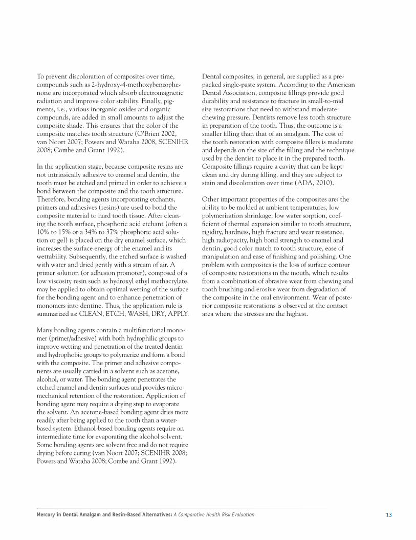
To prevent discoloration of composites over time, compounds such as 2-hydroxy-4-methoxybenzophe-none are incorporated which absorb electromagnetic radiation and improve color stability. Finally, pig-ments, i.e., various inorganic oxides and organic compounds, are added in small amounts to adjust the composite shade. This ensures that the color of the composite matches tooth structure (O’Brien 2002, van Noort 2007; Powers and Wataha 2008, SCENIHR 2008; Combe and Grant 1992).
In the application stage, because composite resins are not intrinsically adhesive to enamel and dentin, the tooth must be etched and primed in order to achieve a bond between the composite and the tooth structure. Therefore, bonding agents incorporating etchants, primers and adhesives (resins) are used to bond the composite material to hard tooth tissue. After clean-ing the tooth surface, phosphoric acid etchant (often a 10% to 15% or a 34% to 37% phosphoric acid solu-tion or gel) is placed on the dry enamel surface, which increases the surface energy of the enamel and its wettability. Subsequently, the etched surface is washed with water and dried gently with a stream of air. A primer solution (or adhesion promoter), composed of a low viscosity resin such as hydroxyl ethyl methacrylate, may be applied to obtain optimal wetting of the surface for the bonding agent and to enhance penetration of monomers into dentine. Thus, the application rule is summarized as: CLEAN, ETCH, WASH, DRY, APPLY.
Many bonding agents contain a multifunctional mono-mer (primer/adhesive) with both hydrophilic groups to improve wetting and penetration of the treated dentin and hydrophobic groups to polymerize and form a bond with the composite. The primer and adhesive compo-nents are usually carried in a solvent such as acetone, alcohol, or water. The bonding agent penetrates the etched enamel and dentin surfaces and provides micro-mechanical retention of the restoration. Application of bonding agent may require a drying step to evaporate the solvent. An acetone-based bonding agent dries more readily after being applied to the tooth than a water-based system. Ethanol-based bonding agents require an intermediate time for evaporating the alcohol solvent. Some bonding agents are solvent free and do not require drying before curing (van Noort 2007; SCENIHR 2008; Powers and Wataha 2008; Combe and Grant 1992).
Dental composites, in general, are supplied as a pre-packed single-paste system. According to the American Dental Association, composite fillings provide good durability and resistance to fracture in small-to-mid size restorations that need to withstand moderate chewing pressure. Dentists remove less tooth structure in preparation of the tooth. Thus, the outcome is a smaller filling than that of an amalgam. The cost of the tooth restoration with composite fillers is moderate and depends on the size of the filling and the technique used by the dentist to place it in the prepared tooth. Composite fillings require a cavity that can be kept clean and dry during filling, and they are subject to stain and discoloration over time (ADA, 2010).
Other important properties of the composites are: the ability to be molded at ambient temperatures, low polymerization shrinkage, low water sorption, coef-ficient of thermal expansion similar to tooth structure, rigidity, hardness, high fracture and wear resistance, high radiopacity, high bond strength to enamel and dentin, good color match to tooth structure, ease of manipulation and ease of finishing and polishing. One problem with composites is the loss of surface contour of composite restorations in the mouth, which results from a combination of abrasive wear from chewing and tooth brushing and erosive wear from degradation of the composite in the oral environment. Wear of poste-rior composite restorations is observed at the contact area where the stresses are the highest.

Mercury in Dental Amalgam and Resin-Based Alternatives: A Comparative Health Risk Evaluation14
Table 2: Summary of chemicals used as constituents in dental composites (SCENIHR 2008, Powers and Wataha 2008)
Product/Process Step Constituent/CAS Number Origin of Constituent/Use
Filler Material –Inorganic Silica glass (SiO2) (CAS #: 60676-86-0)
made of beach sand and ordinary glass, or crystalline quartz, pyrolytic silica and specially engineered aluminium silicates (e.g. barium, strontium or lithium aluminium silicate glass)
Alumina glass (Al2O3) (CAS #: 11092-32-3)
made of crystalline corundum
Glass+Sodium Fluoride e.g., sodium-calcium-aluminafluorosilicate
Matrix Material-Organic Bisphenol A-glycidylmethacrylate (Bis-GMA) (CAS #: 88542-28-3)
different aromatic and diacrylate monomers and oligomers used (some of which is shown here). TEGMA is a monomer used to control the viscosity of unmixed materials.
Ethoxylated bisphenol A-methacrylate (Bis-EMA) (CAS #: 41637-38-1)
Triethylene glycol dimethacrylate (TEGMA) (CAS #: 109-16-0)
Urethane dimethacrylate (UDMA) (CAS #:72869-86-4)
Filler particle incorporation Trialkoxysilane (CAS #: 7783-26-8)
coating of the filler particles with silane coupling agents (e.g., as trialkoxysilane) to ensure covalent coupling between filler and resin matrix
Composite curing (chemical) benzoyl peroxide (CAS #: 94-36-0) and benzene sulphinic acid (CAS #: 98-11-3)
polymerization initiators
aromatic tertiary amine accelerators
Composite curing (light) Camphorquinone (CAS #: 10373-78-1) polymerization initiators
an aliphatic tertiary amine accelerators
Pigments Inorganic oxides and organic compounds used to adjust composite shade
Bonding to enamel and dentine Phosphoric acid, citric acid, and maleic acid
chemical etching solutions,used to demineralize the tooth surface and increase the surface area.
Hydroxyethylmethacrylate a primer solution applied to obtain optimal wetting of the surface for the bonding agent.

15Mercury in Dental Amalgam and Resin-Based Alternatives: A Comparative Health Risk Evaluation
Clinical studies have shown superiority of composites for anterior restorations in which esthetics is essential and occlusal forces are low. However, products devel-oped as posterior or packable composites have better wear resistance than anterior or all-purpose composites (Powers and Wataha 2008).
2.1.2.2 Glass ionomer (Glass polyalkenoate) cements:The first glass-ionomer cement developed by Wilson and Kent (1972) was a product of an acid-base reaction between basic ion-leachable fluoro-alumino-silicate glass powder (proton acceptor) and water-soluble polycarbox-ylic acid (proton donor) in the presence of water, thus consisting of an organic-inorganic complex with high molecular weight (Wilson and Kent 1988; Davidson and Mjör 1999). When the acid and base are mixed together, a viscous paste is formed which subsequently hardens to a solid mass (Combe and Grant 1992).
The filler particles (i.e., glass powder) is prepared by melting alumina (Al2O3), silica (SiO2), metal oxides, metal fluorides, and metal phosphates at 1,100° C-1,300° C, followed by quenching and grinding. The metal ions usually selected are: aluminum (Al), calcium (Ca), strontium (Sr), zinc (Zn), sodium (Na), potassium (K), and lanthanum (La). Lanthanum oxide (La2O3) or strontium oxide (SrO) is added in lieu of Ca to provide radiopaque cement. Barium sulfate (BaSO4) and La2O3, SrO, and zinc oxide (ZnO) can also be added to the glass powder, but not within the glass composition. The primary ingredients of the glass are aluminum oxide and silicon dioxide, which form the skeleton structure of the glass, and their ratio (Al2O3/SiO2) is critical for the correct reactivity. Typical com-position of a glass-ionomer cement powder is shown in Table 3.
Because electric neutrality must be maintained in the total system, alkaline ions or alkaline earth ions (Na+, K+, Ca2+, and Sr2+) exist near the Al3+ ion. These ions work as modifying ions and decrease the molecular weight of the silicate structure. Phosphate and fluo-ride are added to decrease melting temperature in the production process of glass powder and are incorpo-rated into the glass composition to modify the setting characteristics and to improve mechanical properties of cement. Fluoride is an important component due to i) its therapeutic value of the cement; ii) its assistance in the manufacture of the glass by lowering fusion
temperature; and iii) its ability to enhance work-ing characteristics and mechanical properties of the cement. Thus, these negatively charged ions are pres-ent in the glass structure, but not in the skeletal struc-ture. The melted glass is crushed, milled, and powdered to fine particles. The particle size and size distribution of the glass powder are critical in controlling the set-ting characteristics of the cement.
Fluoro-alumino-silicate glass possesses a unique aspect in that it releases fluorine ion without adding fluoride components to the cement. The physical properties of glass-ionomer cement do not deteriorate after fluorine release. Studies suggest that the ability of glass-ionomer cement to recharge fluorine is due to fluorine transport within the cement matrix. In other words, when the level of fluorine ions increases in the proximity of glass-ionomer restoration, fluorine diffuses into and is accumu-lated in the cement. When the concentration of fluorine ions in the oral environment decreases, the accumulated fluorine ions are released again. This steady-state mass balance of fluorine ions maintains constant levels of fluo-rine in the oral environment (Davidson and Mjör 1999; Combe and Grant 1992).
Table 3. Typical composition of a glass-ionomer cement powder (Combe and Grant 1992)
Constituent Mass Percentage
SiO2 30.1
Al2O3 19.9
AlF3 2.6
CaF2 34.5
NaF2 3.7
AlPO4 10.0
In regard to composition and structure of the liquid phase (usually a polycarboxylic acid), acids such as polyvinyl phosphonic acid; polyacrylic acid (originally used); polymaleic acid; acrylic acid-itaconic acid copo-lymer; acrylic acid-maleic acid copolymer; and acrylic acid-2-butene dicarboxylic acid copolymer may be used. The polyacid is either part of the liquid or is often incorporated into the cement powder as a freeze-dried

Mercury in Dental Amalgam and Resin-Based Alternatives: A Comparative Health Risk Evaluation16
powder. Such products are mixed with and dissolved in water or tartaric acid prior to use. Tartaric acid can increase the setting rate. Tannic acid is also incorpo-rated into the mixture as an additive because it can adhere to collagen (Davidson and Mjör 1999, Combe and Grant 1992).
The settling acid-base reaction of glass-ionomer cement starts when the fluoro-alumino-silicate glass powder (i.e., base) and the aqueous solution of polyacrylic acid are mixed, resulting in formation of a polyacid salts matrix. The H+ ions of the acid attack the surface (or outer) layer of the glass particles in the presence of water, decomposing the outer layer and releasing calcium (Ca2+), strontium (Sr2+), and aluminum (Al3+) ions. These metal ions migrate into the aqueous phase, specifi-cally, combining (or cross-linking) with the carboxylic acid groups of the polyacid to form the polyacid salts matrix to cause hardening of the material. The glass surface is changed to a silica hydrogel (i.e., silicon-rich layer). Thus, the product of the cement forming reac-tion is gel-salt. The setting reaction goes to completion slowly and the surface is protected from saliva with an application of varnish after the restorative is placed. The hardened material, i.e., set cement, is heterogeneous in nature. Because only 20-30% of the powder reacts with the liquids, the final set material is composed of a glass core that remains intact (i.e., unreacted powder). The core particles are sheathed by siliceous hyrogel. These are bound together by a matrix of reaction products. The reactivity of the glass surface controls the nature of set cement (Davidson and Mjör 1999; Combe and Grant 1992; Powers and Wataha 2008).
Due to the presence of polyacids, the glass ionomer cement adheres to the tooth structures or metals without the additional step of a special substrate treatment. They offer easy handling, possess a coefficient of thermal expansion similar to that of the tooth, low solubility, fairly high opacity and good biocompatibility. Therefore, a number of different glass ionomers have been devel-oped and used for various clinical applications. Some of these include (Davidson and Mjör 1999):
• Glass ionomers for direct restoration: They are used for pediatric dentistry applications and for restora-tion of Class III and Class V cavities but not recom-mended for permanent filling of occlusal surfaces in adults where there is excessive load because of insufficient resistance to abrasion;
• Metal reinforced glass ionomers: In this case, glass powder contains fluoro-alumino-silicate glass and a silver alloy or the ion-leachable glass is sintered with a fine silver powder to reinforce glass ionomer. The latter is called a cermet (i.e., ceramic, or glass, and metal), which can react with a polyacid to form a set cement. Their good biocompability, strength, wear-resistance, ease of manipulation and sufficient radiopaqueness and adhesive ability to the tooth structure make them appropriate for core buildup and posterior filling applications;
• Highly viscous glass ionomers: They are particu-larly useful for the atraumatic restorative treatment (ART) technique (i.e., a procedure based on exca-vating carious dentin in teeth using hand instru-ments only and restoring the tooth with adhesive filling materials) and as an alternative to amalgam for posterior preventive restorations, due to their manipulative and mechanical characteristics;
• Low viscosity glass ionomers: They are formulated with low powder-liquid ratios and have been devel-oped as highly flowable liners, fissure protection materials, sealing materials, sealing materials for hypersensitive cervical areas and endodontic materi-als (Davidson and Mjör 1999);
• Base and liner and sealants: They are used as occlusal fissure sealants; in cavity lining if cariostatic action is required; and also as a lining under com-posite filing materials; and
• Luting: They are widely used for cementing metal inlays, crowns, and bridges and are considered most suitable luting cements due to their ease of manipu-lation, bonding ability, fluoride release, and low solubility in the oral environment (Davidson and Mjör 1999, Combe and Grant 1992).
Glass ionomers are supplied as powders of various shades and a liquid and can be packaged as unit-dose capsules. The powder and the liquid are mixed rapidly with a total mixing time of 30 to 40 seconds and a typi-cal setting time of 4 minutes. One of the primary disad-vantages of glass ionomers is that they are brittle with low tensile strength. Therefore, they cannot be used for high stress-bearing tooth restorations. They also have poor aesthetic qualities although improvements have been made in this regard (Combe and Grant 1992; Powers and Wataha 2008).

17Mercury in Dental Amalgam and Resin-Based Alternatives: A Comparative Health Risk Evaluation
2.1.2.3 Resin-Modified Glass Ionomer Cement: Certain resin modified cements were developed in the early 1990s to improve functionality and to address inferior mechanical properties (i.e., bending and tensile strength and fracture roughness) of glass ionomer cements. As explained above, in the original form, when the powder (i.e., sodium-calcium-alu-mino- fluoro-silicate glass) and liquid (i.e., polyacrylic acid and tartaric acid) are mixed together, a three phase acid-base reaction occurs, involving calcium and aluminium ions leaching as the acid attacks the glass powder particles, hydrogel formation as the polyacrylic acid molecules crosslink, and polyalkeno-ate salt gelation as the polyalkenoate salt captures un-reacted glass (SCENIHR, 2008). However, in the resin modified cements, water-soluble resin monomers (e.g., 2-hydroxyethylmethacrylate or HEMA, which is capable of free radical polymerization) are added into the aqueous solution of polyacrylic acid to improve functionality with respect to higher strength and water resistance. Thus, resin-modified glass ionomer cement is a material that undergoes both the polymerization reaction and acid-base reaction. In the settling reac-tion, when the powder and liquid are mixed, the H+ ion in the liquid attacks the glass surface. The metal ion released from the glass particles reacts with poly-acrylic acid while HEMA cures concurrently and the surface layer of the glass particle forms a silica gel layer (Davidson and Mjör 1999).
One of the main disadvantages of traditional glass iono-mer cement is that when it comes into contact with water during the early stage of settling, the settling reac-tion is inhibited, damaging the surface of the cement. Water sensitivity could be prevented or reduced by incorporating photopolymerization, which promotes faster setting, which is also an advantage for color stability. That is why the polymerization of HEMA is aided by an oxidation-reduction or a photopolymerizing catalyst or initiator so that light-curing in addition to chemical curing can occur. The setting of resin-modified glass ionomer cement is identical to the polymerization of composite resin (Davidson and Mjör 1999).
The ionic reactivity of a resin-modified glass ionomer to the tooth (an indicator of adhesion of cement to tooth structure) surface is presumed to be lower than that of a conventional glass ionomer cement. However, this can be significantly increased by treating the tooth surface with an acid conditioner (e.g., aqueous solution
of citric acid-ferric chloride or polyacrylic acid-alumi-num chloride). This treatment increases bond strength of resin-modified glass ionomer cement due to improve-ment in tensile strength of the material (Davidson and Mjör 1999).
There are several types of resin-modified glass ionomer cements utilized for different clinical applications. Some of these include:
• Restorative materials: As noted above, one of the main disadvantages of conventional glass ionomer cement as a direct restorative material is the need to avoid polishing immediately after placement in order to prevent deterioration of the material’s physical properties caused by water sensitivity during the initial stage of the setting process. The incorporation of monomers and photo polymeriza-tion resulted in improvements in four major areas: decreased water sensitivity; improved mechanical properties; manipulability; and translucency.
• Base and liner: This was the first clinical application of resin-modified glass ionomer cement. The base and liner applications are often followed by restor-ative and temporary filling procedures, including prior to placement of a composite resin restoration.
• Fissure protection: Although both conventional and resin-modified glass ionomer cement is used for this purpose, the merits of conventional cement as protection material were not accepted in some countries due to their retention rate not being as high as that of a resin sealant and the requirement of moisture prevention in the early stages of setting.
• Luting: The bond strength of conventional glass ionomer cement for luting is not as high as that of resin cement due to frequent failures related to cohesive fractures occurring within the cement. There are many resin-modified types of cement that contain a monomer component in the liq-uid to strengthen the matrix of cured material. Additionally, a major feature of all types of resin-modified glass ionomer cements is the early develop-ment of mechanical strength contributing to the reliability of the resin-modified cement clinically.
• Orthodontic cementing material: Significant improvements made in adhesion of resin-modified glass-ionomer cement allowed its use as cementing material in orthodontic applications (Davidson and Mjör 1999).

Mercury in Dental Amalgam and Resin-Based Alternatives: A Comparative Health Risk Evaluation18
Resin-modified glass ionomers are supplied as powder-liquid or encapsulated forms and are used for restora-tions in low-stress bearing areas and are recommended for patients with high risk of caries. These restorations are more aesthetically appealing than glass ionomers because of the resin content. Resin-modified glass-ionomers release more fluoride than compomers and composites, but release less fluoride than conventional glass ionomers. They have good aesthetic qualities, medium wear resistance, and medium-to-high fluoride rechargeability when exposed to fluoride treatments or fluoride dentrifices (Powers and Wataha 2008).
2.1.2.4 Compomers: They were introduced in 1995 and combine some of the benefits of both resin composites and conventional glass-ionomer cements. In search of a new restora-tion material, an acid monomer was polymerized in the presence of fluoroalumino glass. This resulted in development of a new compound that releases fluo-ride slowly in the oral environment. A compomer is a single-paste formulation in compules and syringes consisting of fillers and a matrix, similar to a composite resin. The filler usually contains fluoro-alumino-silicate glass powder and releases fluoride into the environ-ment by a mechanism similar to that of conventional glass-ionomers and resin-modified glass-ionomers. Metal fluoride is also included in some materials for the same purpose. The glass powder contains strontium or some other metal to make the material radiopaque. A compomer undergoes an acid-base reaction between the acidic monomer (e.g., polymerisable dimethacry-late resins such as urethane dimethacrylate and TCB, which is a reaction product of butane tetracarboxylic acid and hydroxyethylmethacrylate ) and ion-leachable basic glass filler in the presence of water from the saliva. The polymerization reaction of the monomer components, initiated by photo polymerization, forms the basis of the setting reaction of the compomer. The acidic monomer is polymerized with other monomer components of the matrix to the acidic polymer, or the polymer with acidic group in the initial setting. During the setting reaction, HEMA is released while fluoride release occurs after setting. Because there is lower amount of glass ionomer present in compomers, the amount of fluoride released and its duration are lower than those of glass- and hybrid-ionomers. The acid-base reaction is inhibited until the material hardens and absorbs water. The compomers do not contain water and do not self-adhere or bond to hard dental
tissue or tooth structure. They require a bonding agent to bond to tooth structure because of their resin con-tent. These characteristics distinguish compomers from resin modified glass-ionomer. The compomer is often deemed as a resin composite with fluoride releasing potential (SCENIHR 2008; Davidson and Mjör 1999; Powers and Wataha 2008).
Because compomers do not bind to enamel and dentine directly, a specific priming and bonding system was developed. This system includes the use of a tooth conditioner (34% phosphoric acid) and a light curing adhesive consisting of di- and trimethacrylate resins, functionalized amorphous silicon dioxide, dipentae-rythritol penta acrylate monophosphate, photoinitia-tors, stabilizers, cetylamine hydrofluoride and acetone (SCENIHR 2008).
The primary clinical application for compomers is restorative filling because they are not adhesive and require a separate bonding agent. However, they have better mechanical properties and manipulability than glass-ionomer filling materials and their flowability in the cavity is better than that of resin composite. However, the necessity for a bonding agent prior to filling is a disadvantage and mechanical properties of compomers are inferior to those of resin compos-ites. The compomers are classified as an intermediate material between the glass ionomer for filling and the resin composite (Davidson and Mjör 1999), and are recommended for Class I and II restorations in adults in low-stress bearing areas and for patients with medium caries risk.
2.1.2.5 Giomers:They have been recently introduced and feature the hybridization of glass-ionomer and composite resins. They contain an adhesive promoting monomer and a bonding polymer catalyst, which allow bonding to hard tooth tissues.
From Material Safety Data Sheets (MSDSs) of various composite, glass ionomer, and compomer formula-tions, along with preparation and application material formulations (etchants, primers, activators, coupling agents, adhesives, bonding agents) manufactured by different companies in the U.S. (e.g., 3M, Dentsply, Kerr Corp., Ivoclar), the chemical composition was summarized for the different formulations in com-posites (see Table A-1); in preparation/application
19Mercury in Dental Amalgam and Resin-Based Alternatives: A Comparative Health Risk Evaluation
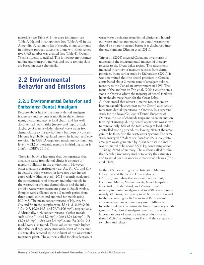
materials (see Table A-2); in glass ionomers (see Table A-3); and in compomers (see Table A-4) in the Appendix. A summary list of specific chemicals found in different product categories along with their respec-tive CAS number was created (see Table 4). Overall, 78 constituents identified. The following environmen-tal fate and transport analysis and acute toxicity data are based on these chemicals.
2.2 Environmental Behavior and Emissions
2.2.1 Environmental Behavior and Emissions: Dental AmalgamBecause about half of the mass of dental amalgam is mercury and mercury is mobile in the environ-ment, bioaccumulates in food chain, and has well documented health risks (neuro- and nephro-toxic), discharge of mercury-laden dental waste water from dental clinics to the environment has been of concern. Mercury is globally regulated due to its human and eco-toxicity. The USEPA-regulated maximum contaminant level (MCL) of inorganic mercury in drinking water is 2 µg/L (USEPA 2011a).
There is a body of literature that demonstrates that amalgam waste from dental clinics is a source of mercury pollution in the environment. However, other amalgam constituents (e.g. Ag, Sn, Cu, and Zn) in dental clinics’ wastewater have not been investi-gated widely. Shraim et al. (2011) recently evaluated the concentrations of mercury and other metals in the wastewater of some dental clinics and the influ-ent of a wastewater treatment plant in Saudi Arabia. Samples were collected over a 2-month period from three dental clinics and analyzed for metals using ICP-MS. The mean concentrations of Hg, Ag, Sn, Cu, and Zn in the samples were 5.3±11.1, 0.49±0.96, 3.0±10.7, 10.0±14.5, and 76.7±106 mg/L, respectively. Additionally, high concentrations of other metals such as Mg (14.4±15.2 mg/L), Mn (3.0±4.6 mg/L), Fe (3.0±4.5 mg/L), Sr (1.6±2.4 mg/L), and Ba (6.9±10.3 mg/L) were also found. These values are much higher than the local regulatory standards. Most of these met-als were also detected in the influent of the wastewater treatment plant. The authors called for classification of
wastewater discharges from dental clinics as a hazard-ous waste and recommended that dental wastewater should be properly treated before it is discharged into the environment (Shraim et al. 2011).
Trip et al. (2004) assessed Canadian inventories to understand the environmental impacts of mercury releases to the Great Lakes region. This assessment included inventory of mercury releases from dental practices. In an earlier study by Richardson (2001), it was determined that the dental practices in Canada contributed about 2 metric tons of amalgam-related mercury to the Canadian environment in 1999. The focus of the analysis by Trip et al. (2004) was the emis-sions in Ontario where the majority of dental facilities lie in the drainage basin for the Great Lakes.Authors stated that almost 1 metric ton of mercury becomes available each year to the Great Lakes ecosys-tems from dental operations in Ontario. In a separate study for the Royal College of Dental Surgeons of Ontario, the use of chairside traps and vacuum-suction filtering of rinsings during dental operations was shown to remove only 40% of the total amalgam load under controlled testing procedures, leaving 60% of the amal-gam to be flushed to the wastewater systems. The same study surveyed 878 dentists. Based on the survey data, amalgam waste generated by 7,000 dentists in Ontario was estimated to be about 2,500 kg, containing about 1,250 kg (50%) of mercury. The authors called for fur-ther detailed inventory studies to verify the estimates and to avoid over- or under-estimation of releases (Trip et al. 2004).
In the U.S., according to the Interstate Mercury Education and Reduction Clearinghouse (IMERC), including the states of Connecticut, Louisiana, Maine, Massachusetts, New Hampshire, New York, Rhode Island, and Vermont, use of mercury in dental amalgam sold in 2001 was approxi-mately 30.8 tons, decreasing to 26.6 tons in 2004 and further decreasing to 16.6 tons in 2007. Increased consumer awareness of mercury use in fillings is hypothesized to drive future declines in mercury amal-gam use. Yet, dental amalgam remained the second largest category of mercury use in products for all three IMERC reporting years (behind the category of switches and relays).

Mercury in Dental Amalgam and Resin-Based Alternatives: A Comparative Health Risk Evaluation20
Many states in the U.S. employ best management practices (BMPs) for dental amalgam waste to prevent mercury from dental amalgam entering wastewater, wastewater sludge, and solid waste. These BMPs include requirements for installing amalgam separa-tors, properly managing solid waste with amalgam, and amalgam recycling (IMERC 2008). Dentists are advised to use dental amalgam separators to catch and hold the excess amalgam waste coming from office spittoons. Publicly-Owned Waste Water Treatment Works (POTWs) have around a 90% efficiency rate for removing amalgam from wastewaters. However, a small amount of waste amalgam is discharged from POTWs into surface waters around the plants. At the treatment plant, the amalgam waste settles out as a component of sewage sludge that is then disposed i) in landfills; ii) through incineration, or iii) by applying the sludge to agricultural land as fertilizer. If the amalgam waste is disposed in a landfill, the mercury may be released into the groundwater or air. If the mercury is incinerated, mercury may be emitted to the atmosphere from the incinerator stacks. And finally, if mercury-contami-nated sludge is used as an agricultural fertilizer, some of the mercury may also evaporate into the air. Through wet or dry precipitation, this airborne mercury eventu-ally gets deposited, contaminating water bodies, land and/or vegetation (USEPA 2011b).
Mercury from dental offices contributes significantly to the overall mercury contamination in wastewater. In 2008, USEPA estimated that there were approximately 122,000 dental offices (comprising approximately 160,000 dentists) that used or removed dental amalgam in the U.S., and that those offices discharged approxi-mately 3.7 tons of mercury each year to POTWs (EPA 2008). Dental offices were found to be the source of 50 percent of all mercury pollution entering POTWs in 2003. A study by the New York Academy of Sciences indicated that as much as 40 percent of total mercury loadings in the New York/New Jersey harbor and water-shed may have come from dental offices (NYAS 2002). In another study in 2002, the National Association of Clean Water Agencies (NACWA) estimated that nearly 40 percent of the mercury in the nation’s wastewater system came from dental offices, and that mercury discharged from dental offices far exceeded all other commercial and residential sources, each of which was below ten percent (EPA 2011b).
In the EU, mercury use for dental amalgams is esti-mated to be more than 90 tons. Approximately 500 million citizens (50-75% of individuals in the EU) have fillings in their mouths. Because the average mouth with fillings in the EU seem to contain 3 to 4 grams of mercury, a ‘human inventory’ of around 1,100 tons is estimated to be found in people’s mouths in the EU. Furthermore, the annual mercury releases, distributed mainly into soil (30 tons), the atmosphere (23 tons), surface water (14 tons) and groundwater (10 tons), are expected to continuously circulate in the biosphere, partially methylate, enter the food chain and detrimentally affect wildlife and human health (EEB 2007).
2.2.2 Environmental Behavior and Presence: Alternative MaterialsKontogianni et al. (2008) reported that, in a typical private dental office, 5 g of solid amalgam waste are disposed daily, while the rate of amalgam over resin-use in restorative procedures is one-third. Although resin-based composite usage has increased significantly within the last two decades, no data was found per-taining to the nature and extent of solid dental waste emissions carrying residues of resin-based restorative materials in unregulated waste. This is mainly due to this waste being classified as municipal waste by the regulatory community. Such municipal waste is often disposed in a landfill. In the absence of knowledge on environmental emis-sions from landfills containing solid dental waste with resin-based restorative materials, the Estimation Program Interface (EPI)TM suite (USEPA 2011c) of physical property and environmental fate estimation models developed by the USEPA’s Office of Pollution Prevention and Toxics and by Syracuse Research Corporation is used to compile data on specific envi-ronmental fate and transport properties of constituents in dental amalgam alternatives listed in Table 4. The EPI uses a single input, such as a compound’s CAS number or SMILES notation, to run a number of esti-mation models, and provides information on estima-tions of physical/chemical properties and environmen-tal fate properties of a given compound. In addition, the EPI suite has a built-in database of property infor-mation compiled from various references (e.g., Merck Index, Beilstein) for 25,000 chemicals (USEPA 2011c).

21Mercury in Dental Amalgam and Resin-Based Alternatives: A Comparative Health Risk Evaluation
The EPI software was run to document environmental fate and transport information such as vapor pressure (VP), Henry’s law constant (H), water solubility (WS), organic-carbon partition coefficient (Koc), octanol-water partition coefficient (Kow), bioconcentration factor (BCF), and rapid biodegradation potential, for each constituent listed in Table 4; and these data are summarized in Table A-5 in the Appendix.
Each compound’s environmental fate was assessed when released into the environment. This informa-tion was utilized to determine how humans might be exposed (i.e., air, water, soil, sediment, and biota), and which exposure pathways (i.e., inhalation, ingestion) may be more relevant and/or significant.
Environmental fate and transport properties are parti-tion coefficients, which indicate in which environmen-tal medium (i.e., air, water, soil) a compound is likely to reside when released to the environment. Thus, these properties provide information about the envi-ronmental behavior of a chemical and are the building blocks of human exposure assessment.
In analyzing the environmental fate and transport data, the following two key references were used: Fate and Transport of Organic Chemicals in the Environment: A Practical Guide (Ney 1998), and Handbook of Chemical Property Estimation Methods (Lyman et al. 1982).
Table 4. Summary of constituents found in formulations of resin-based alternatives as compiled from product Material Safety Data Sheets (MSDSs)
Product/Ingredients CAS # Product/Ingredients CAS #
RESIN COMPOSITES APPLICATION MATERIALS (COUPLING AGENT, PRIMER, ETCHANT, ACTIVATOR, ADHESIVE, BONDING AGENT)
frits chemical; glass filler 65997-18-4 ethanol 64-17-5
glass fibres loose -special purpose; soluble amorphous glass woo
65997-17-3 acetone 67-64-1
silica, dimethylsiloxane treated 67762-90-7 methyl methacrylate 80-62-6
silica amorphous, fumed 68611-44-9 urethane dimethacrylate monomer 105883-40-7
silica amorphous; silicon dioxide 7631-86-9 trimethylolpropane trimethacrylate 3290-92-4
2, 4, 4’ -trichloro-2’ -hydroxydiphenyl ether 3380-34-5
2- hydroxyethyl methacrylate (HEMA) 868-77-9
silanated barium glass filler diphenyl(2, 4, 6- trimethylbenzoyl) phosphine
75980-60-8
silanated silica filler triethylene glycol dimethacrylate 109-16-0
silanated colloidal silica glutaraldehyde 111-30-8
silane treated ceramic 444758-98-9 bisphenol A dimethacrylate 3253-39-2
silane treated silica 248596-91-0 tetrahydrofurfuryl methacrylate 2455-24-5
silane treated zirconium oxide None hexanediol dimethacrylate 6606-59-3
silane treated quartz 100402-89-9 magnesium Salt of N-tolylglycine glycidylmethacrylate (NTG-GMA Salt) (Part A)
211810-95-6
titanium dioxide 13463-67-7 bisphenol A diglycidylmethacrylate (Bis-GMA) (Part B)
1565-94-2

Mercury in Dental Amalgam and Resin-Based Alternatives: A Comparative Health Risk Evaluation22
Product/Ingredients CAS # Product/Ingredients CAS #
2, 2-bis[4-(2-methacryloxy)ethoxy)phenyl]propane
24448-20-2 phosphonic acid acrylate 223681-84-3
bis-GMA; bisphenol A glycidylmethacrylate (Bisphenol-A-bis-(2-hydroxy-3-mehacryloxypropyl) ether)
1565-94-2 phosphoric acid 7664-38-2
triethylene glycol dimethacrylate; TEGDMA; uncured Methacrylate Ester Monomers)
109-16-0 silane treated ceramic 444758-98-9
urethane dimethacrylate (UDMA) 72869-86-4 bisphenol A polyethylene glycol diether dimethacrylate (BISEMA6)
41637-38-1
RESIN COMPOSITES APPLICATION MATERIALS (COUPLING AGENT, PRIMER, ETCHANT, ACTIVATOR, ADHESIVE, BONDING AGENT)
3-trimethoxysilylpropyl methacrylate 2530-85-0 diurethane dimethacrylate (UDMA) 72869-86-4
ethoxylated bisphenol-A-dimethacrylate 56744-60-6 Water 7732-18-5
Tricyclodocandimethanoldimethacrylat 43048-08-4 synthetic amorphous silica 112945-52-5
decamethylene dimethacrylate 6701-13-9 maleic acid 110-16-7
bisphenol A polyethylene glycol diether dimethacrylate (BISEMA6)
41637-38-1 bis-methacrylamidedihydrogenphosphate 911525-18-3
polyethylene glycol dimethacrylate 25852-47-5 isopropanol 67-63-0
2,6-di-tert-butyl-p-cresol (BHT) 128-37-0 acrylamidoaminoacid 72064-86-9
substituted dimethacrylate 27689-12-9 acrylamidosulfonicacid 15214-89-8
ytterbium fluoride (YbF3) 13760-80-0 potassiumfluoride 7789-23-3
3,4-epoxycyclohexylcyclopoly-methylsiloxane
Unknown dimethacrylates 1565-94-2 and 1830-78-0
bis-3,4-epoxycyclohexylethyl-phenyl-methylsilane
154265-59-5 polyethylene glycol dimethacrylate 25852-47-5
borate(1-), tetrakis(pentafluorophenyl)-[4-
178233-72-2 dipentaerythritol pentaacrylate phosphate
(methylethyl)phenyl](4-methylphenyl)iodonium
silane treated silica 122334-95-6
2-benzotriazolyl-4-methylphenol 2440-22-4 copolymer of acrylic and itaconic acid 25948-33-8
urethane modified Bis-GMA dimethacrylate
(dimethylamino)ethyl methacrylate 2867-47-2
Camphorquinone 10373-78-1 camphorquinone 10373-78-1
inorganic iron oxides GLASS IONOMERS
colourants copolymer of acrylic and itaconic acids 25948-33-8

23Mercury in Dental Amalgam and Resin-Based Alternatives: A Comparative Health Risk Evaluation
Product/Ingredients CAS # Product/Ingredients CAS #
COMPOMERS Water 7732-18-5
urethane dimethacrylate 72869-86-4 silane treated glass None
cycloaliphatic dicarboxylic acid dimethacrylate
Unknown silane treated zirconia Unknown
COMPOMERS GLASS IONOMERS
polyethylene glycol dimethacrylate Unknown polyethylene glycol dimethacrylate (PEGDMA)
25852-47-5
polymerizable dimethacrylate resin 105883-40-7 silane treated silica 248596-91-0
polymerizable trimethacrylate resin 3290-92-4 2-hydroxyethyl methacrylate (HEMA) 868-77-9
polymerizable dimethacrylate resin 24448-20-2 glass powder 65997-17-3
polymerizable dimethacrylate resin 109-16-0 bisphenol A diglycidyl ether dimethacrylate (BIS-GMA)
1565-94-2
strontium fluoride 7783-48-4 triethylene glycol dimethacrylate (TEGDMA)
109-16-0
strontium aluminum fluorosilicate glass 65997-18-4 silane treated ceramic 444758-98-9
polymerizable dimethacrylate resin Not Established
diphenyliodonium chloride 1483-72-3
ammonium salt of dipentaerythitol pentaacrylate phosphate
Not Established
ethyl alcohol 64-17-5
silane treated glass None diphenyliodonium hexafluorophosphate 58109-40-3
citric acid dimethacrylate oligomer None
glycerol 1,3-dimethacyrlate 1830-78-0
bisphenol A diglycidyl ether dimethacrylate (bis-GMA)
1565-94-2
silane tretaed silica 248596-91-0
2-benzotriazolyl-4-methylphenol 2440-22-4
2-hydroxyethyl methacrylate 868-77-9
copolymer of acrylic and itaconic acids 25948-33-8
Water 7732-18-5
ethyl alcohol 64-17-5

Mercury in Dental Amalgam and Resin-Based Alternatives: A Comparative Health Risk Evaluation24
The examination of data provided in Table A-5 in the Appendix with respect to the criteria provided above for each environmental fate and transport property reveals that constituents of resin-based restorative materials are complex in their environmental behavior. The table highlights those chemicals with high WS, H, VP, Kow, Koc, BCF in bold and those with medium WS, H, VP, Kow, Koc, BCF in bolded italics, whereas non-highlighted values signify low transport poten-tial. Among the 19 methacrylates listed in Table A-5, some are highly water soluble (e.g., MMA, TEGDMA, HEMA) and some are not (bis-GMA). Some of the methacrylates (BISEMA6, decamethylene dimethac-rylate, MMA) have high to moderate volatilization potential, and they tend to evaporate when released to the water. Thus, for these chemicals, air is the exposure medium of concern for humans. On the other hand, some have low volatilization potential when released.
For many methacrylates with VPs greater than 0.01 mm Hg, inhalation of volatiles is the primary exposure pathway of concern. On the other hand, a few of them (bis-GMA, bisphenol A dimethacrlate) are not readily volatilized when released into the environment. A majority of the methacrylates have Koc values greater than 1,000, indicating that they have high/medium affinity to tightly bind to soil or sediment particles in the environment. For these compounds, which tend to adsorb onto soil/sediment organic carbon, human exposures are often a result of direct exposure pathways such as incidental ingestion of soil and/or inhalation of particles containing sorbed chemicals, and dermal contact with soil/sediment.
As shown in Table A-5, some of the methacrylates (BISEMA6, substituted dimethacrylate) have rela-tively high log Kow values (6-7), suggesting that these compounds are bioaccumulative, immobile,
persistent, and have low water solubility. For these compounds, bioaccumulation through the food chain is of concern. Therefore, human consumption of fish from waters containing polluted sediment and diet, and consumption of other meat (e.g., beef) of animals, which fed on contaminated land, are the exposure pathways of concern. Our research revealed that the majority of the methacrylates has low bioaccumula-tion potential in fish. On the other hand, four of them have medium to high bioaccumulation poten-tial. Lastly, in regards to their biodegradability in the environment, the predictive results are also similarly complex, while some with low molecular weights are rapidly biodegradable when released into the environ-ment while some are persistent.
In regards to solvents and other organic compounds found, particularly in preparation and application materials, the results show that they tend to be highly water soluble and biodegradable with negligible soil adsorption or bioaccumulation potential. Some of them are also volatile (acetone, ethanol, isoproponal). For these compounds, human exposure pathways of con-cern are inhalation and ingestion.
In summary, contamination of environmental media with restorative materials is only feasible when there is an accidental release during transportation of dental waste or leakage or malfunction in landfill liners lead-ing to release of the contaminant to the environment. However, there does not appear to be a significant concern for contamination of environmental media with constituents of resin-based alternatives due to small quantities used and even smaller quantities disposed. However, there needs to be research to verify this assumption, particularly, in areas near landfills which receive large quantities of dental waste through actual environmental measurements.

25Mercury in Dental Amalgam and Resin-Based Alternatives: A Comparative Health Risk Evaluation
III.E X P O S U R E A S S E S S M E N T
3.1. Exposure Assessment: Dental AmalgamDental amalgam has been shown to be the largest single source of continuous metallic Hg exposure for members of the general population who have amalgam fillings. Mercury is released from amalgam fillings in the forms of elemental mercury, mercury ions and in amal-gam particles (Weiner and Nylander 1995). Inhalation of mercury vapor and absorption through the GI tract occur in humans exposed to mercury released from dental fillings. Mercury vapor is released into the oral cavity from dental amalgam containing metallic mer-cury (Hg), causing increased mercury in urine, feces, in exhaled or intra-oral air, saliva, blood, and various organs and tissues including the kidney, pituitary gland, liver, and brain correlated with the amount of oral mercury fillings. The Hg content also increases with maternal amalgam load in amniotic fluid, placenta, cord blood, meconium, various foetal tissues including liver, kidney and brain, in colostrum and breast milk (Richardson et al. 2011). The mercury release rate is dependent on filling size, tooth and surface placement, chewing, food texture, tooth grinding, and brushing teeth, as well as the surface area, composition, and age of the amalgam (Bates et al. 2006).
Eighty percent of inhaled mercury vapors in the oral cavity are readily absorbed in the blood through the lungs and distributed in various organs, mainly in the kidneys, where it may become incorporated before being excreted. Other organs (brain, lungs, liver, gastrointestinal tract, endocrine glands) also show varying degrees of elevated concentrations, although the brain is the site of greatest sensitivity. Because metallic mercury is lipophilic, it can cross the blood–brain and placental barriers where it is oxidized to inorganic mercury. In this state, mercury has limited ability to re-cross these biological membranes, resulting in retainment in the brain and fetal tissues. Mercury from dental fillings may be released to the saliva in ionized form (Hg+2) and as fine particles, which are
then partly absorbed in the gastro-intestinal tract. The amount of mercury from amalgam passing through the gastrointestinal tract may be large but is poorly absorbed, thus, it has only a very minor contribution to systemic exposure (Weiner and Nylander 1995; Levy et al. 2004; SCENIHR 2008). Weiner and Nylander (1995) estimated that the average uptake of mercury from amalgam fillings in Swedish subjects is within 4-19 µg/day.
In addition to this personal absorption due to fillings, the general population is exposed to waste mercury via consumption of contaminated food (e.g., fish), water and air. Dietary intake to methylmercury in fish and other seafood products is the predominant non-occupa-tional exposure source for the general public.
3.1.1 Mercury Exposure Estimates related to Dental Amalgam in General Population and ChildrenWhile inorganic Hg in the human body for which dental amalgam is the primary source is predomi-nately excreted through urine (WHO 2003), organic Hg (methyl mercury) exposure primarily from fish, is measured in hair or whole blood since only 4% of the dose is excreted in the urine. Many studies reported in the literature measured urinary Hg to estimate exposure to amalgam fillings. In addition, other non-invasive biological material such as hair, nails, and saliva has also been used for this purpose (Al-Saleh et al. 2011).
Levy et al. (2004) investigated the effect of amalgam fillings and fish consumption on urine mercury level (U-Hg) in children aged 4–8 years old (n=60) in Montreal, Canada. Children with amalgam fillings were found to have significantly higher urinary mercury levels than children without amalgams (geometric mean=1.412 mg Hg/g versus 0.436 mg Hg/g, respec-tively, P=0.0001). Subjects who reported higher fish consumption also had significantly higher U-Hgs (P=0.004). Univariate analyses provided evidence of an association between elevated U-Hg level and young age (P= 0.009), short height (P=0.024), and low weight

Mercury in Dental Amalgam and Resin-Based Alternatives: A Comparative Health Risk Evaluation26
(P=0.049) in children with amalgam chewing surfaces. A negative correlation between urine mercury and age (-0.378), height (-0.418), and weight (-0.391) was also observed. A multiple logistic regression model showed that the presence of amalgam fillings leads to increased odds of high U-Hg in children (OR=47.18), even after adjusting for high fish consumption (OR=8.66) and height (OR=11.36) (Levy et al. 2004).
Dunn et al. (2008) described levels and correlates/pre-dictors of scalp hair (H-Hg) and urinary (U-Hg) mer-cury in 534 New England Children’s Amalgam Trial (NECAT) participants, who were aged 6–10 years old and without exposure to dental amalgam at baseline, over a 5-year period. Previous research in this area with hair (organic) and urine (inorganic) Hg levels in U.S. children were unable to assess Hg levels while account-ing for exposure to amalgam dental restorations. The mean H-Hg levels ranged between 0.3 and 0.4 mg/g over 5 years with 17–29% of children having H-Hg lev-els ≥0.5 mg/g, and 5.0 to 8.5% of children having levels ≥1 mg/g, in any given study year. Fish consumption fre-quency was the most robust predictor of high H-Hg in adjusted models. U-Hg mean levels varied between 0.7 and 0.9 mg/g creatinine over two years. The percentage of those with U-Hg ≥2.3 mg/g creatinine ranged from 4% to 6%. The number of amalgam restorations had a significant dose-response relationship with U-Hg level. For U-Hg, the number of amalgam-restored surfaces and use of chewing gum in the presence of amalgam were the most robust predictors in the adjusted model. Exposure to amalgam was not associated with detri-mental neuropsychological effects in the wider NECAT study (Dunn et al. 2008).
A recently published study by Al-Saleh et al. (2011) estimated Hg body burden and its association with dental amalgam fillings in 182 children (ages:5–15 years) living in Taif City, Saudi Arabia. Mercury was measured in urine (U-Hg), hair (H-Hg) and toenails (NHg) by the Atomic Absorption Spectrophotometer with Vapor Generator Accessory system. The study revealed that children with amalgam fillings (N=106) had significantly higher U-Hg-C levels than children without (N=76), with means of 3.763 μg/g creatinine versus 3.457 μg/g creatinine, respectively (P=0.019). The results were similar for U-Hg (P=0.01). A similar pattern was also seen for H-Hg, with means of 0.614 μg/g (N=97) for children with amalgam versus 0.242 μg/g (N=74) for those without amalgam fillings (P=0).
After adjusting for many confounders, the multiple logistic regression model showed that UHg-C and H-Hg levels were 2.047 and 5.396 times higher, respec-tively, in children with dental amalgam compared to those without (P<0.01). The authors concluded that amalgam-associated Hg exposure might be related with symptoms of oral health, such as aphthous ulcer, white patches, and a burning-mouth sensation. Furthermore, significant numbers of children with or without amal-gam had Hg levels exceeding the acceptable reference limits (Al-Saleh et al. 2011).
Another recently published study estimated doses related to dental amalgam received by the U.S. popu-lation. Richardson et al. (2011) reported that, based on 2001 to 2004 population statistics, 181.1 mil-lion Americans carry a total of 1.46 billion restored teeth, including children as young as 26 months. The researchers estimated Hg exposure from amalgam fill-ings for five age groups of the US population. While no attempt was made to correlate human heatlh effects, based on the scenario of exposure reflecting the smallest doses evaluated, it was estimated that some 67.2 million Americans would exceed the Hg dose associated with the reference exposure level (REL) of 0.3 μg/m3 established by the USEPA. These exposure estimates were consistent with previ-ous estimates reported by Health Canada in 1995 (Richardson et al. 2011) and amounted to 0.2 to 0.4 μg/day per amalgam-filled tooth surface; or 0.5 to 1 μg/day/amalgam-filled tooth; depending on age and other factors..
3.1.2 Occupational Mercury Exposure Estimates Dental personnel are exposed to higher concentra-tions of mercury while mixing and applying den-tal amalgam and removing amalgam restorations. Exposure of dental personnel to mercury has lessened over the past several decades due to changes in dental amalgam product design and delivery (encapsulated dental amalgam and improvements in amalgam cap-sule design); changes in work practices; and increased regulatory focus on health and safety of dental amal-gam along with environmental rules pertaining to dental waste. Yet, a number of recent studies still pro-vide evidence for increased body burden of mercury in dental personnel as compared to controls. In these studies, the mercury body burden of dental personnel

27Mercury in Dental Amalgam and Resin-Based Alternatives: A Comparative Health Risk Evaluation
was determined to be higher than the general popula-tion’s 1–5 mg Hg/l urine for non-occupational groups. Those personnel with concentrations from 25–50 mg Hg/l urine reported subtle and non-specific symptoms of mercury intoxication (Hörsted-Bindslev 2004).
Morton et al. (2004) investigated low level inorganic Hg exposure using head hair, pubic hair, fingernails, toenails and urine. A cohort of UK dentists (n = 167) and a socioeconomically similar reference population (n = 68) with mercury exposure primarily through diet were evaluated. The mercury content in all biological material was significantly higher in the dental work-ers than in the control population (p < 0.0001). The geometric mean and 90th percentile mercury concen-trations in the urine samples from dentists were 1.7 and 7.3 μmol/mol creatinine, respectively, with only one sample having a value at around the UK’s Health and Safety Executive biological monitoring health guidance level of 20 μmol/mol creatinine.
In a later study, Zolfaghari et al. (2007) evaluated the environmental and occupational exposure to mercury (Hg), and examined various parameters which con-tribute to high levels of mercury of Iranian dentists in Tehran. One hundred hair and nail samples were collected from dentists (n=100); from dental nurses (n=25); and from a control group (n=50). The study included a structured questionnaire designed to pro-vide information about the parameters that influenced their occupational and environmental exposure to Hg. Overall mean concentrations in the hair and nails of the dentists was 2.84 ± 0.47 and 3.56 ± 0.53 mg/kg dry weight respectively. The equivalent values were 0.61 ± 0.07 mg/kg in hair and 0.39 ± 0.06 mg/kg in nails for the control group.. The study showed that use of masks had a significant effect on Hg levels (p = 0.02 for hair and p = 0.03 for nails) and use of gloves only had significant effect on nails Hg (p = 0.05). Overall, hair Hg concentration exceeded the thresh-old value of 5 mg/kg in 22% of dentists and was above the WHO ‘normal’ level in 25% of them (Zolfaghari et al. 2007).
These results as well as others (SCENIHR 2008) provide evidence for continued increased body burden of mercury in dental personnel due to the use of dental amalgam.
3.2 Exposure Assessment: Alternative MaterialsExposure characterization for resin-based materials is challenging due to many factors. The usage of many chemicals in each product, lack of complete composi-tional data, usage of small amounts during teeth resto-ration for brief periods, and insufficient understanding of the reactions/interactions within the mixtures are all impediments. Further, scant information on actual per-sonal or area exposure concentrations in dental offices, lack of record keeping as to the type, the amount, and the duration of material used for different applications and lack of available biological exposure/effect mark-ers for the constituents make specific dose assessments impossible based on available practice data. Thus, there is a critical need to perform systematic research to collect inventory data for usage of various materials in dental clinics and time sequence of these exposures to derive emission estimates that can then be utilized in exposure models to estimate exposure concentrations.
Despite this lack of quantitative data, it is clear that dental professionals are exposed to components from resin-based restorative materials during routine practice as a result of vapors and dusts encountered in place-ment, curing, finishing, and polishing procedures.. These exposures can occur through inhalation and der-mal absorption pathways. While the former is of more concern for patients, occupational exposure in dental clinics may occur via both pathways.
3.2.1 Inhalation ExposureCured composites generally contain some residual monomer that is diffusible and may elute monomer and additives within the patient’s mouth (Allen et al. 2000). Furthermore, respiratory exposure may occur during finishing of the polymerized composite in oral cavity. In general, respiratory exposure to the vapors of the composite resins is not likely to occur at prepara-tion and placement stages since the amount of paste used is minimal, and the time at the monomer stage is very short. Resin-based dental materials release compo-nents, initially due to incomplete polymerization, and later due to degradation.

Mercury in Dental Amalgam and Resin-Based Alternatives: A Comparative Health Risk Evaluation28
One recently published study involved review of the literature on the short- and long-term release of components from resin-based dental materials, and determination of how much of those components may leach out in the oral cavity identifying particu-lar chemicals and amounts to which patients may be exposed (Van Landuyt et al. 2011). Seventy-one studies were identified out of which 22 were included in their analysis. A meta-analytical mean for the evaluated eluates was calculated. In addition to the monomers present in resin-based materials, addi-tives, such as initiators, inhibitors and stabilizers, were included in this assessment. The review noted significantly more release of components in organic than in water-based media. In general, equal or lower quantities of BPA were released compared to the analyzed monomers. Among the monomers, HEMA was released the most, followed by TEGDMA, UDMA and Bis-GMA. The authors concluded that resin-based dental materials might account for the total body burden of orally ingested bisphenol A, and even higher amounts of monomers. Furthermore, the review found data to suggest that even higher amounts of additives might elute, even though com-posites contain very small amounts (Van Landuyt et al. 2011). It is also known that TEGDMA does not completely react in composites and migrates out of polymerized materials over time. However, the eluate containing TEGDMA or other monomers appears to rapidly decrease over time.
These effects have not been monitored clinically, and their health effects have yet to be revealed (Allen et al. 2004). These results indicate that brief exposures to monomers and additives do occur due to release of chemicals from resin-based alternatives, albeit data on variability in human exposure concentrations are cur-rently scant at best.
A focus on BPA is warranted due to increasing evi-dence that BPA and some BPA derivatives can pose health risks attributable to their endocrine-disrupting, estrogenic properties. Fleisch et al. (2010) compiled and critically evaluated the literature characterizing BPA content of dental materials. They assessed BPA exposures from dental materials and potential health risks, and developed evidence-based guidance for reducing BPA exposures while promoting oral health. Though BPA is released from dental resins through salivary enzymatic hydrolysis of BPA derivatives and
BPA is detectable in saliva for up to 3 hours after resin placement, the quantity and duration of systemic BPA absorption is not clear. Based on this review, the authors recommended that the use of resin-based mate-rials should be minimized during pregnancy whenever possible (Fleisch et al. 2010).
Despite this assessment, a joint 2010 FAO/WHO expert meeting on toxicological and health effects of bisphenol-A determined that there was no need to col-lect additional data on BPA levels from dental materi-als due to low levels of BPA in saliva and short-term exposures (FAO/WHO 2010).
Three exposure assessment studies have been pub-lished to date by researchers in Scandinavia, where composite resin restorations have largely replaced amalgam fillings as the treatment of choice for caries during the past 20 years. Marquardt et al. (2009) mea-sured exposure of dental personnel to various vola-tile methacrylates using area sampling. Ambient air concentrations of methacrylates were measured during filling treatment while bonding agents were used in 4 dental practices in Munich, Germany. Methyl methacrylate (MMA), 2-hydroxyethyl methacrylate (HEMA), ethylene glycol dimethacrylate (EGDMA), and triethylene glycol dimethacrylate (TEG-DMA) were detected in the air of all of the dental prac-tices, though exposure levels varied during the filling treatments. The detection of TEG-DMA correlated with the application of bonding agents during place-ment of dental fillings. The maximum levels of MMA measured in this study were at least 200 times lower than the toxicologically relevant maximum allowable concentrations (or exposure limits) defined in various countries. Yet, the authors recommended reduction of the air levels of methacrylates in order to minimize the chance of allergic sensitization of dental personnel since this reaction is not dependent on chronic toxico-logical threshold values but maybe produced by short term over exposure or low dose reexposure (Marquardt et al. 2009).
In an earlier study conducted in Sweden (Hagberg et al. 2005), exposure of dental personnel to 2-hydroxy-ethyl methacrylate and methyl methacrylate in five randomly selected public dental clinics was quanti-fied by taking 21 whole-day and 46 task-specific short-term (1–18 min) samples. The median 8-hour time-weighted averages were 2.5 μg/m3 (dentists)

29Mercury in Dental Amalgam and Resin-Based Alternatives: A Comparative Health Risk Evaluation
and 2.9 μg/m3 (dental nurses) for HEMA, and 0.8 μg/m3 (dentists) and 0.3 μg/m3 (dental nurses) for MMA. The maximum short-term exposure levels were 79 μg/m3 for HEMA and 15 μg/m3 for MMA with similar results in dentists and dental nurses. Authors concluded that, although irritant effects would not be expected in healthy people at these levels, occu-pational respiratory diseases due to sensitization to methacrylates may occur, and, thus, improvements in the handling of these chemicals are warranted (Hagberg et al. 2005).
A Finnish study by Henricks-Eckerman et al. (2001) measured airborne methacrylates and natural rubber latex (NRL) allergens during placement of composite resin restorations in six dental clinics. NRL allergens can be released from surgical protective gloves into the air in hospitals/clinics. Both area and personal sampling were performed, and special attention was paid to measurement of short-term emissions from the patient’s mouth. The median concentration of 2-HEMA was 0.004 mg/m3 close to the dental nurse’s work-desk and 0.003 mg/m3 in the breathing zone of the nurse with a maximum concentration of 0.033 mg/m3. Above the patient’s mouth the con-centration of 2-HEMA was about 0.01 mg/m3 during application of adhesive and composite resins and dur-ing finishing and polishing of the fillings. Maximum concentrations 3–5 times higher than median were also measured. The sampling revealed that triethyl-eneglycol dimethacrylate was released into the air during the removal of old composite resin restorations (0.05 mg/m3) and to a minor extent during finishing and polishing procedures.
The median concentration of the NRL allergen was 0.12 au/m3 (au = arbitrary unit) with a maximum concentration of 1.1 au/m3. In comparison, in health care workers in a hospital in the UK the geometrical mean concentration of NRL allergen was 0.46 μg/m3 as determined by personal sampling. This was much lower than in glove manufacturing factories (up to 17.8 µg/m3) or rubber plantation workers (2.4 µg/m3). Despite the findings that exposure of dental personnel to methacrylates and NRL allergens is low, these authors also called for measures to reduce exposure in order to reduce the risk of sensitization among personnel (Henriks-Eckerman et al. 2001).
In addition to inhalation exposure to methacylates (monomers), there is exposure to aerosols created when composites are finished clinically with high-speed instruments. These aerosols containing crystalline or amorphous silica as well as polymerized resin dusts and additives can accumulate on the operator’s hands, the patient’s face, and on equipment. There is very limited indirect information on aerosol exposure to dental personnel and patients during finishing or removal of composites. Collard et al. (1989) studied the effect of two different abrasives on the size-distribution of composite dusts in the laboratory. The respirable frac-tion of dust particles (i.e., particle aerodynamic size ≤ 4 µm) ranged between 57.2 and 85.7%. The diamond bur created more respirable particles than the carbide bur for each composite tested. The elemental composi-tion of particles of each composite was determined and silicon was detected in amounts ranging from 71-100%. The authors concluded that, based on the composition and particle size distribution, the dust generated during finishing of composite resins containing quartz (crystal-line silica) filler has the potential for causing silicosis in dental personnel (Collard et al. 1989).
A 1991 study by the same authors determined the mass and number of particles released from five composites, with two different shades each. The sampling indi-cated that between 14 and 22% of the dust generated was respirable. Powder x-ray diffraction revealed that respirable dust particles collected from composites reported to contain crystalline silica fillers contain the same crystalline silica. In a clinical environment, factors such as air current, location of restoration, orientation and speed of the hand-piece, proximity of the suction tip to the bur, presence of high speed evacuator and water spray are expected to impact the aerodynamic behavior and the size of the dust to which dental personnel are exposed (Collard et al., 1989, 1991; Nayebzadeh et al. 2000)., While initial data is therefore available with respect to silica, the extent and significance of the total occupational aerosol exposure in the dental setting is currently not known. Nano-particle usage in product formulations has been gaining momentum as well and therefore, there is a need for a comprehensive assessment of the exposure and absorp-tion of all components of these dusts.

Mercury in Dental Amalgam and Resin-Based Alternatives: A Comparative Health Risk Evaluation30
3.2.2 Occupational Inhalation Average Daily Dose Estimates The literature was reviewed to develop a high end and low end model of inhalation exposure to dentist and den-tal technicians. Potential average daily inhalation dose of dental personnel for methacrylates methyl methacrylate (MMA), 2-hydroxyethyl methacrylate (HEMA), ethyl-ene glycol Dimethacrylate (EGDMA), and triethylene glycol dimethacrylate (TEG-DMA) was estimated using the personal exposure measurement data reported in the three Scandinavian studies explained above (Marquardt et al. 2009; Hagberg et al. 2005; and Henricks-Eckerman et al. 2001). In dose estimation, exposure concentration data are integrated with exposure parameters into an estimate of daily dose received by an exposed individual via a specific exposure route. The magnitude of human exposures, in general, is dependent on chemical concen-tration in exposure medium (air), exposure parameters describing human physiology (e.g., inhalation rate, body weight), and population–specific parameters describing exposure behavior (exposure frequency, duration).
When evaluating subchronic or chronic exposures to noncarcinogenic chemicals, dose is averaged over the period of exposure, termed “Average Daily Dose” (ADD), which represent normalized exposure rate in the units of mg of chemical per kg body weight per day, mg/kg-day). The lowest and highest personal exposure concentration measured in these three studies for each monomer (MMA, HEMA, TEGDMA, EDGMA) was identified and a low-end ADD and a high-end ADD were calculated, respectively, with the goal of capturing different exposure conditions in dental clinics/offices. The inhalation ADD is commonly calculated via the following equation (USEPA 1989):
where: Ca: Exposure concentration in air (mg/m3) IRi: Inhalation rate (m3/h) ET: Exposure time (h) EF: Exposure frequency (d/y) ED: Exposure duration (y) BW: Body weight (kg) AT: Averaging time (days) – (ED*365 d/y for noncarcinogens)
It was assumed that dental personnel would be exposed to monomers for 30 minutes on a daily basis, for two days per week for 50 weeks in a year, for a total of 5 years under the low-end exposure scenario. Under the high-end exposure conditions, dental personnel were assumed to be exposed to these volatile monomers for 8 hours on a daily basis, for 5 days per week for 50 weeks in a year, for a total of 30 years. For inhalation rate, 0.4 and 1.2 m3/h corresponding to resting and light-activity conditions as an average value for adult man and woman were used to represent the low- and high-end exposure conditions, respectively (USEPA 1997).
The results of our calculations for each monomer under the low- and high-end scenario are shown in Table 5. We estimated that, under the low-end scenario, mono-mer-specific ADD would range between 8e-08 and 6e-06 mg/kg-d. For high-end scenario, these estimates were approximately three to five orders of magnitude higher, ranging from 1e-03 to 4e-02 mg/kg-d. Because these esti-mates are based on a limited number of measurements in a small sample size of clinics from only three studies, they should be viewed with caution. The representation of both the exposure concentrations measured and ADD estimates derived from these studies for other dental environments is unknown at this time and further expo-sure assessment studies should be conducted to validate these concentrations and ADD estimates.
3.2.3 Dermal Exposure Although most dental materials manufacturers warn that any contact with dermal restorative materials should be followed by glove exchange and hand wash-ing to minimize exposure, this is not always followed in practice, resulting in penetration of monomers from gloves to skin. Several researchers explored the mag-nitude of this problem experimentally. Nakamura et al. (2003) examined the penetration of six monomers used in dentistry through five commonly used dental protective gloves – latex, powder-free latex, coated latex, polychloroprene, and polyvinyl. In order to simulate potential long-term exposure, an unused glove finger tip without pinholes was cut and used to hold 500 mL of monomer while dipped into 99.5% ethanol for 180 minutes at 37˚C. Ethanol was later analyzed by spectrophotometry for monomer penetration. Only the lower molecular weight monomers permeated the gloves tested. The amount of monomers permeating the latex in 10 minutes was 0.8±0.6, 0.6±0.6, 0.07±0.1, 0.07±0.1, 0.1±0.1 and 0.06±0.1 µL/mL for MMA,

31Mercury in Dental Amalgam and Resin-Based Alternatives: A Comparative Health Risk Evaluation
HEMA, EGDMA, TEGDMA, UDMA, and Bis-GMA, respectively. The polyvinyl chloride glove showed the greatest monomer permeability. In conclusion, four of the monomers tested permeated all of the gloves tested in this study. While these experiments did not account for the presence of other prior contaminant materials on the gloves, they demonstrated that gloves do not provide protection against volatile monomers, and dermal exposures in clinical environments are likely (Nakamura et al. 2003).
In another study, Lönnroth et al. (2003) considered potential solvent effect of one monomer for another, by studying monomer mixtures. The permeability and permeation rates were studied and the breakthrough time (BTT, min) as a measure of protection for a mix-ture consisting of 80% MMA, 10% EGDMA and 10% 1,4-BDMA (1,4-butanediol dimethacrylate). Fifteen different gloves (4 natural rubber, 6 synthetic rubber, 4 synthetic polymer, and 1 laminated material) were tested. The lowest molecular weight monomer, MMA, permeated within 3 min through all glove materials. A polyethylene examination glove provided the longest protection period to EGDMA and 1,4-BDMA (> 120 min and 25.0 min), followed by the surgical glove Tactylon (6.0 min and 8.7 min) and the nitrile glove Nitra Touch (5.0 min and 8.7 min).
The glove thickness appeared to affect penetration rates. In order to assess if double gloves provided longer protection, additional tests were carried out with one synthetic rubber (Lirtin) as inner glove and one natural rubber latex (Amanita) as outer glove, using the MMA, EGDMA, and 1,4- BDMA mixture. These
experiments revealed longer protection only when the inner glove was rinsed in water before placing the outer glove on top (Lönnroth et al. 2003). The experimental conditions used in this study were called into question due to poor simulation of clinical exposure conditions. Despite this limitation, this study provided strong evi-dence that no one glove type provided ideal protection, although nitrile gloves performed best under the test conditions (Allen et al. 2004).
Andreasson et al. (2003) assessed the permeability of various types of gloves to methyl methacrylate (MMA), 2-hydroxyethyl methacrylate (HEMA) and triethyleneglycol dimethacrylate (TEGDMA) with special reference to combinations with etha-nol or acetone. The permeation rate and time lag breakthrough (lag-BT) for MMA (neat, or diluted to 30% in ethanol or acetone), HEMA (30% in water, ethanol, or acetone) and TEGDMA (30% in ethanol or acetone) were investigated for nine different types of gloves ((2 vinyl types, polyethylene, 2 nitrile types, 2 latex types, and nitrile/polyethylene combination). The lag-BT for neat MMA was ≤ 2min for all gloves. For 30% MMA in ethanol or acetone, the latex gloves and the polyethene-copolymer glove showed the best protection, but the lag-BTs were short for all gloves. For HEMA and TEGDMA, the lag-BTs were gener-ally longer than for MMA. A neoprene glove seemed to be the best choice for protection against penetra-tion of HEMA and TEGDMA. These studies provide evidence that dermal exposures may occur through penetration of monomers with or without solvents (acetone, ethanol) from gloves.
Table 5. Occupational Inhalation Average Daily Dose Estimates (mg/kg-day)
Monomer Constituent Exposure Concentration (µg/m3)
Average Daily Occupational Inhalation Dose (mg/kg-d)
Low-End High-End Low-End High-End
MMA 0.1 400 8.E-08 4.E-02
HEMA 0.7 79 6.E-07 7.E-03
TEGDMA 1 81 8.E-07 7.E-03
EDGMA 8 13 6.E-06 1.E-03

Mercury in Dental Amalgam and Resin-Based Alternatives: A Comparative Health Risk Evaluation32
4.1 Hazard Identification: Human Health Effects of Dental Amalgam Mercury has long been recognized as a toxic metal due to its adverse effects on humans following acute or chronic high-level occupational exposures. Target organs for mercury exposure are the kidneys, central nervous system and thyroid glands (Holmes et al. 2009). Since the 1990s, several federal agencies around the world have reviewed the scientific literature seek-ing links between dental amalgam and health prob-lems to guide environmental and public health policy decisions. Maths Berlin was assigned by the Swedish government to summarize and evaluate research find-ings related to mercury from amalgam, which were published from November 1997 to November 2002 in order to supplement the risk analysis that was car-ried out for the Swedish Council for Planning and Coordination of Research in 1997 (Berlin, 2002). Some of the important conclusions of this review were: i) identification of the thyroid as the target organ for the toxic effect of mercury in occupational exposure to mercury vapor in low doses; ii) identification and quantification of neuropsychological symptoms at low exposure levels in occupationally-exposed workers; iii) observed gender differences in the toxicokinetics of mercury; iv) potential effect of the mercury vapor on human fetal development; and v) clinical demonstra-tion of variance in high sensitivity in individuals who are exposed to small quantities of mercury through skin exposure or inhalation (Berlin, 2002).
In the U.S., the Life Sciences Research Office (LSRO) tasked by the Centers for Disease Control and Prevention (CDC) in the U.S. analyzed the health effects data for dental amalgam in 2004 (LSRO 2004) by examining the peer-reviewed, primary scientific and medical literature published between January 1, 1996 and December 31, 2003 relating to dental amalgam and human health. About 950 scientific and medical
studies were considered in this evaluation and approxi-mately 300 of the studies met criteria for scientific merit and study design and were used to support the policy decisions. The analysis concluded that there is insufficient evidence to support a correlation between dental amalgam exposure and kidney, cognitive dys-function including Alzheimer’s and Parkinson’s disease, or autoimmune disease, including multiple sclerosis. Further, the various non-specific symptoms attributed by some to dental amalgam have not been shown to be due to increased mercury release and absorption. Finally, dental amalgam is capable of producing delayed hypersensitivity reactions in some individuals. For these individuals, the removal of dental amalgam resto-rations and their replacement with composite materials is suggested to promote the resolution of the allergic symptomatology. Despite the existence of rare allergic hypersensitity, the CDC concluded that there is little evidence to support a causal relationship between mer-cury fillings and human health problems in the general population (LSRO 2004).
In 2009, the U.S. Federal Drug Administration (FDA) updated the CDC report and reviewed the best avail-able scientific evidence to determine whether the low levels of mercury vapor associated with dental amalgam fillings are a cause for health concern for individuals with fillings. Specifically, an additional 29 human stud-ies and 5 animal studies were reviewed and evaluated. Compared to previous analyses performed by CDC, no significant new information was discovered from the review of additional information that would change the risk estimates by FDA for the use of dental amalgam. Thus, the FDA concluded that there is insufficient evidence to support an association between exposure to mercury from dental amalgams and adverse health effects in humans, including sensitive subpopulations. (FDA 2009). It was noted that data was still lacking due to the existence of only very limited informa-tion related to long-term health outcomes in vulner-able subpopulations (e.g., pregnant women and their developing fetuses, and children under the age of six, including infants who are breastfed).
IV.H A Z A R D I D E N T I F I C AT I O N

33Mercury in Dental Amalgam and Resin-Based Alternatives: A Comparative Health Risk Evaluation
In December 2010, FDA announced the formation of another review panel to assess health risks of dental amalgam to the U.S. population. The Panel noted the potential existence of a susceptible subpopulation that is prone to adverse health effects after receiving amalgams, but qualified that this population could not be easily pre-identified at this time (FDA 2010).
Bates et al. (2004) performed a retrospective cohort study, the largest of its kind, containing people in the New Zealand Defence Force (NZDF) between 1977 and 1997. The authors reviewed annual dental treat-ment histories, including amalgam filling placements, and data on dental amalgam were compiled from individual records. The cohort was linked with morbid-ity records. The final cohort contained 20,000 people, with 84% males. Of conditions allegedly associated with amalgam, multiple sclerosis had a non significant elevated adjusted hazard ratio (HR) of 1.24 (95% CI: 0.99, 1.53, P = 0.06), but there was no elevation of chronic fatigue syndrome (HR = 0.98, 95% CI: 0.94, 1.03), or kidney diseases. There were insufficient cases for investigation of Alzheimer’s or Parkinson’s diseases. The authors concluded that there was only limited evidence of an association between amalgam and dis-ease but further follow-up of the cohort would permit investigation of diseases more common in the elderly (Bates et al. 2004).
The same research group (Bates et al. 2006) provided a comprehensive review of the epidemiologic evi-dence for the safety of dental amalgam fillings, with an emphasis on methodological issues and identifying gaps in the literature. Studies showed little evidence of effects on general chronic disease incidence or mortal-ity. The authors reported that limited evidence exists for an association with multiple sclerosis. The pre-ponderance of evidence suggested no renal effects and that ill-defined symptom complexes, including chronic fatigue syndrome, are not caused by amalgams. They found little direct evidence to assess reproductive haz-ards since there were only a few relevant epidemiologic studies available for review (Bates et al. 2006).
In another review article, Holmes et al. (2009) evalu-ated the strength of the epidemiological evidence on the effects of prolonged low-level exposure to the various forms of mercury. For dental mercury, the authors reported that the UK Committee on Toxicity of Chemicals in Food, Consumer Products (COT)
concluded that any risk of neurotoxicity as a result of exposure to mercury vapor stemming from dental amal-gam was of greater relevance to occupational cohorts, and that there was no available evidence indicating that the use or removal of dental amalgam fillings during pregnancy was harmful. The EC Scientific Committee on Emerging and Newly Identified Health Risks (SCENIHR, 2008) reviewed the existing data-base for dental amalgam and its alternatives compre-hensively and concluded that there is no evidence that current use of dental amalgam or other dental materials is associated with systemic disease (SCENIHR 2008).
Ye et al. (2009) evaluated impacts of low level mer-cury exposure on renal function and neurobehavioral and neuropsychological performance among children in Shanghai, China. Dental histories for 403 chil-dren aged 7–11 years in five schools were checked by dentists. Of these, 198 with confirmed amalgam fillings were recruited as participants (exposure group). Control group (N =205) never had dental amal-gam treatment. Each child provided a urine sample for measurements of total mercury, N-acetyl-b-D-glucosaminidase activity, microalbumin, and creati-nine (Cr). The Child Behavior Checklist, Eysenck Personality Questionnaire, and an intelligence screen-ing test were also administered. The geometric mean urinary mercury concentration was 1.6 mg/g Cr for children with and 1.4 mg/g Cr for children without amalgam fillings. No differences were found between children with and without fillings for either renal func-tion biomarker, or on neurobehavioral, neuropsycho-logical, or intelligence tests.
A randomized clinical trial was conducted in Europe in 1997-2005 in which children needing dental restorative treatment were randomly assigned to either amalgam (n=253) or resin composite (n=254) groups for posterior restorations (DeRouen et al. 2006). Subjects (n=507) were 8-10 years old children living in Lisbon, Portugal and had at least one carious lesion on a permanent tooth and no previous exposure to amal-gam; and had no interfering health conditions. The researchers performed neurobehavioral assessments of memory, attention/ concentration, and motor/visuomo-tor domains, and measured nerve conduction veloci-ties. While children had a mean of 18.7 tooth surfaces (median=16) restored in the amalgam group, this mea-sure was 21.3 (median=18) in the composite group dur-ing the 7-year trial period. There were no statistically

Mercury in Dental Amalgam and Resin-Based Alternatives: A Comparative Health Risk Evaluation34
significant differences in measures of memory, atten-tion, visuomotor function, or nerve conduction veloci-ties for the amalgam and composite groups over all 7 years of follow-up. However, starting at 5 years after initial treatment, the need for additional restorative treatment in children treated with resin composite was approximately 50% higher. (DeRouen et al. 2006).
In a follow-up study of the same cohort, Lauterbach et al. (2008) reported no significant differences between treatment groups in any neurological measures. The groups did not differ with respect to Neurologic Hard Signs, tremor, or Neurologic Soft Signs SS. (Lauterbach et al. 2008).
A similar study conducted in the U.S., the New England Children’s Amalgam Trial (NECAT) involved a randomized trial involving 6- to 10-year-old children (n=534) as subjects. Bellinger et al. (2006) compared the neuropsychological and renal function of children whose dental caries were restored using amalgam or mercury-free materials in the NECAT study involving six community health dental clinics in Massachusetts. Children with no prior amalgam restorations and two or more posterior teeth with caries were randomly assigned to receive dental restoration using either amalgam (n=267) or resin composite (n=267) materials for these and subsequent carries. The results showed that assignment to the amalgam group was associated with a significantly higher mean urinary mercury level (0.9 vs. 0.6 μg/g of creatinine at year 5). The investigators found no statistically significant differences between children in the amalgam and composite groups in 5-year change in full-scale IQ score (3.1 vs. 2.1, P=.21) after adjust-ing for randomization stratum and other covariates. Similarly, no statistical differences between two groups were observed for general memory index, visuomotor composite or urinary albumin (median=7.5 vs. 7.4 mg/g of creatinine, P=0.61). (Bellinger et al. 2006).
The same researchers investigated the hypothesis that restoration of caries using dental amalgam resulted in worse psychosocial outcomes than restoration using mercury-free composite resin. All significant associa-tions favored the amalgam group. No evidence was found that exposure to mercury from dental amalgams was associated with adverse psychosocial outcomes over the five-year period following initial placement of amalgams (Bellinger et al. 2008).
In another NECAT analysis by the same research-ers (Bellinger et al. 2007), the neuropsychological outcomes of subjects whose caries were restored using dental amalgam were compared with the outcomes of those whose caries were restored using mercury-free resin-based composite. The primary intention-to-treat analyses did not reveal significant differences between the treatment groups on: Full-Scale IQ score, General Memory Index, and Visual-Motor Composite (Bellinger et al. 2007).
The negative findings in Portugal and U.S. cohorts have recently been scrutinized by the International Academy of Oral Medicine and Toxicology (IAOMT). IAOMT pointed out a number of deficiencies and limi-tations in study designs (e.g., low statistical power, poor control of confounders) that may explain the negative associations, and they called for a new meta-analysis of data by combining the NECAT and Casa Pia study data sets, thus providing increased statistical power for detecting differences in incidence of neurological effects between higher dose and lower dose members of the combined amalgam cohorts (IAOMT 2010).
A recently published study by Mutter et al. (2010) inves-tigated evidence for Alzheimer’s disease (AD) as related to inorganic mercury (IM) exposure as the main focus, albeit it examined effects associated with other forms of mercury, including dental amalgam. One thousand, and forty one references were scrutinized, and 106 studies fulfilled the inclusion criteria. The authors noted that studies on health effects in persons with amalgams have been largely negative though also significantly flawed methodologically. (Mutter et al. 2010).
The above summary highlights the largely negative literature with respect to adverse health effects of dental amalgams and the scientific controversy as to whether the inherent faults in these studies leave room for concern about unidentified effects in the subjects. Regardless, due to the toxicology and amount of mercury released into the environment from this use, the World Health Organization called to reduce or, wherever pos-sible, eliminate the use of mercury containing dental amalgam due to potential risks to the human popula-tion from current background environmental levels (http://www.who.int/phe/news/Mercury-flyer.pdf 2007).

35Mercury in Dental Amalgam and Resin-Based Alternatives: A Comparative Health Risk Evaluation
4.2 Hazard Identification: Alternative Materials As less is known about the alternatives, a scientific weight of evidence analysis is performed to determine whether constituents of resin-based alternatives used as they are in dental restoration are linked to adverse health effects. In order to be able to perform this analy-sis, we compiled and evaluated available toxicology data from NIH’s TOXNET Database maintained by the National Library of Medicine (NLM) in the U.S. and from scientific literature.
4.2.1 Acute Toxicity Data (LD50, LC50)We used NLM’s Hazardous Substances Data Bank (HSDB), which provides human and animal toxic-ity data for about 5,000 chemicals. In addition, we used NLM’s ChemicIDplus Advanced database, which allows users to obtain acute toxicity data for over 370,000 chemicals. From these databases, the dose (LD50) or concentration (LC50) of constituents of resin-based alternatives listed in Table 4 causing 50% mortality in test species in animal models was compiled and presented in Table A-6 in Appendix. A separate table for MMA was created (see Table A-7 in Appendix) due to availability of a large set pertaining to this chemical. Target organ or specific target organ identified in toxicity testing was also documented for each constituent.
Acute toxicity data for inhalation and oral pathways, as shown in Table A-6 and A-7, reveal that toxicity benchmarks vary significantly by several orders of mag-nitude across different constituents. In addition, there are large data gaps even in acute toxicity information since only 22 of 78 constituents (i.e., 28%) were found to have any acute toxicity data. However, available information does show some consistent results. A majority of the methacrylates are skin-sensitizers. Fillers used in formulations have respiratory health effects. Furthermore, some of the monomers (TEGDMA, trimethylolpropane trimethacrylate monomer, HEMA) are associated with, central nervous system effects including somnolence, tremor, and ataxia. In addition inhalation of MMA has been associated with induction of asthma in animal testing.
4.2.2 CytotoxicityMany in vitro tests have provided evidence of the toxic-ity of amalgam alternatives although the mechanisms of this cytotoxicity have not been elucidated. It has been hypothesized that monomers of methacrylate are the primary component responsible for the cyto-toxic effect of resin containing materials. The focus of cytotoxicity testing in these studies was often directed to the hazards to the dental pulp and screening for biocompatibility. We have limited this hazard identi-fication analysis to those studies with human health implications.
In early 1970s, structure–toxicity relationship of 18 acrylic and methacrylic compounds was investigated by Lawrence et al. (1972) and Bass et al. (1974). In the former, the toxicity of a series of esters of acrylic and methacrylic acids was determined in groups of mice, and the resultant LD50 values were analyzed with the use of the mathematical models of Free and Wilson and of Hansch to relate structure of the compounds to toxicity. In the latter study, eight additional compounds (four acrylate esters and four methacrylate esters) were evaluated by the same procedures. In both studies, the authors found that acute toxicity correlated with water solubility (Yoshii 1997).
Spanberg et al. (1973) hypothesized that acrylic mono-mers in the catalyst system were the causative agents for damage. The change of the composite materials to a two-paste system (Adaptic, Concise) resulted in the reduction in the level of toxicity in vitro, indicating a more complete chemical binding of toxic components. However, the 24-hour experiments demonstrated that irritant components of the material were still released to the environment. The authors concluded that ante-rior tooth filling materials contain biologically reactive chemicals which are not bound when the material is introduced into the cavity and the biologic properties of these materials should be evaluated before they are recommended for common use (Spanberg et al. 1973).
Dillingham et al. (1983) investigated the dependence of hemolytic activity and LD50 (mice) on physical properties (lipophilicity, molar refraction, and molecu-lar volume) of the esters (acrylates and methacrylates) using multiple regression analysis. The hemolytic activity of acrylates and methacrylates was found to be related to lipophilicity (inversely related to water solubility) and that the mechanism of the action of

Mercury in Dental Amalgam and Resin-Based Alternatives: A Comparative Health Risk Evaluation36
the esters was membrane-mediated and relatively nonspecific. Furthermore, in vivo biotransformation was not a significant factor (Dillingham et al. 1983). Because the residual monomer of the BIS-GMA type resins was found to be more hemolytic than that of the MMA type resins despite the lower elution potency of the former monomer, Fujisawa et al. (1978) under-took a study to explain the high hemolytic activity of BIS-GMA from the structure-activity relationship using BIS-GMA, and various types of methacrylates by employing hemolysis test. This study found that the strong hemolytic potency of BIS-GMA was due to the high hydrophobic nature of the compound and its hav-ing a reasonably high affinity for erythrocytes (Fujisawa et al. 1978).
Hanks et al. (1991) performed a study to determine the cytotoxic concentrations of 11 components of resin composites on monolayers of cultured Balb/c 3T3 fibroblasts, to study the inhibitory effects of these components on DNA synthesis, total protein content, and protein synthesis, and to determine whether effects were reversible when the components were withdrawn from the medium. These data were reported as concen-trations which inhibited 10% (ID10) and 50% (ID50) of a particular metabolic process as well as the range of concentrations over which cell metabolism was irreversibly inhibited. For any individual component, the ID50 values for all three metabolic parameters were of the same magnitude as was the ranges of irreversibil-ity. Ethoxylated Bis-phenol A dimethacrylate (E-BPA) was the most toxic molecule of the group (ID50 being between 1 and 10 µmol/L). The ID50 concentrations for three of the components, including Bis-GMA, UDMA, TEGDMA, and Bis-phenol A, ranged between 10 and 100 µmol/L, while the ID50 values of three components (N,N dihydroxyethyl-p-toluidine, camphoroquinone, and N,N dimethylaminoethyl methacrylate) were above 100 µmol/L. The results showed that the components of resin composites are hazardous in that they all cause significant toxicity in direct contact with fibroblasts. However, the potencies of the resin components varied by greater than 100 times and total protein, protein syn-thesis and DNA synthesis were roughly equivalent in measuring these potencies for most components (Hanks et al. 1991).
HEMA have also been found to be cytotoxic in a variety of different in vitro models. Bouillaguet et al. (1996) determined the cytotoxicity of HEMA using BALB/c 3T3 mouse fibroblasts in direct contact with HEMA for 12 or 24 h in an in vitro diffusion chamber. Concentrations of HEMA diffused through dentin were measured by ultraviolet spectroscopy, and the effects of initial HEMA concentration, dentin thickness, and back pressure on diffusion were assessed. Although HEMA diffused rapidly through dentin under all condi-tions, increased thickness, back pressure, or decreased initial concentration all reduced diffusion. It was concluded that the risk of acute cytotoxicity to HEMA through dentin was probably low, but that decreased dentin thickness, lack of polymerization, or extended exposure times might increase the risk significantly. Furthermore, the authors cautioned that the occur-rence of other types of adverse reactions, such as hypersensitivity, complement activation, or alteration of gene expression in odontoblasts, could not be ruled out (Bouillaguet et al. 1996).
There have only been a few studies that reported a relationship between molecular structures and cytotox-icity of dental resin monomers. Yoshii (1997) aimed to reduce this data gap by investigating the toxicity of acrylates and methacrylates using a cytotoxicity test and determining the structure–cytotoxicity relation-ships, such as the cytotoxic effects of monomers on alkyl substituents, a hydroxyl group, and on oxyeth-ylene. The author evaluated thirty-nine acrylates and methacrylates used in dental resin materials and all the acrylates evaluated were found to be more toxic than corresponding methacrylates. In both the acrylates and methacrylates, a hydroxyl group seemed to enhance cytotoxicity. The cytotoxicity ranking of monomers was BIS-GMA> UDMA> TEGDMA>HEMA > MMA. In acrylates, methacrylates, and ethylmeth-acrylates with ether substituents, the lipophilicity of substituents affected their cytotoxicity.
Wataha et al. (1994) has also summarized the literature and reported that the resin components of composites, metal ions and hydrogen peroxide, all of which are released from dental restorative materials, were found to be cytotoxic in vitro in sufficient concentrations. Geurtsen et al. (1998) undertook a larger toxicity scale study and determined cytotoxic effects (ED50 con-centrations) of 35 monomers or additives identified in commercial dental resin composites. Monolayers of

37Mercury in Dental Amalgam and Resin-Based Alternatives: A Comparative Health Risk Evaluation
permanent 3T3 cells and three primary human fibro-blast types derived from oral tissues (gingiva, pulp, and periodontal ligament) were used as test systems. In general, ED50 values varied from 0.06 to >5 mM. This screening showed that, for several of the highly cyto-toxic composite components, less cytotoxic alterna-tives are available. Furthermore, there was no cell type identified which was consistently less or more sensitive to the toxic effects of the tested compounds than the others (Geurtsen et al. 1998).
Schedle et al. (1998) compared the cytotoxic effects of six different light-cured dental composites, one compomer, one advanced glass–ionomer, two glass–ionomer cements, two zinc phosphate cements, one calcium hydroxide liner, one composite cement and one carboxylate cement with the same standardized cell-culture system. Although differences in toxicologi-cal potency between various commonly used dental materials were observed, all dental materials tested were cytotoxic immediately after production and this toxicity disappeared after preincubation in a biologi-cal medium for 7 days in most cases. Additionally, this study also demonstrated that combinations of compos-ites and compomers with adhesive systems lose their cytotoxicity after 6 weeks’ preincubation in a biological medium (Schedle et al. 1998).
The cytotoxic potentials of the dental composite compo-nents TEGDMA and HEMA as well as mercuric chlo-ride (HgCl2) and methyl mercury chloride (MeHgCl) were investigated by Kehe et al. (2001) and Reichl et al. (2001). Proliferating A549 (human bronchoalveolar carcinoma derived) and L2 cell (rat bronchoalveolar) monolayers were cultured in the absence or presence of composite components or mercurials. All tested sub-stances induced a dose-dependent loss of viability in A549 and L2 cells after 24 h. The EC50 values of both mercurials were significantly (p<0.05) lower compared to the values of both composite components. TEGDMA was about 5-fold (A549 cells) and about 2-fold (L2 cells) more toxic compared to HEMA. The toxic effect of HgCl2 and MeHgCl from the L2 cells was about 100±700-fold higher than those of the dental composite components. The authors emphasized that, although the concentrations of organic composite components that pose cytotoxic hazards was identified in the study, the question of whether these concentrations are large enough to cause in vivo effects needed to be clarified (Kehe et al. 2001; Reichl et al. 2001).
Emmler et al. (2008) assessed the relevant cytotoxic concentrations of selected TEGDMA-associated metabolites in human pulmonary A549 cells. Metabolic by-products associated with TEGDMA deg-radation include triethylene glycol (TEG), methacrylic acid (MA), 2,3-epoxymethacrylic acid (2,3-EMA), and formaldehyde. Within 24 h, all tested metabolites (exception TEG) induced a dose-dependent loss of viability in exposed A549 cells. However, 2,3-EMA was identified to have the highest cytotoxicity in pul-monary A549 cells. In vivo, TEGDMA-intermediates are excreted via the lungs, but found not to reach cyto-toxic levels. On the other hand, the authors suggested further studies to assess possible mutagenic effects of 2,3-EMA (Emmler et al. 2008).
Samuelsen et al. (2008) investigated potential cellular responses following long-term exposure to relatively low and potentially more clinically-relevant HEMA concentrations by exposing 20-600 µM to a subman-dibular gland cell line for up to 72 h. The impact on cell proliferation, apoptosis, and possible underlying mechanisms was assessed. Exposure to HEMA (600 µM) resulted in reduced cell proliferation after 24 h and increased apoptosis after 60 h. The authors sug-gested that these results might provide some mecha-nistic explanations to observed oral lichenoid lesions observed near dental restorative materials (Samuelsen et al. 2008).
Basic cellular functions such as synthesis of protein and DNA as well as enzyme activities have also been reported to be altered following exposure to methac-rylate monomers (Bouillaguet et al. 1996; Hanks et al. 1991; and Noda et al. 2002). However, there is scant information about effects associated with repeated exposures. To investigate effects of low-level chronic exposure to released dental polymers in oral environ-ment, Noda et al. (2000) exposed human THP-1 monocytes to sublethal concentrations of HEMA and TEGDMA for two weeks and then assessed the monocytic response to subsequent 24-h challenge with the same components at higher concentrations. Chronic (2 week) exposures of monocytes to HEMA significantly altered monocyte response to short-term (24 h) secondary exposures, even when overt effects of the chronic exposures were not apparent. However, cellular responses were highly variable depending on the exposure concentrations. For TEGDMA, no effects were observed. These results demonstrated that the

Mercury in Dental Amalgam and Resin-Based Alternatives: A Comparative Health Risk Evaluation38
chronic effects of materials must be considered even when the chronic exposure has no initial overt effect (Noda et al. 2000).
Due to the lack of data concerning the potency of important individual resin compounds to generate apoptosis or necrosis in normal human cells, Janke et al. (2003) attempted to determine what concentrations of TEGDMA would cause cell death due to apoptosis in human gingival fibroblasts (HGF) as biopsies from healthy volunteers. The finding of Noda et al. (2000) that TEGDMA leaching from dentin adhesives might reach concentrations up to 4 mmol/L in the pulp was also validated in this study. TEGDMA at 5 and 7.5 mM inhibited proliferation after 24 hrs. No increased frequency of apoptosis or necrosis was observed with 1 mM or 2.5 mM TEGDMA after 24 hrs. Apoptosis and Annexin V-positive cells were observed with 5 mM and 7.5 mM TEGDMA by light microscopy after 24 hrs. A dramatic increase in apoptotic cells was detected by FACS after 24 hrs with 7.5 mM TEGDMA. In conclu-sion, TEGDMA was clearly cytotoxic and “apoptotic” in a dose- and time-dependent manner (Janke et al. 2003; Allen et al. 2004).
Schweikl et al. (2005) investigated effects of TEGDMA on the various phases of the cell cycles deficient and proficient of a functional p53 tumor suppressor protein. V79 Chinese hamster lung fibroblasts (p53 deficient), N1 human skin fibroblasts (p53 proficient), and primary human pulp fibroblasts (p53 proficient) were exposed to increasing TEGDMA concentrations (0–3 mmol/l). The results showed TEGDMA-caused cell cycle delays through p53-dependent and independent pathways in the various cell lines (Schweikl et al. 2005).
Resin monomers have also been identified as chemi-cals that can influence the cellular redox balance by increasing the level of reactive oxygen species (ROS) and depleting the level of glutathione (GSH) (Noda et al. 2005; Ansteinsson et al. 2011). These events have been associated with reduced cell proliferation and increased apoptosis. Glutathione balance between reduced (GSH) and oxidized (GSSG) is a major mechanism by which cells maintain redox balance.
Noda et al. (2005) studied GSH:GSSG balance in a study in which THP-1 human monocytic cells were exposed to hydroxyethyl methacrylate (HEMA), benzoyl peroxide (BPO), camphorquinone (CQ), or
triethyelene glycol dimethacrylate (TEGDMA) for 24 h at sublethal doses, then GSH and GSSG levels were measured. The results indicated that these dental resin compounds act, at least, partly via oxidative stress by increasing GSH levels at sublethal concentrations. These increases occurred for all four resin compounds at sub-TC50 doses, and ranged from approximately 20% for CQ to 50% for TEGDMA. The GSH:GSSG ratio was relatively unaffected. Only BPO altered the GSH-GSSG ratio at 24 h, again at sublethal levels (7.5–15 μmol/L). The authors concluded that resin-induced oxidative stress may play an important role in the toxicity of these compounds or their ability to induce changes in cell function (Noda et al. 2005).
In conclusion, resin monomer alternatives have shown to have cytotoxic properties in various studies. However, significance of this finding to human health has not yet been understood. The alternatives to dental amalgam may cause some cytotoxicity which translates into local irritation in human clinical terms. Although this is likely to be of little overall significance to human health, it remains to be confirmed.
4.2.3 CarcinogenicityBoth HEMA and TEGDMA are known to cause geno-toxic effects and TEGDMA has tested positive in a gene mutation assay (Schweikl et al., 1998). Components of resin materials have been shown to damage DNA, lead-ing to genetic alterations in mammalian cells.
Schweikl et al. (2001) analyzed the resin monomers for the induction of chromosomal aberrations indi-cated by micronuclei induced in mammalian V79 cells (Chinese hamster lung fibroblasts). The potential of the same substances to cause gene mutations in bacte-rial and mammalian cells had been investigated earlier by Schweikl et al. (1998) and Schweikl and Schmalz (1999). A dose-related increase in the numbers of micronuclei was observed with TEGDMA, HEMA, and GMA. These effects were reduced, however, by a meta-bolically active microsomal fraction from rat liver. Very low activity of Bis-GMA and UDMA and the elevated numbers of micronuclei caused by high concentrations of methyl methacrylate and bisphenol A were associ-ated with cytotoxicity. This study provided evidence for the induction of micronuclei by TEGDMA, HEMA, and GMA under physiological conditions, indicating clastogenic activity of these chemicals in vitro.

39Mercury in Dental Amalgam and Resin-Based Alternatives: A Comparative Health Risk Evaluation
Other studies have indicated that methacrylate monomers can cause DNA damage in bacteria and mammalian cells. Kleinsasser et al. (2004) assessed genotoxic potentials of TEGDMA, HEMA, Bis-GMA, and UDMA on human peripheral lymphocytes in the Comet assay to control for mutagenicity and mutagen sensitivity of individuals. At higher concentration lev-els, all tested substances induced significant but minor enhancement of DNA migration in the Comet assay indicating a possible sign for limited genotoxic effects. However, with the highest levels of DNA migration combined with elevated cytotoxic effects, a low in vivo genotoxic strain appeared to be posed by the resin components (Kleinsasser et al. 2004).
In a follow-up study, Kleinsasser et al. (2006) investi-gated genotoxic and cytotoxic effects of three common methacrylates (TEGDMA, UDMA, HEMA, MNNG) in human samples of salivary glands and peripheral lymphocytes. At higher concentration levels, all tested substances induced significant enhancement of DNA migration in the Comet assay as a possible sign for genotoxic effects in human salivary glands and lympho-cytes. The authors concluded that these results added to the results of prior studies in human peripheral lym-phocytes and give evidence of a possible risk factor for tumor initiation in human salivary glands (Kleinsasser et al. 2006).
Although mutagenicity of single compounds of dental resin materials has been investigated previously, the induction of mutagenic effects by extracts of clinically used composites was not known. Schweikl et al. (2005) studied cytotoxic effects and the formation of micro-nuclei in V79 fibroblasts after exposure to extracts of modern composite filling materials (Solitaire, Solitaire 2, Tetric Ceram, Dyract AP, Definite). For cytotoxic-ity testing, test specimens were aged for various time periods (0, 24, and 168 h), and V79 cells were then exposed to dilutions of the original extracts for 24, 48, and 72 h. The ranking of the cytotoxic effects of the composites according to EC50 values after a 24-h exposure period was as follows: Solitaire (most toxic)=Solitaire 2<Tetric Ceram<Dyract AP<Definite (least toxic). Cytotoxicity was independent of the period of aging for each composite, but varied with exposure periods. The cytotoxic effect of Solitaire increased about two-fold between exposure periods of 24, 48, and 72 h, whereas no changes were observed with Solitaire 2. Cytotoxicity of Tetric Ceram, Dyract
AP, and Definite were reduced. Even eight-fold diluted original extracts of freshly mixed Solitaire test speci-mens increased the numbers of micronuclei about 10-fold, and Solitaire 2 was slightly less effective. The authors concluded that mutagenic components of bio-logically active composite resins should be replaced by more biocompatible substances to avoid risk factors for the health of patients and dental personnel (Schweikl et al. 2005).
Schweikl et al. (2006) reviewed the literature on the induction of genotoxic stress associated with altera-tions in the normal cell cycle as a reaction to resin monomers, with a specific focus on the role of ROS as a source of DNA damage and cell death via apoptosis. The authors concluded that, although the details of the mechanisms leading to cell death, genotoxicity, and cell-cycle delay are not completely understood, resin monomers may be able to alter the functions of the cells of the oral cavity.
In another study, Schweikl et al. hypothesized that ROS might contribute to the generation of genotox-icity by TEGDMA and HEMA as well. To test this hypothesis, they examined the formation of micro-nuclei in V79 cells by both resin monomers in the presence of the antioxidant N acetylcysteine (NAC), which scavenges ROS. In addition, they analyzed the effects of TEGDMA and HEMA on the normal cell cycle in the presence of NAC. A dose-related increase in the number of micronuclei in V79 cells-induced by TEGDMA and HEMA indicated genotoxicity of both chemicals. However, the formation of micronuclei was reduced in the presence of 10 mmol/L NAC, indicat-ing its protective role. These results suggested that genotoxic effects and the modification of the cell cycle caused by TEGDMA and HEMA are mediated, at least, in part, by oxidative stress (Schweikl et al. 2007).
Bakopoulou et al. (2008) investigated eluates derived from commercially available composite materials used for direct (Tetric Ceram/Ivoclar-Vivadent, Simile/Pentron, Filtek Z-250/3M ESPE) and indirect (Adoro/Ivoclar-Vivadent and Conquest Sculpture/Pentron) dental resins for their genotoxic effects on human peripheral lymphocytes obtained from blood samples of three healthy donors. The results showed that eluates derived from the three direct composites (Filtek Z-250, Simile and Tetric Ceram) increased the frequencies of SCE and CAs and markedly reduced PRI and MI. Tetric

Mercury in Dental Amalgam and Resin-Based Alternatives: A Comparative Health Risk Evaluation40
Ceram’s eluate was the most genotoxic. In contrast, eluates derived from the laboratory-processed compos-ites (Adoro and Conquest Sculpture) induced much less cytogenetic damage. These results indicated that the newer composite resins used for direct and indirect dental restorations were substantially less cytotoxic and genotoxic than the older ones highlighting the impact of improved polymerization (Bakopoulou et al. 2008).
Poplawski et al. (2009) investigated cytotoxicity and genotoxicity of glycidyl methacrylate (GMA) in the human peripheral blood lymphocytes and the CCR-CM human cancer cells by employing a battery of tests.. GMA can be used to produce a modification of poly(methyl methacrylate) (PMMA) in order to increase hydrophobicity and chemico-physical resis-tance of this compound, resulting in a versatile mate-rial for dental filler application. GMA at concentra-tions up to 5 mM evoked a concentration-dependent decrease in the viability of the lymphocytes up to about 80%, as assessed by flow cytometry. GMA did not induce strand breaks in the isolated plasmid DNA, but evoked concentration-dependent DNA damage in the human lymphocytes evaluated by the alkaline and neutral comet assay. This damage included oxidative modifications to the DNA bases, as well as single and double DNA strand breaks. The lymphocytes exposed to GMA at 2.5 µM were able to remove about 90% of damage to their DNA in 120 min. The ability of GMA to induce DNA double-strand breaks was confirmed by pulsed field gel electrophoresis. Authors recommended that, due to broad spectrum of GMA genotoxicity, its use should be accompanied by precautions, reduc-ing the chance of its release into the blood stream and the possibility to induce adverse biological effects (Poplawski et al. 2009).
In another study, Urcan et al. (2010) tested the genotoxic action of Bis-GMA, HEMA, TEGDMA, and UDMA in gingival fibroblasts using the sensi-tive g-H2AX DNA repair focus assay. Additionally, cytotoxicity was investigated in order to determine the cytotoxic effects of these monomers/co- monomers in primary human gingival fibroblasts (HGFs), which are highly exposed to monomers/co-monomers after release from composites into the human oral cavity. The results showed increasing monomer cytotoxicities in the order of Bis-GMA > UDMA > TEGDMA > HEMA, an order that was also observed for their capac-ity to induce double strand breaks (DSBs). This study
showed for the first time that exposure to dental resin monomers at EC50 concentrations for a relatively short period could induce DNA DSBs in primary human oral cavity cells, which demonstrate their genotoxic capac-ity (Urcan et al. 2010).
Ansteinsson et al. (2011) investigated the mechanisms of HEMA-induced toxicity in the human lung epithe-lial cell line BEAS-2B and tested the hypothesis that HEMA induced cell-cycle arrest and apoptosis are related to non-oxidative DNA-damage. Depletion of cellular glutathione (GSH) and an increased level of reactive oxygen species (ROS) were seen after 2 h of exposure. However, the levels were restored to control levels after 12 h. After 24 h, inhibited cell prolifera-tion and apoptotic cell death were found. The authors indicated that the results suggested that the toxicity of HEMA is mediated by DNA damage of non-oxidative origin (Ansteinsson et al. 2011).
In a clinical comparison, Di Pietro et al. (2008) assessed the potential genotoxicity of dental restorative com-pounds in peripheral blood lymphocytes of 20 males and 24 females with dental fillings compared with 24 male and female controls. The age of the subjects ranged between 18 and 27 years. Within the exposed group, 45.5% had composite fillings only, 22.7% had amal-gams only, and 31.8% had fillings of both materials. In the 44 exposed subjects, the mean numbers of restored surfaces was 3.0 and 3.8 in males and females, respec-tively. Interestingly, all parameters were found to be significantly two-fold higher in the exposed group than in unexposed controls. Though, on average, methacry-late restorations showed a higher level of lymphocyte DNA damage than amalgams, no significant differences were observed between amalgam and composite groups. Multivariate analysis revealed that the association between dental fillings of either type and DNA damage was enhanced by the number fillings (i.e., dose) and by the exposure time. The authors postulated that the main mechanism underlying the genotoxicity of the restorative materials might be ascribed to the ability of both amalgams and methacrylates to trigger the genera-tion of cellular reactive oxygen species, able to cause oxidative DNA lesions (Di Pietro et al. 2008).

41Mercury in Dental Amalgam and Resin-Based Alternatives: A Comparative Health Risk Evaluation
4.2.4 EstrogenicitySöderholm and Mariotti (1999) critically surveyed research dealing with the release of resin components from dental composites and the potential of these agents to mimic or disrupt estrogenic cell responses. The estrogenic effect of bisphenol A was targeted because bisphenol A is present as an impurity in some resins (BIS-GMA) and as a degradation product from other resins (bisphenol A dimethacrylate, or BIS-DMA). This evaluation revealed that short-term administra-tion of BIS-GMA and/or bisphenol A in animals or cell cultures can induce changes in estrogen-sensitive organs or cells. However, considering the dosages and routes of administration and the modest response of estrogen-sensitive target organs, the authors concluded that the short-term risk of estrogenic effects from treat-ments using bisphenol A–based resins is insignificant (Söderholm and Mariotti 1999; Allen et al. 2000).
Another study investigated the estrogenicity of eluates of 24 commercially available resin composites and 18 chemicals identified from the GC analysis composites (Wada et al. 2004). Among the 24 composites, 6 prod-ucts were estrogenic, and among the 18 constituents, 1 photostabilizer, 2-hydroxy-4-methoxy-benzophenone (HMBP), 1 photoinitiator, 2,2-dimethoxy-2-phenyl-acetophenone (DMPA), and 1 inhibitor, 2,6-di-tert-butyl-p-cresol (BHT) had significant estrogenic activ-ity. The authors suggested that the observed estrogenic activity of 6 composites is associated with the elution of either HMBP or DMPA, and concluded that the amount leached appears to be far below the levels required to cause estrogenicity in humans (Wada et al. 2004; Allen et al. 2005).
4.2.5 Allergic ReactionsTosti et al. (1992) summarized the agents and jobs associated with occupational contact dermatitis from exposure to epoxy resins and acrylates. Dentists, dental assistants and dental laboratory personnel were identified as occupational groups of concern. Marks et al. (2002) also identified dental personnel as one of the occupations commonly associated with contact dermatitis.
Kanerva and Alanko (1998) published a case study, which was the first report of non-occupational aller-gic symptoms from epoxy di(meth)acrylates in den-tal acrylics. A patient showed extreme sensitivity
to BIS-GMA. The authors cautioned dentists that patients allergic to acrylics may be able to have acrylic dental fillings, but uncured acrylic monomers from drilled acrylic dust or from new fillings/coatings may cause problems. Furthermore, patients suspected of getting symptoms from dental acrylic resins recom-mended to be patch-tested with a (meth)acrylate series. In another clinical report published by Martin et al. (2003), a patient had a type IV delayed hypersensitiv-ity reaction to methacrylate constituents of the dental materials to which she was exposed. Authors cautioned dental and medical staff about contact allergy risks of methacrylates because repeated exposures may lead to increasingly severe reactions.
In a questionnaire study in Denmark, 27% of the studied dentists reported work-related skin reactions, and 2% of these reactions were considered potentially caused by polymer-based dental materials (Munksgaard et al. 1996). In another Scandinavian study, Örtengren et al. (1999) investigated the prevalence of self-reported hand eczema as well as subjective associations between skin symptoms and composite/bonding or other dental materials among licensed Swedish dentists (n~3,500) through a questionnaire on skin symptoms, atopy, occupational experience, and other background factors. Seven percent reported skin symptoms when working with acrylic resins, and 15% had experienced rapid itching related to protective gloves. Most of these reports concerned symptoms when in contact with cold-curing acrylic resins (i.e., chemically curing acrylic resins) (Örtengren et al. 1999).
Wrangjö et al. (2001) investigated the occurrence of contact allergy and IgE-mediated allergy to NRL in dental personnel (n-174) referred for examination at an occupational dermatology department in Stockholm. After clinical examination, 131 of the subjects were patch-tested with the Swedish standard series and 109 with a dental screening series. Furthermore, 137 were tested for IgE-mediated allergy to natural rubber latex (NRL). Hand eczema was diagnosed in 109/174 (63%), 73 (67%) being classified as irritant contact dermatitis and 36 (33%) as allergic. 77/131 (59%) had positive reactions to substances in the standard series and 44/109 (40%) to substances exclusive to the dental series. Contact allergy to (meth)acrylate was seen in 22% of the patch-tested patients, with reactions to 3 predominant test substances (HEMA, EGDMA and MMA) (Wrangjö et al. 2001).

Mercury in Dental Amalgam and Resin-Based Alternatives: A Comparative Health Risk Evaluation42
In another study focusing on dental nurses in Finland (Alanko et al. 2004), 799 female dental nurses out of 923 in the Helsinki district were administered a structured questionnaire to inquire about skin, respi-ratory symptoms, atopy, work history and methods, and exposure at work. . In total, there were 29 cases of allergic contact dermatitis, 15 of contact urticaria, 12 of irritant contact dermatitis, and 1 case of ony-chomycosis. Rubber chemicals and NRL in protective gloves, as well as dental-restorative plastic materials, i.e., methacrylates, were found to be the most common causes of allergy (Alanko et al. 2004).
Self-reported data collected by the Dental Biomaterials Adverse Reaction Unit at the University of Bergen in Norway since 1993 was abstracted and compared reported objective intraoral findings with those clini-cal findings found during dental and medical exami-nation at the unit. From 1993 to 1999, a total of 899 reports were received while 253 patients were referred and examined at the unit. The reports involved mainly reactions related to amalgam fillings (84%), metals in fixed dentures (11%), resin-based materi-als and cements (4%), materials used in removable dentures (2%), and endodontic materials (2%) (Lygre et al. 2003).
In the U.K (Scott et al. 2004), the UK Adverse Reactions Reporting Project collected data on adverse reactions to dental materials. The largest proportion of patient-related adverse reactions were due to met-als (n= 175). These were mainly amalgam associated oral lichenoid reactions (n = 124). Dental techni-cians reported acrylic resin as the causal factor of hand dermatitis in 44 out of a total 72 cases reported. Finally, dental surgery staff reported gloves as causing hand dermatitis in 75% 398 out of a total 531cases (Scott et al. 2004).

43Mercury in Dental Amalgam and Resin-Based Alternatives: A Comparative Health Risk Evaluation
Dose-response assessment takes the toxicity data gathered in the hazard identification step from ani-mal studies and exposed human population studies and describes the quantitative relationship between the amount of exposure to a chemical (or dose) and the extent of toxic injury or disease (or response). In this step, human toxicity benchmarks for carcinogens (Cancer Slope Factor) and non-carcinogens (Reference Dose or Reference Concentration) are established mathematically using dose-response relationship: A chronic RfD is defined as an estimate (with uncertainty spanning perhaps an order of magnitude or greater) of a daily exposure level for the human population, including sensitive subpopulations, that is likely to be without an appreciable risk of deleterious effects during a lifetime (USEPA 1989). The derivation of toxicity value, RfD/RfC, for noncarcinogens assumes that they are threshold chemicals, i.e., there is a threshold below which no adverse effects are observed in test species.
A number of regulatory agencies responsible for environmental and public health protection have devoted resources in developing and documenting
toxicity values for noncarcinogens (RfDs/RfCs) and carcinogens (CSFs/URFs). We used a variety of sources to compile RfD/RfC and CSF/URF data for constitu-ents of dental amalgam and of resin-based alternatives (USEPA 2011d; ORNL 2009; USEPA 2011e) and present them below.
5.1 Dose-Response Assessment: Dental AmalgamTable 6 provides a summary of available toxicity values (RfD/RfC) for the constituents of dental amalgam listed in Table 1, specifically, for mercury, silver, tin, copper and zinc. Target organs for these constituents are as follows: mercury (CNS); zinc (blood); copper (gastrointestinal system); tin (kidney, liver); and silver (skin). No cancer slope factor estimates are available for any of the constituents.
Table 6. Summary of Available Toxicity Values for Constituents of Dental Amalgam
Chemical CAS #
Chronic Oral RfD
(mg/kg-day)
Subchronic Oral RfD
(mg/kg-day)
Short-term Oral RfD
(mg/kg-day)
Acute Oral RfD
(mg/kg-day)
Chronic Inhalation
RfC (mg/m3)
Subchronic Inhalation
RfC (mg/m3)
Acute Inhalation
RfC (mg/m3)
Mercury (elemental) 7439-97-6 1.60E-04 (CALEPA)
- - 3.00E-04 (IRIS)
3.00E-04 (HEAST)
6.00E-04 (CALEPA)
Silver 7440-22-4 5.00E-03 (IRIS)
5.00E-03 (HEAST)
-
Tin 7440-31-5 6.00E-01 (HEAST)
3.00E-01 (ATSDR)
3.00E-01 (ATSDR)
Copper 7440-50-8 4.00E-02 (HEAST)
1.00E-02 (ATSDR)
1.00E-02 (ATSDR)
1.00E-02 (ATSDR)
1.00E-01 (CALEPA)
Zinc and compounds
7440-66-6 3.00E-01 (IRIS)
3.00E-01 (ATSDR)
3.00E-01 (ATSDR)
Note: IRIS: EPA IRIS; PPRTV: EPA’s Provisional Peer-Reviewed Toxicity Values; ATSDR: Agency for Toxic Substances and Disease Registry; CALEPA: California Environmental Protection Agency; HEAST: Health Effects Summary Tables
V.D O S E - R E S P O N S E A S S E S S M E N T

Mercury in Dental Amalgam and Resin-Based Alternatives: A Comparative Health Risk Evaluation44
5.2 Dose-Response Assessment: Alternative MaterialsTable 7 provides a summary of available toxicity values (RfD/RfC) for the constituents of resin-based alterna-tive materials listed in Table 4. Among 78 constituents, only seven of them have established human toxicity
values, specifically non-cancer RfDs/RfCs. Among methacylates, only the lower molecular weight MMA has RfD/RfC estimates derived from dose-response data. Target organs for these seven constituents are as follows: MMA and glutaraldehyde (respiratory system, eyes, skin); acetone, ethanol and isoproponal (CNS, liver, kidney, cardiovascular system); silica (respiratory system). Table 7 clearly demonstrates deficiencies in human toxicity data for specific constituents in resin-based composite. The human toxicity of mixtures of these chemicals is also not known.
Table 7. Summary of Available Toxicity Values for Constituents of Resin-based Alternative Materials
Chemical CAS #
Chronic Oral RfD
(mg/kg-day)
Subchronic Oral RfD
(mg/kg-day)
Short-term Oral RfD
(mg/kg-day)
Chronic Inhalation
RfC (mg/m3)
Short-term Inhalation
RfC (mg/m3)
Acute Inhalation
RfC (mg/m3)
Methyl Methacrylate
80-62-6 1.4e+00 (IRIS)
8.00E-02 (HEAST)
7.0e-01 (IRIS)
Silica (crystalline, respirable)
7631-86-9 0.003 (CALEPA)
Glutaraldehyde 111-30-8 0.00008 (CALEPA)
Phosphoric Acid 7664-38-2 4.86E+01 (PPRTV)
4.86E+01 (PPRTV)
- 1.00E-02 (IRIS)
- -
Acetone 67-64-1 9.00E-01 (IRIS)
1.00E+00 (IRIS)
2.00E+00 (ATSDR)
3.09E+01 (ATSDR)
3.09E+01 (ATSDR)
6.18E+01 (ATSDR)
Ethanol 64-17-5 - - - - - -Isopropanol 67-63-0 - - - 7.00E+00
(CALEPA)- 3.20E+00
(CALEPA)Note: IRIS: EPA IRIS; PPRTV: EPA ‘ Provisional Peer-Reviewed Toxicity Values; ATSDR: Agency for Toxic Substances and Disease Registry; CALEPA: California Environmental Protection Agency; HEAST: Health Effects Summary Tables

45Mercury in Dental Amalgam and Resin-Based Alternatives: A Comparative Health Risk Evaluation
It is now widely recognized that dental amalgams are a significant source of human exposure to inorganic mercury. While we are primarily concerned about the low level neurologic effects of organic mercury, inhala-tion of elemental mercury elevates the body burden of both elemental and organic mercury as evidenced by its detection in various human body tissues. As described in Section 4.1, elemental mercury vapor can be released from dental amalgam in low quanti-ties for a long time. Dose-response relationships have been established linking mercury concentration in urine in both occupationally and non-occupationally exposed individuals to the number of amalgam fillings. The release of Hg into the oral cavity is a function of several factors, including temperature, chewing, brushing, biological corrosion due to bacteria, elec-trochemical corrosion, and saliva pH (Holmes et al. 2009; Di Pietro et al. 2008). While elemental mercury is extremely poorly absorbed by the gastrointestinal tract, some of the released Hg vapor is inhaled and distributed throughout the body primarily to the kid-ney. A relatively high amount may accumulate as well in the brain as both the metallic and organic mercury can cross the blood brain barrier easily (Holmes et al 2009). Reported adverse effects of this exposure have been limited to localized hypersensitivity reactions. IARC classifies elemental Hg as well as inorganic Hg compounds as not classifiable as to carcinogenicity to humans. The identification of human health effects of these exposures has not been well established.
As a result, the human toxicity of chronic exposure to Hg from amalgam fillings is still being debated across the world. This debate is driven largely by the inability of epidemiological studies to demonstrate a statistical association between exposure and any disease end-point including neurological diseases in study cohorts, including children. Despite this scientific uncertainty, a number of EU countries have taken steps to reduce the exposure of their population to dental mercury. Specifically, Denmark, Sweden, and Norway have banned dental amalgam, and several other countries (e.g., Canada, Italy, Australia) have taken steps to reduce amalgam use by warning about the use of
mercury in a number of vulnerable populations includ-ing children, pregnant women, nursing mothers, people with impaired kidney function, and people with allergy to amalgam.
Health risks of mercury exposure from dental amalgam are not limited to those of the patient with the dental restorative. In 2008, USEPA estimated that there were approximately 122,000 dental offices (with approxi-mately 160,000 dentists) that used or removed dental amalgam in the U.S., and that those offices discharged approximately 3.7 tons of mercury each year (USEPA 2011b). Dental offices were found to be the source of 50 percent of all mercury pollution s in 2003. A study by the New York Academy of Sciences indicated that as much as 40 percent of total mercury burden in the New York/New Jersey harbor and watershed may have come from dental offices (USEPA 2011b). These stud-ies and others demonstrate that treated dental waste water serves as a significant source of mercury to the environment. This mercury is readily converted in that environment to methyl mercury and enters the food chain producing a proportion of child bearing women with mercury levels above those recommended during pregnancy.
In September 2010, USEPA announced the start of a new regulatory development program (an effluent guideline) to reduce discharges of mercury from dental offices. As can be gleaned from historical programs and this new initiative, in the case of mercury in dental amalgam, the focus for emission reduction has been on the application of best management practices rather than source control. As a result, the relative environmental mercury burden attributable to dental amalgam implantation has increased over the years, contributed by the reduction of mercury emissions from other industrial sources. This highlights the importance of further reducing and/or eliminating mercury use in dental practices.
Recognizing the presence of mercury in the environ-ment as a public health risk primarily due to its ability to impact on fetal neurologic development in its
VI.D I S C U S S I O N A N D C O M PA R AT I V E A S S E S S M E N T

Mercury in Dental Amalgam and Resin-Based Alternatives: A Comparative Health Risk Evaluation46
organic form, WHO has called for the reduction or, wherever possible, elimination of the use of mercury due to potential risks to the human population from current background environmental levels (Holmes et al. 2009).
Concerns about the safety of mercury based dental amalgams and the need for more aesthetically pleas-ing materials in the last three decades has led to the increased usage of resin-based restorative alternative materials (composites, glass ionomers, compomers, etc.). Our review of MSDS information for different formulations manufactured by companies in the U.S. revealed 78 different compounds/constituents in the form of monomers, additives, and adhesives in resin-based alternative materials including preparation and application materials (e.g., etchants, bonding agents). The increased usage of these new sets of materials occurred in the absence of systematic animal toxicity studies employing a battery of tests for different health end-points.
With increased clinical usage, case reports on hyper-sensitivity reactions to composites emerged in the literature. Furthermore, a host of studies have been published providing the evidence for cytotoxicity due to monomer (methacrylates) released from the filling. This release is, primarily, due to incomplete polymeriza-tion, during a short time after setting, and to degrada-tion processes in the oral environment (Di Pietro et al. 2008). Released monomer is hypothesized to diffuse into the tooth pulp and gingival tissue and then reach salivary glands, saliva and circulating blood. Although methacrylates are categorized by IARC as not classifi-able as to their carcinogenicity to humans, in vitro stud-ies have shown the genotoxicity of TEGDM, MMA and HEMA and their metabolites. However it is not known whether the low-level exposures due to short oral exposures after implantation are associated with health effects in people with resin-based fillings.
A hazard quotient (HQ) is an indicator of risks associ-ated with health effects other than cancer. If HQ > 1 (i.e., average daily dose is greater than the safe dose level), there may be concern for potential adverse systemic health effects in the exposed individuals. We estimated non-cancer inhalation risks (i.e., HQs) associated with sub-chronic and chronic exposure to methyl metacrylate (MMA) by dividing average daily dose (ADD) estimates presented in Table 5 by the toxicity values (i.e., Reference Concentrations) presented in Table 7. The ADD estimates are based on the exposure levels measured in three Scandinavian personal exposure assessment studies aforementioned. Our inhalation HQ estimate varied from 4e-07 under low-end exposure scenario to 0.2 under the high-end exposure scenario. Using the median 8-hr average con-centration measured in the breathing-zone of dentists as a surrogate for the patient exposure (Hagberg et al. 2005), HQ estimate corresponding to an average expo-sure scenario was 2e-04. All of these HQ estimates are significantly less than 1.
However, MMA in the breathing zone co-exists with other methacrylates as shown in a limited number of exposure assessment studies. Furthermore, it is likely to be present in a mixture containing other chemi-cals, such as solvents and other organic molecules. Information on complete exposure profile in the breathing zone of dental personnel in occupational settings is needed so that cumulative risk assessments can be performed. Given the fact that non-cancer risk associated with one chemical is in the order of 10-4, it is conceivable that cumulative non-cancer risk could exceed 1 if exposure concentration and toxicity data were available for other constituents. However, no con-clusive statements could be made currently on accept-ability of health risks posed by resin-based alternative materials. More exposure data verifying the measure-ments collected in the three studies cited and more human toxicity data need to be developed to enable a more robust human health risk estimate for resin-based restorative materials.

47Mercury in Dental Amalgam and Resin-Based Alternatives: A Comparative Health Risk Evaluation
We identified only one study in the published litera-ture that attempted to estimate health risks posed by dental resin composites (Richardson 1997). The author performed a probabilistic assessment for adult expo-sures to BIS-GMA and two degradation products of BlSGMA (formaldehyde and methacrylic acid). With the assumption that the Canadian adult population with fillings had only composite resin materials, aver-age exposures to formaldehyde and methacrylic acid were 10,000 times and 1,600,000 times lower, respec-tively, than their respective reference doses. The results of this probabilistic risk assessment study focusing on oral exposure to metabolites of BIS-GMA in the gen-eral population in Canada are not comparable to our deterministic risk estimates for inhalation exposures to dental personnel, which are based on actual exposure measurements.
Unlike dental amalgam, environmental releases of con-stituents found in resin-based alternatives are expected to be very small, except in very special circumstances (e.g., leakage from landfills receiving large quantities of dental waste). Thus, exposure to resin-based alternative materials is expected to be, mostly, limited to patients and their dental care providers.

Mercury in Dental Amalgam and Resin-Based Alternatives: A Comparative Health Risk Evaluation48
Based on the evaluation of scientific literature pertain-ing to environmental emissions, human exposures, and health effects of dental amalgam and resin-based alternatives for restoration of teeth, we propose the following approach, with specific actions to be followed and implemented in tandem:
• Although health effects of dental amalgam for patients that have these restoratives are still being debated, the environmental persistence of mercury and significant contribution of dental waste to the total global environmental mercury burden are cer-tain. Due to this contribution, the use of dental mer-cury amalgams should be phased out. This process must take into account the importance of dental restoration to the health of children and adults, especially in poor communities suffering chronic malnutrition, and assure the practical availability of alternative materials.
• A timetable for this phase-out should accompany an organized effort to train dental personnel in the proper usage of alternative materials, since a higher skill level is often required in applying these materi-als to tooth structure.
• The regulatory and scientific communities should continue to improve exposure and toxicity informa-tion pertaining to both constituents and mixtures of resin-based alternatives. Exposure assessment studies both under laboratory-controlled conditions as well as in the workplace should be conducted to understand the fingerprinting of the mixture and compositional profile of exposure. These studies should be accompanied by toxicity studies aimed at understanding both cancer and non-cancer effects in humans.
• Given the demonstrated genotoxicity of some monomers released while resin-based restoratives are placed, dental personnel should take proper exposure control measures while employing resin-based materials. Some of these include careful planning in the tooth restoration process to reduce exposure time; having adequate ventilation in the workplace; careful selection of glove material to reduce penetration of monomers; and strict adher-ence to glove usage during handling resin-based materials to reduce dermal exposures and contact dermatitis risks.
• The search for environmentally inert material that is also not detrimental to human health should con-tinue. Advances in materials science, particularly in the area of nanotechnology and biomaterials, and in tissue engineering should be taken advan-tage of in this regard by multidisciplinary research efforts aimed at developing new materials that are biologically compatible but also environmentally sustainable. Safety of nanomaterials, biomaterials, or other materials for humans should be investigated thoroughly prior to adoption of such materials as dental restorative agents in clinical applications. Environmental and occupational health and safety should be an integral part of such a multidisciplinary research effort from the beginning.
• Access to dental hygiene and caries prevention programs and public health interventions and cost-effective safe dental materials in underdeveloped or developing countries and/or urban neighborhoods in industrialized nations with socio-economically disadvantaged subpopulations should be made a priority to advocate oral health justice policy.
VII.P O L I C Y R E C O M M E N D AT I O N S

49Mercury in Dental Amalgam and Resin-Based Alternatives: A Comparative Health Risk Evaluation
ADA (American Dental Association). Oral Health Topics: Composites (Dental Filling Options). Available at: http://www.ada.org/4700.aspx?currentTab=1 (Accessed in August 2011)
Alanko, K., Susitaival, P., Jolanki, R., Kanerva, L. 2004. Occupational skin diseases among dental nurses. Contact Dermatitis. 50:77-82.
Allen, E.P., Bayne, S.C., Becker, I.M., Donovan, T.E., Hume, W.R., Kois, J.C. 2000.Annual Review of Selected Dental Literature: Report of the Committee on Scientific Investigation of the American Academy of Restorative Dentistry. The Journal of Prosthetic Dentistry. 84:59-92.
Allen, E.P, Bayne, S.C., Cronin Jr., R.J., Donovan, T.E., Kois, J.C., Summitt, J.B. 2004. Annual review of selected dental literature: report of the committee on scientific investigation of the American academy of restorative dentistry. The Journal of Prosthetic Dentistry. 92:39-71.
Allen, E.P., Brodine, A.H., Cronin Jr., R.C., Donovan, T.E., Rouse, J.S., Summitt, J.B. 2005. Annual review of selected dental literature: Report of the Committee on Scientific Investigation of the American Academy of Restorative Dentistry. The Journal of Prosthetic Dentistry. 94:146-176.
Al-Saleh, I., Al-Sedairi, A. Mercury (Hg) burden in children: The impact of dental amalgam. Science of the Total Environment. 409:3003-3015.
Andreasson, H., Boman, A., Johnsson, S., Karlsson, S., Barregard, L. 2003. On permeability of methyl methacrylate, 2-hydroxyethyl methacrylate and triethyleneglycol dimethacrylate through protective gloves in dentistry. Eur. J. Oral Sci. 111:529-35.
Ansteinsson, V., Solhaug, A., Samuelsen, J.T., Holme, J.A., Dahl, J.E. 2011. DNA-damage, cell-cycle arrest and apoptosis induced in BEAS-2B cells by 2-hydroxyethyl methacrylate (HEMA). Mutation Research/Genetic Toxicology and Environmental Mutagenesis. 723:158-164.
Bakopoulou, A., Mourelatos, D., Tsiftsoglou, A.S., Mioglou, E., Garefis, P. 2008. Sister-chromatid exchange, chromosomal aberrations and delays in cell-cycle kinetics in human lymphocytes induced by dental composite resin eluates. Mutation Research/Genetic Toxicology and Environmental Mutagenesis. 649:79-90.
Bass, G.E., Lawrence, W.H., Purcell, W.P., Autian, J. 1974. Further evaluation of a quantitative mathematical model for predicting acute toxicity of acrylate and methacrylate esters. J. Dent. Res. 53: 756.
Bates, M.N. 2006. Mercury amalgam dental fillings: An epidemiologic assessment. International Journal of Hygiene and Environmental Health. 209:309-316
Bates, M.N., Fawcett, J., Garrett, N., Cutress, T., Kjellstrom, T., 2004. Health effects of dental amalgam exposure: A retrospective cohort study. Int. J. Epidemiol. 33:894-902
Bellinger, D.C., Trachtenberg, F., Barregard, L., Tavares, M., Cernichiari, E., Daniel, D., McKinlay, S. 2006. Neuropsychological and renal effects of dental amalgam in children: a randomized clinical trial. JAMA. 295:1775-83.
Bellinger, D.C., Trachtenberg, F., Daniel, D., Zhang, A., Tavares, M.A., McKinlay, S. 2007. A dose-effect analysis of children’s exposure to dental amalgam and neuropsychological function: The New England Children’s Amalgam Trial. JADA.138:1210-12166.
Bellinger, D.C., Trachtenberg, F., Zhang, A., Tavares, M., Daniel, D., McKinlay, S. 2008. Dental amalgam and psychosocial Status: The New England Children’s Amalgam Trial. J. Dent. Res. 87: 470-474.
Berlin, M. Dental Materials and Health. Mercury in dental-filling materials ––an updated risk analysis in environmental medical terms. The Dental Material Commission––Care and Consideration, Stockholm, Sweden, 2002 (included as an annex in a final report submitted 3 June 2003, SOU 2003:53). Available at: http://www.sweden.gov.se/content/1/c6/01/76/11/fb660706.pdf (Accessed in October, 2011)
Bouillaguet, S., Wataha, J.C., Hanks, C.T., Ciucchi, B., Holz, H. 1996. In vitro cytotoxicity and dentin permeability of HEMA, J. Endod. 22:244–248.
Collard, S.M., McDaniel, R.K., Johnston, D.A. 1989. Particle size and composition of composite dusts. Am J Dent. 2:247-253.
Collard ,S.M., Vogel, J.J., Ladd, G.D. 1991. Respirability, microstructure and filler content of composite dusts. Am J Dent. 4:143-152.
Combe, E.C. and Grant, A.A. 1992. Notes on dental materials. Sixth Edition. Churchill Livingstone, Medical Division of Longman Group, UK Limited.
Davidson, C.L. and Mjör, I.A. 1999. Advances in glass-ionomer cements. Quintessence Publishing, Inc. Carol Stream, IL, USA.
DeRouen, T.A., Martin, M.D., Leroux, B.G., Townes, B.D., Woods, J.S., Leitão, J., Castro-Caldas, A., Luis, H., Bernardo, M., Rosenbaum, G., Martins, I.P. 2006. Neurobehavioral effects of dental amalgam in children: a randomized clinical trial. JAMA. 295:1784-92.
Dillingham, E.O., Laurence, W.H., Autian, J. 1983. Acrylate and methacrylate esters: Relationship of hemolytic activity and in vivotoxicity. J. Biomed. Mater. Res. 17:945–957.
R E F E R E N C E S

Mercury in Dental Amalgam and Resin-Based Alternatives: A Comparative Health Risk Evaluation50
Di Pietro, A., Visalli, G., La Maestra, S., Micale, R., Baluce, B., Matarese, G., Cingano, L., Scoglio, M.E. 2008. Biomonitoring of DNA damage in peripheral blood lymphocytes of subjects with dental restorative fillings. Mutation Research/Genetic Toxicology and Environmental Mutagenesis. 650: 115-122.
Dunn, J.E., Trachtenberg, F.L., Barregard, L., Bellinger, D., McKinlay, S. 2008. Scalp hair and urine mercury content of children in the Northeast United States: The New England Children’s Amalgam Trial. Environmental Research. 107:79-88.
EEB (European Environmental Bureau). Report from the Conference Dental Sector as a Source of Mercury Contamination. Health and Environmental Alliance. Brussels. May 25. 2007.
Emmler, J. Seiss, M., Kreppel, H., Reichl, Hickel, R., Kehe, K. 2008. Cytotoxicity of the dental composite component TEGDMA and selected metabolic by-products in human pulmonary cells. Dental Materials. 24:1670–1675.
FAO/WHO. 2010. Toxicological and Health Effects of Bisphenol-A. A final report of joint FAO/WHO expert meeting in 2-5 November 2010, including report of stakeholder meeting on bisphenol A, 1-5 November 2010, Ottawa, Canada.
FDA (U.S. Federal Drug Administration). 2009. White Paper: FDA Update/Review of Potential Adverse Health Risks Associated with Exposure to Mercury in Dental Amalgam. National Center for Toxicological Research. U.S. Food and Drug Administration. July.
FDA (Food and Drug Administration). 2010. 24 hours summary December 14-15, 2010 Dental Products Panel Meeting. Prepared by Olga I. Claudio, Ph.D.
Fleisch, A.F., Sheffield, P.E., Chinn, C., Edelstein, B.L., Landrigan, P.J. 2010. Bisphenol A and Related Compounds in Dental Materials. Pediatrics. 126:760–768.
Fujisawa, S., Imai, Y., Kojima, K., Masuhara, E. 1978. Studies on hemolytic activity of bisphenol A diglycidyl methacrylate (BIS-GMA). J. Dent. Res.57:98–102.
Geurtsen, W., Lehmann, F., Sphal, W., Leyhausen, G. 1998. Cytotoxicity of 35 dental resins composite monomers/additives in permanent 3T3 and three human primary fibroblast cultures. J. Biomed. Mater. Res.41:474– 480.
Hagberg, S., Ljungkvist, G., Andreasson, A., Karlsson, S., Barregård, L. 2005. Exposure to Volatile methacrylates in dental personnel. Journal of Occupational and Environmental Hygiene. 2:302-306.
Hanks, C.T., Strawn, S.E., Wataha, J.C., Craig, R.G. 1991. Cytotoxic effects of resin components on cultured mammalian fibroblasts. J. Dent. Res., 70:1450–1455.
Henriks-Eckerman, M-L, Alanko, K., Jolanki, R., Kerosuo, H., Kanerva, L. 2001. Exposure to airborne methacrylates and natural rubber latex allergens in dental clinics. J. Environ. Monit. 3:302-305
Holmes, P., James, K., Levy, L. 2009. Is low-level environmental mercury exposure of concern to human health? Science of the Total Environment. 408:171-182.
Hörsted-Bindslev, P. 2004. Amalgam toxicity – environmental and occupational hazards. J. Dent. 32:359-365.
IAOMT (International Academy of Oral Medicine and Toxicology). 2010. Mercury Exposure and Risks from Dental Amalgam, Part 1: Updating Exposure, Reexamining Reference Exposure Levels, and Critically Evaluating Recent Studies. Prepared by SNC-Lavalin Environment
Ottawa, Ontario for Academy of Oral Medicine and Toxicology. November 8.
IMERC (Interstate Mercury Education & Reduction Clearinghouse). 2008. Trends in Mercury Use in Products: Summary of the Interstate Mercury Education and Reduction Clearinghouse (IMERC) Mercury-added Products. Prepared by Northeast Waste Management Officials’ Association. June.
Janke, V., von Neuhoff, N., Schlegelberger, B., Leyhausen, G., Geurtsen, W. 2003. TEGDMA causes apoptosis in primary human gingival fibroblasts, J. Dent. Res. 82:814–818.
Kanerva, L.., Alanko, K. 1998. Stomatitis and perioral dermatitis caused by epoxy diacrylates in dental composite resins. Journal of the American Academy of Dermatology, 38:116-120.
Kehe, K., Reich, F.X., Durner, J., Walther, U., Hickel, R., Forth, W. 2001. Cytotoxicity of dental composite components and mercury compounds in pulmonary cells. Biomaterials. 22:317-322.
Kleinsasser, N.H., Wallner, B.C., Harreus, U.A., Kleinjung, T., Folwaczny, M., Hickel, R., Kehe, K., Reichl, F.X. 2004. Genotoxicity and cytotoxicity of dental materials in human lymphocytes as assessed by the single cell microgel electrophoresis (comet) assay. J. Dent. 32: 229–234.
Kleinsasser, N.H., Schmid, K., Sassen, A.W., Harreus, U.A., Staudenmaier, R., Folwaczny, M., Glas, J., Reichl, F.X. 2006. Cytotoxic and genotoxic effects of resin monomers in human salivary gland tissue and lymphocytes as assessed by the single cell microgel electrophoresis (comet) assay. Biomaterials. 27:1762–1770.
Kontogianni, S., Xirogiannopoulou, A., Karagiannidis, A. 2008. Investigating solid waste production and associated management practices in private dental units. Waste Management. 28:1441-1448.
Lauterbach, M., Martins, I.P., Castro-Caldas, A., Bernardo, M., Luis, H., Amaral, H., Leitão, J., Martin, M.D., Townes, B., Rosenbaum, G., Woods, J.S., DeRouen, T. 2008. Neurological outcomes in children with and without amalgam-related mercury exposure: Seven years of longitudinal observations in a randomized trial. J. Am. Den. Assoc.139:138-45.
Lawrence, W.H., Bass, G.E., Purcell, W.P., Autian, J. 1972. Use of mathematical models in the study of structure-toxicity relationships of dental compounds. I. Esters of acrylic and methacrylic acids. J. Dent. Res. 51:526–535.
Levy, M., Schwartz, S., Dijak, M., Weber, J.-P., Tardif, R., Rouah, F. 2004. Childhood urine mercury excretion: dental amalgam and fish consumption as exposure factors. Environmental Research. 94:283-290.
LSRO (Life Sciences Research Office). 2004. Review and analysis of the literature on the potential health effects of dental amalgam. FASEB Special Publications. Bethesda, MD. pp. 1-165.

51Mercury in Dental Amalgam and Resin-Based Alternatives: A Comparative Health Risk Evaluation
Lönnroth, E.C., Wellendorf, H., Ruyter, E. 2003. Permeability of different types of medical protective gloves to acrylic monomers. Eur. J. Oral Sci. 111:440-6.
Lygre, G.B., Gjerdet, N.R., Grönningsaeter, A.G., Björkman, L. 2003. Reporting on adverse reactions to dental materials – intraoral observations at a clinical follow-up. Community Dent. Oral Epidemiol. 31:200-206.
Lyman, W.J., Reehl, W.F., Rosenblatt, D.H. Handbook of Chemical Property Estimation Methods: Environmental Behavior of Organic Compounds. McGraw-Hill, Inc., 1982.
Marquardt, W., Seiss, M., Hickel, R., Reichl, F.X. 2009. Volatile methacrylates in dental practices. J. Adhes. Dent. 11:101–107.
Martin, N., Bell, H.K., Longman, L.P., King, C.M. 2003. Orofacial reaction to methacrylates in dental materials: A clinical report. The Journal of Prosthetic Dentistry. 90:225-227.
Morton, J., Mason, H.J., Ritchie, K.A., White, M. 2004. Comparison of hair, nails and urine for biological monitoring of low level inorganic mercury exposure in dental workers. Biomarkers. 9:47-55.
Munksgaard, E.C., Hansen, E.K., Engen, T., Holm, U. 1996. Self- reported occupational dermatological reactions among Danish dentists. Eur. J. Oral Sci. 104: 396-402.
Mutter, J. Curth, A., Naumann, J., Deth, R., Walach, H. 2010. Does inorganic mercury play a role in Alzheimer’s disease? A systematic review and an integrated molecular mechanism. Journal of Alzheimer’s Disease. 22:1–18.
Mutter, J. 2011. Is dental amalgam safe for humans? The opinion of the scientific committee of the European Commission. Journal of Occupational Medicine and Toxicology. 6:2.
Nakamura, M. Oshima, H., Hashimoto, Y. 2003. Monomer permeability of disposable dental gloves. J Prosthet. Dent. 90:81-85.
NAS (National Academy of Sciences). 1983. Risk Assessment in the Federal Government: Managing the Process. National Academy Press, Washington, DC.
Nayebzadeh, A., Stangel, I., Dufresne, A. 2000. Risk of respiratory exposure of dental personnel to amalgam alternatives. Science of the Total Environment. 247:33-39.
Ney, RE. Fate and Transport of Organic Chemicals in the Environment: A Practical Guide. Third Edition. Government Institutes, Inc.: Rockville, Maryland 1998.
Noda, M., Wataha, J.C., Lockwood, P.E., Volkmann, K.R., Kaga, M., Sano, H. 2002. Low-dose, long-term exposures of dental material components alter human monocyte metabolism. J. Biomed. Mater. Res. 62:237–243.
Noda, M., Wataha, J.C., Lewis, J.B., Kaga, M., Lockwood, P.E., Messer, R.L., Sano, H. 2005. Dental adhesive compounds alter glutathione levels but not glutathione redox balance in human THP-1 monocytic cells. J. Biomed. Mater. Res. B: Appl. Biomater. 73:308–314.
O’Brien WJ. Dental materials and their selection, Quintessence Publishing Co., Inc., Chicago. 2002.
Örtengren, U., Andreasson, H., Karlsson, S., Meding, B., Barregard, L. 1999. Prevalence of self-reported hand eczema and skin symptoms associated with dental materials among Swedish dentists. Eur. J. Oral Sci.107:496-505.
Poplawski, T., Pawlowska, E., Wisniewska-Jarosinska, M., Ksiazek, D., Wozniak, K., Szczepanska, J., Blasiak, J. 2009. Cytotoxicity and genotoxicity of glycidyl methacrylate. Chemico-Biological Interactions. 180: 69-78.
Powers, J.M., Wataha J.C. Dental Materials: Properties and Manipulation. Mosby Elsevier. 2008.
Reichl, F.X., Walther, U.I., Durner, J., Kehe, K., Hickel, R., Kunzelmann, K.H., Spahl, W., Hume, W.R., Benschop, H., Forth, W. 2001. Cytotoxicity of dental composite components and mercury compounds in lung cells. Dental Materials. 17: 95-101.
Richardson, G.M. 1997. An assessment of adult exposure and risks from components and degradation products of composite resin dental material. Human Ecolog. Risk Assessment. 3/4: 683-697.
Richardson, G.M., Wilson, R., Allard, D., Purtill, C., Douma, S., Gravière, J. 2011. Mercury exposure and risks from dental amalgam in the US population, post-2000. Science of The Total Environment. 409:4257-4268.
Samuelsen, J.T., Holme, J.A., Becher, R., Karlsson, S., Morisbak, E., Dahl, J.E. 2008. HEMA reduces cell proliferation and induces apoptosis in vitro, Dent. Mater. 24:134–140.
SCENIHR (Scientific Committee on Emerging and Newly Identified Health Risks-EU Health and Consumer Protection Directorate-General). The safety of dental amalgam and alternative dental restoration materials for patients and users. 2008.
Schedle, A., Franz, A., Rausch-Fan, X., Spittler, A., Lucas, T., Samorapoompichit, P., Sperr, W., Boltz-Nitulescu, G. 1998. Cytotoxic effects of dental composites, adhesive substances, compomers and cements. Dental Materials. 14:429-440.
Schweikl, H., Schmalz, G., Rackebrandt, K. 1998. The mutagenic activity of unpolymerized resin monomers in Salmonella Iyphimurium and V79 cells. Mutat. Res. 415:119-130.
Schweikl, H., Schmalz, G. 1999. Triethylene glycol dimethacrylate induces large deletions in the hprt gene of V79 cells. Mutat. Res. 438:71-78.
Schweikl, H., Schmalz, G., Spruss, T. 2001. The induction of micronuclei in vitro by unpolymerized resin monomers. J. Dent. Res. 80:1615–1620.
Schweikl, H., Altmannberger, I., Hanser, N., Hiller, K.A., Bolay, C., Brockhoff, G., Spagnuolo, G., Galler, K., Schmalz, G. 2005. The effect of triethylene glycol dimethacrylate on the cell cycle of mammalian cells. Biomaterials. 26:4111–4118.
Schweikl, H., Hiller, K-A, Bolay, C., Kreissl, M., Kreismann, W., Nusser, A., Steinhauser, S., Wieczorek, J, Vasold, R., Schmalz. G. 2005. Cytotoxic and mutagenic effects of dental composite materials Biomaterials. 26:1713-1719.
Schweikl, H., Spagnuolo, G., Schmalz, G. 2006. Genetic and cellular toxicology of dental resin monomers. J. Dent. Res. 85:870–877.

Mercury in Dental Amalgam and Resin-Based Alternatives: A Comparative Health Risk Evaluation52
Schweikl, H., Hartmann, A., Hiller, K.A., Spagnuolo, G., Bolay, C., Brockhoff, G., Schmalz, G. 2007. Inhibition of TEGDMA and HEMA-induced genotoxicity and cell cycle arrest by N-acetylcysteine. Dent. Mater. 23:688–695.
Scott, A., Egner, W., Gawkrodger, D.J., Hatton, P.V., Hatton, P.V., Sherrif, M., van Noort, R., Yeoman, C., Grummitt, J. 2004. The national survey of adverse reactions to dental materials in the UK: a preliminary survey by the UK Adverse Reactions Reporting Project. Br. Dent. J. 196:471-477.
Shraim, A., Alsuhaimi, A., Al-Thakafy, J.T. 2011. Dental clinics: A point pollution source, not only of mercury but also of other amalgam constituents. Chemosphere. 84:1133-1139
Spangberg, L. Rodrigues, H., Langeland, L., Langeland, K. 1973. Biologic effects of dental materials: 2. Toxicity of anterior tooth restorative materials on HeLa cells in vitro. Oral Surgery, Oral Medicine, Oral Pathology, 36:713-724.
Söderholm, K-J, Mariotti, 1999. A. BIS-GMA-based resins in dentistry: are they safe? J. Am. Dent. Assoc.130:201-209.
Tosti, A., Guerra, L., Bardazzi, F. 1992. Occupational contact dermatitis from exposure to epoxy resins and acrylates. Clinics in Dermatology. 10:133-140.
Trip, L., Bender, T., Niemi, D. Assessing Canadian inventories to understand the environmental impacts of mercury releases to the Great Lakes region. Environmental Research. 95:266-271.
Urcan, E., Scherthan, H., Styllou, M., Haertel, U., Hickel, R., Reichl, F.X. 2010. Induction of DNA double-strand breaks in primary gingival fibroblasts by exposure to dental resin composites. Biomaterials. 31:2010–2014.
USEPA (U.S. Environmental Protection Agency). 1989. Risk Assessment Guidance for Superfund, Volume I: Human Health Evaluation Manual (Part A) (Interim Final). EPA/540/1-89002. Office of Emergency and Remedial Response, Washington, D.C.
USEPA (U.S. Environmental Protection Agency). 1995. Guidance for Risk Characterization. Science Policy Council. February.
USEPA (U.S. Environmental Protection Agency). 1997. Exposure Factors Handbook, Volume I – General Factors. EPA/600/P-95/002Fa. National Center for Environmental Assessment, Office of Research and Development, Washington, DC, August.
USEPA (U.S. Environmental Protection Agency). 2000. Science Policy Council Handbook: Risk Characterization. Office of Science Policy. Office of Research Development. EPA-100-B-00-002. Washington, D.C. December.
USEPA (U.S. Environmental Protection Agency). 2011a. National Drinking Water Regulations. Available at: http://water.epa.gov/drink/contaminants/index.cfm. (Accessed in August 2011)
USEPA (U.S. Environmental Protection Agency). 2011b. Mercury in Dental Amalgam. Available at: http://www.epa.gov/hg/dentalamalgam.html.
USEPA (U.S. Environmental Protection Agency). 2011c. Estimation Program Interface (EPI) Suite. Office of Pollution Prevention and Toxic Substances. Washington, D.C. 2011. Available at: http://www.epa.gov/opptintr/exposure/pubs/episuite.htm
USEPA (U.S. Environmental Protection Agency). 2011d. EPA Region 9 Regional Screening Levels. Available at: http://www.epa.gov/region9/superfund/prg/ (Accessed in September, 2011).
USEPA IRIS (U.S. Environmental Protection Agency). 2011e. Integrated Risk Information System (IRIS). Available at: http://www.epa.gov/IRIS/search_keyword.htm (Accessed in August-October 2011).
Van Landuyt, K.L., Nawrot, T., Geebelen, B., De Munck, J., Snauwaert, J., Yoshihara, K., Scheers, H., Godderis, L., Hoet, P., Van Meerbeek, B. 2011. How much do resin-based dental materials release? A meta-analytical approach. Dental Materials. 27:723-747.
Van Noort, R. Introduction of Dental Materials. Third Edition. Mosby Elsevier. 2007.
Wada, H., Tarumi, H., Imazato, S., Narimatsu, M., Ebisu, S. 2004. In vitro estrogenicity of resin composites. J Dent Res 83:222-226.
Wataha, J.C., Hanks, C.T., Strawn, S.E., Fat, J.C. 1994. Cytotoxicity of components of resins and other dental restorative materials. J. Oral. Rehab. 21:453–462.
Weiner, J.A. and Nylander, M. 1995. An estimation of the uptake of mercury from amalgam fillings based on urinary excretion of mercury in Swedish subjects. Science of the Total Environment. 168: 255-265.
WHO (World Health Organization). 2003. CICAD 50. Elemental Mercury and Inorganic Mercury Compounds: Human Health Aspects, IPCS. Geneva, Switzerland.
WHO (World Health Organization). 2009. Future Use of Materials for Dental Restoration: Report of the Meeting convened at WHO HQ, Geneva, Switzerland, November 16-17.
Wilson, A.D., Kent, B.E. 1972. A new translucent cement for dentistry. British Dental Journal. 132:133-135.
Wilson, A.D., McLean, J.W. 1988. Glass-ionomer cement. Quintessence Publishing, Inc. Chicago.
Wrangsjö, K., Swartling, C., Meding, B. 2001. Occupational dermatitis in dental personnel: contact dermatitis with special reference to (meth)acrylates in 174 patients. Contact Dermatitis. 45:158-163.
Ye, X., Qian, H., Xu, P., Zhu, L., Longnecker, M.P., Fu, H.. 2009. Nephrotoxicity, neurotoxicity, and mercury exposure among children with and without dental amalgam fillings. Int. J. Hyg. Environ. Health. 212:378–386.
Yoshii, E. 1997. Cytotoxic effects of acrylates and methacrylates: relationships of monomer structures and cytotoxicity. J. Biomed. Mater. Res. 37:517-524.
Zolfaghari, G., Esmaili-Sari, A., Ghasempouri, S.M., Faghihzadeh, S. 2007. Evaluation of environmental and occupational exposure to mercury among Iranian dentists. Science of the Total Environment. 381:59-67.

53Mercury in Dental Amalgam and Resin-Based Alternatives: A Comparative Health Risk Evaluation
Table A-1. Chemical composition of dental resin composites commercially available in the U.S., as reported in MSDSs
Product/Ingredients CAS # Percentage
DENTSPLY ESTHETX FLOW LIQUID MICRO HYBRID RESTORATIVE
frits chemical 65997-18-4 >60
2, 2-bis[4-(2-methacryloxy)ethoxy)phenyl]propane 24448-20-2 <15
bisphenol A glycidylmethacrylate 1565-94-2 <15
triethylene glycol dimethacrylate 109-16-0 <10
urethane modified Bis-GMA dimethacrylate <10
silica, dimethylsiloxane treated 67762-90-7 <5
titanium dioxide 13463-67-7 <2
DENTSPLY ESTHETX HD HIGH DEFINITION MICRO MATRIX RESTORATIVE
glass fibres loose -special purpose 65997-17-3 <50
frits chemical 65997-18-4 <30
triethylene glycol dimethacrylate 109-16-0 <20
urethane modified bis-GMA dimethacrylate <10
silica amorphous, fumed 68611-44-9 <5
silica amorphous 7631-86-9 <5
DENTSPLY ESTHET X MIRCO MATRIX RESTORATIVE
frits chemical 65997-18-4 >70
urethane modified Bis-GMA dimethacrylate <25
silica, dimethylsiloxane treated 67762-90-7 <3
titanium dioxide 13463-67-7 <1
DENTSPLY TPH 3 MICRO MATRIX RESTORATIVE
soluble amorphous glass wool 65997-17-3 <50
frits chemical 65997-18-4 <30
2,2-bis[4-(2-methacryloxy)ethoxy)phenyl]propane 24448-20-2 <10
triethylene glycol dimethacrylate 109-16-0 <10
urethane modified Bis-GMA dimethacrylate <10
silica amorphous, fumed 68611-44-9 <3
silica, dimethylsiloxane treated 67762-90-7 <3
titanium dioxide 13463-67-7 <1
inorganic iron oxides Not Specified
colourants Not Specified
DENTSPLY SUREFIL HIGH DENSITY POSTERIOR COMPOSITE
frits chemical 65997-18-4 >60
urethane modified Bis-GMA dimethacrylate 10-15
2, 2-bis[4-(2-methacryloxy)ethoxy)phenyl]propane 24448-20-2 5-10
silica amorphous, fumed 68611-44-9 1-2
A P P E N D I X

Mercury in Dental Amalgam and Resin-Based Alternatives: A Comparative Health Risk Evaluation54
Product/Ingredients CAS # Percentage
KURARAY CLEARFIL AP-X, CLEARFIL AP-X PLT
bisphenol A diglycidylmethacrylate 1565-94-2 <12%
triethylene glycol dimethacrylate 109-16-0 <5%
Other ingredients:
Silanated barium glass filler
Silanated silica filler
Silanated colloidal silica
dl-Camphorquinone
Catalysts
Accelerators
Pigments
Others
KERR CORP. HERCULITE XRV PASTE PRODUCTS
Uncured Methacrylate Ester Monomers 109-16-0 20-35
Other Ingredients
Non-hazardous inert mineral fillers, non-hazardous activators and stabilizers
KERR CORP. SONICFILL DENTAL RESTORATIVE MATERIAL
glass, oxide, chemicals 65997-17-3 10-30
3-trimethoxysilylpropyl methacrylate 2530-85-0 10-30
Silicon dioxide 7631-86-9 5-10
Ethoxylated bisphenol-A-dimethacrylate 56744-60-6 1-5
Bisphenol-A-bis-(2-hydroxy-3-mehacryloxypropyl) ether 1565-94-2 1-5
Triethyleneglycoldimethacrylate 109-16-0 1-5
KERR CORP. HERCULITE ULTRA RESTORATIVE COMPOSITE
uncured Methacrylate Ester Monomers 109-16-0 20-45
Other ingredients:
Non-hazardous inert mineral fillers, non-hazardous activators and stabilizers
KERR CORP. KOLOR+ COLOR MODIFIER AND OPAQUER
uncured methacrylate ester monomers 109-16-0 80-95
Other Ingredients: Inert mineral fillers, photoinitiators and stabilizing additives
KERR CORP. POINT 4 OPTIMIZED PARTICLE COMPOSITE SYSTEM
uncured methacrylate ester monomers 109-16-0 20-35
Other Ingredients: Inert mineral fillers, activators and stabilizers
KERR CORP. PREMISE LOW SHRINKAGE COMPOSITE SYSTEM OR PREMISE FLOWABLE OPTIMIZED PARTICLE COMPOSITE SYSTEM OR PRODIGY COMPOSITE RESTORATIVE SYSTEM OR PRODIGY CONDENSABLE RESTORATIVE SYSTEM
uncured methacrylate ester monomers 109-16-0 20-35
Other Ingredients Inert mineral fillers, activators and stabilizers
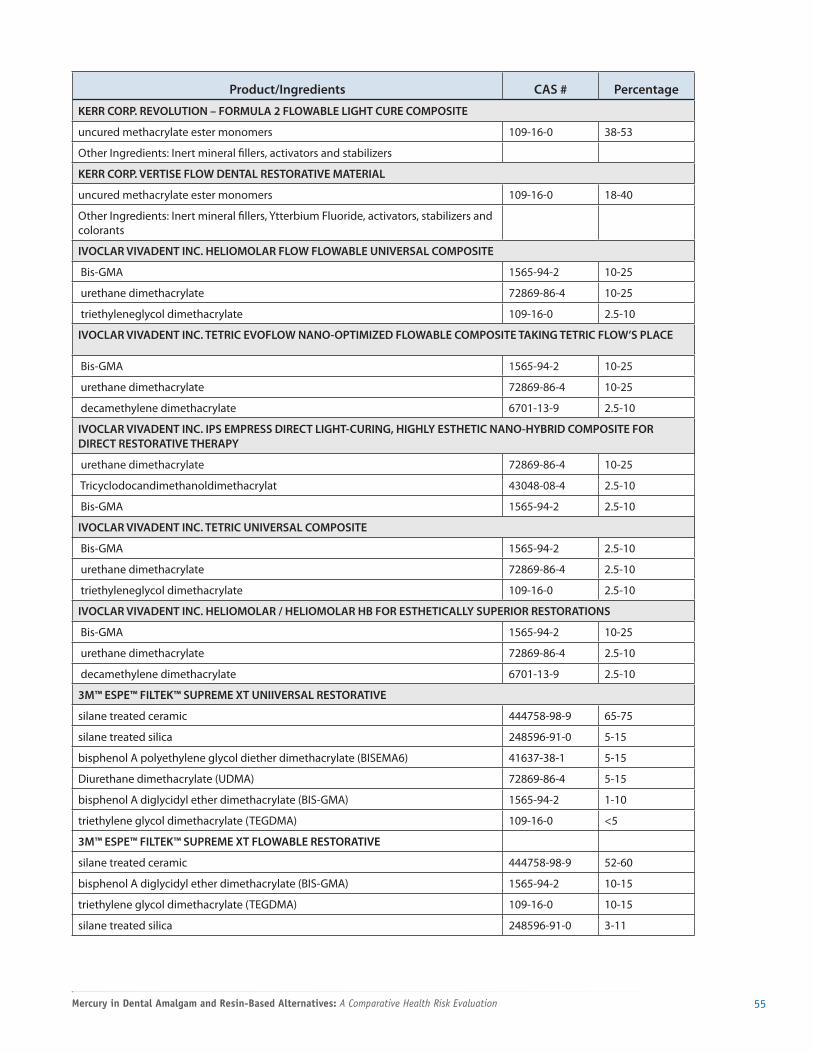
55Mercury in Dental Amalgam and Resin-Based Alternatives: A Comparative Health Risk Evaluation
Product/Ingredients CAS # Percentage
KERR CORP. REVOLUTION – FORMULA 2 FLOWABLE LIGHT CURE COMPOSITE
uncured methacrylate ester monomers 109-16-0 38-53
Other Ingredients: Inert mineral fillers, activators and stabilizers
KERR CORP. VERTISE FLOW DENTAL RESTORATIVE MATERIAL
uncured methacrylate ester monomers 109-16-0 18-40
Other Ingredients: Inert mineral fillers, Ytterbium Fluoride, activators, stabilizers and colorants
IVOCLAR VIVADENT INC. HELIOMOLAR FLOW FLOWABLE UNIVERSAL COMPOSITE
Bis-GMA 1565-94-2 10-25
urethane dimethacrylate 72869-86-4 10-25
triethyleneglycol dimethacrylate 109-16-0 2.5-10
IVOCLAR VIVADENT INC. TETRIC EVOFLOW NANO-OPTIMIZED FLOWABLE COMPOSITE TAKING TETRIC FLOW’S PLACE
Bis-GMA 1565-94-2 10-25
urethane dimethacrylate 72869-86-4 10-25
decamethylene dimethacrylate 6701-13-9 2.5-10
IVOCLAR VIVADENT INC. IPS EMPRESS DIRECT LIGHT-CURING, HIGHLY ESTHETIC NANO-HYBRID COMPOSITE FOR DIRECT RESTORATIVE THERAPY
urethane dimethacrylate 72869-86-4 10-25
Tricyclodocandimethanoldimethacrylat 43048-08-4 2.5-10
Bis-GMA 1565-94-2 2.5-10
IVOCLAR VIVADENT INC. TETRIC UNIVERSAL COMPOSITE
Bis-GMA 1565-94-2 2.5-10
urethane dimethacrylate 72869-86-4 2.5-10
triethyleneglycol dimethacrylate 109-16-0 2.5-10
IVOCLAR VIVADENT INC. HELIOMOLAR / HELIOMOLAR HB FOR ESTHETICALLY SUPERIOR RESTORATIONS
Bis-GMA 1565-94-2 10-25
urethane dimethacrylate 72869-86-4 2.5-10
decamethylene dimethacrylate 6701-13-9 2.5-10
3M™ ESPE™ FILTEK™ SUPREME XT UNIIVERSAL RESTORATIVE
silane treated ceramic 444758-98-9 65-75
silane treated silica 248596-91-0 5-15
bisphenol A polyethylene glycol diether dimethacrylate (BISEMA6) 41637-38-1 5-15
Diurethane dimethacrylate (UDMA) 72869-86-4 5-15
bisphenol A diglycidyl ether dimethacrylate (BIS-GMA) 1565-94-2 1-10
triethylene glycol dimethacrylate (TEGDMA) 109-16-0 <5
3M™ ESPE™ FILTEK™ SUPREME XT FLOWABLE RESTORATIVE
silane treated ceramic 444758-98-9 52-60
bisphenol A diglycidyl ether dimethacrylate (BIS-GMA) 1565-94-2 10-15
triethylene glycol dimethacrylate (TEGDMA) 109-16-0 10-15
silane treated silica 248596-91-0 3-11

Mercury in Dental Amalgam and Resin-Based Alternatives: A Comparative Health Risk Evaluation56
Product/Ingredients CAS # Percentage
3M™ ESPE™ FILTEK™ SUPREME XT UNIIVERSAL RESTORATIVE (continued)
silane treated zirconium oxide None 3-11
bisphenol A polyethylene glycol diether dimethacrylate (BISEMA6) 41637-38-1 1-5
functionalized dimethacrylate polymer None 1-5
3M™ ESPE™ FILTEK™ ULTIMATE UNIVERSAL RESTORATIVE and 3M™ ESPE™ FILTEK™ SUPREME XTE UNIVERSAL
silane treated ceramic 444758-98-9 60-80
silane treated silica 248596-91-0 1-10
Diurethane dimethacrylate (UDMA) 72869-86-4 1-10
bisphenol a polyethylene glycol diether dimethacrylate 41637-38-1 1-10
bisphenol A diglycidyl ether dimethacrylate (BIS-GMA) 1565-94-2 1-10
silane treated zirconia Unknown 1-10
polyethylene glycol dimethacrylate 25852-47-5 <5
triethylene glycol dimethacrylate (TEGDMA) 109-16-0 <5
2,6-di-tert-butyl-p-cresol (BHT) 128-37-0 <0.5
3M™ ESPE™ FILTEK™ ULTIMATE FLOWABLE RESTORATIVE and 3M™ ESPE™ FILTEK™ SUPREME XTE FLOWABLE RESTORATIVE
silane treated ceramic 444758-98-9 50-60
substituted dimethacrylate 27689-12-9 15-25
bisphenol A diglycidyl ether dimethacrylate (BIS-GMA) 1565-94-2 5-10
silane treated silica 248596-91-0 5-10
triethylene glycol dimethacrylate (TEGDMA) 109-16-0 5-10
ytterbium fluoride (YbF3) 13760-80-0 <5
functionalized dimethacrylate polymer None <5
titanium dioxide 13463-67-7 <0.5
3M FILTEK Z250 UNIVERSAL RESTORATIVE PASTE
silane treated ceramic 444758-98-9 75-85
bisphenol A polyethylene glycol diether dimethacrylate (BISEMA6) 41637-38-1 1-10
Diurethane dimethacrylate (UDMA) 72869-86-4 1-10
bisphenol A diglycidyl ether dimethacrylate (BIS-GMA) 1565-94-2 1-10
triethylene glycol dimethacrylate (TEGDMA) 109-16-0 <5
3M™ ESPE™ FILTEK™ FLOW RESTORATIVE PASTE
silane treated ceramic 444758-98-9 55-65
bisphenol A diglycidyl ether dimethacrylate (BIS-GMA) 1565-94-2 10-20
triethylene glycol dimethacrylate (TEGDMA) 109-16-0 10-20
silane treated silica 248596-91-0 5-10
functionalized dimethacrylate polymer None <5
3M™ ESPE™ FILTEK P90 LOW SHRINK POSTERIOR RESTORATIVE
silane treated quartz 100402-89-9 60-70
3,4-epoxycyclohexylcyclopolymethylsiloxane Unknown 5-15
bis-3,4-epoxycyclohexylethyl-phenyl-methylsilane 154265-59-5 5-15
ytterbium fluoride (YbF3) 13709-49-4 5-15

57Mercury in Dental Amalgam and Resin-Based Alternatives: A Comparative Health Risk Evaluation
Product/Ingredients CAS # Percentage
3M™ ESPE™ FILTEK P90 LOW SHRINK POSTERIOR RESTORATIVE (continued)
mixture of other by-products Mixture <5
mixture of epoxy-mono-silanole by-products Mixture <5
mixture of epoxyfunctional di- and oligo-siloxane byproducts Mixture <5
mixture of alpha-substituted by-products Mixture <5
borate(1-), tetrakis(pentafluorophenyl)-[4- 178233-72-2 <1
(methylethyl)phenyl](4-methylphenyl)iodonium
3M™ ESPE™ EXI-697 RESTORATIVE PASTE and 3M™ ESPE™ VALUX PLUS RESTORATIVE
silane treated ceramic 444758-98-9 80-90
triethylene glycol dimethacrylate 109-16-0 5-10
bisphenol A diglycidyl ether dimethacrylate (BIS-GMA) 1565-94-2 5-10
2-benzotriazolyl-4-methylphenol 2440-22-4 <1
3M™ ESPE™ FILTEK™ P60 POSTERIOR RESTORATIVE PASTE
silane treated ceramic 444758-98-9 75-85
bisphenol A polyethylene glycol diether dimethacrylate (BISEMA6) 41637-38-1 1-10
Diurethane dimethacrylate (UDMA) 72869-86-4 1-10
bisphenol A diglycidyl ether dimethacrylate (BIS-GMA) 1565-94-2 1-10
triethylene glycol dimethacrylate (TEGDMA) 109-16-0 <5
3M™ ESPE™ FILTEK™ SUPREME UNIVERSAL RESTORATIVE and 3M™ ESPE™ FILTEK™ SUPREME PLUS UNIVERSAL RESTORATIVE and 3M™ ESPE™ FILTEK™ Z350 UNIVERSAL RESTORATIVE
silane treated ceramic 444758-98-9 65-75
silane treated silica 248596-91-0 5-15
bisphenol A polyethylene glycol diether dimethacrylate (BISEMA6) 41637-38-1 5-15
Diurethane dimethacrylate (UDMA) 72869-86-4 5-15
bisphenol A diglycidyl ether dimethacrylate (BIS-GMA) 1565-94-2 1-10
triethylene glycol dimethacrylate (TEGDMA) 109-16-0 <5
3M™ ESPE™ Z100™ RESTORATIVE PASTE
silane treated ceramic 444758-98-9 80-90
bisphenol A diglycidyl ether dimethacrylate (BIS-GMA) 1565-94-2 1–10
triethylene glycol dimethacrylate (TEGDMA) 109-16-0 1–10
2-benzotriazolyl-4-methylphenol 2440-22-4 <1
Table A-2. Chemical composition of dental resin composite preparation and application materials commercially available in the U.S., as reported in MSDSs
Product/Ingredients CAS # Percentage
DENTSPLY CALIBRA SILANE COUPLING AGENT
ethanol 64-17-5 92.6
acetone 67-64-1 7.4
DENTSPLY PROBOND PRIMER
acetone 67-64-1 <80

Mercury in Dental Amalgam and Resin-Based Alternatives: A Comparative Health Risk Evaluation58
Product/Ingredients CAS # Percentage
DENTSPLY PROBOND PRIMER (continued)
ethanol 64-17-5 <25
dipentaerythritol pentaacrylate phosphate <10
DENTSPLY PRIME & BOND NT
acetone 67-64-1 >60
methacrylate, typically 30
methyl methacrylate 80-62-6
DENTSPLY PRIME & BOND NT PREMIX
acetone 67-64-1 >60
urethane dimethacrylate monomer 105883-40-7 <15
trimethylolpropane trimethacrylate 3290-92-4 <10
dipentaerythritol pentaacrylate phosphate <10
2, 4, 4’ -trichloro-2’ -hydroxydiphenyl ether 3380-34-5 <5
DENTSPLY PRIME & BOND NT SELF CURE ACTIVATOR
acetone 67-64-1 59
ethanol 64-17-5 39.8
non hazardous ingredients [manufacturer] NotSpec
DENTSPLY SELF CURE ACTIVATOR
acetone 67-64-1 50-100
urethane dimethacrylate monomer 105883-40-7 10-25
2- hydroxyethyl methacrylate 868-77-9 2.5-10
diphenyl(2, 4, 6- trimethylbenzoyl)phosphine 75980-60-8 <2.5
DENTSPLY PROBOND ADHESIVE
urethane dimethacrylate monomer 105883-40-7 <55
triethylene glycol dimethacrylate 109-16-0 <30
dipentaerythritol pentaacrylate phosphate <7
glutaraldehyde 111-30-8 <5
DENTSPLY PRISMA UNIVERSAL BOND
urethane dimethacrylate resin 1-60
triethylene glycol dimethacrylate 109-16-0 1-30
bisphenol A dimethacrylate 3253-39-2 1-12
dipentaerythritol pentaacrylate phosphate <7
DENTSPLY TRIAD VLC BONDING AGENT
methyl methacrylate 80-62-6 60
tetrahydrofurfuryl methacrylate 2455-24-5 30
hexanediol dimethacrylate 6606-59-3 10
BISCO, Inc. ALL-BOND 3
ethanol(Part A) 64-17-5 >50
magnesium Salt of N-tolylglycine glycidylmethacrylate (NTG-GMA Salt) (Part A) 211810-95-6 >1.0
bisphenol A diglycidylmethacrylate (Bis-GMA) (Part B) 1565-94-2 >20
HydroxyEthylMethAcrylate (HEMA) (Part B) 868-77-9 >20

59Mercury in Dental Amalgam and Resin-Based Alternatives: A Comparative Health Risk Evaluation
Product/Ingredients CAS # Percentage
BISCO, Inc. ALL-BOND 3 (continued)
BPDM(Part B) Proprietary >1.0
BiISCO, Inc. ALL-BOND 3 Resin
bisphenol A diglycidylmethacrylate (Bis-GMA) 1565-94-2 >10
urethane Dimethacrylate. 72869-86-4 >10
triethyleneglycol Dimethacrylate. 109-16-0 >10
glass Filler 65997-18-4 >40
KERR CORP. OPTIGUARD SURFACE SEALANT
uncured Methacrylate Ester Monomers 109-16-0 90-95
Other Ingredients: Photoinitiators and stabilizers 5-10
KERR CORP. OPTIBOND XTR PRIMER DENTAL ADHESIVE
acetone 67-64-1 25-35
ethyl alcohol 64-17-5 4-15
hydroxyethylmethacrylate (HEMA) 868-77-9 30-50
KERR CORP. OPTIBOND SOLO PLUS SINGLE COMPONENT DENTAL ADHESIVE
ethyl alcohol 64-17-5 20-25
alkyl dimethacrylate resins 55-60
barium aluminoborosilicate glass 5-10
fumed silica (silicon dioxide) 5-10
sodium hexafluorosilicate 0.5-1
IVOCLAR VIVADENT Inc. AdheSE PRIMER
phosphonic acid acrylate 223681-84-3 <40
IVOCLAR VIVADENT INC. TOTAL ETCH ETCHING GEL
phosphoric acid 7664-38-2 37
silane treated ceramic 444758-98-9 75-85
bisphenol A polyethylene glycol diether dimethacrylate (BISEMA6) 41637-38-1 1-10
diurethane dimethacrylate (UDMA) 72869-86-4 1-10
bisphenol A diglycidyl ether dimethacrylate (BIS-GMA) 1565-94-2 1-10
triethylene glycol dimethacrylate (TEGDMA) 109-16-0 <5
3M™ ESPE™ SCOTCHBOND™ ETCHANT DELIVERY SYSTEM
water 7732-18-5 55-65
phosphoric acid 7664-38-2 30-40
synthetic amorphous silica 112945-52-5 10-May
IVOCLAR VIVADENT INC. SYNTAC PRIMER — BONDING AGENT
acetone 67-64-1 <42
maleic acid 110-16-7 4
mixture of water, acetone, maleic acid and dimethacrylate
IVOCLAR VIVADENT Inc. AdheSE DC ACTIVATOR
ethanol 64-17-5 50-100
IVOCLAR VIVADENT Inc. AdheSE One F SELF-ETCH DENTAL ADHESIVE
bis-methacrylamidedihydrogenphosphate 911525-18-3 5-25

Mercury in Dental Amalgam and Resin-Based Alternatives: A Comparative Health Risk Evaluation60
Product/Ingredients CAS # Percentage
IVOCLAR VIVADENT Inc. AdheSE One F SELF-ETCH DENTAL ADHESIVE (continued)
isopropanol 67-63-0 5-15
acrylamidoaminoacid 72064-86-9 5-20
acrylamidosulfonicacid 15214-89-8 1-10
potassiumfluoride 7789-23-3 <1
Mixture of derivatives of bis-acrylamide, water, alcohol, bis-
methacrylamide dihydrogen phosphate, acrylamido amino acid,
hydroxy alkyl methacrylamide, acrylamido suflonic acid, highly
dispersed silicon dioxide, initiators, catalysts and potasium fluoride.
IVOCLOR VIVADENT Inc. ExciTE F LIGHT-CURING TOTAL-ETCH ADHESIVE
bis-GMA 1565-94-2 25-50
ethanol 64-17-5 10-25
2-hydroxyethyl methacrylate 868-77-9 10-25
phosphonic acid acrylate 223681-84-3 10-25
urethane dimethacrylate 72869-86-4 2.5-10
potassium fluoride 7789-23-3 ≤2.5
IVOCLOR VIVADENT Inc. ExciTE F DUAL-CURING TOTAL-ETCH ADHESIVE
bis-GMA 1565-94-2 25-50
ethanol 64-17-5 10-25
2-hydroxyethyl methacrylate 868-77-9 10-25
phosphonic acid acrylate 223681-84-3 10-25
potassium fluoride 7789-23-3 ≤2.5
IVOCLAR VIVADENT Inc AdheSE BONDING
HEMA 868-77-9 <25
dimethacrylates 1565-94-2 and 1830-78-0
<75
IVOCLAR VIVADENT INC. SYNTAC ADHESIVE — BONDING AGENT
Polyethylene glycol dimethacrylate 25852-47-5 20-40
Glutaraldehyde 111-30-8 5
Mixture of water, glutaraldehyde, maleic acid and polyethyleneglycoldimethacrylate
3M™ ESPE™ ADPER™ EASY BOND VIAL
bisphenol A diglycidyl ether dimethacrylate (BIS-GMA) 1565-94-2 15-25
2-hydroxyethyl methacrylate 868-77-9 15-25
ethanol 64-17-5 10-15
water 7732-18-5 10-15
phosphoric acid-6-methacryloxy-hexylesters Mixture 5-15
silane treated silica 122334-95-6 8-12
1,6-hexanediol dimethacrylate 6606-59-3 5-10
copolymer of acrylic and itaconic acid 25948-33-8 1-5
(dimethylamino)ethyl methacrylate 2867-47-2 1-5
camphorquinone 10373-78-1 1-3
2,4,6-trimethylbenzoyldiphenylphosphine oxide 75980-60-8 1-3

61Mercury in Dental Amalgam and Resin-Based Alternatives: A Comparative Health Risk Evaluation
Product/Ingredients CAS # Percentage
3M(TM) ESPE(TM) SINGLE BOND ADHESIVE
ethanol 64-17-5 30-40
bisphenol A diglycidyl ether dimethacrylate (BIS-GMA) 1565-94-2 15-25
2-hydroxyethyl methacrylate 868-77-9 10-20
glycerol 1,3 dimethacrylate 1830-78-0 5-15
copolymer of acrylic & itaconic acids 25948-33-8 5-15
diurethane dimethacrylate 72869-86-4 2-8
water 7732-18-5 2-8
Table A-3. Chemical composition of glass ionomers commercially available in the U.S., as reported in MSDSs
Product/Ingredients CAS # Percentage
3M ESPE VITREMER CORE BUILDUP/RESTORATIVE LIQUID
copolymer of acrylic and itaconic acids 25948-33-8 45-50 by Weight
water 7732-18-5 25-30 by Weight
2-hydroxyethyl methacrylate 868-77-9 15-20 by Weight
3M™ ESPE™ KETAC™ N100 LIGHT-CURE GLASS IONOMER RESTORATIVE PASTE A
silane treated glass None 40-55
silane treated zirconia Unknown 20-30
polyethylene glycol dimethacrylate (PEGDMA) 25852-47-5 5-15
silane treated silica 248596-91-0 5-15
2-hydroxyethyl methacrylate (HEMA) 868-77-9 1-15
glass powder 65997-17-3 <5
bisphenol A diglycidyl ether dimethacrylate (BIS-GMA) 1565-94-2 <5
triethylene glycol dimethacrylate (TEGDMA) 109-16-0 <1
3M™ ESPE™ KETAC™ N100 LIGHT-CURE GLASS IONOMER RESTORATIVE PASTE B
silane treated ceramic 444758-98-9 40-60 by wt
copolymer of acrylic and itaconic acids 25948-33-8 20-30 by wt
water 7732-18-5 10-20 by wt
2-hydroxyethyl methacrylate (HEMA) 868-77-9 1-10 by wt
3M™ ESPE™ VITREBOND™ GLASS IONOMER LIQUID
copolymer of acrylic and itaconic acids 25948-33-8 35-45 by wt
water 7732-18-5 30-40 by wt
2-hydroxyethyl methacrylate (HEMA) 868-77-9 20-30 by wt
3M™ ESPE™ VITREBOND™ GLASS IONOMER POWDER
glass powder 65997-17-3 >95
diphenyliodonium chloride 1483-72-3 <2
diphenyliodonium hexafluorophosphate 58109-40-3 <1

Mercury in Dental Amalgam and Resin-Based Alternatives: A Comparative Health Risk Evaluation62
Table A-4. Chemical composition of compomers commercially available in the U.S., as reported in MSDSs
Product/Ingredients CAS # Percentage
IVOCLAR VIVADENT INC. COMPOGLASS F COMPOMER
urethane dimethacrylate < 12
cycloaliphatic dicarboxylic acid dimethacrylate Unknown < 7
polyethylene glycol dimethacrylate Unknown < 5
paste of dimethacrylates, Ba-fluorosilicate glass, mixed oxides,
ytterbiumtrifluoride, initiators, stabilizers and pigments
IVOCLAR VIVADENT INC. COMPOGLASS FLOW COMPOMER
urethane dimethacrylate 72869-86-4 < 21
cycloaliphatic dicarboxylic acid dimethacrylate Unknown < 6
polyethylene glycol dimethacrylate Unknown < 7
DENTSPLY/CAULK DYRACT EXTRA UNIVERSAL COMPOMER RESTORATIVE
polymerizable dimethacrylate resin 105883-40-7 < 35 by wt
polymerizable trimethacrylate resin 3290-92-4 < 10 by wt
polymerizable dimethacrylate resin <35 by Weight 24448-20-2 < 35 by wt
polymerizable dimethacrylate resin <10 by Weight 109-16-0 < 10 by wt
strontium Fluoride <10 by Weight 7783-48-4 < 10 by wt
other Information: Other colorants are inorganic iron oxides
DENTSPLY/CAULK DYRACT FLOW FLOWABLE COMPOMER RESTORATIVE
strontium aluminum fluorosilicate glass 65997-18-4 < 65 %
polymerizable dimethacrylate resin Not Established < 25 %
ammonium salt of dipentaerythitol pentaacrylate phosphate Not Established < 20 %
3M ESPE F2000 COMPOMER RESTORATIVE PASTE
silane treated glass None 80 - 90 by wt
citric acid dimethacrylate oligomer None 1 - 10 by wt
glycerol 1,3-dimethacyrlate 1830-78-0 1 - 10 by wt
triethylene glycol dimethacrylate (TEGDMA) 109-16-0 < 5 by wt
bisphenol A diglycidyl ether dimethacrylate (BIS-GMA) 1565-94-2 < 5 by wt
silane tretaed silica 248596-91-0 < 5 by wt
2-benzotriazolyl-4-methylphenol 2440-22-4 < 1 by wt
3M™ ESPE™ F2000™ COMPOMER PRIMER SIDE A
2-hydroxyethyl methacrylate 868-77-9 60 - 70
copolymer of acrylic and itaconic acids 25948-33-8 15 - 25
Water 7732-18-5 5 - 10
ethyl alcohol 64-17-5 1 - 5

63Mercury in Dental Amalgam and Resin-Based Alternatives: A Comparative Health Risk Evaluation
Table A-5. Environmental fate and transport properties of constituents of resin-based alternative materials
Product/Ingredients CAS # MWVP
(mm Hg)
H (atm-m3/
mole) WS (mg/L) log Kow Koc (L/kg)
BCF (L/kg wet
weight)
Rapid Biodegra-
dation?
Fillers
silica, dimethylsiloxane treated
67762-90-7 625.06 3.83E-19 1.81E-24 1.74E+05 -3.22 1.60E-03 3.162 n
silica amorphous; silicon dioxide
7631-86-9 60.08 5.36E-08 8.75E-14 4.84E+04 0.53 2.88E+00 3.162 y
titanium dioxide 13463-67-7 79.87 2.45E+03 4.89E-02 1.63E+03 2.23 8.61E+01 13.73 n
Methacrylates
2, 2-bis[4-(2-methacryloxy)ethoxy)phenyl]propane
24448-20-2 452.55 2.17E-09 4.15E-07 3.11E-02 6.63 3.74E+04 1.11E+04 n
bisphenol A diglycidyl ether dimethacrylate (bis-GMA)
1565-94-2 512.61 1.50E-15 2.84E-14 3.56E-02 4.94 6.54E+02 471.9 n
TEGDMA; uncured Methacrylate Ester Monomers)
109-16-0 286.33 9.44E-04 9.73E-07 3.66E+02 1.88 4.49E+01 8.08 y
urethane dimethacrylate (UDMA)
72869-86-4 470.57 3.99E-09 2.27E-08 1.09E-01 4.69 1.51E+03 24.41 n
3-trimethoxysilylpropyl methacrylate
2530-85-0 248.35 1.27E-02 7.56E-07 5.49E+03 0.75 1.88E+01 3.162 n
ethoxylated bisphenol-A-dimethacrylate
56744-60-6 540.66 8.78E-12 2.50E-09 2.50E-03 6.08 1.22E+04 n
tricyclodocandi-methanoldim ethacrylat
43048-08-4 332.44 6.71E-05 1.38E-04 2.13E-01 5.35 5.66E+03 1577 n
decamethylene dimethacrylate
6701-13-9 310.44 2.47E-04 1.65E-03 6.12E-02 6.14 1.55E+04 219.9 y
bisphenol A polyethylene glycol diether dimethacrylate (BISEMA6)
41637-38-1 310.44 2.47E-04 1.65E-03 6.12E-02 6.14 1.55E+04 219.9 y
polyethylene glycol dimethacrylate
25852-47-5 374.43 1.13E-05 1.13E-08 4.95E+02 1.11 1.11E+01 2.513 n
substituted dimethacrylate
27689-12-9 480.61 3.48E-10 7.36E-07 2.99E-04 7.61 1.30E+05 6649 n
methyl methacrylate 80-62-6 100.12 3.67E+01 6.24E-04 7.75E+03 1.38 4.20E+01 3.78 y
trimethylolpropane trimethacrylate; polymerizable trimethacrylate resin
3290-92-4 338.4 1.37E-04 4.70E-05 1.30E+00 4.39 1.67E+03 366 n
2-hydroxyethyl methacrylate (HEMA)
868-77-9 130.14 7.91E-02 1.15E-07 1.18E+05 0.47 5.11E+00 3.162 y
bisphenol A dimethacrylate
3253-39-2 364.44 8.54E-07 4.91E-06 8.34E-02 5.6 7.79E+03 2304 n
tetrahydrofurfuryl methacrylate
2455-24-5 170.21 1.61E-01 2.01E-05 1.79E+03 1.8 5.82E+01 7.123 y
hexanediol dimethacrylate
6606-59-3 254.33 4.80E-03 2.63E-04 6.11E+00 4.17 1.26E+03 263.3 y
glycerol 1,3-dimethacrylate
1830-78-0 228.25 4.79E-04 1.40E-08 1.03E+04 1.16 1.02E+00 2.713 y
(dimethylamino)ethyl methacrylate
2867-47-2 157.21 7.21E-01 1.41E-06 1.06E+05 0.97 2.14E+01 3.162 n

Mercury in Dental Amalgam and Resin-Based Alternatives: A Comparative Health Risk Evaluation64
Product/Ingredients CAS # MWVP
(mm Hg)
H (atm-m3/
mole) WS (mg/L) log Kow Koc (L/kg)
BCF (L/kg wet
weight)
Rapid Biodegra-
dation?
Others
2-benzotriazolyl-4-methylphenol
2440-22-4 225.25 1.87E-07 2.17E-09 2.56E+01 4.31 3.54E+03 324.1 n
Inorganic iron oxides
2,6-di-tert-butyl-p-cresol (BHT)
128-37-0 220.36 1.77E-03 8.92E-05 5.75E+00 5.1 8.18E+03 645.6 n
ethanol 64-17-5 46.07 6.09E+01 4.66E-06 7.92E+05 -0.31 2.20E+00 3.162 y
acetone 67-64-1 58.08 2.49E+02 8.65E-05 2.20E+05 -0.24 9.73E+00 3.162 y
Camphorquinone 10373-78-1 166.22 4.85E-04 3.29E-08 3.23E+03 0.75 1.31E+00 4.676 n
2, 4, 4’ -trichloro-2’ -hydroxydiphenyl ether
3380-34-5 289.55 4.65E-06 3.83E-07 4.62E+00 4.76 8.42E+03 642.2 n
diphenyl(2, 4, 6- trimethylbenzoyl)phosphine
75980-60-8 348.38 2.92E-08 4.28E-09 3.13E+00 3.87 4.08E+02 166.7 n
glutaraldehyde 111-30-8 100.12 1.88E+00 1.48E-06 1.67E+05 -0.18 2.33E+00 3.162 y
phosphoric acid 7664-38-2 98 6.09E-11 1.46E-17 5.39E+05 -0.77 4.00E+00 3.162 n
maleic acid 110-16-7 116.07 9.43E-12 1.38E-17 1.04E+05 0.46 2.57E+00 3.162 y
isopropanol 67-63-0 60.1 4.96E+01 9.75E-06 4.02E+05 0.05 3.48E+00 3.162 y
acrylamidosulfonicacid 15214-89-8 207.25 6.75E-09 1.84E-15 1.00E+06 -2.19 4.88E-01 3.162 n
potassium fluoride 7789-23-3 58.1 5.58E-16 6.79E-23 6.29E+05 -0.77 2.14E-01 3.162 y
In analyzing the environmental fate and transport data, the following two key references were used: Fate and Transport of Organic Chemicals in the Environment: A Practical Guide (Ney 1998), and Handbook of Chemical Property Estimation Methods (Lyman et al. 1982).

65Mercury in Dental Amalgam and Resin-Based Alternatives: A Comparative Health Risk Evaluation
Table A-6. Acute toxicity information for constituents of resin-based alternative materials (HSDB-NLM)
Product/Ingredients CAS #Lethal Dose (LD or Lethal
Concentration (LC) Toxic Effect/Target Organ
Fillers
glass fibres loose -special purpose ; soluble amorphous glass wool, glass powder
65997-17-3 mouse: > 20mg/kg (intratracheal)
lungs, thorax, or other respiration
silica amorphous; silicon dioxide 7631-86-9 rat: LC:> 200000 mg/m3 (inh)
rat: LCLo:> 2190 mg/m3 (inh) lungs, thorax, or respiration: dyspnea
rat: LDLo:> 5000 mg/kg (oral)
Methacrylates
bisphenol A diglycidyl ether dimethacrylate (BIS-GMA) 1565-94-2 skin sensitizerTEGDMA; (uncured Methacrylate Ester Monomers)
109-16-0 mouse: LD50: 10750 mg/kg (oral); rat: LD50: 10837 mg/kg (oral)
behavioral: somnolence ; lungs, thorax, or respiration: other changes (mouse-oral); skin sensitizer
urethane modified Bis-GMA dimethacrylate
urethane dimethacrylate (UDMA)
72869-86-4 skin sensitizer
3-trimethoxysilylpropyl methacrylate
2530-85-0 rabbit: LD-skin: >20mL/kg; rat: LD50: 22.6 mL/kg (oral); rat: LDLo: 226 mg/kg (intravenous)
skin sensitizer
methyl methacrylate (MMA) 80-62-6 see Table A-7 asthma; dermatotoxin (skin burns); skin sensitizer
trimethylolpropane trimethacrylate; polymerizable trimethacrylate resin
3290-92-4 mouse: LD50: 2889 mg/kg (intraperitoneal); rabbit: LD50: 16 mL/kg (skin); rat: LD50: 3100 mg/kg (intraperitoneal); rat: LD50: 5.66 mL/kg (oral)
behavioral: tremor, ataxia; lungs, thorax, or respiration: dyspnea (rat-intraper.); skin sensitizer
2-hydroxyethyl methacrylate (HEMA)
868-77-9 dog: LDLo: 99200 mL/kg (intravenous); guinea pig: LD50: 4680 mg/kg (oral); mouse:LD50: 497 mg/kg (intraperitoneal); mouse: LD50: 3275 mg/kg (oral); rat: LD50: 1250 mg/kg (intraperitoneal); rat: LD50: 5050 mg/kg (oral)
behavioral: coma (guinea pig-oral; mouse-oral; rat-oral); contact dermatitis; skin sensitizer
dimethacrylates (bisphenol A-glycidyl methacrylate and glycerol 1,3-dimethacrylate)
1565-94-2 and 1830-78-0
skin sensitizer
(dimethylamino)ethyl methacrylate
2867-47-2 dog: LDLo: 20800 mL/kg (intravenous) mouse: LC50: 1800 mg/m3 (inh); mouse: LD50: 25 mg/kg (intraperitoneal); rat: LC50: 620 mg/m3 (inh); rat: LD50: 1751 mg/kg (oral)

Mercury in Dental Amalgam and Resin-Based Alternatives: A Comparative Health Risk Evaluation66
Product/Ingredients CAS #Lethal Dose (LD or Lethal
Concentration (LC) Toxic Effect/Target Organ
Others
2,6-di-tert-butyl-p-cresol (BHT) 128-37-0 rat: LD-Lo:940 mg/kg (oral); guinea pig: LD50: 10,700 mg/kg (oral); mouse: LD50: 138 mg/kg (intraperitoneal); mouse: LD50: 180 mg/kg (intravenous); mouse: LD50: 650 mg/kg (oral); rabbit: LDLo: 2100 mg/kg (oral); rat: LD50: 890 mg/kg (oral); women: TDLo: 80 mg/kg (oral)
gastrointestinal: hypermotility, diarrhea; behavioral: tremor; lungs, thorax, or respiration: respiratory depression (rat and guinea pig), pulmonary edema (mouse-intraper./oral); blood: hemorrhage (mouse-intraper.); behavioral: sleep (mouse-intraven.); tremor (mouse-oral); hypermotility, diarrhea, respiratory depression, tremor (rabbit-oral); skin sensitizer; hepatotoxin (secondary); gastrointestinal: gastritis, nausea or vomiting (women); behavior: coma (women)
ytterbium fluoride (YbF3) 13760-80-0 mammal (unspecified): LDLo: 10 mg/kg (intraperitoneal)
2-benzotriazolyl-4-methylphenol
2440-22-4 mouse: LD50: 6500 mg/kg (oral)
2, 4, 4’ -trichloro-2’ -hydroxydiphenyl ether
3380-34-5 mouse: LD50: 84 mg/kg (intraperitoneal); mouse: LD50: 4530 mg/kg (oral); mouse: LD50: 3800 mg/kg (subcutaneous); rabbit: LD50: 9300 mg/kg (skin); rat: LD50: 89 mg/kg (intraperitoneal); rat: LD50: 29 mg/kg (intravenous); rat: LD50: 3700 mg/kg (oral); rat: LD50: 3900 mg/kg (subcutaneous)
Dermatotoxin: contact dermatitis, Photoallergic; skin sensitizer
phosphoric acid 7664-38-2 man: LDLo: 220mg/kg (unreported); rabbit: LD50: 2740 mg/kg (skin); rat: LC50: >850 mg/m3 (inhalation); rat: LD50: 1530 mg/kg (oral)
behavioral: somnolence (general depressed activity); behavioral: excitement (rabbit-skin); behavioral: somnolence (general depressed activity); kidney, ureter, and bladder: hematuria; skin and appendages (skin): hair: other (rat-oral); toxic pneumonitis; dermatotoxin: skin burns
maleic acid 110-16-7 mouse: LD50: 2400 mg/kg (oral) ; rabbit: LD50: 1560 mg/kg (skin); rat: LC50: >720mg/m3 (inhalation); rat: LD50 708 mg/kg (oral)
behavioral: tremor (rabbit-skin);behavioral: convulsions or effect on seizure threshold; behavioral: muscle weakness; gastrointestinal: ulceration or bleeding from stomach (rat-oral)
acrylamidosulfonicacid 15214-89-8 rat: LD50: 5400mg/kg (oral) behavioral: somnolence (general depressed activity), convulsions or effect on seizure threshold, ataxia (rat-oral)
potassiumfluoride 7789-23-3 guinea pig: LDLo: 250mg/kg (oral); mouse: LD50: 40.03 mg/kg (intraperitoneal); rat: LD50: 64 mg/kg (intraperitoneal); rat: LD50: 245 mg/kg (oral)
A skin, eye, and mucous membrane irritant

67Mercury in Dental Amalgam and Resin-Based Alternatives: A Comparative Health Risk Evaluation
Product/Ingredients CAS #Lethal Dose (LD or Lethal
Concentration (LC) Toxic Effect/Target Organ
Others (continued)
copolymer of acrylic and itaconic acids
25948-33-8 mouse:LD50: 300 mg/kg (intraperitoneal)
strontium fluoride 7783-48-4 guinea pig: LDLo: > 5000mg/kg (oral); guinea pig: LDLo: > 5000mg/kg (subcutaneous); mouse: LD50: 4400 mg/kg (intraperitoneal); rat: LD50: 10600mg/kg (oral); rat: LDLo: 625 mg/kg (intravenous)
behavioral: somnolence (general depressed activity), ataxia; lungs, thorax, or respiration: respiratory depression (mouse-intraper.; rat-oral)
copolymer of acrylic and itaconic acids
25948-33-8 mouse:LD50: 300 mg/kg (intraperitoneal)
titanium dioxide 13463-67-7 rat: LD: 0.1 mg/kg (intratracheal)
lungs, thorax, or respiration: structural or functional change in trachea or bronchi; blood: “changes in serum composition (e.g., tp, bilirubin, cholesterol)”
diphenyliodonium chloride 1483-72-3 rat: LD50: 60 mg/kg (oral) gastrointestinal: hypermotility, diarrhea

Mercury in Dental Amalgam and Resin-Based Alternatives: A Comparative Health Risk Evaluation68
Table A-7. Acute toxicity information for methyl methacrylate (MMA) (HSDB-NLM)
OrganismTest Type Route
Normalized Dose Effect
cat LDLo subcutaneous 7 mL/kg peripheral nerve and sensation: spastic paralysis with or without sensory changekidney, ureter, and bladder: changes primarily in glomeruli
dog LCLo inhalation 41200 mg/m3 behavioral: somnolence (general depressed activity)behavioral: ataxiagastrointestinal: changes in structure or function of salivary glands
dog LD50 oral 4725 mg/kg behavioral: somnolence (general depressed activity)behavioral: ataxiagastrointestinal: changes in structure or function of salivary glands
dog LD50 subcutaneous 4252 mg/kg behavioral: somnolence (general depressed activity)dog LDLo intravenous 0.12 mL/kg guinea pig LCLo inhalation 19000 mg/m3 behavioral: muscle weakness
behavioral: comalungs, thorax, or respiration: respiratory depression
guinea pig LD50 intraperitoneal 1890 mg/kg behavioral: somnolence (general depressed activity)guinea pig LD50 oral 5954 mg/kg behavioral: somnolence (general depressed activity)
behavioral: ataxiagastrointestinal: changes in structure or function of salivary glands
guinea pig LD50 subcutaneous 5954 mg/kg behavioral: somnolence (general depressed activity)human TCLo inhalation 60 mg/m3 behavioral: sleep
behavioral: excitementvascular: bp lowering not characterized in autonomic section
human TCLo inhalation 125 ppm behavioral: sleepbehavioral: excitementbehavioral: anorexia (human
mammal (species unspecified)
LC50 inhalation 20000 mg/m3
mouse LC50 inhalation 18500 mg/m3 mouse LD50 intraperitoneal 945 mg/kg behavioral: somnolence (general depressed activity)mouse LD50 oral 3625 mg/kg mouse LD50 subcutaneous 5954 mg/kg behavioral: somnolence (general depressed activity)rabbit LCLo inhalation 17500 mg/m3 behavioral: muscle weakness
lungs, thorax, or respiration: respiratory depressionbehavioral: coma
rabbit LD50 oral 8700 mg/kg rabbit LD50 skin 5000 mg/kg skin and appendages (skin): “dermatitis, other: after systemic
exposure”rabbit LDLo subcutaneous 14 mL/kg behavioral: somnolence (general depressed activity)
kidney, ureter, and bladder: hematurialungs, thorax, or respiration: other changes
rat LC50 inhalation 78000 mg/m3 rat LD50 intraperitoneal 1328 mg/kg
rat LD50 oral 7872 mg/kg
behavioral: muscle weaknessbehavioral: comalungs, thorax, or respiration: respiratory depression
rat LD50 subcutaneous 7088 mg/kg behavioral: somnolence (general depressed activity)

Health Care Without Harm (www.noharm.org)Healthier Hospitals Initiative (www.healthierhospitals.org)12355 Sunrise Valley Dr., Suite 680Reston, VA 20191
Environmental and Occupational Health SciencesUniversity of Illinois at Chicago School of Public Health835 S. Wolcott, Suite E-144, MC 684Chicago, IL 60612