mergedfile - wordpress.com · (a university under section 3 of ugc act 1956) tirumalaisamudram,...
TRANSCRIPT

DEVELOPMENT OF ELECTROCHEMICAL DNA BIOSENSOR TO
DETECT SHORT DNA SEQUENCES
Dissertation submitted to the SASTRA University
in partial fulfillment of the requirements
for the award of the degree of
Bachelor of Technology in Bioengineering
By
G.S.SANTHOSHI SADHANAA (117011018)
V.VIJAYALAKSHMI (117011023)
SCHOOL OF CHEMICAL AND BIOTECHNOLOGY
SASTRA UNIVERSITY
(SHANMUGHA ARTS SCIENCE TECHNOLOGY & RESEARCH ACADEMY)
THANJAVUR – 613401, TAMILNADU, INDIA
April 2017

SASTRA UNIVERSITY
(SHANMUGHA ARTS SCIENCE TECHNOLOGY & RESEARCH ACADEMY)
(A University under section 3 of UGC Act 1956)
Tirumalaisamudram, Thanjavur-613 401, Tamil Nadu, India
BONAFIDE CERTIFICATE
This is to certify that the thesis, entitled “Development of electrochemical DNA biosensor to
detect short DNA sequences”, submitted to SASTRA University, in partial fulfillment of the
requirements for the award of the Degree of Bachelor of Technology in Bioengineering, is a
bonafide record of the work done by Ms. G.S. Santhoshi Sadhanaa (117011018) &
Ms. V. Vijayalakshmi (117011023) during the Final Semester of the academic year 2016-2017,
in the School of Chemical and Biotechnology, under my supervision and guidance. The
dissertation has not formed the basis for the award of any Degree / Diploma /Associateship /
Fellowship or other similar title to any candidate of any University.
Project Supervisor Associate Dean
Internal Examiner External Examiner

ACKNOWLEDGEMENTS
We take immense pleasure in thanking Prof. R. Sethuraman, our beloved Vice-Chancellor for
having permitted us to carry out this project work. We express our profound gratitude to Dr. S.
Swaminathan, Dean, (Sponsored Research) SASTRA for his overwhelming support throughout
our project.
We take it the rarest opportunity and proud privilege to express our profound sense of
immeasurable and debt of gratitude to Dr. R. John Bosco Balaguru, Associate Dean (Research),
SEEE, SASTRA University for his guidance, duly evaluating my progress and always
encouraging me by his invaluable suggestions and encouraging me during this project work. I also
thank all other members in the institute for providing me all the information, suggestions and
improvements at every stage of my project.
We owe deepest thanks to Dr. G. Arun Kumar, Professor SCBT and Dr. N. Noel, Professor,
SCBT, SASTRA University for their advice and support. We also extend our thanks for the lively
association and affection showered on us by Ph.D. scholars Ms. G. Manju Bhargavi and Ms.
Bhat Lakshmishri Ramachandra.
We wish to express my deep sense of gratitude to all the faculty members, my friends and others
who involved directly or indirectly for their encouragement during the project.
We sincerely thank Nanosensors laboratory, ASK-I, SASTRA University for their support in our
project work.
G.S. Santhoshi Sadhanaa (117011018)
V.Vijayalakshmi (117011023)

LIST OF CONTENTS LIST OF TABLES i
LIST OF FIGURES ii
LIST OF SYMBOLS iv
LIST OF ABBREVIATIONS v
ABSTRACT vi
CHAPTER 1: INTRODUCTION 1
1.1 Biosensors 1
1.2 Components of Biosensor 1
1.3 Classification of Biosensor 2
1.4 Methods Of Immobilization 7
1.4.1 Physical Immobilization 8
1.4.2 Chemical Immobilization 8
1.5 Applications of Biosensor
1.6 DNA Biosensor 8
1.6.1 Nucleic acid hybridization 9
1.7 Metal oxide and their sensing application 10
1.7.1 Significance of Zinc Oxide 11
1.7.1.1 Properties of Zinc Oxide 11
1.8 Scope of DNA Biosensor 11
1.8.1 HPV and its impacts 11
1.8.2 Importance of DNA Sensor for detection of HPV 12
CHAPTER 2: REVIEW OF LITERATURE 13
2.1 Review of HPV 13
2.2 Review of Electrochemical Sensing 13
2.3 Review of Zinc Oxide 14
2.4 Review of Microwave assisted synthesis 15
CHAPTER 3: AIMS AND OBJECTIVES 17
CHAPTER 4: MATERIALS AND METHODS 18
4.1 Materials and Reagents 18
4.1.1 Electrode System 18
4.2 Sample Preparation 18
4.3 Synthesis of Zinc Oxide Nanoparticles 19
4.4 Pre-treatment of working electrode 19
4.5 Fabrication of Electrode 20
4.6 Characterization Techniques 20
4.6.1 Scanning Electron Microscopy (SEM) 20
4.6.1.1 Experimental Setup 21
4.6.1.2 Advantages of SEM 21

4.6.2 X-Ray Diffraction (XRD) 21
4.6.2.1 Advantages 21
4.7 Electrochemical Sensing 22
4.7.1 Cyclic Voltammetry 22
4.7.2 Amperometry 22
4.7.3 Impedometry 23
CHAPTER 5: RESULTS AND DISCUSSIONS 24
5.1 Characterization Results
5.1.1 SEM Analysis for Zinc Oxide Nanoparticles 24
5.1.2 XRD data for Zinc Oxide Nanoparticles 25
5.2 Electrochemical Studies 26
5.2.1 Cyclic Voltammetry Studies
5.2.1.1 Effect of different electrode system 26
5.2.1.2 Effect of scan rates 28
5.2.1.3 Effect of Varying Concentrations 29
5.2.2 Amperometric Studies 31
5.2.3 Impedometric Studies 31
CHAPTER 6: CONCLUSION 32
CHAPTER 7: REFERENCES 33

ABSTRACT
In order to identify specific DNA sequences, an electrochemical DNA biosensor was developed
by modifying the electrode surface with zinc oxide nanoparticles. Here, the sensing was based on
the principle of nucleic acid hybridization, which involves base pairing between two
complementary sequences of nucleic acids. Zinc oxide nanoparticles were synthesized by
microwave method with zinc chloride and sodium hydroxide as precursors. These nanoparticles
were characterized using SEM and XRD. Fabrication of electrode was done by coating of zinc
oxide nanoparticles on the surface of working electrode (gold electrode) followed by
immobilization of the probe sequences specific to the target sequences. Zinc oxide nanoparticle
has good biocompatibility and high IEP (~ 9.5) to obtain strong physical adsorption.
Electrochemical detection of DNA hybridization will be done based on three parameters such as
potential, current and impedance based on cyclic voltammetric, amperometric and impedance
studies respectively. Electrochemical sensing of DNA sequence was done in the absence and
presence of zinc oxide nano-interface. 200 nucleotides and 20 nucleotides sequences were sensed.
Cyclic voltammograms of a probe of 200 nucleotides sequences and its respective target sequences
in the concentration range of 0.1 to 0.7 nM were studied with an optimized scan rate of 0.09 Vs-1.
Similarly a probe of 20 nucleotides and its target sequences of 96 pM, 100 pM and 108 pM were
also sensed in a similar way. Enhanced electron transfer offered by the ZnO nano-interface helped
to achieve better hybridization. As a result, rapid detection of DNA hybridization was observed.
Thus, sequence-selective DNA hybridization was examined in different cases by varying the
number of nucleotides and targets with different concentrations of target sequences. With the help
of these features, electrochemical DNA biosensor was tested for its efficacy to detect specific DNA
hybridization.
KEYWORDS: Zinc oxide nanoparticles, microwave, probe, target, electrochemical,
hybridization

i
LIST OF TABLES
Table 1: Comparison of different electrode system for 200 nucleotides and 20 nucleotides – 27
ii
LIST OF FIGURES
FIGURE NUMBER FIGURE NAME PAGE NUMBER
Figure 1 Components of a biosensor 1
Figure 2 Flowchart representation of
biosensors based on type of
transducer
2
Figure 3 Electrochemical cell setup 3
Figure 4 Schematic representation of an
optical biosensor
4
Figure 5 Classification of an optical
biosensor based on A) Optical geometry B) Transduction
5
Figure 6 Schematic representation of a
thermal biosensor
6
Figure 7 Schematic representation of an
acoustic biosensor
7
Figure 8 Methods of immobilization 7
Figure 9 Physical immobilization 8
Figure 10 Chemical immobilization 8
Figure 11 Schematic representation of a
DNA biosensor
9

iii
Figure 12 Microwave synthesis of ZnO
nanoparticles
19
Figure 13 Fabrication of working (gold)
electrode
20
Figure 14 Cyclic voltammogram 22
Figure 15 The FE-SEM micrograph of
the zinc oxide particle
24
Figure 16 X-ray diffraction pattern of
ZnO particle
25
Figure 17 CV for DNA sequences
showing the effect of different
electrode system
A) 200 nucleotides
B) 20 nucleotides
27
Figure 18 CV for DNA sequences
showing the effect of scan rate
A) 200 nucleotides
B) 20 nucleotides
28
Figure 19 CV for 200 nucleotide
sequences showing the A)
Effect of varying concentration
B) Bar graph representation
versus current
29
Figure 20 CV for 20 nucleotide
sequences showing the A)
Effect of varying concentration
B) Bar graph representation
versus current
30
Figure 21 Amperometric studies of 200
nucleotide sequences
31
Figure 22 Impedometric studies of 200
nucleotide sequences
31

iv
LIST OF SYMBOLS (If applicable)
pM Picomolar
µΩ Micro Molar
β Beta
nM Nano Molar
Ґ Surface coverage
Q Charge obtained by integration of anodic peak
n Number of electrons
F Faraday’s constant
A Area of the electrode
W Watt
C Coulomb
C Celsius
M Molar
mm Millimeter
λ Wavelength of the x-ray
θ Angle between the incident ray and the surface of the crystal
d Spacing between the layers of atoms
n Integer

v
LIST OF ABBREVIATIONS
DNA Deoxyribo Nucleic Acid
HPV Human Papilloma Virus
HIV Human Immunodeficiency Virus
IEP Iso Electric Point
ZnO– Zinc Oxide
ZnCl2 Zinc Cholride
NaOH Sodium Hydroxide
PCR Polymerase Chain Reaction
Au Gold
Ag Silver
AgCl Silver Chloride
CA Concentration of solution before dilution
CB Concentration of solution before dilution
VA Volume of solution after dilution.
VB Volume of solution before dilution
Rpm Revolutions per minute
SEM Scanning Electron Microscope
XRD X –ray diffraction
CV Cyclic Voltammetry

1
CHAPTER 1: INTRODUCTION
In this project, we have developed a nano-interface based biosensor for detection of short DNA
sequences. In order to perform this, the working electrode was fabricated with zinc oxide
nanoparticles along with probe DNA sequences and then electrochemical sensing was done.
We will discuss about the importance of biosensors in various fields, different types of
biosensors, various methods of immobilization and also the significance of zinc oxide
nanoparticles in electrochemical DNA biosensors.
1.1 BIOSENSORS:
Biosensors are analytical device which are used for the detection of an analyte by conversion
of biological response into electrical signal. Here the analyte or samples could be blood, urine,
saliva, cell cultures and food samples. The various types of biosensors are enzyme sensor,
immuno sensor, DNA sensor, thermal sensor and piezoelectric biosensor.
1.2 COMPONENTS OF A BIOSENSOR:
The three main components of a biosensor are as follows:
Bio receptor
Transducer
Signal processing unit
Data processing unit
Bio receptor is the region wherein the desired analyte is made to bind and then the biological
signal is converted into electrical signal by a transducer. This electrical signal is then amplified
with the help of signal processing unit and the obtained information or data are analysed using
data processing unit.
Fig.1: Components of a biosensor.

2
1.3 CLASSIFICATION OF BIOSENSORS:
Biosensors can be classified into different types based on various criteria’s as follows:
A) Based on the nature of interaction between biological material and the desirable analyte
B) Based on the types of transducer
A) BASED ON THE NATURE OF INTERACTION:
There are two types of biosensors based on the type of nature of interaction between the
biological molecules and the desired analyte. They are:
1) Catalytic biosensor
2) Affinity biosensor
1) CATALYTIC BIOSENSOR:
This type of biosensor employs the reaction between enzyme and substrate and the result is
analysed by the formed product. Here the enzyme will act as the bioreceptor with substrate
acting as the analyte.
2) AFFINITY SENSOR:
Here the bio receptor could be single stranded nucleic acids or antibodies. The possible analyte
could be either the other strand of nucleic acids or antigen respectively. For example, DNA
sensor is a type of affinity sensor. DNA sensor mainly employs binding between the two
strands of DNA which is based on electrostatic interaction between the nucleotides.
B) BASED ON THE TYPES OF TRANSDUCER:
BIOSENSORS BASED ON TYPES OF TRANSDUCER
ELECTROCHEMICAL
POTENTIOMETRIC
AMPEROMETRIC
CONDUCTOMETRICOPTICAL
THERMAL
RESONANT
Fig.2: Flowchart representation of biosensors based on type of transducer.

3
ELECTROCHEMICAL BIOSENSOR:
This sensor involves quantification of the analyte by means of redox reaction. Electrochemical
sensing is done based on the measurement of current or potential difference or conductance
obtained when the analyte makes contact with the bio receptor. The basic principle in
electrochemical sensing is the consumption or production of electrons as a result of chemical
reaction between the analyte and the bio receptor. Electrodes play a very important role in
electrochemical sensing. In most cases, three electrode system is preferred. Three electrode
system consists of working electrode, reference electrode and counter electrode. Working
electrode could be gold electrode, glassy carbon electrode, silver electrode, platinum electrode
and copper electrode. The function of working electrode is to transfer electrons to signal
processing unit. Bio receptor was placed on the surface of working electrode. There will be
production of ions and as a result, potential difference is created with respect to reference
electrode. The changes in the electron conduction may be due to pH changes, ionic strength
and redox reaction.
Fig.3: Electrochemical cell setup
A) POTENTIOMETRIC:
In this type of electrochemical sensing, the parameter taken into consideration will be
either oxidation potential or reduction potential. The principle behind electrochemical
sensing is there will be production of current as a result of an applied voltage. Based on
the applied voltage, detection is made.
B) AMPEROMETRIC:
At a constant potential, due to oxidation or reduction of electro active species, there will be
production of current. The magnitude of current is directly proportional to the concentration
of an analyte. In some cases, there will be a mediator which takes up the electrons and then
transfers it to the electrode. Amperometric sensors can work both in two or three electrode
configurations. Two electrode system consists of working electrode and reference electrode.
The drawback of two electrode configuration is maintaining the potential at the surface of
working electrode in the case of higher currents. In case of three electrode system, the third
electrode will be a counter electrode. Potential is applied between working and reference

4
electrodes and there will be current flow between the working electrode and counter
electrode.
C) CONDUCTOMETRIC:
In this type of electrochemical sensing, the parameter to be measured is resistance or
conductance of the solution. There will be change in resistivity or conductivity of the
solution when there is production of ions or electrons. Other way of measuring
conductance is by direct means of immobilization of the bio receptor onto the electrode
surface.
OPTICAL BIOSENSORS:
The important components of an optical biosensor are light source, transmission medium,
bioreceptor region and light detection system. Mostly, fibre optics are used as transducer in
optical sensing. When the incident light hits the sample, there will be change in optical
properties such as absorbance, reflection, fluorescence, refractive index, wavelength,
amplitude and also phase shift during transduction process.
Fig.4: Schematic representation of an optical biosensor
The features of optical biosensors are:
Selectivity and specificity
No electromagnetic interference
Biocompatibility
Remote sensing
CLASSIFICATION OF OPTICAL BIOSENSORS:
Optical biosensors can be classified based on the following characteristics:
1) Transduction
5
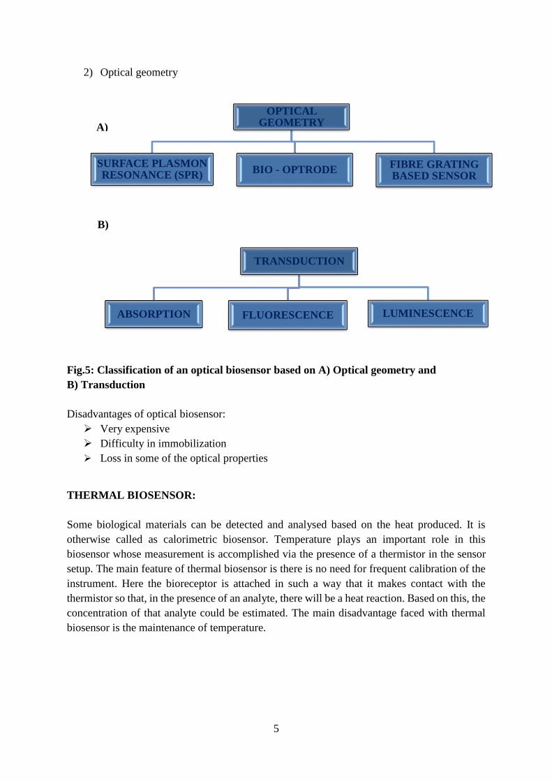
2) Optical geometry
B)
Fig.5: Classification of an optical biosensor based on A) Optical geometry and
B) Transduction
Disadvantages of optical biosensor:
Very expensive
Difficulty in immobilization
Loss in some of the optical properties
THERMAL BIOSENSOR:
Some biological materials can be detected and analysed based on the heat produced. It is
otherwise called as calorimetric biosensor. Temperature plays an important role in this
biosensor whose measurement is accomplished via the presence of a thermistor in the sensor
setup. The main feature of thermal biosensor is there is no need for frequent calibration of the
instrument. Here the bioreceptor is attached in such a way that it makes contact with the
thermistor so that, in the presence of an analyte, there will be a heat reaction. Based on this, the
concentration of that analyte could be estimated. The main disadvantage faced with thermal
biosensor is the maintenance of temperature.
A)
OPTICAL GEOMETRY
SURFACE PLASMON RESONANCE (SPR)
BIO - OPTRODEFIBRE GRATING BASED SENSOR
TRANSDUCTION
ABSORPTION FLUORESCENCE LUMINESCENCE

6
RESONANT BIOSENSOR:
Resonant biosensor is otherwise called as acoustic biosensor. Since it employs the principle of
piezoelectric effect, it is also called as piezoelectric sensor. In the presence of an analyte, the
mass of bio receptor changes which in turn results in change in frequency. In other words, with
the help of piezoelectric crystals, the sound waves are excited at an input transducer and
detected with an output transducer.
1.4 METHODS OF IMMOBILIZATION:
There are two methods of immobilization – physical and chemical immobilization.
Fig.6: Schematic representation of a thermal biosensor [Source: Google images]
Fig.7: Schematic representation of an acoustic biosensor. [Source: Google images]
7
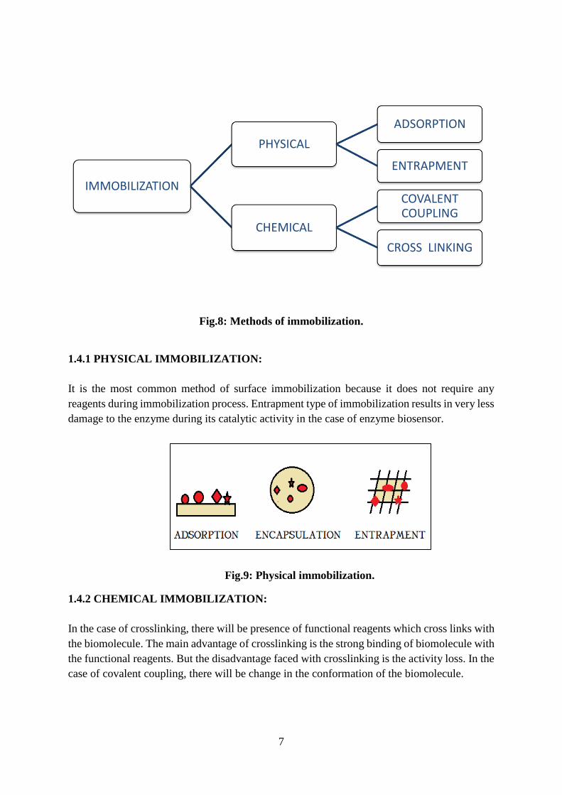
1.4.1 PHYSICAL IMMOBILIZATION:
It is the most common method of surface immobilization because it does not require any
reagents during immobilization process. Entrapment type of immobilization results in very less
damage to the enzyme during its catalytic activity in the case of enzyme biosensor.
1.4.2 CHEMICAL IMMOBILIZATION:
In the case of crosslinking, there will be presence of functional reagents which cross links with
the biomolecule. The main advantage of crosslinking is the strong binding of biomolecule with
the functional reagents. But the disadvantage faced with crosslinking is the activity loss. In the
case of covalent coupling, there will be change in the conformation of the biomolecule.
IMMOBILIZATION
PHYSICAL
ADSORPTION
ENTRAPMENT
CHEMICAL
COVALENT COUPLING
CROSS LINKING
Fig.8: Methods of immobilization.
Fig.9: Physical immobilization.

8
1.5 APPLICATIONS OF BIOSENSORS:
Biosensors has numerous applications owing to its small size, sensitivity, selectivity, low cost
and quicker detection. The main applications of biosensors are as follows:
1. Biosensors play an important role in maintaining the environment (i.e.,) in controlling
the environmental pollution. Pesticide sensor is an example for environmental type of
sensor. The need for oxygen is also measured by this type of biosensors. Biosensors are
helpful to evaluate the amount of environmental pollutants.
2. Biosensors is helpful in estimating various ions and also fermentation products. To
obtain a better yield. In order to measure the odour of food samples, biosensors are
highly beneficial.
3. They are highly advantageous in detection of hazardous gases especially for military.
4. One of the most important application of biosensor is healthcare related. They play a
very crucial role in estimating different biological ssubstances present inside the body.
Glucose biosensor is one of the best-known biosensor in the field of medicine and
healthcare. These biosensors are highly useful in diagnosis of numerous diseases.
1.6 DNA BIOSENSORS:
DNA biosensor is a type of genosensor and the sensing depends on nucleic acid hybridization.
DNA sensing has gained importance owing to its major application in the field of medicine and
healthcare.
Fig.10: Chemical immobilization.

9
1.6.1 NUCLEIC ACID HYBRIDIZATION:
Nucleic acid hybridization is a process where the single strand nucleic acid molecule is made
to interact with their synthesized complementary sequences. The principle of nucleic acid
hybridization is the formation of hydrogen bond due to affinity between the two strands. It is
the measure of sequence similarity and from that process we can also detect the specific type
of sequences. Earlier, nucleic acids were detected by visualizing under the electron microscope.
Here, the process can be carried out in a liquid buffer or some type of gels or nitrocellulose
paper where one strand of nucleic acid (probe which are radio labelled or with fluorescent dye)
are immobilized.
Three types of nucleic acid hybridization are possible:
DNA-DNA hybrid
DNA-RNA hybrid
RNA-RNA hybrid
1.7 METAL OXIDES AND THEIR SENSING APPLICATION:
In recent years, nanomaterials play a crucial role in the development of biosensor. Among
different nanomaterials, metal oxides have its own characteristics which can be listed as
follows:
• Good biocompatibility
• Morphological structure in nano level
• Better sensing characteristics
• Strong physical adsorption properties
• Enhanced electron transfer
• Non – toxicity
• Highly suitable for immobilization of biomolecules
Fig.11: Schematic representation of a DNA biosensor

10
Following are the various methods by which these metal oxides can be synthesized in nano
scale:
• Sol-gel methods
• Sputtering technology
• Hydrothermal technique
From either of these techniques, metal oxide nanoparticles can be synthesized with uniform
size and appropriate morphology.
The main challenge in the development of a biosensor is the fabrication of electrode. An
excellent fabrication is possible with the help of the metal oxide nanoparticles.
DIFFERENT METAL OXIDES FOR BIOSENSING APPLICATION:
Cerium oxide
Cupric oxide
Manganese oxide
Magnesium oxide
Zinc oxide
Iron oxide
Titanium oxide
Tin oxide
Zirconia
SALIENT FEATURES OF THE METAL OXIDES:
1. BIOCOMPATIBILITY: This is the most important feature of most of the
nanoparticles based on metal oxides. Biocompatibility is the foremost property
required for a metal oxide to be used in a biosensing application with metal oxides
as the interface between the electrochemical and biological parts.
2. ISOELECTRIC POINT: It is defined as the pH exhibited by a molecule at which it
does not possess any electric charge.
3. ELECTRON TRANSFER: The main benefit of metal oxides is they help to promote
the electron transfer
1.7.1 SIGNIFICANCE OF ZINC OXIDE:
Many researchers proved that, zinc oxide is the best among the various metal oxides which is
suitable for the biosensing application.
1.7.1.1 PROPERTIES OF ZINC OXIDE:
Nanostructured zinc oxide is a semiconductor based nanomaterial

11
Researchers identified a strong electrostatic interaction between the zinc oxide which
is positively charged and the DNA sequences which is negatively charged.
Very low detection limit for biological samples and offers higher sensitivity
They possess larger excitation energy of around 60 meV
Zinc oxides has wider band gap which is almost equal to 3.37 eV
Very good biocompatibility which is highly necessary for biosensors
They offer high isoelectric point of about 9.5 and this is perfect in the case of DNA
biosensors
Increased surface area for DNA immobilization and hybridization
1.8 SCOPE OF DNA BIOSENSORS:
1.8.1 HPV AND ITS IMPACTS:
Human papilloma virus is the most common cause of sexually transmitted disease affecting
both men and women throughout the world. It is found to be a very significant topic due to its
high rate of morbidity and mortality and is found to be rapidly increasing nowadays. More than
200 different types of HPV types are recognized based on DNA sequence data. Among these
types, 85 HPV genotypes are found to be very well characterized and 120 other HPV types are
partially characterized. In general, HPV mainly infects basal epithelial cells of skin and
anogenital epithelium. It also targets the inner lining of tissues. To be more specific, it infects
the cutaneous (hands and feet) and also the mucosal types (lining of mouth, throat and
respiratory tract).
Sexually transmitted HPV types are categorized into two types: Low risk HPV genotypes and
high risk HPV genotypes. Low risk HPV genotypes are as follows:
HPV -6, 11, 42, 43, 44
High risk HPV genotypes are as follows:
HPV – 16,18,31,33,34,35,39 etc.
Though low risk HPV genotypes does not cause cancer but it can cause Condylomata
acuminata (skin warts) around the genitals and anus. It also targets the respiratory tract causing
benign tumors. High risk HPV genotypes are highly known for causing cancer. Among various
high risk HPV genotypes, HPV -16 and HPV -18 causes cervical cancer.
1.8.2 IMPORTANCE OF DNA SENSOR FOR DETECTION OF HPV:
Though many conventional techniques are used for the detection of HPV and cervical cancer,
the level of effectiveness is not assured. Some of the common conventional techniques
available for the detection of HPV are Pap smear test (cytological test), analysis of protein
expression, VIA test and DNA test. These above mentioned techniques suffer from many
disadvantages. Cytological test does not provide clear information at molecular level and hence
fails for the diagnosis in molecular level. VIA test is less accurate and also shows moderate
specificity and the quality is not assured. DNA test is highly time consuming. Pap test also has
a drawback that some of the cells are lost during fixation step of histological analysis and other

12
disadvantage is the accurate collection of cells from the infected area. Owing to these
disadvantages, biosensors is the best alternative for the detection of HPV sequences since they
offer good sensitivity and specificity.
Therefore, the main aim of this project work is to develop an electrochemical DNA sensor
based on ZnO platform for the detection of DNA sequences by means of hybridization principle
and in future, this sensor can be used for the detection of HPV sequences, thereby cervical
cancer can be diagnosed at a very early stage itself [8].

13
CHAPTER 2: REVIEW OF LITERATURE
2.1 REVIEW ON HPV:
1) Elaine VM Souza et al., [16] explained about the prevailing risk for the development
of cervical cancer which is mainly due to HPV type 16 sequences. This paper has
described about the electrochemical sensing for the sequence specific detection of E1
HPV 1 (type 16) in the presence of methylene blue as the indicator. They concluded
that the detection limit of the HPV sequences respective to its complementary
sequences was found to be 1.49 pM.
2) Isaac A.M. Frias et al., [13] stated about the conventional strategies available for the
detection of HPV sequences. Since these techniques are very complex, sensing is
preferred. It involves the interaction between the biological molecule and its transducer
for detection of the HPV sequences. Different methodologies have been adopted to
study the detection which are as follows: electrochemical sensors, optical sensors and
thermal biosensors. They concluded that future researches need to be conducted in order
to increase the rates of immobilization and also to increase the stability of probe
sequences.
3) Danielly S.Campos-Ferreira et al.,[7] developed an electrochemical sensor for
detection of HPV type 6 sequences in real samples. Here, the electrochemical sensing
was studied by differential pulse voltammetry in the presence of methylene blue
indicator. Conventional techniques for detection of HPV sequences has many
disadvantages owing to their very low specificity. Few advantages of electrochemical
DNA sensor are cheaper, simpler and effective sensing. Due to difficulties faced with
conventional techniques, diagnosis of HPV type 16 sequences should be done using
sensing and nano technology.
2.2 REVIEW ON ELECTROCHEMICAL SENSING:
1) Ana Maria Olveira Brett [2] stated in a review article that the major advantage of
choosing electrochemical method for sensing of DNA sequences is because of the rapid
response time, quantitative analysis, sensitivity, less expensive. The behaviour of DNA
sequences upon electrochemical technique paves way for detection of damaged DNA
sequences. This enables us to study the interaction of DNA immobilized on the surface
of electrode with the target sequences in solution.
2) T. Gregory Drummond et al., [10] proposed that electrochemical based sensors has
features such as sensitivity, selectivity and also cheaper for the detection of DNA
sequences. This review article focused on three properties as follows: Direct DNA
electrochemistry, indirect DNA electrochemistry, enhanced electrochemical response
depending upon the presence of specific nanoparticles, detection of DNA sequences
based on redox indicators. Some of the most important challenges faced with
electrochemical sensing is the fabrication of electrode and complexity with the

14
biological samples. But it offers a huge advantage of having inherent sensitivity for the
corresponding DNA sequences.
3) Eric Bakker et al., [8] explained about different types of electrochemical sensors and
their applications in biological perspective. Researchers prefer electrochemical sensing
due to their quick, simple and inexpensive characteristics for detection of biological
molecules. Several detection approaches were recognized as follows: direct detection
technique, use of some mediators, labelling with enzymes and other labelling
compounds.
4) Lam Dai Tran et al., [16] carried out their research based on HIV sequences by means
of electrochemical detection based on iron oxide nanoparticles. The main feature of this
study is their micro environment which results in enhanced electron transfer and good
sensitivity which was confirmed in the presence of methylene blue indicator. Though
iron oxide nanoparticles have certain advantages such as highly sensitive and enhancing
electron transfer, there could be biocompatibility issues.
2.3 REVIEW ON ZINC OXIDE:
ZINC OXIDES:
Zinc oxides are classified under the category of metal oxides which have the most stunning
unique property for the areas of the sensing application especially in bio sensing for nano level
sensing. From the literature survey, the properties of the zinc oxides are listed as having high
ratio of surface to volume, high IEP, nontoxicity, good biocompatibility, faster electron transfer
between the analyte and the working electrode and also provide the nano structure stability.
1. Shafiquzzaman Siddique et al., [15] Zinc oxide nanoparticles were taken to be one of the
most famous as well as important nanoparticle, because of the unique characteristics such as
electronic, metallic and structural properties. Most of the research work in sensing have
concerned about the high electron transfer property of the zinc oxide nanoparticle to promote
the conductance between the electrically active species and the working electrode at nano
interface. The zinc oxide nanoparticle also has the great advantage to increase the surface area
to promote the more binding of the receptor and the target in order to enhance the sensitivity
and the rapid occurrence of the process.
2. Maumita Das et al., [5] zinc oxide particle does have property of compatible with biological
environment in case of sensing with the nucleic acids, antibodies and other biological elements.
It also have the isoelectric point in the range of (~9.5) which gives good and strong physical
adsorption between the nucleic acids (DNA) (~4.5) by the high electrostatic interaction. Other
than those properties, it has the high electron transport with no toxicity, chemically stable
which also enhance the good immobilization of the receptor on to the surface of the electrode
and it can be recruited for most of the bio sensing techniques.
3. Tugrul Yumak et al., [18] Recent works are more eager about the some unique character
of the zinc oxide nanoparticle such as broad band gap and high excitation energy. Several types
of nanostructure of zinc oxide particle such as nanorods, nanosphere, nanoflowers, nanosheets,

15
nanospikes can also be easily modified, because it provides good and stable nanomorphology.
It also has several advantages in the application such as biosensor, batteries, nanolasers etc.
2.4 REVIEW ON MICROWAVE ASSISTED SYNTHESIS:
MICROWAVE SYNTHESIS:
This technique is one of the simple, faster and easy method of hydrothermal synthesis of
nanoparticle. It was considered to be more eco-friendly and effective in consumption of energy.
Furthermore, it promotes the material quality and uniform structure, and then it also enhance
the uniform temperature distribution and yield of the particle. By this method, different types
of nano structure are produce by varying different parameters.
1. M. Hasanpoor et al., [3] Nowadays, currently undergoing research shows interest on
microwave assisted hydrothermal method to produce the particle such as hydroxide, sulphide
and oxides. This type of microwave assisted technique prevents the problem related to thermal
gradient factor. In comparison with the other ancient technique for hydrothermal synthesis, it
provides simple and easily controlling environment. It makes the reaction to be fast and within
that simple environment suitable temperature and pressure for the reaction was easily attained
with short time. It also makes the uniform structure and size distribution of the preferred
nanoparticle in controlled manner.
2. G. Barreto et al., [4] In order to overcome the disadvantage of the ancient method of
hydrothermal techniques, microwave assisted technique was employed in current studies and
research. Here, by the use of the microwave radiation the difficulty created by the thermal
factors are easily eliminated. In the microwave assisted method, microwave heating generally
employs mainly two types of mechanism such as dipole-dipole vibration and conduction
between the ionic species. The process of the reaction linked directly to the chemical mixture
of the preferred particles, where the reaction happened because of the conversion of
electromagnetic microwave radiation to the thermal radiation as energy.
3. Ashok K. Singh et al., [3] Among the different types of nanomaterial synthesis, microwave
method of preparation provides effectiveness in the reaction process and also have simple
reaction mechanism, where the thermal energy by electromagnetic radiation are directly
applied to the samples, where in case of convectional hydrothermal nanoparticle synthesis there
is loss of heat due to the enlarged medium for reaction process. By this technique, the heat
energy was penetrated over the particle, results in uniform supply of heat energy throughout
the surface and volume of the particle.
16
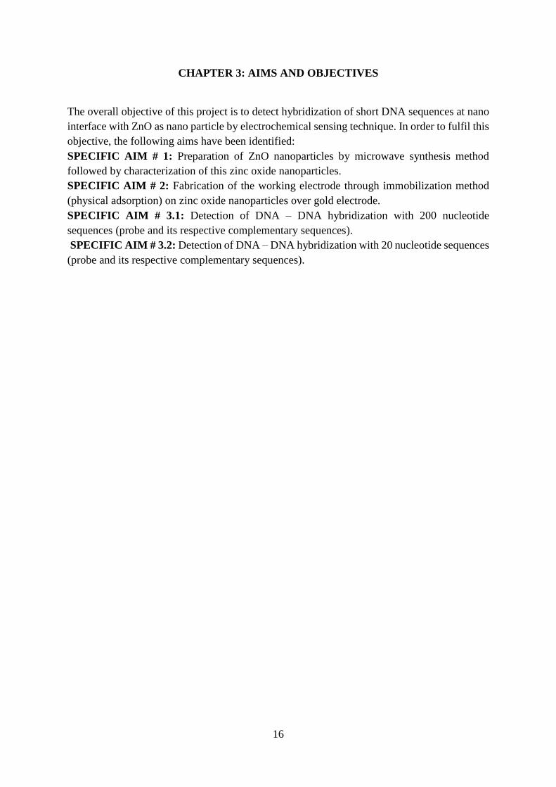
CHAPTER 3: AIMS AND OBJECTIVES
The overall objective of this project is to detect hybridization of short DNA sequences at nano
interface with ZnO as nano particle by electrochemical sensing technique. In order to fulfil this
objective, the following aims have been identified:
SPECIFIC AIM # 1: Preparation of ZnO nanoparticles by microwave synthesis method
followed by characterization of this zinc oxide nanoparticles.
SPECIFIC AIM # 2: Fabrication of the working electrode through immobilization method
(physical adsorption) on zinc oxide nanoparticles over gold electrode.
SPECIFIC AIM # 3.1: Detection of DNA – DNA hybridization with 200 nucleotide
sequences (probe and its respective complementary sequences).
SPECIFIC AIM # 3.2: Detection of DNA – DNA hybridization with 20 nucleotide sequences
(probe and its respective complementary sequences).

19
CHAPTER 4: MATERIALS AND METHODS
4.1 MATERIALS AND REAGENTS:
Zinc chloride powder (ZnCl2) and sodium hydroxide pellets (NaOH) were purchased from
Merck Specialities Private Limited, Mumbai, India. The purity of zinc chloride and sodium
hydroxide are 95% and 97% respectively. Distilled water (obtained from Nano fluids
laboratory, SASTRA University) was used for dilution purpose during preparation of zinc
oxide solution. Deionised water was purchased from Aqua purification system, Salem, India.
The electrical conductivity of deionised water is <1µΩ. 20 µL of oligonucleotides (DNA- PCR
products) were extracted from β- globin which was obtained from Genomics laboratory,
SASTRA University. Alumina powder was used for micro polishing.
4.1.1 ELECTRODE SYSTEM:
In this biosensor, three electrode system was used. The working electrode, reference electrode
and counter electrode used for electrochemical sensing were gold (Au), Ag/AgCl and platinum
wire respectively. The surface area of the working electrode used was 0.0314 cm2.
4.2 SAMPLE PREPARATION:
DNA forward PCR product was used as immobilizing DNA probe. This solution was prepared
by dilution of 20 µL forward PCR product in 5 mL of deionised water. DNA reverse PCR
product was used as target (analyte). This solution was prepared by dilution of 20 µL reverse
PCR product in 5 mL of deionised water. Seven different concentrations such as 0.1, 0.2, 0.3,
0.4, 0.5, 0.6, 0.7 nM of target were prepared using volumetric equation as follows:
CBVB = CAVA
where,
CB & VB – Concentration and volume of solution before dilution.
CA & VA – Concentration and volume of solution after dilution.
These probe and target consisted of approximately 200 nucleotides. Similarly, these steps were
repeated for 20 nucleotides. The concentration of the electro active species DNA probe and
target on the electrode surface were calculated with the help of surface coverage analysis
technique and thereby the molecular weight was also calculated using the following equation:
Surface coverage analysis:
Ґ = 𝑄
𝑛𝐹𝐴
where,
Ґ – Surface coverage
Q – Charge obtained by integration of anodic peak
n – Number of electrons.
F – Faraday’s constant (96485 sA/mol).
A – Area of the electrode.

20
4.3 SYNTHESIS OF ZINC OXIDE NANOPARTICLES:
Zinc oxide nanoparticles was synthesized by microwave technique. Zinc oxide solution was
prepared by mixing the zinc chloride solution with sodium hydroxide (0.5 M), under the
magnetic stirrer for 30 min, where the chemicals are purchased from MERCK chemical private
ltd. After that, it was kept in microwave oven under 800W for 10 min. Nanoparticles get
separated by centrifuge method (Conditions: 1200 rpm for 15 min- repeated for 3 times). And
then it was dried in a hot air oven for 24 hours under 80˚C. It was annealed in muffle furnace
under 200˚C for 4 hours.
4.4 PRETREATMENT OF WORKING ELECTRODE:
The working electrode used here is gold electrode of 3mm diameter. First, the electrode surface
was cleaned and then it was polished with alumina powder using wet pad until a mirror glossy
appearance is seen on the surface of this electrode. After sometime, the electrode was gently
washed in running water. It is then dried in order to prevent crystallization of the particles at
the electrode surface.
4.5 FABRICATION OF ELECTRODE:
First, the working electrode is pretreated and then zinc oxide nanoparticles are coated over the
surface of it. Then the 200 nucleotide sequences are attached to the nano interface.
Hybridization event is facilitated when the probe sequence binds with its respective target
Fig.12: Microwave synthesis of ZnO nanoparticles

21
sequences. Similar steps are followed in the case of 20 nucleotide sequences also and
hybridization is analysed.
4.6 CHARACTERIZATION TECHNIQUES:
4.6.1 SCANNING ELECTRON MICROSCOPY (SEM):
Scanning electron microscopy is the most versatile microscopy which is used for the
exploration of surface morphology, grain size and also many micro level features. In
conventional microscope, light waves are used for imaging. On the other hand, SEM uses
electrons for imaging purpose. So, this type of electron microscope produces highly magnified
image of the surface of the respective material. First, the electrons are produced from electron
gun and then passed onto the corresponding sample to be analysed. These accelerated electrons
will produce kinetic energy. There will be many types of electrons coming out of the electron
gun. When these electrons hit the sample, there will be dissipation of kinetic energy in the form
of various signals. The various signals could be back scattered electrons and also in some cases,
diffracted back scattered electrons for determination of the crystal structure. From this,
information about the surface topography, composition and distribution of the particle is
revealed. In this type of microscopy, sample should have the conductive surface for the electron
transfer to occur for imaging the surface topography. In the case of non-conductive material,
the material should have been coated with conductive material before imaging by low vacuum
sputter coating or high vacuum evaporation. This is mainly done to prevent the accumulation
of the static charges on the surface of the sample during electron irradiation. This can produce
the high resolution image up to x300000 with great clarity.
Fig.13: Fabrication of working (gold) electrode.

22
4.6.1.1 EXPERIMENTAL SETUP:
The very basic components of scanning electron microscope are as follows:
• Electron gun
• Lenses
• Stage for samples
• Detectors (secondary electron detector)
• Output unit
4.6.1.2 ADVANTAGES OF SEM:
High resolution image of the samples
Three-dimensional morphology of samples
Very high and powerful magnification
4.6.2 X-RAY DIFFRACTION (XRD):
X-ray diffraction pattern of the sample contain the information about the crystalline nature,
size and orientation of the crystallites and also the phase composition of the samples. When the
incident beam of x-ray (electromagnetic radiation) hits the sample, the process of scattering
occurs. The diffraction of the x-ray occurs when the scattered rays are in phase with other
scattered rays from other atomic planes. The diffraction of x-ray was based on the principle of
Bragg’s equation.
According to Bragg’s law,
n λ= 2d sin(θ)
where,
λ is the wavelength of the x-ray
θ is the angle between the incident ray and the surface of the crystal
d is the spacing between the layers of atoms
n is the integer
Crystalline materials such as semi crystalline polymer, metal and metal oxide nanoparticle,
layered silicate nanoclay has unique x-ray diffraction characteristics patterns.
4.6.2.1 ADVANTAGES:
Effective tool for identification of unknown material
Very low quantity of samples for determination
Easier data interpretation

23
4.7 ELECTROCHEMICAL SENSING TECHNIQUES:
4.7.1 CYCLIC VOLTAMMETRY:
This technique shows the information about the thermodynamics of the charges released or
absorbed during the redox reaction, adsorption process and the kinetics of the charge transfer.
In this technique, the graph was plotted against the potential linearly versus time, which gives
the information about chemical reaction happened at the electrode interface as redox peaks.
Depend upon the requirements of the certain analysis or the specific experiment control, single
or several scan can be done. During this type of technique, under the different potential and
current, the analyte or the substance in the electrolyte solution or at the electrode interface
undergo electron communication, this may be directly proportional to the concentration of the
substance which acts as the analyte.
4.7.2 AMPEROMETRY:
The method of amperometry was as same as the cyclic voltammetry where the current was
produced when the constant potential was applied between the two electrodes such as reference
and the counter electrode. The current was measured between the counter electrode and the
working electrode. This produced current was proportional to the change in the density of the
species involved in the chemical reaction. The maintaining of the potential was achieved by
varying the potentiostat.
4. 4.7.3 IMPEDOMETRY:
The impedometric technique of the electrochemical sensing comprises the both resistance and
capacitive nature of the substance at the electrode interface. In this technique, the constant
potential was maintained to determine the impedance of the system, where the current can be
varied. It is the effective tool for interrogating the electrode and the electrolyte kinetics
Fig.14: Cyclic voltammogram
24
CHAPTER 5: RESULTS AND DISCUSSION
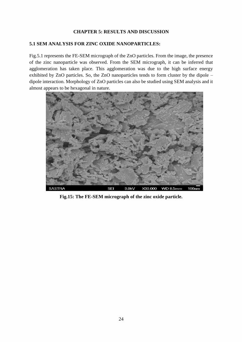
5.1 SEM ANALYSIS FOR ZINC OXIDE NANOPARTICLES:
Fig.5.1 represents the FE-SEM micrograph of the ZnO particles. From the image, the presence
of the zinc nanoparticle was observed. From the SEM micrograph, it can be inferred that
agglomeration has taken place. This agglomeration was due to the high surface energy
exhibited by ZnO particles. So, the ZnO nanoparticles tends to form cluster by the dipole –
dipole interaction. Morphology of ZnO particles can also be studied using SEM analysis and it
almost appears to be hexagonal in nature.
Fig.15: The FE-SEM micrograph of the zinc oxide particle.

25
5.2 XRD DATA FOR ZnO NANOPARTICLES:
The X-ray diffraction pattern of ZnO particles were synthesized at 800 W under microwave
synthesis were represented in the fig.16. There were number of Bragg’s reflections with two-
theta values of 33°, 35°, 37°, 48°, 57°, 64°, 66°, 69°, 70°, 77° corresponding to Miller indices
plane such as (110), (002), (101), (102), (110), (103), (112), (201), (202) which confirmed the
crystalline nature of the zinc oxide particles and was found to be similar with [JCPDS 36-
1451]. The hexagonal structure of ZnO was further confirmed by means of diffraction peaks
obtained in XRD analysis. It was also found that, the broader the peak, the smaller the size of
the particle.
0 10 20 30 40 50 60 70 80 90
0
100
200
300
400
500
600
700
ZnO
A
ZnO
Fig 16: X-ray diffraction pattern of ZnO particle.

26
5.3 ELECTROCHEMICAL STUDIES:
5.3.1 CYCLIC VOLTAMMETRY STUDIES:
5.3.1.1 EFFECT OF DIFFERENT ELECTRODE SYSTEM:
From figure 17, the effect of different electrode system was studied. The CV was run in three
cases as follows: i) with the bare electrode in target sequences solution, ii) bare electrode with
immobilized probe in the target sequences solution and iii) bare electrode with coated ZnO
interface immobilized with probe in the target solution.
A) 200 NUCLEOTIDES:
Figure 17 A) represents the CV plot for probe and target sequences consisting of 200
nucleotides. From this graph, it can be seen that, when compared with bare electrode system
and also with bare – probe electrode system, working electrode with nano interface (ZnO
particles) showed a higher peak. This is because of enhanced electron transfer in the presence
of ZnO particles.
-0.8 0.0 0.8
0.000000
0.000002
0.000004
Cu
rren
t(A
)
Potential(V)
BARE
BARE+PROBE
BARE+PROBE+NPS
0
B) 20 NUCLEOTIDES:
Figure 17 B) represents the CV plot for probe and target sequences consisting of 20
nucleotides. From this graph, it can be seen that, when compared with bare electrode system
and also with bare – probe electrode system, working electrode with nano interface (ZnO
particles) showed a higher peak. This is because of enhanced electron transfer in the
presence of ZnO particles.

27
-0.7 0.0 0.7
-0.000002
0.000000
0.000002
0.000004
Cu
rren
t(A
)
Potential(V)
BARE
BARE+PROBE+NPS
BARE+PROBE
COMPARISION
BARE ELECTRODE
BARE + PROBE
BARE + PROBE +
NANOPARTICLE
NUMBER OF BASES
200
BASES
20 BASES 200
BASES
20 BASES 200
BASES
20 BASES
FWHM 2.6856 2.6856 3.3380 3.2142 3.0323 3.3581
KS 2.4982 2.4982 0.08828 4.3552 0.08509 0.07695
Ґ (µmol/cm2) 177 177 191 121 744 509
EP (V) -0.4405 -0.4405 0.44 -0.2432 -0.571 -0.704
IP (µA) 2.6856 2.6856 0.1025 3.2142 0.3838 0.2378
TABLE 1: Comparison of different electrode system for 200 bases and 20 bases.
Table 1 represents the comparison of different electrode system for both 200 nucleotide
sequences and 20 nucleotide sequences with various electrochemical parameters such as
FWHM (full width –half maximum), KS (Reaction rate), Ґ (surface coverage), Ep (peak
potential) and Ip (peak current).
5.3.1.2 EFFECT OF SCAN RATES:
From figure 18, it can be observed that a linearity was obtained in terms of scan rates in the
range of 0.01 to 0.09 V/S. It was found that 0.09 V/S was found to be the optimized scan rate.
Fig.17: CV for DNA sequences showing the effect of different
electrode system A) 200 nucleotides B) 20 nucleotides

28
A) 200 NUCLEOTIDES:
B) 20 NUCLEOTIDES:
0.00 0.04 0.08 0.12
0
10
20
30
Cu
rre
nt
(µA
)
Scan rate(V/s)
Slope= 32.004
Fig.18: CV for DNA sequences showing the effect of scan
rates A) 200 nucleotides B) 20 nucleotides

29
5.3.1.3 EFFECT OF VARYING CONCENTRATIONS:
I) 200 NUCLEOTIDES:
-1.0 -0.8 -0.6 -0.4 -0.2 0.0 0.2 0.4 0.6 0.8 1.0
-0.000002
0.000000
0.000002
0.000004
0.000006
0.000008
0.000010
curr
ent
(A)
Potential (v)
0.1 nM
0.2 nM
0.3 nM
0.4 nM
0.5 nM
0.7 nM
0.0 0.2 0.4 0.6 0.80
1
2
3
4
5
Cu
rre
nt
(A)
Concentration (nM)
concentration
A)
Fig.19: CV for 200 nucleotide sequences showing the A) effect of
varying concentration B) Bar graph for concentration versus current

30
II) 20 NUCLEOTIDES:
-1.0 -0.8 -0.6 -0.4 -0.2 0.0 0.2 0.4 0.6 0.8 1.0
-0.000002
0.000000
0.000002
0.000004
0.000006
0.000008
0.000010
0.000012
0.000014
Cu
rren
t (A
)
Potential (V)
96 pM
108 pM
100 pM
B)
96 102 108
0
1
2
3
4
5
Cu
rren
t (A
)
Potential (V)
Concentration
Fig.20: CV for 20 nucleotide sequences showing the A) effect of varying
concentration B) Bar graph for concentration versus current
A)
31
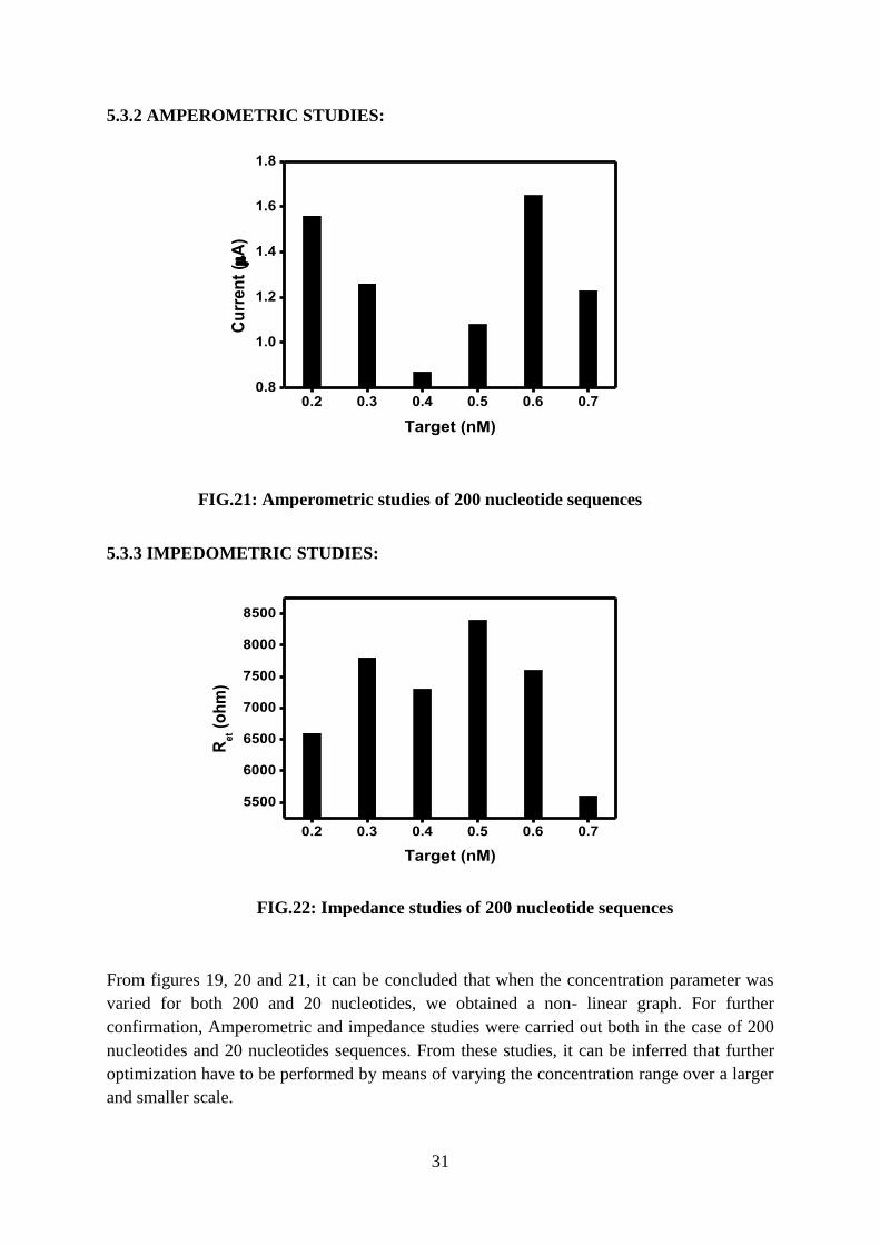
5.3.2 AMPEROMETRIC STUDIES:
0.2 0.3 0.4 0.5 0.6 0.70.8
1.0
1.2
1.4
1.6
1.8
Cu
rre
nt
(A
)
Target (nM)
5.3.3 IMPEDOMETRIC STUDIES:
0.2 0.3 0.4 0.5 0.6 0.7
5500
6000
6500
7000
7500
8000
8500
Re
t (o
hm
)
Target (nM)
From figures 19, 20 and 21, it can be concluded that when the concentration parameter was
varied for both 200 and 20 nucleotides, we obtained a non- linear graph. For further
confirmation, Amperometric and impedance studies were carried out both in the case of 200
nucleotides and 20 nucleotides sequences. From these studies, it can be inferred that further
optimization have to be performed by means of varying the concentration range over a larger
and smaller scale.
FIG.21: Amperometric studies of 200 nucleotide sequences
FIG.22: Impedance studies of 200 nucleotide sequences

32
CHAPTER 6: CONCLUSION
In this project work, ZnO particles were synthesized in a nano scale by microwave synthesis.
The morphology, size and distribution of zinc oxide nanoparticles were studied by SEM and
XRD analysis. From this characterization results, it was confirmed that the zinc oxide powder
had a tetragonal zincite structure.
This approach combines the influence of both nanotechnology and electrochemistry. From this,
it can be concluded that, after inclusion of zinc oxide nanoparticles, electrochemical sensing of
nucleotide sequences increased significantly. Zinc oxide nanoparticles play a very crucial role
in such a way that it helps to enhance the transfer of electrons for hybridization of the probe
and its corresponding complementary target sequences. Zinc oxide nanoparticles also offers
good biocompatibility and has high isoelectric point (IEP ~9.5) in order to obtain strong
physical adsorption of DNA sequences. Electrochemical sensing was performed using cyclic
voltammetric, amperometric and impedance studies. This has resulted in the development of
very simple and inexpensive electrochemical DNA biosensor with nano-interface and hence it
can be used for rapid detection of various defective sequences and this, in turn, is highly useful
for diagnostic applications. For example, by checking for hybridization of HpV type 16
sequences, cervical cancer can be detected at an earlier stage.

33
CHAPTER 7: REFERENCES
1) Applications of nanostructured materials in fabrication of electrochemical genosensors
Amid Rahi et al., Journal of Advanced Medical Sciences and Applied Technologies
(2015) 1(1)
2) Electrochemical DNA Sensors for Detection of DNA Damage Ana Maria Olveira Brett
et al., 5(6) (2005) 377-393
3) Microwave synthesis, characterization and photocatalytic properties of SnO2
nanoparticles Ashok K singh, Umesh T. Nakate, Advances in particles 2 (2013) 66-70
4) Microwave assisted synthesis of ZnO tridimensional nanostructures
G. Barreto et al., Procedia material and science 8 (2015) 535-540
5) Electrochemical DNA biosensor for the detection of human papillomavirus E6 gene
inserted in recombinant plasmid Danielly S.Campos - Ferreira et al.,Arabian Journal
of Chemistry 9(3) (2016) 443 – 450.
6) Application of nanostructured ZnO films for electrochemical DNA biosensor M. Das
et al., Thin solid films 519 (2010) 1196-1201
7) Electrochemical sensors Eric Bakker, Yu Qin, ACS publications 78(12) (2006) 3965 -
3984
8) Trends in Biosensors for HPV: Identification and diagnosis Isaac A.M. Frias et al.,
Journal of sensors (2015)
9) Electrochemical Biosensor-Sensor principles and architectures D. Grieshaber et al.,
Sensors 8 (2008) 1400-1458
10) Electrochemical DNA sensors T. Gregory Drummond et al., Nature biotechnology 21
(2003) 1192-1199
11) Microwave assisted synthesis of zinc oxide nanoparticles
M. Hsanpoor et al., Procedia material and science 11 (2015) 320-325
12) Biomedical detection via macro and nano-sensors fabricated with metallic and semi
conducting oxides Jong-In Hahm, Biomed Nanotechnol. 9(1) (2013) 1-25
13) Nanostructured metal oxide-based biosensors Pratima R. Solanki et al., NPG Asia
materials 3(1) 17-24 (2011)
14) Nanoparticle-enhanced electrochemical biosensor with DNA immobilization and
hybridization of Trichoderma harzianum gene S. Siddiquee et al., Sensing and Bio-
Sensing Research 2 (2014) 16 -22
15) Electrochemical detection of short HIV sequences on chitosan/ Fe3O4 nanoparticle
based screen printed electrodes L.D. Tran et al., Materials science and Engineering C
31 (2011) 477-485
16) ’Spotted Nanoflowers’: Gold seeded Zinc oxide nanohybrid for selective bio-capture
Veeradasan Perumal et al., Scientific reports – Nature (2015)
17) Preparation and characterization of zinc oxide nanoparticle and their sensor
applications for electrochemical monitoring of nucleic acid hybridization T. Yumak et
al., Colloids and surfaces B: Biointerfaces 86 (2011) 397-403