mesenchymal stem cells delivered in a microsphere-based engineered skin contribute to cutaneous...
TRANSCRIPT

Journal of Dermatological Science 66 (2012) 29–36
Mesenchymal stem cells delivered in a microsphere-based engineered skincontribute to cutaneous wound healing and sweat gland repair
Sha Huang a,b,c,1, Gang Lu b,d,1, Yan Wu b,e, Enhe Jirigala b,f, Yongan Xu b, Kui Ma b, Xiaobing Fu a,b,*a Wound Healing and Cell Biology Laboratory, Institute of Basic Medical Sciences, General Hospital of PLA, Beijing 100853, PR Chinab Burns Institute, the First Affiliated Hospital, General Hospital of PLA, Trauma Center of Postgraduate Medical College, Beijing, 100048, PR Chinac Military Training Related Medical Sciences Institute, the 150 Hospital of Chinese PLA, Luoyang, 471031, PR Chinad Department of Burn and Plastic Surgrey, General Hospital of Beijing PLA Military Region, Beijing 100025, PR Chinae Mudanjing Medical College, Mudanjing, 157011, PR Chinaf Inner Mongolia Medical College, Huhhot, 010059, PR China
A R T I C L E I N F O
Article history:
Received 22 October 2011
Received in revised form 31 January 2012
Accepted 4 February 2012
Keywords:
Stem cells
Transplantation
Wound healing
Sweat glands
Skin
A B S T R A C T
Background: Bone-marrow-derived mesenchymal stem cells (BM-MSCs) can contribute to wound
healing after skin injury. However, the role of BM-MSCs on repairing skin appendages in renewal tissues
is incompletely explored. Moreover, most preclinical studies suggest that the therapeutic effects
afforded by BM-MSCs transplantation are short-lived and relatively unstable.
Objective: To assess whether engrafted bone-marrow-derived mesenchymal stem cells via a delivery
system can participate in cutaneous wound healing and sweat-gland repair in mice.
Methods: For safe and effective delivery of BM-MSCs to wounds, epidermal growth factor (EGF)
microspheres were firstly developed to both support cells and maintain appropriate stimuli, then cell-
seeded microspheres were incorporated with biomimetic scaffolds and thus fabricated an engineered
skin construct with epithelial differentiation and proliferative potential. The applied efficacy was
examined by implanting them into excisional wounds on both back and paws of hind legs in mice.
Results: After 3 weeks, BM-MSC-engineered skin (EGF loaded) treated wounds exhibited accelerated
healing with increased re-epithelialization rates and less skin contraction. Furthermore, histological and
immunofluorescence staining analysis revealed sweat glands-like structures became more apparent in
BM-MSC-engineered skin (EGF loaded) treated wounds but the number of implanted BM-MSCs were
decreased gradually in later phases of healing progression.
Conclusions: Our study suggests that BM-MSCs delivered by this EGF microspheres-based engineered skin
model may be a promising strategy to repair sweat glands and improve cutaneous wound healing after
injury and success in this study might provide a potential benefit for BM-MSCs administration clinically.
� 2012 Japanese Society for Investigative Dermatology. Published by Elsevier Ireland Ltd.
All rights reserved.
Contents lists available at SciVerse ScienceDirect
Journal of Dermatological Science
jou r nal h o mep ag e: w ww .e lsev ier . co m / jds
1. Introduction
A wound healing process that begins immediately after skininjury involves multiple coordinated steps including inflammation,cell proliferation, cell migration, angiogenesis, and extracellularmatrix (ECM) production. Bone-marrow-derived mesenchymalstem cells (BM-MSCs), also referred to as multipotent stromalprogenitor cells, can promote tissue repair during wound healing[1,2]. Specifically, considerable evidence suggests that BM-MSCshave a strong propensity to ameliorate cutaneous damage inresponse to injury by contributing to enhanced growth of
* Corresponding author at: Burns Institute, the First Affiliated Hospital, General
Hospital of PLA, 51 Fu Cheng Road, Beijing 100048, PR China.
Tel.: +86 01 066867391; fax: +86 10 68989955..
E-mail addresses: [email protected], [email protected] (X. Fu).1 These authors contributed equally to this work.
0923-1811/$36.00 � 2012 Japanese Society for Investigative Dermatology. Published b
doi:10.1016/j.jdermsci.2012.02.002
epidermal cells, angiogenesis, wound contraction and collagendeposition [3–8].
Additionally, several groups have attempted clinical trials ofBM-MSCs for treating neurological diseases, spinal injury, andmyocardial infarction; damaged tissue in these diseases is difficultto heal by normal tissue regeneration [9–11]. Despite the broadtherapeutic efficacy of BM-MSCs shown in vivo, they have notachieved wide use in clinical treatment; presumably, by relativelypoor viability of the transplanted cells. Furthermore, use of suchcells strictly requires a suitable microenvironment to promoteproliferation and differentiation; otherwise their use alone oftenfails to form a biologically complete tissue [12].
Recent advances in cellular tissue-engineered skin constructshave delivered considerable benefits to patients with skin damagedue to burns, accidents, infections and chronic wounds. Increasingattention is being placed on the use of adult stem cells as a sourceof in vitro generation of skin [13,14]. However, stem cells also need
y Elsevier Ireland Ltd. All rights reserved.

S. Huang et al. / Journal of Dermatological Science 66 (2012) 29–3630
to be delivered safely and effectively to a wound. Within anengineered skin model, they can remain in contact with the woundbed and be kept viable in the often-hostile wound microenviron-ment. In particular, a large number of studies have assessed thehuge potential for using MSCs via tissue engineering strategies torepair or replace important damaged tissues [15].
Evidence from our laboratory and others also suggests thatMSCs, particularly those derived from BM, may contributesignificantly to promoting the repair and regeneration of skin byaltering the tissue microenvironment and BM-MSCs can differen-tiate into other cells within injured tissue [16–18]. Although theuse of BM-MSCs for research purposes and for clinical applicationshas been vigorously investigated, the extent to which these cellscontribute to functional recovery such as sweat gland repairremains uncertain. The presence of skin appendages such as sweatglands in repaired skin is of major clinical importance formaintaining skin homeostasis and regulating body temperature.However, because of poor intrinsic healing capacity, skinappendages cannot be successfully repaired by traditionaltreatment after severe skin injury.
To address this issue, we considered that BM-MSCs could be acandidate for repair and regeneration of both skin tissue and sweatglands. Additionally, an engineered collagen-based scaffoldincorporating specific stimulating factors-loaded microsphereswould be developed to characterize the microenvironmentalinfluences in directing stem cell differentiation and proliferation,paracrine activity and regulation of ECM deposition. Based on ourprevious findings, epidermal growth factor (EGF) was selected asstimulating factors due to its significant role in development ofsweat glands. Because BM-MSCs can differentiate into tissue-specific cells in response to cues provided by different organs, theywould be then implanted in excisional wounds on both the backand the paws of hind legs in a Murine model.
2. Materials and methods
2.1. Isolation and culture of MSCs from mouse bone-marrow
Bone-marrow-derived cells were collected from the femurs andtibias of 3–5 week-old male C57 – green fluorescent protein (GFP)+/
+ transgenic mice (C57Bl/6-TgN(ACTbEGFP)1Osb, Jackson Labs).Cells were cultured in MesenCult basal medium containing MSCstimulatory supplements (StemCell Technologies). After 48 h, thenonadherent cells were removed, and fresh medium was added tothe cells. Medium was changed every 2 or 3 days. The adherentspindle-shaped cells were further propagated for 3 passages.
2.2. Flow cytometry analysis
Cultured BM-MSCs were analyzed by flow cytometry (FACSCalibur; BD Biosciences). Cells were incubated with antibodies forCD31, CD34, CD44, CD90, CD29 (Cymbus Biotechnology), andsecondary FITC-conjugated antibodies (BD Biosciences, St. Jose, CA).
2.3. Differentiation ability assays
BM-MSCs were placed in basic medium consisting of DMEM(Invitrogen Life Technologies), 10% FBS, 1% penicillin, 1% strepto-mycin, 1% amphotericin B, and then specific supplements formesenchymal lineage differentiation were added [19]. Adipogenicdifferentiation was induced by basic medium with 0.5 mMdexamethazone, 0.5 mM 3-isobutyl-1-methylxanthine, and0.1 mM indomethacine (Sigma–Aldrich) [20]. Osteogenic differen-tiation was achieved by use of basic medium containing 0.1 mMdexamethazone, 50 mM ascorbic acid, and 10 mM b-glyceropho-sphate (Sigma–Aldrich) [21]. Chondrogenic differentiation was
induced by use of basic medium containing 50 mM ascorbic acid,0.1 mM dexamethazone, 10 ng/ml TGF-b (R&D Systems), 40 mg/mlL-proline (Sigma–Aldrich), and 100 mg/ml sodium pyruvate(Wako) [22]. Each differentiation medium was changed every2–3 days. Confirmation of differentiation of the cells to adipocytes,osteocytes and chondrocytes involved staining with oil-red O,alizarin red, and toluidine blue, respectively.
2.4. Construction of stem cell-seeded microspheres
Gelatin microspheres were generated as previously described[23]. Gelatin with an isoelectric point of 5.0 and recombinant EGFwere from Sigma–Aldrich (St. Louis, MO). All other reagents orchemicals were of analytical grade (Shanghai Chemical Co., China)and were used without any further treatment or purification. Inbrief, preparing an aqueous solution including EGF (50 mg in 1 mlphosphate buffer, pH 7.4) and dropping it onto the resulting gelatinmicrospheres (100 mg), allowing for sorption of the protein to thematrix. To test the effect of EGF on stem cell growth, EGF-freemicrospheres were also created as controls. As the carrier for cellculture, the resulting EGF-microspheres were screened with adiameter ranged from 50 mm to 200 mm and sterilized by use ofethylene oxide gas for 6 h and then washed with PBS 3 times. Then,2.1 � 106 GFP+ BM-MSCs were inoculated into 1.3 ml of culturemedium containing 26 mg microspheres in siliconized 12-well cellculture plates. After a culture time of 24 and 72 h, GFP+ BM-MSC–microsphere complexes were formed and analyzed by fluorescencemicroscopy (Olympus IX 70, Tokyo, Japan) at 100 magnifications.
2.5. Constitution of engineered skin constructs
Engineered skin constructs were generated as previouslydescribed [24]. In brief, the cold collagen type I solution andMatrigelTM (2:1) (BD Biosciences, San Jose, CA) were mixed with10 � Dulbecco’s modified Eagle’s medium (DMEM), 10% fetalbovine serum (FBS), 200 mM L-glutamine solution and 72 g/lsodium bicarbonate solution on ice. The pH was adjusted to 7.4with 1 M NaOH solution. Subsequently, GFP+ BM-MSC – micro-sphere complexes (EGF-loaded or not) were delivered into theabove-mentioned matrix via injection under standard cultureconditions. After 14-day organotypic co-culture, GFP+ BM-MSC-seeded engineered skin constructs were created for in vitro
investigation and in vivo implantation. For histological analysis,specimens of constructs were embedded in OCT. Sections 6 mmthick were stained with hematoxylin and eosin (H&E) according toroutine histology protocols for light microscopy (Olympus BX-41,Japan). Sections were also incubated with monoclonal antibodiesto Keratin 5 (rabbit anti-, Abcam, Cambridge, MA) and weredetected with Cy3-labeled goat anti- rabbit IgG (Beyotime, China)for the presence of epithelial development.
2.6. Wound healing model and BM-MSC-seeded engineered skin
implantation
All animal procedures were approved under the guidelines of theInstitutional Animal Care and Use Committee of the GeneralHospital of PLA. A total of 54 C57BL/6 mice (GFP�; 8 weeks old;female; body weight 20–23 g) were used to generate the excisionalwound models. In brief, after hair was removed from the dorsalsurface and after anesthesia, a 12 mm, full-thickness excisional skinwound was created on the midline of the back. Animals wererandomized to 4 groups for treatment: control, GFP+ BM-MSCinjection, GFP+ BM-MSC -seeded engineered skin (EGF-loaded ornot) implantation. In control animals receiving no graft, topicalantibiotic ointment (Bacitracin Zinc e Neomycin Sulfate e PolymyxinB Sulfate, Alphapharma USPD, Baltimore, MD, USA) was applied to

S. Huang et al. / Journal of Dermatological Science 66 (2012) 29–36 31
the wound in a thin layer, and the surgery was completed. GFP+ BM-MSC injection involved 1 � 106 cells (GFP+ BM-MSCs) with PBSinjected intravenously into the tail vein of mice. GFP+ BM-MSC-engineered skin (with the same amount of cells as for injection) wereoverlaid with a piece of nonadherent dressing placed orthotopicallyon the wound and secured to the wound margin with sutures. Wetested the dressing on the skin in mice before this experiment andobserved no skin irritation or allergic reaction. After surgery, micewere housed individually. Postoperative recovery was closelymonitored for 15 min, and then mice were returned to individualcages and cared for as routine.
For analysis of daily wound sizes, standardized images ofwounds were recorded by use of a digital camera. Wound area wasmeasured by tracing the wound margin and calculating by use ofMetamorph software (Molecular Devices, Sunnyvale, CA). Theproportion of wound closure was calculated as follows: (area oforiginal wound – area of actual wound)/area of originalwound � 100. The investigators measuring samples were blindedto group and treatment. Time to wound closure was defined as thetime when the wound bed was completely re-epithelialized andfilled with new tissue. The wound perimeters were traced at thetime of surgery and weekly from 1 to 4 weeks postoperatively.
To test the effect of sweat gland repair, full-thickness woundswere also created on both paws of hind legs according to thedistribution features of sweat glands in the mice (totally 16animals, a 2 mm, full-thickness excisional skin wound wascreated on the both paws of hind legs) and wounds were treatedaccording to the same protocol as above back wounds. Addition-ally, special equipments were prepared previously to keep thedressing remain intact wounds. 2 and 3 weeks after surgery,biopsies of tissue from each graft group, along with adjoiningmouse skin, were collected. Specimens were stained with H&Eaccording to routine histology protocols and viewed under amicroscope. To monitor the survival of implanted cells in thewound, tissue sections were embedded in OCT and observed by
Fig. 1. Characterization of isolated bone-marrow-derived mesenchymal stem cells (BM
(scale bars: 50 mm). (B) Cell-surface markers of BM-MSCs were assessed by FACS. MSCs e
was revealed by oil-red O staining (scale bars: 10 mm). (D) Osteogenic differentiation
potential of MSCs was determined by staining for toluidine blue (scale bars: 20 mm).
fluorescence microscopy. In addition, skin sections were treatedwith primary antibodies for CEA (Neomarker, Fremont, CA), a signof developing sweat glands, as we previously described [25]. Thensecondary antibodies conjugated to rhodamine-isothiocyanate(Southern Biotechnology) were used for fluorescence stainingdetection and confocal laser scanning fluorescence microscopy(Olympus FV10i, Tokyo, Japan)
2.7. Statistical analysis
All values are represented as means � standard deviation (SD).Analysis involved ANOVA and SPSS 17.0 (SPSS Inc., Chicago, IL, USA). Ap < 0.05 was considered statistically significant.
3. Results
3.1. Characterization of isolated BM-MSCs
Discrete colonies of fibroblast-like cells attached to the plasticwere evident at days 4–5 after initial seeding. After 10 days, mostcell lines were composed of cells with a characteristic spindleshape, whereas others had cells with polygonal morphology(Fig. 1A). Cell-surface markers were assessed by flow cytometry tocharacterize isolated MSCs. BM-MSCs expressed CD29, CD44, andCD90 but not CD34 and CD31 (Fig. 1B), which is consistent withprevious reports [19,20]. BM-MSCs were further characterized byconfirming their ability to undergo specific adipogenic, osteogenic,and chondrogenic differentiation by positive staining with oil-redO, alizarin red, and toluidine blue (Fig. 1C–E). Only cells that metthese criteria were used in subsequent experiments.
3.2. Characterization of BM-MSCs-seeded microspheres
Notably, owing to the use of the microsphere culture techniqueand sustained-release effect of EGF (data not shown) [24,26], it is
-MSCs). (A) Morphologic appearance of BM-MSCs at 10th day after initial seeding
xpressed CD29, CD44, and CD90 but not CD34, CD31. (C) Adipogenic differentiation
was confirmed by alizarin red staining (scale bars: 20 mm). (E) The chondrogenic
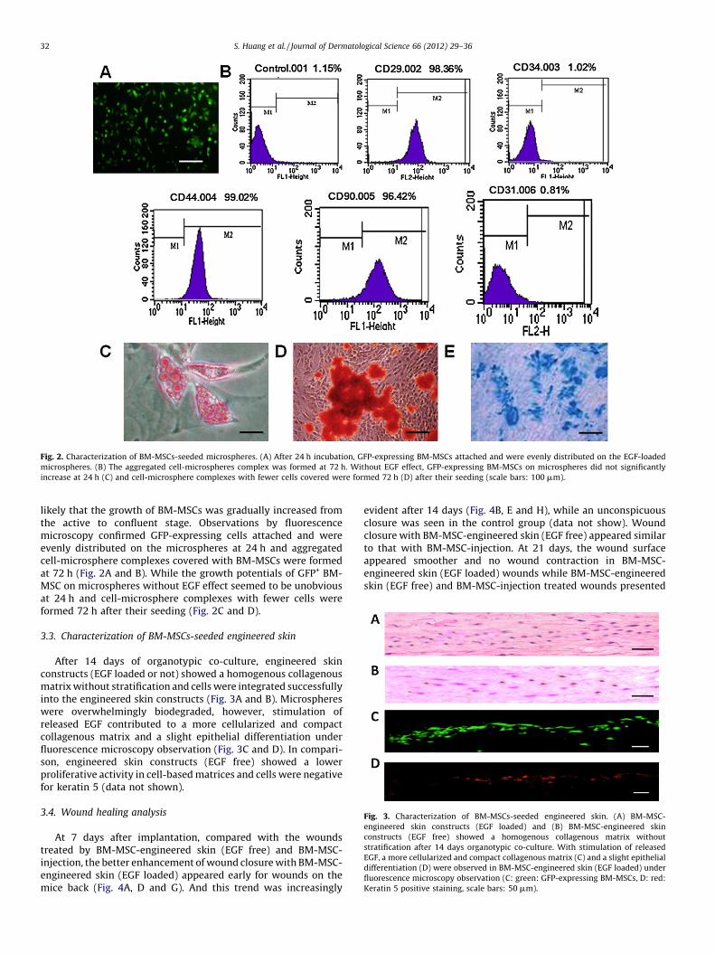
Fig. 2. Characterization of BM-MSCs-seeded microspheres. (A) After 24 h incubation, GFP-expressing BM-MSCs attached and were evenly distributed on the EGF-loaded
microspheres. (B) The aggregated cell-microspheres complex was formed at 72 h. Without EGF effect, GFP-expressing BM-MSCs on microspheres did not significantly
increase at 24 h (C) and cell-microsphere complexes with fewer cells covered were formed 72 h (D) after their seeding (scale bars: 100 mm).
Fig. 3. Characterization of BM-MSCs-seeded engineered skin. (A) BM-MSC-
engineered skin constructs (EGF loaded) and (B) BM-MSC-engineered skin
constructs (EGF free) showed a homogenous collagenous matrix without
stratification after 14 days organotypic co-culture. With stimulation of released
EGF, a more cellularized and compact collagenous matrix (C) and a slight epithelial
differentiation (D) were observed in BM-MSC-engineered skin (EGF loaded) under
fluorescence microscopy observation (C: green: GFP-expressing BM-MSCs, D: red:
Keratin 5 positive staining, scale bars: 50 mm).
S. Huang et al. / Journal of Dermatological Science 66 (2012) 29–3632
likely that the growth of BM-MSCs was gradually increased fromthe active to confluent stage. Observations by fluorescencemicroscopy confirmed GFP-expressing cells attached and wereevenly distributed on the microspheres at 24 h and aggregatedcell-microsphere complexes covered with BM-MSCs were formedat 72 h (Fig. 2A and B). While the growth potentials of GFP+ BM-MSC on microspheres without EGF effect seemed to be unobviousat 24 h and cell-microsphere complexes with fewer cells wereformed 72 h after their seeding (Fig. 2C and D).
3.3. Characterization of BM-MSCs-seeded engineered skin
After 14 days of organotypic co-culture, engineered skinconstructs (EGF loaded or not) showed a homogenous collagenousmatrix without stratification and cells were integrated successfullyinto the engineered skin constructs (Fig. 3A and B). Microsphereswere overwhelmingly biodegraded, however, stimulation ofreleased EGF contributed to a more cellularized and compactcollagenous matrix and a slight epithelial differentiation underfluorescence microscopy observation (Fig. 3C and D). In compari-son, engineered skin constructs (EGF free) showed a lowerproliferative activity in cell-based matrices and cells were negativefor keratin 5 (data not shown).
3.4. Wound healing analysis
At 7 days after implantation, compared with the woundstreated by BM-MSC-engineered skin (EGF free) and BM-MSC-injection, the better enhancement of wound closure with BM-MSC-engineered skin (EGF loaded) appeared early for wounds on themice back (Fig. 4A, D and G). And this trend was increasingly
evident after 14 days (Fig. 4B, E and H), while an unconspicuousclosure was seen in the control group (data not show). Woundclosure with BM-MSC-engineered skin (EGF free) appeared similarto that with BM-MSC-injection. At 21 days, the wound surfaceappeared smoother and no wound contraction in BM-MSC-engineered skin (EGF loaded) wounds while BM-MSC-engineeredskin (EGF free) and BM-MSC-injection treated wounds presented

Fig. 4. Wound closure on the back of mice at days 7, 14 and 21 after transplantation (A–C). BM-MSC injection; (D–F) BM-MSC-engineered skin (EGF free); (G–I) BM-MSC-
engineered skin (EGF loaded) treatment (scale bars: 5 mm). (J) Measurement of wound closure of different groups at weeks 1, 2 and 3 after transplantation (*p < 0.05).
S. Huang et al. / Journal of Dermatological Science 66 (2012) 29–36 33
different degrees of contraction (Fig. 4C, F and I). At 28 days, allwounds in BM-MSC-engineered skin treated mice (EGF or not)achieved complete wound closure; with no completely closedwounds seen with BM-MSC injection or control treatment (datanot shown). Based on the measurements and statistical analysis,wound size in the mice with BM-MSC-injection was notsignificantly different from that of control mice. BM-MSC-engineered skin treated wounds showed accelerated woundclosure in mice; in addition, BM-MSC-engineered skin (EGFloaded) further encouraged wound repair and repaired woundsfaster than did BM-MSC-engineered skin (EGF free) (Fig. 4J).
To monitor the repair effect of sweat glands, we collected thebiopsies of healing wounds on the paws of hind legs from each graftgroup after 2 and 3 weeks. Compared with BM-MSC-engineered skin(EGF free) or BM-MSC injection, histological evaluation of wounds inmice at 14 days revealed that enhanced cellularity and increasedvasculature with BM-MSC-engineered skin (EGF loaded). In addi-tion, slight sweat gland-like structures in superficial layer of dermiscould also be observed (Fig. 5A–C). Analysis of 21-day woundsindicated that, as compared with BM-MSC-engineered skin (EGF
free) or BM-MSCs injection treated wounds, BM-MSC-engineeredskin (EGF loaded) treated wounds had enhanced re-epithelializationand granulation tissue appeared to be thicker and larger. Further-more, sweat gland-like structures became apparent in wounds thatBM-MSC-engineered skin treated (Fig. 5D–F). Consistent with thesefindings, immunofluorescence staining at 21 and 28 days showedthat CEA – positive cells densely aggregated in sweat gland-likestructures only with BM-MSC-engineered skin (EGF loaded), whichsuggests better cutaneous regeneration (Fig. 6A and B). However,these wounds exhibited decreased proportions of GFP-expressingcells with healing progression (Fig. 6C and D). In contrast, absence ofsweat gland expression in other wounds was observed during thesame period (data not shown).
4. Discussion
In the last years, although the use of BM-MSCs for researchpurposes and for clinical applications has been vigorouslyinvestigated, they have not achieved wide use in clinical treatment.This situation may be explained, at least in part, by short-term

Fig. 5. Histological evaluation of wounds on the paws of mice at 14 days and 21 days (A and D). BM-MSC injection; (B and E) BM-MSC-engineered skin (EGF free); (C and F) BM-
MSC-engineered skin (EGF loaded) treatment (scale bars: 100 mm) (arrow: sweat glands-like structures).
S. Huang et al. / Journal of Dermatological Science 66 (2012) 29–3634
effect and related instability of BM-MSCs transplantation. Further-more, use of such stem cells strictly requires a suitablemicroenvironment to promote cell survival and proliferation;otherwise their use alone often fails to ensure therapeutic activityin vivo.
In this study, we probed the role of BM-MSCs in wound repairinvolving repair of skin appendages, sweat glands, by deliveringcells via EGF microspheres in an instructive biomaterial-basedengineered skin. Our previous studies demonstrated that micro-spheres could be used as both a slow-release depot for growthfactors and as a delivery vehicle for cells in stimulating skinregeneration [24,26,27]. EGF was also demonstrated to play a keyrole in development of epithelialization and sweat glands [28,29].By microsphere-based technology, BM-MSCs could be expandedefficiently and provided sustained stimuli to maintain theproliferated and epithelial differentiated potential; within anengineered skin model, these potential cells can be delivered safelyand effectively to a wound and remained in contact with thewound bed and kept viable in the complicated wound microenvi-ronment. Additionally, a collagen-based matrix constructed tosimulate a skin-specific microenvironment was conducive to cellspreading and ECM formation. Therefore, this established systemto deliver BM-MSCs could support their proliferation and may playan instructive role in regulation of the final tissue structure.
As expected, BM-MSC transplantation delivered by this systemshowed a substantial efficacy in cutaneous wound healing in mice.From our data, it is clear that accelerated wound closure andenhanced healing quality with EGF loaded BM-MSC-engineeredskin treatment as compared with other treatments. This result isconsistent with the in vitro analysis of more aggregated cell-microsphere complexes and more cellularized and compactcollagenous matrix as well as the slight epithelial differentiationunder EGF stimulation, which may help in rapid re-epithelializa-tion and organized collagen deposition. This finding also supportsthe hypothesis that 3D bioengineering of skin model may help inthe formation of a niche-like microenvironment for cell differenti-ation and stratification.
To determine the capacity of BM-MSCs in sweat glands repair,excisional wounds were created on the paw of the mice due tosweat gland-specific regenerative niches. Excitingly, it was shownthat sweat gland-like structures appeared on the paw of the mousewith loaded with EGF loaded BM-MSC-engineered skin treatmentas compared with other treatments. Thus, these findings indicatethat this instructive engineered skin together with EGF micro-spheres provides a reliable and safe means for assessing thefunction of BM-MSCs in aspects of stimulating the repair processincluding sweat gland repair in injured skin.
Furthermore, to track the BM-MSCs applied to wounds, we usedGFP� mice to monitor GFP+ autologous BM-MSCs isolated fromsyngeneic mice. Even at later times, GFP-expressing cells could befound with BM-MSC-engineered skin treatment, so they partici-pated in the whole process of wound healing. Of note, after 21 days,our data showed a gradual reduction of the GFP-expressing cells inEGF loaded BM-MSC-engineered skin treated wounds. Consistentwith our findings, a recent study showed a marked decrease of BM-MSCs in acutely infarcted myocardia after engraftment [30]. Themechanisms involved with the decrease in number of implantedBM-MSCs are not fully understood. More probably, a significantnumber of BM-MSCs can differentiate into cells of skin-lineage andfacilitate wound healing. And this phenomenon might partly bedue to the changes in the microenvironment with the woundhealing process.
In addition, EGF loaded BM-MSC-engineered skin treatmentsignificantly increased the number of regenerating sweat gland-like structures. No such structures occurred with BM-MSC-engineered skin (EGF free) treatment or BM-MSC injection. Thisresult corresponds to our analysis to the BM-MSC-engineered skin(EGF loaded), which showed a slight of epithelial change beforetransplantation, known potent tendency to sweat gland regenera-tion. Also, this result is consistent with the previous study in whichsweat glands could be induced by EGF in Murine model [29].Interestingly, the remaining GFP-expressing cells were aggregatedwithin the CEA – positive cell areas or adjacent to the majority ofthese cells, which suggest that delivered BM-MSCs or paracrine

Fig. 6. Immunofluorescence staining analysis of BM-MSC-engineered skin (EGF loaded) treated wounds on the paws of mice at 21 and 28 days. (A and B) CEA positive cells
(red) were densely aggregated in sweat gland-like structures (blue: nucleus). (C and D) GFP-expressing cells (green) were decreased in wounds with healing progression
(scale bars: 100 mm).
S. Huang et al. / Journal of Dermatological Science 66 (2012) 29–36 35
factors released by BM-MSCs in the wound contribute to cells inthe development of sweat glands. Several reports demonstratedthat high levels of growth factors released by BM-MSCs could bebeneficial to wound healing [31,32]. Moreover, BM-MSCs can alsodifferentiate into tissue-specific cells in response to cues providedby different organs [18,19,21,22]. Because of lacking a specificmarker to clearly define skin cells derived from BM-MSCs, we canonly reason 2 main possibilities to explain these findings based onthe present data at this point. One is that a small proportion ofcultured cells direct differentiation from BM-MSCs to sweat glandcells under appropriate microenvironment, thus contributing tosweat gland repair. As well, BM-MSCs may participate instimulating sweat gland repair by producing cytokines and/orstimulating endogenous resident cells to build new sweat glandtissue. Further studies are ongoing to answer some of thesequestions.
5. Conclusions
The key finding in this study is that BM-MSCs delivered via aninstructive microsphere-based engineered skin model has signifi-cant effects on enhancement of healing quality and sweat glandrepair during the whole healing process. As well, the inclusion ofEGF in a skin model is essential to control stem cell properties in
vitro for potential in vivo therapies. Hence, this novel deliverysystem could potentially serve as a feasible and effective templatefor cell-based therapy in regenerative medicine. Furthermore, thismodel underscores the need to identify the mechanisms thatgovern cell fate in vivo as well as practical and relevant biomarkersthat can be used to monitor the activity of MSCs afteradministration. More significantly, delivered BM-MSCs, or para-crine factors released by BM-MSCs in this way can contribute toregenerating sweat gland-like structures in healed wounds. These

S. Huang et al. / Journal of Dermatological Science 66 (2012) 29–3636
findings indicate that BM-MSCs and its related functions duringwound healing are more complex than initially envisioned.Although the detailed mechanisms of specific cell-type differenti-ation from BM-MSCs still remain to be identified, their functionalcomplexity explains, in part, the therapeutic efficacy exhibited in
vivo. To better handle this potentially useful cell and providepromising, novel regenerative cell therapies, greater knowledge ofBM-MSCs’ complex functions is required.
Acknowledgments
This study was supported by the National Basic Science andDevelopment Program (973 Program 2012CB518105), the Post-doctoral Science Foundation (20080440225) and the NationalNatural Science Foundation of China (81121004, 81000843).
References
[1] Minguell JJ, Erices A. Mesenchymal stem cells and the treatment of cardiacdisease. Exp Biol Med 2006;231:39–49.
[2] Chernykh ER, Shevela EY, Leplina OY, Tikhonova MA, Ostanin AA, Kulagin AD,et al. Characteristics of bone-marrow cells under conditions of impairedinnervation in patients with spinal trauma. Bull Exp Biol Med 2006;141:117–20.
[3] Satoh H, Kishi K, Tanaka T, Kubota Y, Nakajima T, Akasaka Y, et al. Transplantedmesenchymal stem cells are effective for skin regeneration in acute cutaneouswounds. Cell Transplant 2004;13:405–12.
[4] Nakagawa H, Akita S, Fukui M, Fujii T, Akino K. Human mesenchymal stem cellssuccessfully improve skin-substitute wound healing. Br J Dermatol 2005;153:29–36.
[5] Schneider RK, Neuss S, Stainforth R, Laddach N, Bovi M, Knuechel R, Perez-BouzaA. Three-dimensional epidermis-like growth of human mesenchymal stem cellson dermal equivalents: contribution to tissue organization by adaptation ofmyofibroblastic phenotype and function. Differentiation 2008;76:156–67.
[6] Neuss S, Schneider RK, Tietze L, Knuchel R, Jahnen-Dechent W. Secretion offibrinolytic enzymes facilitates human mesenchymal stem cell invasion intofibrin clots. Cells Tissues Organs 2010;191:36–46.
[7] Fathke C, Wilson L, Hutter J, Kapoor V, Smith A, Hocking A, Isik F. Contributionof bone-marrow-derived cells to skin: collagen deposition and wound repair.Stem Cells 2004;2:812–22.
[8] Wu Y, Chen L, Scott PG, Tredget EE. Mesenchymal stem cells enhance woundhealing through differentiation and angiogenesis. Stem Cells 2007;25:2648–59.
[9] Halkos ME, Zhao ZQ, Kerendi F, Wang NP, Jiang R, Schmarkey LS, et al.Intravenous infusion of mesenchymal stem cells enhances regional perfusionand improves ventricular function in a porcine model of myocardial infarction.Basic Res Cardiol 2008;103:525–36.
[10] Tang YL, Zhao Q, Zhang YC, Cheng L, Liu M, Shi J, et al. Autologous mesenchy-mal stem cell transplantation induce VEGF and neovascularization in ischemicmyocardium. Regul Pept 2004;117:3–10.
[11] Mazzini L, Mareschi K, Ferrero I, Vassallo E, Oliveri G, Boccaletti R, et al.Autologous mesenchymal stem cells: clinical applications in amyotrophiclateral sclerosis. Neurol Res 2006;28:523–6.
[12] Schneider RK, Anraths J, Kramann R, Bornemann J, Bovi M, Knuchel R, et al. Therole of biomaterials in the direction of mesenchymal stem cell properties and
extracellular matrix remodelling in dermal tissue engineering. Biomaterials2010;31:7948–59.
[13] Auger FA, Berthod F, Moulin V, Pouliot R, Germain L. Tissue-engineered skinsubstitutes: from in vitro constructs to in vivo applications. Biotechnol ApplBiochem 2004;39:263–75.
[14] Garlick JA. Engineering skin to study human disease tissue models for cancerbiology and wound repair. Adv Biochem Eng Biotechnol 2007;103:207–39.
[15] Caplan AI. Adult mesenchymal stem cells for tissue engineering versus regen-erative medicine. J Cell Physiol 2007;213:341–7.
[16] Fu X, Li H. Mesenchymal stem cells and skin wound repair and regeneration:possibilities and questions. Cell Tissue Res 2009;335:317–21.
[17] Sheng Z, Fu X, Cai S, Lei Y, Sun T, Bai X, et al. Regeneration of functional sweatgland-like structures by transplanted differentiated bone-marrow mesenchy-mal stem cells. Wound Repair Regen 2009;17:427–35.
[18] Schneider RK, Puellen A, Kramann R, Raupach K, Bornemann J, Knuechel R,et al. The osteogenic differentiation of adult bone-marrow and perinatalumbilical mesenchymal stem cells and matrix remodelling in three-dimen-sional collagen scaffolds. Biomaterials 2010;31:467–80.
[19] Dezawa M, Kanno H, Hoshino M, Cho H, Matsumoto N, Itokazu Y, et al. Specificinduction of neuronal cells from bone-marrow stromal cells and applicationfor autologous transplantation. J Clin Invest 2004;113:1701–10.
[20] Tropel P, Noel D, Platet N, Legrand P, Benabid AL, Berger F, et al. Isolation andcharacterisation of mesenchymal stem cells from adult mouse bone-marrow.Exp Cell Res 2004;295:395–406.
[21] Kawada H, Fujita J, Kinjo K, Matsuzaki Y, Tsuma M, Miyatake H, et al. Non-hematopoietic mesenchymal stem cells can be mobilized and differentiateinto cardiomyocytes after myocardial infarction. Blood 2004;104:3581–7.
[22] Steck E, Bertram H, Abel R, Chen B, Winter A, Richter W, et al. Induction ofintervertebral disc-like cells from adult mesenchymal stem cells. Stem Cells2005;23:403–11.
[23] Huang S, Deng T, Wang Y, Deng Z, He L, Liu S, et al. Multifunctional implantableparticles for skin tissue regeneration: preparation, characterization, in vitroand in vivo studies. Acta Biomater 2008;4:1057–66.
[24] Huang S, Xu Y, Wu C, Sha D, Fu X. In vitro constitution and in vivo implantationof engineered skin constructs with sweat glands. Biomaterials 2010;31:5520–5.
[25] Li HH, Zhou G, Fu XB, Zhang L. Antigen expression of human eccrine sweatglands. J Cutan Pathol 2009;36:318–24.
[26] Huang S, Zhang Y, Tang L, Deng Z, Lu W, Feng F, et al. Functional bilayered skinsubstitute constructed by tissue-engineered extracellular matrix and micro-sphere-incorporated gelatin hydrogel for wound repair. Tissue Eng Part A2009;15:2617–24.
[27] Huang S, Fu X. Naturally derived materials-based cell and drug deliverysystems in skin regeneration. J Control Release 2010;142:149–59.
[28] Shirakata Y, Kimura R, Nanba D, Iwamoto R, Tokumaru S, Morimoto C, et al.Heparin-binding EGF-like growth factor accelerates keratinocyte migrationand skin wound healing. J Cell Sci 2005;118:2363–70.
[29] Blecher SR, Kapalanga J, Lalonde D. Induction of sweat glands by epidermalgrowth factor in murine X-linked anhidrotic ectodermal dysplasia. Nature1990;345:542–4.
[30] Noiseux N, Gnecchi M, Lopez-Ilasaca M, Zhang L, Solomon SD, Deb A, et al.Mesenchymal stem cells overexpressing Akt dramatically repair infractedmyocardium and improve cardiac function despite infrequent cellular fusionor differentiation. Mol Ther 2006;14:840–50.
[31] Chen L, Tredget EE, Wu PY, Wu Y. Paracrine factors of mesenchymal stem cellsrecruit macrophages and endothelial lineage cells and enhance wound heal-ing. PLoS One 2008;2;3:e1886.
[32] Yew TL, Hung YT, Li HY, Chen HW, Chen LL, Tsai KS, et al. Enhancement ofwound healing by human multipotent stromal cell conditioned medium: theparacrine factors and p38 MAPK activation. Cell Transplant 2011;20:693–706.