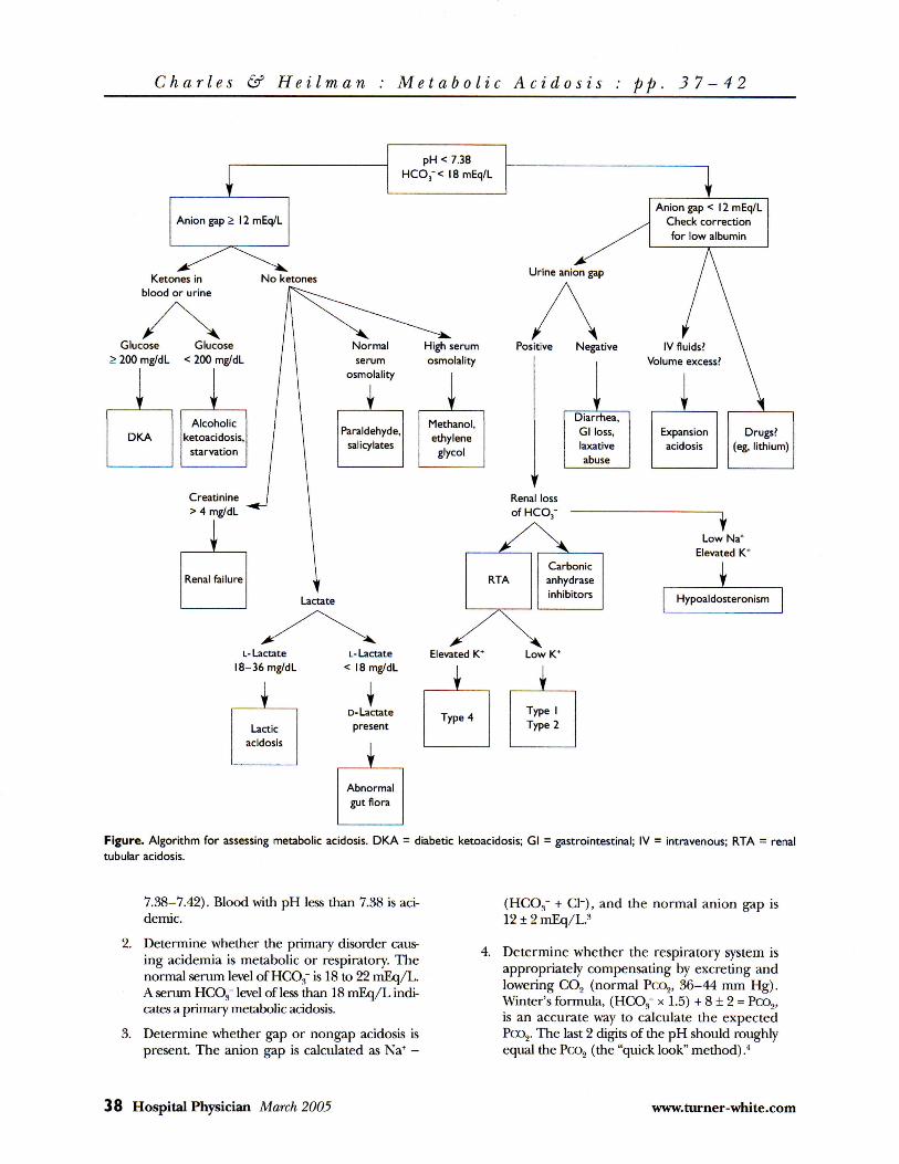
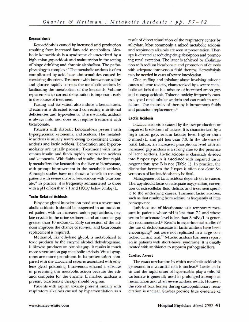
metabolic acidosis · figure provides an algorithm for assessing metabolic acidosis. 1. determine...
TRANSCRIPT
Clinical Review Article
Metabolic Acidosis
Joseph C. Charles, MD Raymond L. Heilman, AID
M etabolic acidosis, the most common acid-base disorder, is associated with many life-threatening conditions. Metabolic acidosis is a state produced by excessive acid produc
tion, reduced acid excretion, or consumption or loss of body alkali. Arterial blood gas analysis typically shows the p H to be less than 7.35 and serum bicarbonate (HCO.,~) to be less than 18 mEq/L. The signs and symptoms of metabolic acidosis are nonspecific, and its diagnosis relies on analysis of laboratory data. Delay in diagnosis is associated with increased mortality and morbidity.1 Early recognition and prompt initiation of treatment are therefore critical. This article discusses the evaluation and management of this important acid-base disorder.
PATHOPHYSIOLOGY Cellular metabolism produces carbon dioxide. By a
reversible intracellular process, CO, combines with water to form carbonic acid (H 2 C0 3 ~). Carbonic acid is able to dissociate into hydrogen ions and HC0 3 ~ ions in a reversible manner. Acidemia is the state of elevated H + concentration and is measured in units of p H . Cells have a narrow p H tange within which they function optimally.
There are 2 major mechanisms whereby cells maintain a constant H + concentration. The C 0 2 - H C 0 3 ~ buffering system is the most important. The primary response to a metabolic acidosis is an increase in ventilation, resulting in increased C 0 2 excretion by diffusion in the lungs. This results in a drop in the blood p H . Additionally, an excess of H + can be excreted by conversion to CO,. The formula representing this buffering system is: H + + HC0 3 ~ «-> H 2CO,~ <-> C 0 2 + H 9 0 . The second mechanism for maintaining p H is a 2-tiered response by the kidneys. First, I T ions are excreted in the proximal tubules, where they combine with HC0 3 ~ to form carbonic acid (H 2 C0 3 ~). In the brush borders of the tubular cells, carbonic acid is converted to C 0 2 and water, and these are reabsorbed. Second, bicarbonate can be regenerated by a reverse process of the buffering system in the lungs ( C 0 2 +
H 2 0 <-> H 2 C 0 3 <-> H ' + HC0 3 ~). A metabolic acidosis can result when either or both of these compensatory responses fails or is overwhelmed.
CLINICAL SIGNS AND SYMPTOMS Metabolic acidosis may be asymptomatic. If present,
the signs and symptoms of metabolic acidosis are relatively nonspecific and may include fatigue, anorexia, confusion, tachycardia, tachypnea, and dehydration. Other manifestations depend on the underlying cause of the disorder.
The adverse hemodynamic effects of a deteriorating metabolic acidosis are profound and, if untreated, can be life threatening. An increase in acidity causes pulmonary vasoconstriction and an increase in pulmonary vascular pressures. These developments can lead to right ventricular failure. At an arterial p H less than 7.2, generalized myocardial depression eventually occurs.2
In arteriolar smooth muscle, a decrease in p H leads to systemic vasodilation, which can cause hypotension and circulatory failure. In patients with underlying lung disease, the burden imposed by the compensatory increase in minute ventilation will progress to respiratory muscle fatigue and failure. The metabolic consequences include hyperkalemia, hypercalcemia, and hypercalciuiia, a calabolic state caused by accelerated amino acid oxidation.
BLOOD GAS ANALYSIS AND INTERPRETATION Metabolic acidosis can be identified by following the
5 steps below, using information from arterial blood gas analysis and serum electrolyte concentrations. The Figure provides an algorithm for assessing metabolic acidosis.
1. Determine whether the patient is alkalemic or acidemic on the basis of arterial pH (normal,
Dr. Charles is division- education coordinator and consultant, Division of Hospital Internal Medicine, Mayo Clinic Hospital, Phoenix, AZ. Dr. Heilman is an assistant professor of medicine, Division of Transplantation Medicine and Nephrology', Mayo Clink, Scottsdale, AZ.
www.turner-white.com Hospital Physician March 2005 37
Charles & Heilman : Metabolic Acidosis : pp. 37-42
Anion gap > 12 mEq/L
Ketones in blood or urine
No ketones
Glucose Glucose > 200 mg/dL < 200 mg/dL
Alcoholic DKA ketoacidosis,
starvation
pH < 7.38 H C C y < 18 mEq/L
Creatinine > 4 mg/dL
Renal failure
Positive Negative
Carbonic anhydrase inhibitors
Anion gap < 12 mEq/L Check correction for low albumin
Diarrhea, Gl loss, Expansion Drugs? laxative acidosis (eg, lithium) abuse
Low Na* Elevated K*
Hypoaldosteronism
i-Lactate 18-36 mg/dL
Lactic acidosis
L-Lactate < 18 mg/dL
D-Lactate present
Abnormal gut flora
Figure. Algorithm for assessing metabolic acidosis. DKA = diabetic ketoacidosis; Gl = gastrointestinal; IV = intravenous; RTA = renal tubular acidosis.
7.38-7.42). Blood with pH less than 7.38 is aci-demic.
2. Determine whether the primary disorder causing acidemia is metabolic or respiratory. The normal serum level of HCO,," is 18 to 22 mEq/L. A serum HCO.f level of less than 18 mEq/L indicates a primary metabolic acidosis.
3. Determine whether gap or nongap acidosis is present. The anion gap is calculated as Na+ -
(HCOs~ + CI"), and the normal anion gap is 12 ± 2 mEq/L.s
4. Determine whether the respiratory system is appropriately compensating by excreting and lowering CO, (normal Pco2, 36-44 mm Hg). Winter's formula, (HC03~ x 1.5) + 8 ± 2 = Pco2, is an accurate way to calculate the expected Pco2. The last 2 digits of the pH should roughly equal the Pco2 (the "quick look" method) .4
38 Hospital Physician March 2005 www.turner-white.com
Charles & Heilman Metabolic Acidosis : pp. 37-42
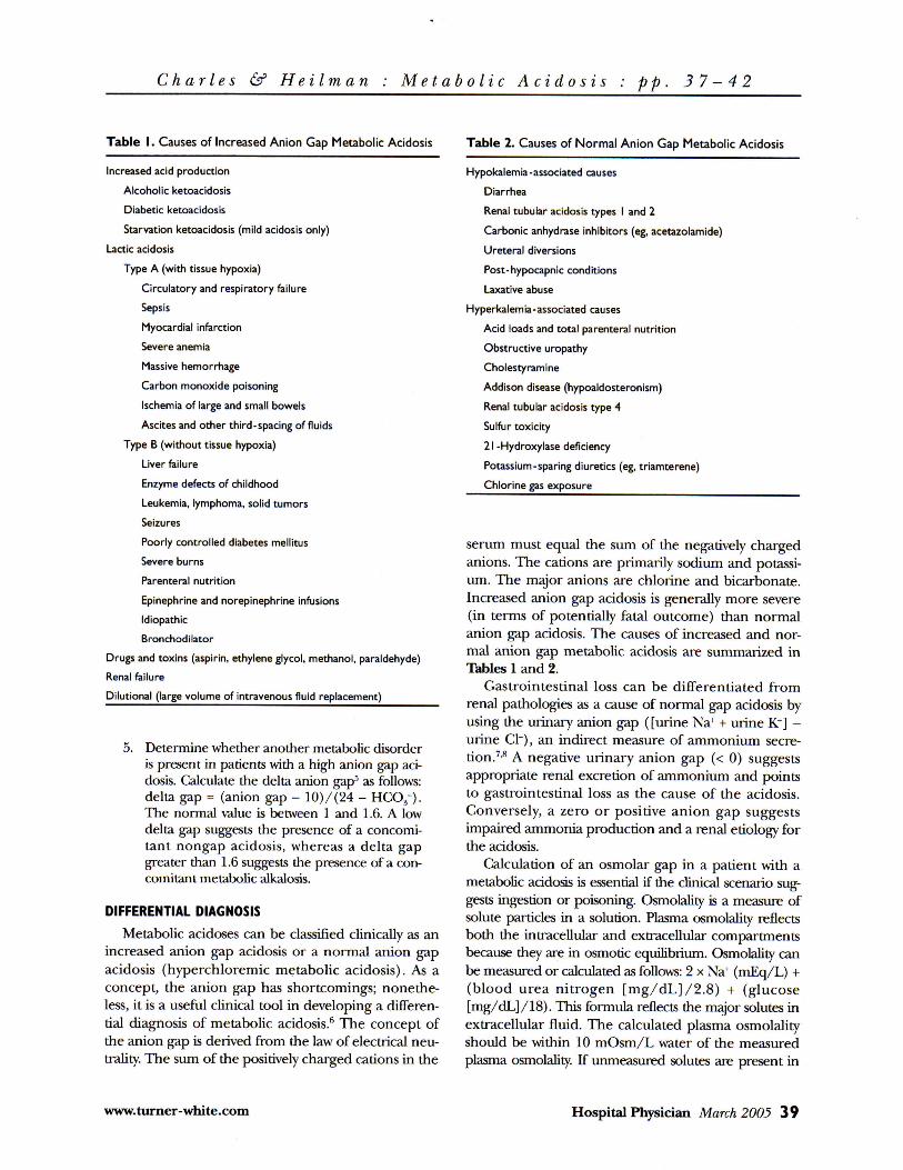
Table I. Causes of Increased Anion Gap Metabolic Acidosis
Increased acid production Alcoholic ketoacidosis Diabetic ketoacidosis Starvation ketoacidosis (mild acidosis only)
Lactic acidosis Type A (with tissue hypoxia)
Circulatory and respiratory failure Sepsis Myocardial infarction Severe anemia Massive hemorrhage Carbon monoxide poisoning Ischemia of large and small bowels Ascites and other third-spacing of fluids
Type B (without tissue hypoxia) Liver failure
Enzyme defects of childhood Leukemia, lymphoma, solid tumors Seizures Poorly controlled diabetes mellitus Severe burns Parenteral nutrition Epinephrine and norepinephrine infusions Idiopathic Bronchodilator
Drugs and toxins (aspirin, ethylene glycol, methanol, paraldehyde) Renal failure Dilutional (large volume of intravenous fluid replacement)
5. Determine whether another metabolic disorder is present in patients with a high anion gap acidosis. Calculate the delta anion gap3 as follows: delta gap = (anion gap - 10)/(24 - HCO,-). The normal value is between 1 and 1.6. A low delta gap suggests the presence of a concomitant nongap acidosis, whereas a delta gap greater than 1.6 suggests the presence of a concomitant metabolic alkalosis.
DIFFERENTIAL DIAGNOSIS Metabolic acidoses can be classified clinically as an
increased anion gap acidosis or a normal anion gap acidosis (hyperchloremic metabolic acidosis). As a concept, the anion gap has shortcomings; nonetheless, it is a useful clinical tool in developing a differential diagnosis of metabolic acidosis.6 The concept of the anion gap is derived from the law of electrical neutrality. The sum of the positively charged cations in the
Table 2. Causes of Normal Anion Gap Metabolic Acidosis
Hypokalemia-associated causes Diarrhea Renal tubular acidosis types I and 2 Carbonic anhydrase inhibitors (eg, acetazolamide) Ureteral diversions Post-hypocapnic conditions Laxative abuse
Hyperkalemia-associated causes Acid loads and total parenteral nutrition Obstructive uropathy Cholestyramine Addison disease (hypoaldosteronism) Renal tubular acidosis type 4 Sulfur toxicity 21 -Hydroxylase deficiency Potassium-sparing diuretics (eg, triamterene) Chlorine gas exposure
serum must equal the sum of the negatively charged anions. The cations are primarily sodium and potassium. The major anions are chlorine and bicarbonate. Increased anion gap acidosis is generally more severe (in terms of potentially fatal outcome) than normal anion gap acidosis. The causes of increased and normal anion gap metabolic acidosis are summarized in Tables 1 and 2.
Gastrointestinal loss can be differentiated from renal pathologies as a cause of normal gap acidosis by using the urinary anion gap ([urine Na + + urine K + ] -urine Cl~), an indirect measure of ammonium secretion.7-8 A negative urinary anion gap (< 0) suggests appropriate renal excretion of ammonium and points to gastrointestinal loss as the cause of the acidosis. Conversely, a zero or positive anion gap suggests impaired ammonia production and a renal etiology for the acidosis.
Calculation of an osmolar gap in a patient with a metabolic acidosis is essential if the clinical scenario suggests ingestion or poisoning. Osmolality is a measure of solute particles in a solution. Plasma osmolality reflects both the intracellular and extracellular compartments because they are in osmotic equilibrium. Osmolality can be measured or calculated as follows: 2 x Na + (mEq/L) + (blood urea nitrogen [mg/dL]/2 .8) + (glucose [mg/dL]/18). This formula reflects the major solutes in extracellular f luid. The calculated plasma osmolality should be within 10 mOsm/L water of the measured plasma osmolality. If unmeasured solutes are present in
www.turner-white.com Hospital Physician March 2005 3 9
Charles & Heilman : Metabolic Acidosis pp. 3 7-42
the plasma, the measured osmolality'will be much higher than the calculated osmolality; this is called an osmolar gap. An osmolar gap in metabolic acidosis should laise suspicion for ethylene glycol or methanol poisoning.
If the anion gap is being used to assess a metabolic acidosis, the anion gap must be adjusted if the patient has h)poalbuminemia. Albumin is a negatively charged protein, and thus hypoalbuminemia falsely lowers the anion gap. The adjustment is made by adding 2.5 to the gap for every 1 g/dL that the albumin is below normal. 9
BICARBONATE THERAPY Treatment strategies for metabolic acidosis are pri
marily directed toward the underlying cause. Bicarbonate therapy is a temporary measure used for severe acidosis (pH < 7.1). The rationale for bicarbonate therapy is that at extracellular p H levels lower than 7.1, small decreases in the level of H C 0 3 " or increases in Pco, are poorly tolerated.
The use of bicarbonate therapy continues to be con-troversial.4 ,10 Risks associated with bicarbonate theiapy include hypernatremia, hyperosmolality, volume overload, and overshoot alkalosis. Also, bicarbonate paradoxically shifts the hemoglobin-oxygen dissociation curve unfavorably, potentially resulting in a worsened cerebral metabolic acidosis.
I f bicarbonate is used, i t should be given cautiously, with frequent acid-base monitor ing and a goal of returning the p H to approximately 7.2. I t should be given as a slow infusion to lessen the effect of CO, generation during buffering. The goal is to adjust the serum HC0 3~ level to 8 to 10 mEq/L. A useful formula for calculating the bicarbonate requirement is: dose of bicarbonate = (desired HC0 3 ~ - serum HC0 3~) (mEq/L) x weight (kg) x 0.5.11 Intravenous bicarbonate is the main alkalinizing agent.
Although the controversy surrounding bicarbonate therapy in metabolic acidosis probably overstates its risks, it has led to a search for alternative agents with fewer adverse effects. Such agents include carbicarb, which consists of equimolar concentrations of sodium bicarbonate and sodium carbonate; tris-hydroxymethyl aminomethane (THAM); and Tiibonat, a mixture of T H A M , acetate, bicarbonate, and phosphate. Only T H A M , however, is currently available in the United States, and the benefits of these agents have not been confirmed. 1 2 " 1 4
MANAGEMENT OF SPECIFIC CONDITIONS Renal Failure
When renal failure progresses to the point that ure
mia is present, metabolic acidosis results. Studies suggest that this leads to protein malnutrition, depressed myocardial contractility, increased bone resorption, and decreased thyroid hormone secretion. 1 8 Correcting the acidosis improves the nutritional status by preventing muscle protein breakdown. 1 6 This correction can be achieved by the oral administration of either sodium bicarbonate or sodium citrate 10% solution. Sodium citrate has fewer gastrointestinal adverse effects than sodium bicarbonate; however, caution is required with long-term use of oral citrate in patients with advanced chronic renal failure because i t has been shown to increase intestinal absorption of aluminum, which can result in chronic bone toxicity. In short-term situations in which serum HC0 3 ~ is less than 15 mEq/L, it can be slowly corrected with intravenous bicarbonate. Too-rapid correction of acidosis can lead to tetany and arrhythmias. In addition, hypernatremia, hyper tension, and edema may occur.
Hyperkalemia is sometimes present in patients with some degree of renal failure or a disturbance of tubular secretion of potassium. Management depends on the degree of hyperkalemia present.
Renal Tubular Acidoses Hyperkalemic hyperchloremic metabolic acidosis
(type 4 renal tubular acidosis) results from either aldosterone deficiency or renal tubules not responding to aldosterone. In cases caused by drug-related nephrotoxicity, removal of the offending agent is indicated. A decrease in serum potassium concentrations often improves the acidosis; the decision to treat is based on the degree of hyperkalemia. A cation exchange resin (sodium polystyrene sulfonate) with restriction of dietary potassium is effective. Hypovolemia should be corrected. Oral bicarbonate may be beneficial. Cases caused by mineralocorticoid deficiency may require replacement therapy.
Type 1 (distal) renal tubular acidosis is frequendy associated with renal stone formation and hypokalemia. The goal of treatment is to eliminate acidosis, which will decrease the hypercalciuria. Alkalinization with oral sodium bicarbonate or Shohl's solution (sodium and potassium citrate) is effective. Potassium supplementation is usually not required.
Type 2 (proximal) renal tubular acidosis is caused by defective H C 0 3 " resorption in the proximal renal tubules. Treatment with oral bicarbonate or citrate salts is beneficial. Potassium supplementation is required in the rare severely acidotic cases that require alkali therapy.
40 Hospital Physician March 2005 www. turner - white. com
Charles & Heilman Metabolic Acidosis : pp. 37-42
Ketoacidosis Ketoacidosis is caused by increased acid production
resulting from increased fatty acid metabolism. Alcoholic ketoacidosis is a syndrome characterized by a high anion gap acidosis and malnutrition in the setting of binge drinking and chronic alcoholism. The pathophysiology is complex.1 7 The metabolic acidosis is often complicated by acid-base abnormalities caused by coexisting disorders. Treatment with intravenous saline and glucose rapidly corrects the metabolic acidosis by facilitating the metabolism of the ketoacids. Volume replacement to correct dehydration is important early in the course of treatment.
Fasting and starvation also induce a ketoacidosis. Treatment is directed toward correcting nutritional deficiencies and hypovolemia. The metabolic acidosis is always mild and does not require treatment with bicarbonate.
Patients with diabetic ketoacidosis present with hyperglycemia, ketonemia, and acidosis. The metabolic acidosis is usually severe owing to coexistent uremic acidosis and lactic acidosis. Dehydration and hyperos-molarity are usually present. Treatment with intravenous insulin and fluids rapidly reverses the acidosis and ketonemia. With fluids and insulin, the liver rapidly metabolizes the ketoacids in the liver to bicarbonate, with prompt improvement in the metabolic acidosis. Although studies have not shown a benefit to treating patients with severe diabetic ketoacidosis with bicarbonate,18 in practice, it is frequently administered to those with a p H of less than 7.1 and HC0 3~ below 8 mEq/L.
Toxin-Related Acidosis Ethylene glycol intoxication produces a severe met
abolic acidosis. It should be suspected in an intoxicated patient with an increased anion gap acidosis, oxy-late crystals in the urine sediment, and an osmolar gap greater than 10 mOsm/L. Early correction of the acidosis improves the chance of survival, and bicarbonate replacement is required.
Methanol, like ethylene glycol, is metabolized to toxic products by the enzyme alcohol dehydrogenase. It likewise produces an osmolar gap. It results in much more severe anion gap metabolic acidosis. Visual symptoms are more prominent in its presentation compared with the ataxia and seizures associated with ethylene glycol poisoning. Intravenous ethanol is effective in preventing this metabolic action because the ethanol competes for the enzyme. I f marked acidosis is present, bicarbonate therapy should be given.
Patients with aspirin toxicity present initially with respiratory alkalosis caused by hyperventilation as a
result of direct stimulation of the respiratory center by saliq'late. Most commonly, a mixed metabolic acidosis and respiratory alkalosis are seen at presentation. Therapy is directed at reducing drug absorption and promoting renal excretion. The latter is achieved by alkaliniza-tion with sodium bicarbonate and promotion of diuresis with adequate inffavenous fluid therapy. Hemodialysis may be needed in cases of severe intoxication.
Glue sniffing and inhalant abuse involving toluene causes toluene toxicity, characterized by a severe metabolic acidosis that is a mixture of increased anion gap and n on gap acidosis. Toluene toxicity frequently causes a type I renal tubular acidosis and can result in renal failure. The mainstay of therapy is intravenous fluids and potassium replacement.19
Lactic Acidosis L-Lactic acidosis is caused by the overproduction or
impaired breakdown of lactate. It is characterized by a high anion gap, serum lactate level higher than 5 mmol/L, and p H less than 7.3. In the absence of renal failure, an increased phosphorus level with an increased gap acidosis is a strong clue to the presence of lactic acidosis. Lactic acidosis is classically divided into 2 types: type A is associated with impaired tissue oxygenation; type B is not (Table 1). In practice, the distinction between the 2 types is often not clear. Severe cases of lactic acidosis may be fatal.
Management of lactic acidosis depends on its causes. Therapy should focus on adequate oxygenation, correction of extracellular fluid deficits, and treatment specific to the underlying causes. Transient lactic acidosis, such as that resulting from seizure, is frequently of little consequence.
Judicious use of bicarbonate as a temporary measure in patients whose p H is less than 7.1 and whose serum bicarbonate level is less than 8 mEq/L is generally recommended.2 0 Results in experimental studies of the use of dichloroacetate in lactic acidosis have been encouraging2 1 but were not replicated in a large controlled clinical trial. 2 2 D-Lactic acidosis has been reported in patients with short-bowel syndrome. I t is usually treated with antibiotics to suppress pathogenic flora.
Cardiac Arrest The exact mechanism by which metabolic acidosis is
generated in myocardial cells is unclear.23 Lactic acidosis and the rapid onset of hypercarbia play a role. Bicarbonate is generally used in prolonged attempts at resuscitation and when severe acidosis results. However, the role of bicarbonate during cardiopulmonary resuscitation is unclear. Studies provide litde evidence of
www.turner-white.com Hospital Physician March 2005 4 I
Charles & Heilman : Metabolic Acidosis : pp . 37-42
b e n e f i t . 2 3 2 4 Coronary perfusion pressure, n o t myocardial p H , seems to determine the success o f resuscitation. Efforts are best directed at establishing adequate oxygenation and effective circulation.
Dilutional Acidosis I n patients receiving intravenous solutions o f lactate,
acetate, or citrate i n large volumes, an increased gap acidosis can develop as a result o f incomplete conversion to bicarbonate. The addit ion o f bicarbonate to the i n f u sion prevents this. By a similar mechanism, the negatively charged salts o f some antibiotics (eg, carbenicil l in) given i n large quantities can cause a metabolic acidosis.
CONCLUSION Metabolic acidosis may be the result o f a transient
and easily reversible condi t ion such as a seizure. M o r e severe metabolic acidoses require precise diagnosis and timely t rea tment o f the u n d e r l y i n g c o n d i t i o n . T h e focus o f any ueatment plan is the underlying disease process; however, i f the p H is lower than 7.2, the effects o f acidemia can dominate c l inical decision m a k i n g . The goal o f alkali therapy is to reverse severe acidemia and protect against the detr imental effects on the cardiovascular system. Intravenous sodium bicarbonate is the mainstay o f alkali therapy and is given as a cont inuous infusion to prevent the effects o f "overshoot" alkalosis. Alternative alkalizing agents such as sodium lactate a n d citrate are no t as reliable because their effects d e p e n d o n o x y g e n a t i o n to b i c a r b o n a t e . Research efforts a i m e d at f i n d i n g alternatives to bicarbonate therapy continue, as do studies to better identify those subgroups o f metabolic acidosis that benefit f r o m alka-linization therapy. HP
REFERENCES 1. Hamblin PS, Topliss DJ, Chosich N, et al. Deaths associ
ated with diabetic ketoacidosis and hyperosmolar coma. 1973-1988. Med J Aust 1989:151:439, 441-2, 444.
2. Orchard CH, Cingolani HE. Acidosis and arrhythmias in cardiac muscle. Cardiovasc Res 1994;28:1312-9.
3. Winter SD, Pearson JR, Gabow PA et al. The fall of the serum anion gap. Arch Intern Med 1990;150:311-3.
4. Fulop M . A guide for predicting arterial CO, tension in metabolic acidosis. AmJ Nephrol 1997;17:421-4.
5. Fall PJ. A stepwise approach to acid-base disorders. Practical patient evaluation for metabolic acidosis and other conditions. Postgrad Med 2000;107:249-50, 253-4, 257-8.
6. Emmett M , Nanus RG. Clinical use of the anion gap. Medicine (Baltimore) 1977;56:38-54.
7. Batlle DC, Hizon M, Cohen E, et al. The use of the urinary anion gap in the diagnosis of hyperchlorcmic meta-
Copyright 2005 by Turner White Communi'
bolic acidosis. N Engl J Med 1988;318:594-9. 8. Goldstein MB, Bear R, Richardson RM, et al. The urine
anion gap: a clinically useful index of ammonium excretion. AmJ Med Sci 1986;292:198-202.
9. Figge J, Jabor A, Kazda A, Fencl V. Anion gap and hypoalbuminemia CritCare Med 1998;26:1807-10.
10. Gabow PA. Sodium bicarbonate: a cure or curse for metabolic acidosis? J Critical Illness 1989;4(5):13-28.
11. Bersin RM, Arieff A l . Improved hemodynamic function during hypoxia with Carbicarb, a new agent for the management of acidosis. Circulation 1988;77:227-33.
12. LeungJM, Landow L, Franks M, et al. Safety and efficacy of intravenous Carbicarb in patients undergoing surgery comparison with sodium bicarbonate in the ueatment of mild metabolic acidosis [published erratum appears in Crit Care Med 1995;23:420]. SPI Research Group. Study of Perioperative Ischemia. CritCare Med 1994;22:1540-9.
13. Brasch H , Thies E, Iven H . Pharmacokinetics of TRIS (hydroxymethyl-)aminomethane in healthy subjects and in patients with metabolic acidosis. Eur J Clin Pharmacol 1982;22:257-64.
14. Bjerneroth G. Alkaline buffers for correction of metabolic acidosis during cardiopulmonary resuscitation with focus on Tribonat—a review. Resuscitation 1998;37: 161-71.
15. Mitch WE. Uremia and the control of protein metabolism. Nephron 1988;49:89-93.
16. Walls J. Effect of correction of acidosis on nutritional status in dialysis patients. Miner Electrolyte Metab 1997; 23:234-6.
17. Wrenn KD, Slovis CM, Minion GE, Rutkowski R. The syndrome of alcoholic ketoacidosis. A m J Med 1991;91: 119-28.
18. Viallon A, Zeni F, Lafond P, et al. Does bicarbonate therapy improve the management of severe diabetic ketoacidosis? CritCare Med 1999;27:2690-3.
19. Carlisle EJ, Donnelly SM, Vasuvattakul S, et al. Glue-sniffing and distal renal tubular acidosis: sticking to the facts. J Am Soc Nephrol 1991;1:1019-27.
20. Adrogue HJ, Madias NE. Management of life-threatening acid-base disorders. Fii'st of two parts [published erratum appears in N Engl J Med 1999:340:247]. N Engl J Med 1998;338:26-34.
21. Stacpoole PW, Lorcnz AC, Thomas RG, Harman EM. Dichloroacetate in die treatment of lactic acidosis. Ann Intern Med 1988;108:58-63.
22. Stacpoole PW, Wright EC, Baumgartner TG, et al. A controlled clinical trial of dichloroacetate for U'eatment of lactic acidosis in adults. The Dichloroacetate-Lactic Acidosis Study Group. N Engl J Med 1992;327:1564-9.
23. Shapiro JI. Pathogenesis of cardiac dysfunction dining metabolic acidosis: therapeutic implications. Kidney Int Suppl 1997;61:S47-51.
24. Kette F, Weil M H , von Planta M, et al. Buffer agents do not reverse intramyocardial acidosis during cardiac resuscitation. Circulation 1990;81:1660-6.
'ions Inc., Wayne, PA. All rights reserved.
42 Hospital Physician March 2005 www.turner-white.com