metagenomic characterization of indoor dust bacterial and
TRANSCRIPT

HAL Id: hal-02930076https://hal.archives-ouvertes.fr/hal-02930076
Submitted on 4 Sep 2020
HAL is a multi-disciplinary open accessarchive for the deposit and dissemination of sci-entific research documents, whether they are pub-lished or not. The documents may come fromteaching and research institutions in France orabroad, or from public or private research centers.
L’archive ouverte pluridisciplinaire HAL, estdestinée au dépôt et à la diffusion de documentsscientifiques de niveau recherche, publiés ou non,émanant des établissements d’enseignement et derecherche français ou étrangers, des laboratoirespublics ou privés.
Distributed under a Creative Commons Attribution| 4.0 International License
Metagenomic Characterization of Indoor Dust Bacterialand Fungal Microbiota in Homes of Asthma and
Non-asthma Patients Using Next Generation SequencingJ-P Gangneux, Mohamed Sassi, Pierre Lemire, Pierre Le Cann
To cite this version:J-P Gangneux, Mohamed Sassi, Pierre Lemire, Pierre Le Cann. Metagenomic Characterization ofIndoor Dust Bacterial and Fungal Microbiota in Homes of Asthma and Non-asthma Patients Us-ing Next Generation Sequencing. Frontiers in Microbiology, Frontiers Media, 2020, 11, pp.1671.�10.3389/fmicb.2020.01671�. �hal-02930076�

fmicb-11-01671 July 30, 2020 Time: 11:56 # 1
ORIGINAL RESEARCHpublished: 30 July 2020
doi: 10.3389/fmicb.2020.01671
Edited by:Sara Gago,
The University of Manchester,United Kingdom
Reviewed by:Raquel Sabino,
National Institute of Health Dr. RicardoJorge, Portugal
Danielle Weaver,The University of Manchester,
United Kingdom
*Correspondence:Jean-Pierre Gangneux
Specialty section:This article was submitted toFungi and Their Interactions,
a section of the journalFrontiers in Microbiology
Received: 25 February 2020Accepted: 25 June 2020Published: 30 July 2020
Citation:Gangneux J-P, Sassi M, Lemire P
and Le Cann P (2020) MetagenomicCharacterization of Indoor DustBacterial and Fungal Microbiota
in Homes of Asthma and Non-asthmaPatients Using Next Generation
Sequencing.Front. Microbiol. 11:1671.
doi: 10.3389/fmicb.2020.01671
Metagenomic Characterization ofIndoor Dust Bacterial and FungalMicrobiota in Homes of Asthma andNon-asthma Patients Using NextGeneration SequencingJean-Pierre Gangneux* , Mohamed Sassi, Pierre Lemire and Pierre Le Cann
Univ Rennes, CHU Rennes, Inserm, EHESP, Institut de Recherche en Santé, Environnement et Travail (Irset) – UMR_S 1085,Rennes, France
Background: The exposure of house occupants to indoor air pollutants has increasedin recent decades. Among microbiological contaminants, bacterial and fungal aerosolsremain poorly studied and the debate on the impact of these aerosols on respiratoryhealth is still open. This study aimed to assess the diversity of indoor microbialcommunities in relationship with the health of occupants.
Methods: Measurements were taken from dwellings of 2 cohorts in Brittany (France),one with children without any pathology and the other with children and adults withasthma. Thirty dust samples were analyzed by next generation sequencing with a 16Sand 18S targeted metagenomics approach. Analysis of sequencing data was performedusing qiime 2, and univariate and multivariate statistical analysis using R software andphyloseq package.
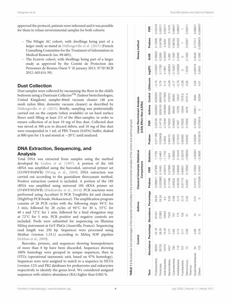
Results: A total of 2,637 prokaryotic (589 at genus level) and 2,153 eukaryotictaxa were identified (856 fungal taxa (39%) and 573 metazoa (26%)). The four mainbacterial phyla were identified: Proteobacteria (53%), Firmicutes (27%), Actinobacteria(11%), Bacteroidetes (8%). Among Fungi, only 136 taxa were identified at genus level.Three main fungal phyla were identified: Ascomycota (84%), Basidiomycota (12%)and Mucoromycota (3%). No bacterial nor fungal phyla were significantly associatedwith asthma versus control group. A significant over representation in control groupversus asthma was observed for Christensenellaceae family (p-value = 0.0015, adj.p-value = 0.033). Besides, a trend for over representation in control group was observedwith Dermabacteraceae family (p-value = 0.0002, adj. p-value = 0.815).
Conclusions: Our findings provide evidence that dust samples harbor a high diversityof human-associated bacteria and fungi. Molecular methods such as next generation
Frontiers in Microbiology | www.frontiersin.org 1 July 2020 | Volume 11 | Article 1671

fmicb-11-01671 July 30, 2020 Time: 11:56 # 2
Gangneux et al. Dust Microbiota and Asthma Patients
sequencing are reliable tools for identifying and tracking the bacterial and fungal diversityin dust samples, a less easy strategy for the detection of eukaryotes at least using18Smetagenomics approach. This study showed that the detection of some bacteria mightbe associated to indoor air of asthmatic patients. Regarding fungi, a higher number ofsamples and sequencing with more depth could allow reaching significant signatures.
Keywords: microbiota, mycobiota, asthma, indoor environment, dust, next-generation sequencing,metagenomics, fungi
INTRODUCTION
Asthma is a common chronic inflammatory airway diseasein children and adults from developed countries. In France,it affects more than 10% of children and 6% of adults(Delmas and Fuhrman, 2010).
Asthma is a very complex disease characterized byenhanced bronchial reactivity leading to airway obstructionand respiratory difficulties (Martinez and Vercelli, 2013).Exposure to environmental chemical or biological factors canlead to exacerbation of asthma and prevention measures are stillneeded in order to reduce the exposure to these agents (Beasleyet al., 2015; Shaheen, 2019). The airway microbiome in asthmapatients results from numerous factors. Environmental exposure,a complex relationship with the gastrointestinal microbiome,the development of immune function, and a predisposition toallergic sensitization and asthma are among the most referredfactors (Huang and Boushey, 2015).
Indoor fungal exposure has been associated to thedevelopment of asthma (Chen et al., 2014). But, according to Egeet al. (2011), children who grow up in the environments with awide range of microbial exposures, like farming environments,are more likely protected from childhood asthma and atopy thanurban children. Feng et al. (2016) reported that parental allergicdiseases, atopy, diet and early life exposures might explain thehigher prevalence of asthma in the urban environment. Also,there is a complex interplay between genetic predispositionand environmental exposures, including microbes and allergens(Anderson and Jackson, 2017). Regarding exposure to molds, itis estimated that in France about 125,000 individuals suffer fromsevere asthma with fungal sensitization (SAFS) episodes (189cases/100,000 adults per year) (Gangneux et al., 2016).
Among biological agents, bacteria have been less studied evenif endotoxins produced by gram negative bacteria have beensignificantly correlated with wheezing in children (Horick et al.,2006). Moreover, many studies have suggested that exposure tolipopolysaccharide (LPS), in non-rural environments, is a riskfactor for increased asthma prevalence and severity of disease(Thorne et al., 2005).
Culture-based studies have been in use for a long timefor the detection of various microorganisms indoor (Rintalaet al., 2008, 2012; Méheust et al., 2014). However, while cultureisolation focuses on the presence of particular bacteria or fungi,next generation sequencing (NGS) studies unveil microbialcommunities comprising thousands of uncultured microbes.Recent advances in microbiological methods show promise indetermining whether the microbes recovered during indoor
sampling campaigns may in fact be causative agents as such, oronly surrogates of an underlying harmful or beneficial exposure.Given the lack of knowledge on the specificities of the relevantexposures, assessments at this stage need to be kept broad andcomprehensive (Mensah-Attipoe et al., 2017).
According to Singanayagam et al. (2017), who underlined therole of microbes in the exacerbation of asthma, the emergence ofnext generation molecular sequencing techniques to characterizethe microbiota has facilitated renewed interest. They concludedthat longitudinal studies that characterize the changes in lowerrespiratory tract microbiota from birth up to development ofasthma are currently unavailable. Additionally, few studies havefocused on the interactions between bacteria and the immunesystem in driving development of nonatopic asthma phenotypes.
Thus, despite the recognition of the importance of microbialexposure for human health, the precise role of microbes in
FIGURE 1 | Bacterial alpha-diversity, measured by Shannon diversity Index isplotted for samples with asthma (red) and controls (blue). The line inside thebox represents the median, while the whiskers represent the lowest andhighest values within the 1.5 interquartile range (IQR). Outliers as well asindividual sample values are shown as dots. Mann-Whitney statistical testingshowed no significant difference in diversity between the two groups(pShannon = 0.5393).
Frontiers in Microbiology | www.frontiersin.org 2 July 2020 | Volume 11 | Article 1671

fmicb-11-01671 July 30, 2020 Time: 11:56 # 3
Gangneux et al. Dust Microbiota and Asthma Patients
the development and exacerbation of respiratory symptoms andallergies remains poorly understood.
The objective of our study was to use 16S and 18S rRNAsequencing approaches to characterize and identify indoorbacterial and eukaryotic communities that may be associatedwith the exacerbation of asthma. This pilot study compared twogroups of asthmatic and non-asthmatic people’s dwellings.
MATERIALS AND METHODS
Study Population and Home VisitsThirty dust samples were collected from dwellings of 2 Frenchcohorts and were analyzed by next-generation sequencing:
(i) 15 dwellings of asthma patients (ASTHMA) from TheEcenvir cohort, which aims to evaluate the clinical andthe economic impact of Indoor Environment Counselor(IEC) on the symptoms of severe asthma. Patients answered
a questionnaire during an interview with the IEC ondwellings characteristics (localization in cities or rural,apartment or house, . . .) and cleaning habits of patients.Asthma was graded with the GINA (Global Initiative forAsthma) scale and patients had three clinical check-ups.
(ii) 15 control dwellings of control patients (CONTROL) fromthe Pélagie AC cohort, which follows more than 3,500pregnant women in Brittany from pregnancy to age of20 of children. The Pélagie AC project aims to assess theeffects on children respiratory health of chemicals andbiological pollutants exposure. One of the check-up ofPélagie AC cohort occurs once the children are 6 years old.A subset of families without asthma answered a detailedquestionnaire about their dwellings characteristics and lifefor the Pélagie AC study.
According to the French Public Health Law, protocols ofthis type are exempt from the requirement for formal informedconsent. However, The INSERM and Rennes Ethical Committees
FIGURE 2 | Hierarchical clustering analysis: Dendrogram of Bray-Curtis dissimilarity matrices between samples based on 16S data at feature level with UPGMAmethod.
Frontiers in Microbiology | www.frontiersin.org 3 July 2020 | Volume 11 | Article 1671
fmicb-11-01671 July 30, 2020 Time: 11:56 # 4
Gangneux et al. Dust Microbiota and Asthma Patients
approved the protocol, patients were informed and it was possiblefor them to refuse environmental samples for both cohorts:
– The Pélagie AC cohort, with dwellings being part of alarger study as stated in Dallongeville et al. (2015) (FrenchConsulting Committee for the Treatment of Information inMedical Research (no. 09.485),
– The Ecenvir cohort, with dwellings being part of a largerstudy as approved by the Comité de Protection desPersonnes de Rennes Ouest V (8 january 2013, N◦ID RCB2012-A01414-39).
Dust CollectionDust samples were collected by vacuuming the floor in the child’sbedroom using a Dustream CollectorTM (Indoor biotechnologies,United Kingdom) sampler-fitted vacuum cleaner (40 µmmesh nylon filter, domestic vacuum cleaner) as described byDallongeville et al. (2015). Briefly, sampling was preferentiallycarried out on the carpets (when available) or on hard surfacefloors until filling at least 2/3 of the filter-sampler, in order toensure collection of at least 10 mg of fine dust. Collected dustwas sieved at 300 µm to discard debris, and 10 mg of fine dustwere resuspended in 1 mL of PBS-Tween (0.05%) buffer, shakedat 800 rpm for 1 h and stored at −20◦C until analyzed.
DNA Extraction, Sequencing, andAnalysisTotal DNA was extracted from samples using the methoddeveloped by Godon et al. (1997). A portion of the 16SrRNA was amplified using the barcoded, universal primer set(515WF/918WR) (Wang et al., 2009). DNA extraction wascarried out according to the guanidium thiocyanate method.Positive extraction control is included. A portion of the 18SrRNA was amplified using universal 18S rRNA primer set(574WF/952WR) (Hadziavdic et al., 2014). PCR reactions wereperformed using AccuStart II PCR ToughMix kit and cleaned(HighPrep PCR beads, Mokascience). The amplification programconsists of 28 PCR cycles with the following steps: 94◦C for3 min, followed by 28 cycles of 94◦C for 30 s, 53◦C for40 s and 72◦C for 1 min, followed by a final elongation stepat 72◦C for 5 min. PCR positive and negative controls areincluded. Pools were submitted for sequencing on IlluminaMiSeq instrument at GeT-PlaGe (Auzeville, France). Sequencingread length was 250 bp. Sequences were processed usingMothur (version 1.33.1) according to MiSeq SOP pipeline(Schloss et al., 2009).
Barcodes, primers, and sequences showing homopolymersof more than 8 bp have been discarded. Sequences showing100% homology were grouped in unique sequences, then inOTUs (operational taxonomic unit, based on 97% homology).Sequences were next assigned to match to a sequence in SILVA(version 123) and PR2 databases for prokaryotes and eukaryotesrespectively to identify the genus level. We considered assignedsequences with relative abundance (RA) higher than 0.002 %. TA
BLE
1|A
bund
ance
san
dpe
rcen
tage
sof
the
bact
eria
lcom
mun
ities
inth
een
viro
nmen
tals
ampl
esof
the
cont
rola
ndas
thm
agr
oups
.
Co
ntro
lA
sthm
aC
lass
ical
Uni
vari
ate
Sta
tist
ical
Co
mp
aris
ons
Line
arD
iscr
imin
ant
Ana
lysi
s(L
DA
)Eff
ect
Siz
e(L
EfS
e)D
eseq
met
hod
Taxa
Ab
und
ance
Per
cent
age
Ab
und
ance
Per
cent
age
Pva
lues
FDR
Sta
tist
ics
Pva
lues
FDR
Ast
hma
Co
ntro
lLD
Asc
ore
log
2FC
lfcS
EP
valu
esFD
R
Pro
teob
acte
ria39
305
42.1
867
401
58.8
40.
0866
110.
2799
51.
7759
0.07
1185
0.23
728
5335
600
4209
300
−5.
75−
0.54
907
0.36
825
0.13
595
0.45
318
Firm
icut
es35
733
38.3
527
883
24.3
40.
0209
820.
1049
1−
2.44
610.
0294
360.
1471
826
3910
038
5970
05.
790.
1778
20.
2959
0.54
788
0.93
211
Act
inob
acte
ria12
415
13.3
210
543
9.2
0.25
007
0.50
014
−1.
1745
0.29
020.
7254
910
5280
013
1780
05.
120.
1867
0.28
297
0.50
940.
9321
1
Bac
tero
idet
es44
214.
7475
426.
580.
1119
80.
2799
51.
641
0.60
413
0.79
302
8506
0047
2570
−5.
28−
0.90
264
0.38
90.
0203
20.
1016
Cya
noba
cter
ia79
00.
8579
10.
690.
9363
20.
9363
20.
0806
20.
9482
50.
9482
589
040
8606
5−
3.17
−0.
1635
71.
1595
0.88
782
0.98
647
Aci
doba
cter
ia11
80.
1317
30.
150.
9018
50.
9363
20.
1244
50.
7161
30.
7957
1375
612
537
−2.
79−
0.54
658
2.20
490.
8042
20.
9864
7
Fuso
bact
eria
101
0.11
154
0.13
0.67
713
0.84
641
0.42
078
0.60
465
0.79
302
1234
475
76.6
−3.
380
01
1
Dei
noco
ccus
_Th
erm
us73
0.08
510.
040.
5005
0.73
593
−0.
6825
40.
6344
20.
7930
244
7881
31.4
3.26
1.25
332.
1463
0.55
927
0.93
211
Eps
ilonb
acte
raeo
ta53
0.06
120.
010.
5151
50.
7359
3−
0.65
921
0.55
026
0.79
302
2283
.160
843.
28−
0.85
519
2.95
280.
7721
10.
9864
7
Oth
ers
172
0.18
00
0.01
5326
0.10
491
−2.
5826
0.00
7561
90.
0756
190
2029
44.
015.
5643
2.15
850.
0099
431
0.09
9431
Frontiers in Microbiology | www.frontiersin.org 4 July 2020 | Volume 11 | Article 1671

fmicb-11-01671 July 30, 2020 Time: 11:56 # 5
Gangneux et al. Dust Microbiota and Asthma Patients
FIGURE 3 | Proportion of bacterial community at phylum level in the control and asthma groups at the phylum level. *0.01 < p ≤ 0.05, based on Mann-WhitneyU-test.
Sequencing Data AnalysisAnalysis of sequencing data was performed using QIIME 21, andunivariate and multivariate statistical analysis using R softwareand phyloseq package.
RESULTS
Comparison of Microbiota in theEnvironmental SamplesThe sequencing of the 16S rRNA allowed the identification of2,637 bacterial taxa, 589 at genus level with a predominanceof Proteobacteria (53%), Firmicutes (27%) and Actinobacteria(11%). Regarding 18S, 2,153 eukaryotic taxa were identified:856 fungal taxa (39%) and 573 metazoa (26%). AmongFungi, only 136 taxa were identified at genus level. The16S and 18S sequencing data have been uploaded at EBIMetagenomics under the accession numbers PRJEB37043 andPRJEB37050 respectively.
BacteriaThe Shannon index was used to evaluate the intra-groupdiversity in the environmental samples of the two groups;it was 3.68 ± 0.67 in the control group and 3.94 ± 0.22in the asthma group, showing no significant difference indiversity between the two groups (Mann-Whitney statisticalp = 0.5393) (Figure 1). The beta-diversity results using
1https://qiime2.org
ANOSIM method indicated that the bacterial compositionin the two groups was different as the global R-value was0.109, with a p-value of 0.004. Hierarchical clustering analysis(Figure 2) indicated no distinct difference in the OTUs forthe environmental samples of the two groups. There was nosignificant difference between the compositions of the bacterialcommunities in the environmental samples of the two groups,when analyzed at feature level (Supplementary Figure 1).When analyzed at the phylum level the abundance for thephylum Firmicutes was 38.35% in the control group, significantlyhigher than and 24.34% in the asthma group (24.34%,p = 0.02) (Table 1). Further, Proteobacteria, Actinobacteriaand Bacteroides showed an abundance of 42.18, 13.32, and4.74% in the control group and 58.84, 9.20, and 6.58% inthe asthma group, respectively, with no significant differencebetween the compositions of the bacterial communities in theenvironmental samples of the two groups (Table 1, Figure 3 andSupplementary Figure 1).
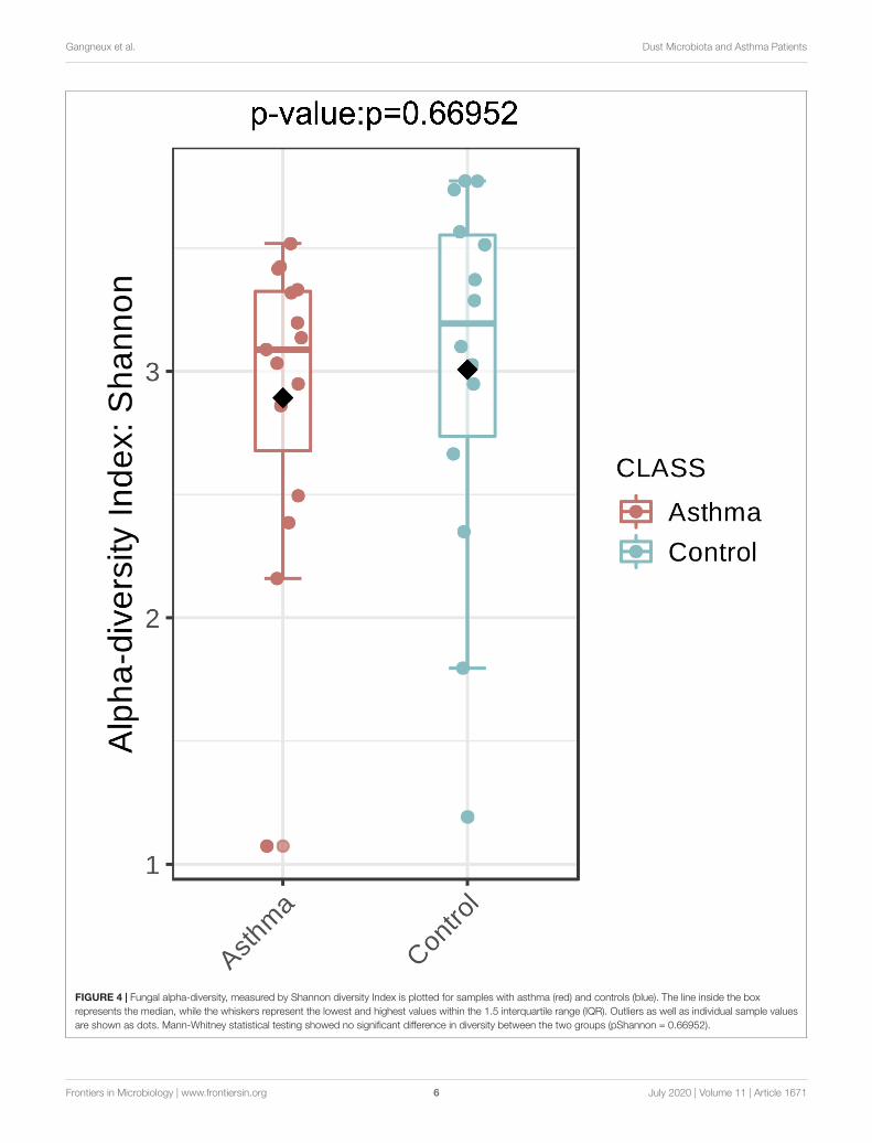
FungiThe Shannon index was used to evaluate the intra-groupdiversity in the environmental samples of the two groups; itwas 3.00 ± 0.77 in the control group and 2.89 ± 0.64 in theasthma group, showing no significant difference in diversitybetween the two groups (Mann-Whitney statistical p = 0.66952)(Figure 4). The beta-diversity results using ANOSIM methodindicated that the fungal composition in the two groups wasnot different as the global R- value was -0.003, with a p-valueof 0.859. There was no significant difference between the
Frontiers in Microbiology | www.frontiersin.org 5 July 2020 | Volume 11 | Article 1671
fmicb-11-01671 July 30, 2020 Time: 11:56 # 6
Gangneux et al. Dust Microbiota and Asthma Patients
FIGURE 4 | Fungal alpha-diversity, measured by Shannon diversity Index is plotted for samples with asthma (red) and controls (blue). The line inside the boxrepresents the median, while the whiskers represent the lowest and highest values within the 1.5 interquartile range (IQR). Outliers as well as individual sample valuesare shown as dots. Mann-Whitney statistical testing showed no significant difference in diversity between the two groups (pShannon = 0.66952).
Frontiers in Microbiology | www.frontiersin.org 6 July 2020 | Volume 11 | Article 1671

fmicb-11-01671 July 30, 2020 Time: 11:56 # 7
Gangneux et al. Dust Microbiota and Asthma Patients
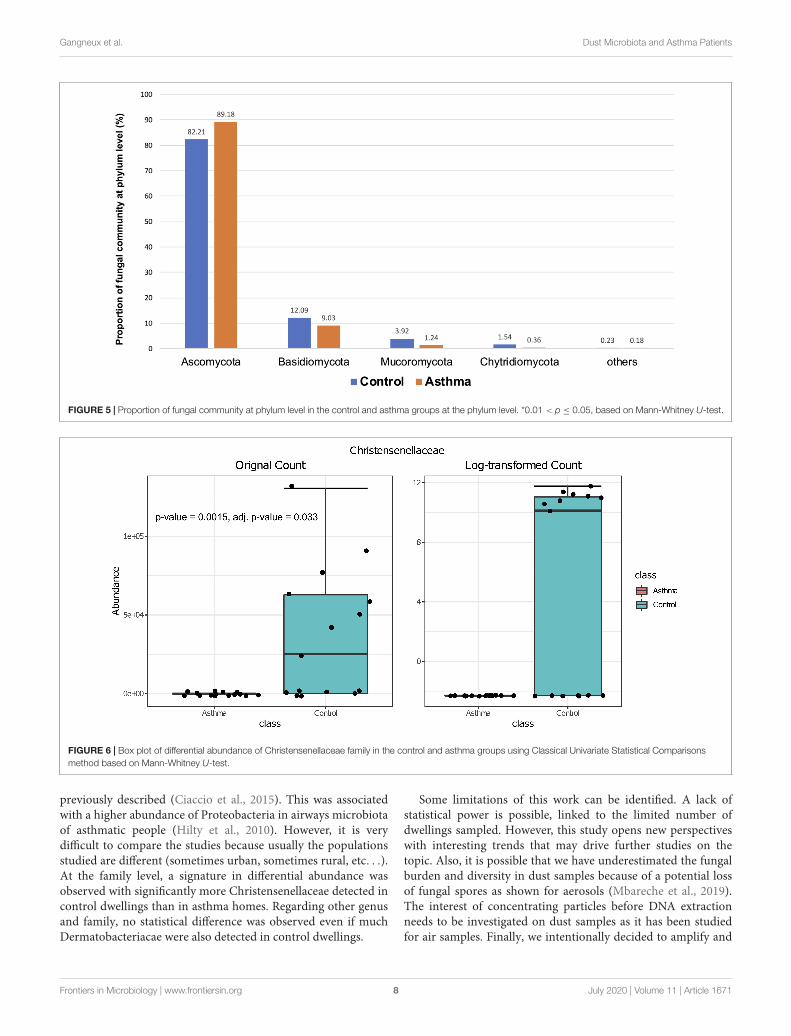
compositions of the fungal communities in the environmentalsamples of the two groups, when analyzed at feature level(Supplementary Figure 2) and phylum level (Table 2). Forthe phylum Ascomycota, the abundance was 82.21% in thecontrol group and 89.18% in the asthma group. Furthermore,Basidiomycota and Mucoromycota showed an abundance of12.09 and 3.92% in the control group and 9.03 and 1.24% in theasthma group, respectively (Figure 5).
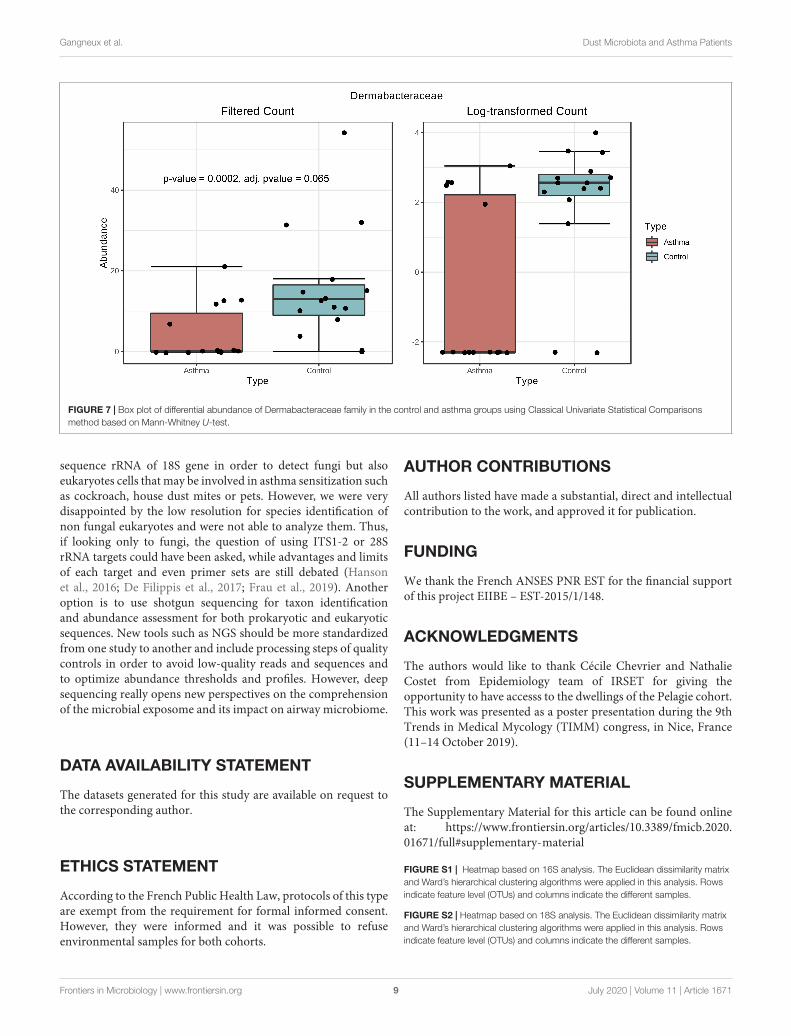
Differential Abundance AnalysisBacterial and fungal taxon-levels (OTU) differences in relativeabundance were examined between the asthma and controlgroups. A significant over representation of specific bacteriawas observed in control patients’ group for Christensenellaceaefamily (p-value = 0.0015, adj. p-value = 0.033) (Figure 6) andonly a trend for Dermabacteraceae family (p-value = 0.0002, adj.p-value = 0.065) (Figure 7).
DISCUSSION
Health effects of environmental microbes are still debated andnot completely understood. A first approach is to examinerelationships between genes, microbes in airways and gut, andthe environment in asthma causes. This is a complex, dynamicand very heterogeneous process still unresolved (Huang andBoushey, 2015). Besides, healthy environments may have impacton the exacerbation of asthma and the usefulness of allergenavoidance on asthma control have shown various evidence oftheir efficacy on the clinical improvement of patients (Le Cannet al., 2017; Gangneux et al., 2020). In this work, our aim wasto compare microbial communities in asthma patients dwellingscompared to non-asthma control homes, and to characterizeand identify indoor bacterial and eukaryotic communities thatmay be associated with the exacerbation of asthma. In theliterature, exposure to low fungal and bacterial richness inhouse dust is associated with an increased risk of asthmadevelopment (Ege et al., 2011; Dannemiller et al., 2013). Air,dust and surface sampling strategies to detect bacteria and fungihave alternately advantages and limits, as well as the differentmethods of detection, identification and quantification (Méheustet al., 2014; Gangneux et al., 2019). Because our objective forthis work was to use NGS, we decided to sample dust byvacuuming the floor rather than aerosols in order to gain insensitivity. Another valuable technical option described in theliterature relies on the use of electrostatic dust cloths for furthermolecular, immunological or cultural analysis (Cox et al., 2017;Kristono et al., 2019).
In our work, the alpha- and beta-diversity results indicatedthat the bacterial and fungal composition in the two groups werenot different, without significant difference when analyzed atthe phylum level. On a global point of view, our results are inaccordance to others as the main phylum detected in both groupis Proteobacteria, then Firmicutes, Actinobacteria, Bacteroidetes(Hewitt et al., 2012), with Firmicutes over represented in thecontrol group compared to asthma dwellings. Besides, there wasa trend of more Proteobacteria detected in asthma population as TA
BLE
2|A
bund
ance
san
dpe
rcen
tage
sof
the
fung
alco
mm
uniti
esin
the
envi
ronm
enta
lsam
ples
ofth
eco
ntro
land
asth
ma
grou
ps.
Co
ntro
lA
sthm
aC
lass
ical
Uni
vari
ate
Sta
tist
ical
Co
mp
aris
ons
Line
arD
iscr
imin
ant
Ana
lysi
s(L
DA
)Eff
ect
Siz
e(L
EfS
e)D
eseq
met
hod
Taxa
Ab
und
ance
Per
cent
age
Ab
und
ance
Per
cent
age
Pva
lues
FDR
Sta
tist
ics
Pva
lues
FDR
Ast
hma
Co
ntro
lLD
Asc
ore
log
2FC
lfcS
EP
valu
esFD
R
Asc
omyc
ota
1364
9282
.21
1697
4789
.18
0.11
207
0.31
014
142
0.10
635
0.29
345
8744
400
8042
200
−5.
55−
0.39
748
0.40
435
0.32
560.
5150
3
Bas
idio
myc
ota
2007
812
.09
1719
49.
030.
1860
80.
3101
474
0.17
607
0.29
345
9963
7013
8810
05.
290.
2240
10.
2231
30.
3154
10.
5150
3
Muc
orom
ycot
a65
153.
9223
571.
240.
9125
10.
9125
110
80.
8951
10.
8951
120
1920
3580
104.
89−
1.28
480.
9958
10.
1969
80.
5150
3
Chy
trid
iom
ycot
a25
641.
5469
10.
360.
1655
40.
3101
475
0.15
849
0.29
345
3327
518
5200
4.88
0.99
249
1.48
680.
5044
40.
5150
3
Oth
ers
376
0.23
352
0.18
0.33
805
0.42
257
123.
50.
3248
10.
4060
124
060
2647
33.
08−
1.29
741.
9928
0.51
503
0.51
503
Frontiers in Microbiology | www.frontiersin.org 7 July 2020 | Volume 11 | Article 1671
fmicb-11-01671 July 30, 2020 Time: 11:56 # 8
Gangneux et al. Dust Microbiota and Asthma Patients
FIGURE 5 | Proportion of fungal community at phylum level in the control and asthma groups at the phylum level. *0.01 < p ≤ 0.05, based on Mann-Whitney U-test.
FIGURE 6 | Box plot of differential abundance of Christensenellaceae family in the control and asthma groups using Classical Univariate Statistical Comparisonsmethod based on Mann-Whitney U-test.
previously described (Ciaccio et al., 2015). This was associatedwith a higher abundance of Proteobacteria in airways microbiotaof asthmatic people (Hilty et al., 2010). However, it is verydifficult to compare the studies because usually the populationsstudied are different (sometimes urban, sometimes rural, etc. . .).At the family level, a signature in differential abundance wasobserved with significantly more Christensenellaceae detected incontrol dwellings than in asthma homes. Regarding other genusand family, no statistical difference was observed even if muchDermatobacteriacae were also detected in control dwellings.
Some limitations of this work can be identified. A lack ofstatistical power is possible, linked to the limited number ofdwellings sampled. However, this study opens new perspectiveswith interesting trends that may drive further studies on thetopic. Also, it is possible that we have underestimated the fungalburden and diversity in dust samples because of a potential lossof fungal spores as shown for aerosols (Mbareche et al., 2019).The interest of concentrating particles before DNA extractionneeds to be investigated on dust samples as it has been studiedfor air samples. Finally, we intentionally decided to amplify and
Frontiers in Microbiology | www.frontiersin.org 8 July 2020 | Volume 11 | Article 1671
fmicb-11-01671 July 30, 2020 Time: 11:56 # 9
Gangneux et al. Dust Microbiota and Asthma Patients
FIGURE 7 | Box plot of differential abundance of Dermabacteraceae family in the control and asthma groups using Classical Univariate Statistical Comparisonsmethod based on Mann-Whitney U-test.
sequence rRNA of 18S gene in order to detect fungi but alsoeukaryotes cells that may be involved in asthma sensitization suchas cockroach, house dust mites or pets. However, we were verydisappointed by the low resolution for species identification ofnon fungal eukaryotes and were not able to analyze them. Thus,if looking only to fungi, the question of using ITS1-2 or 28SrRNA targets could have been asked, while advantages and limitsof each target and even primer sets are still debated (Hansonet al., 2016; De Filippis et al., 2017; Frau et al., 2019). Anotheroption is to use shotgun sequencing for taxon identificationand abundance assessment for both prokaryotic and eukaryoticsequences. New tools such as NGS should be more standardizedfrom one study to another and include processing steps of qualitycontrols in order to avoid low-quality reads and sequences andto optimize abundance thresholds and profiles. However, deepsequencing really opens new perspectives on the comprehensionof the microbial exposome and its impact on airway microbiome.
DATA AVAILABILITY STATEMENT
The datasets generated for this study are available on request tothe corresponding author.
ETHICS STATEMENT
According to the French Public Health Law, protocols of this typeare exempt from the requirement for formal informed consent.However, they were informed and it was possible to refuseenvironmental samples for both cohorts.
AUTHOR CONTRIBUTIONS
All authors listed have made a substantial, direct and intellectualcontribution to the work, and approved it for publication.
FUNDING
We thank the French ANSES PNR EST for the financial supportof this project EIIBE – EST-2015/1/148.
ACKNOWLEDGMENTS
The authors would like to thank Cécile Chevrier and NathalieCostet from Epidemiology team of IRSET for giving theopportunity to have accesss to the dwellings of the Pelagie cohort.This work was presented as a poster presentation during the 9thTrends in Medical Mycology (TIMM) congress, in Nice, France(11–14 October 2019).
SUPPLEMENTARY MATERIAL
The Supplementary Material for this article can be found onlineat: https://www.frontiersin.org/articles/10.3389/fmicb.2020.01671/full#supplementary-material
FIGURE S1 | Heatmap based on 16S analysis. The Euclidean dissimilarity matrixand Ward’s hierarchical clustering algorithms were applied in this analysis. Rowsindicate feature level (OTUs) and columns indicate the different samples.
FIGURE S2 | Heatmap based on 18S analysis. The Euclidean dissimilarity matrixand Ward’s hierarchical clustering algorithms were applied in this analysis. Rowsindicate feature level (OTUs) and columns indicate the different samples.
Frontiers in Microbiology | www.frontiersin.org 9 July 2020 | Volume 11 | Article 1671

fmicb-11-01671 July 30, 2020 Time: 11:56 # 10
Gangneux et al. Dust Microbiota and Asthma Patients
REFERENCESAnderson, H. M., and Jackson, D. J. (2017). Microbes, allergic sensitization, and
the natural history of asthma. Curr. Opin. Allergy Clin. Immunol. 17, 116–122.doi: 10.1097/ACI.0000000000000338 doi: 10.1097/aci.0000000000000338
Beasley, R., Semprini, A., and Mitchell, E. A. (2015). Risk factors for asthma:is prevention possible ? Lancet 386, 1075–1085. doi: 10.1016/S0140-6736(15)00156-157 doi: 10.1016/s0140-6736(15)00156-7
Chen, C. H., Chao, H. J., Chan, C. C., Chen, B. Y., and Guo, Y. L. (2014). Currentasthma in schoolchildren is related to fungal spores in classrooms. Chest 146,123–134. doi: 10.1378/chest doi: 10.1378/chest.13-2129
Ciaccio, C. E., Barnes, C., Kennedy, K., Chan, M., Portnoy, J., and Rosenwasser, L.(2015). Home dust microbiota is disordered in homes of low-income asthmaticchildren. J. Asthma 52, 873–880. doi: 10.3109/02770903.2015.1028076
Cox, J., Indugula, R., Vesper, S., Zhu, Z., Jandarov, R., and Reponen, T. (2017).Comparison of indoor air sampling and dust collection methods for fungalexposure assessment using quantitative PCR. Environ Sci. Process Impacts 19,1312–1319. doi: 10.1039/c7em00257b
Dallongeville, A., Le Cann, P., Zmirou-Navier, D., Chevrier, C., Costet, N., Annesi-Maesano, I., et al. (2015). Concentration and determinants of molds andallergens in indoor air and house dust of French dwellings. Sci. Total Environ.536, 964–972. doi: 10.1016/j.scitotenv.2015.06.039
Dannemiller, K. C., Murphy, J. S., Dixon, S. L., Pennell, K. G., Suuberg, E. M.,Jacobs, D. E., et al. (2013). Formaldehyde concentrations in household air ofasthma patients determined using colorimetric detector tubes. Indoor Air. 23,285–294. doi: 10.1111/ina.12024
De Filippis, F., Laiola, M., Blaiotta, G., and Ercolini, D. (2017). Differentamplicon targets for sequencing-based studies of fungal diversity. Appl.Environ. Microbiol. 83, e905–e917. doi: 10.1128/AEM.00905-917
Delmas, M. C., and Fuhrman, C. (2010). Asthma in France: a reviewof descriptive epidemiological data. Rev. Mal. Respir. 27, 151–159. doi:10.1016/j.rmr.2009.09.001
Ege, M. J., Mayer, M., Normand, A. C., Genuneit, J., Cookson, W. O. C. M., Braun-Fahrländer, C., et al. (2011). Exposure to environmental microorganisms andchildhood asthma. N. Engl. J. Med. 364, 701–709. doi: 10.1056/NEJMoa1007302
Feng, M., Yang, Z., Pan, L., Lai, X., Xian, M., Huang, X., et al. (2016). Associationsof early life exposures and environmental factors with asthma among childrenin rural and urban areas of Guangdong, China. Chest 149, 1030–1041. doi:10.1016/j.chest.2015.12.028
Frau, A., Kenny, J. G., Lenzi, L., Campbell, B. J., Ijaz, U. Z., Duckworth, C. A., et al.(2019). DNA extraction and amplicon production strategies deeply influencethe outcome of gut mycobiome studies. Sci. Rep. 9:9328.
Gangneux, J. P., Bougnoux, M. E., Hennequin, C., Godet, C.,Chandenier, J., Denning, D. W., et al. (2016). An estimation ofburden of serious fungal infections in France. J. Med. Mycol. 26,385–390.
Gangneux, J. P., Bouvrais, M., Frain, S., Morel, H., Deguen, S., Chevrier, C.,et al. (2020). Asthma and indoor environment: usefulness of a global allergenavoidance method on asthma control and exposure to molds. Mycopathologia185, 367–371. doi: 10.1007/s11046-019-00417-419
Gangneux, J. P., Guegan, H., Vandenborght, L. E., Buffet-Bataillon, S.,Enaud, R., and Delhaes, L. (2019). A European ECMM-ESCMIDsurvey on goals and practices for mycobiota characterisation usingnext-generation sequencing. Mycoses 62, 1096–1099. doi: 10.1111/myc.12999
Godon, J. J., Zumstein, E., Dabert, P., Habouzit, F., and Moletta, M. (1997).Molecular microbial diversity of an anaerobic digestor as determined by small-subunit rDNA sequence analysis. Appl. Environ. Microbiol. 63, 2802–2813. doi:10.1128/aem.63.7.2802-2813.1997
Hadziavdic, K., Lekang, K., Lanzen, A., Jonassen, I., Thompson, E. M., andTroedsson, C. (2014). Characterization of the 18S rRNA gene for designinguniversal eukaryote specific primers. PLoS One 9:e87624. doi: 10.1371/journal.pone.0087624
Hanson, B., Zhou, Y., Bautista, E. J., Urch, B., Speck, M., Silverman, F., et al. (2016).Characterization of the bacterial and fungal microbiome in indoor dust andoutdoor air samples: a pilot study. Environ. Sci. Process Impacts 18, 713–724.doi: 10.1039/c5em00639b
Hewitt, K. M., Gerba, C. P., Maxwell, S. L., and Kelley, S. T. (2012). Officespace bacterial abundance and diversity in three metropolitan areas. PLoS One7:e37849. doi: 10.1371/journal.pone.0037849
Hilty, M., Burke, C., Pedro, H., Cardenas, P., Bush, A., Bossley, C., et al. (2010).Disordered microbial communities in asthmatic airways. PLoS One 5:e8578.doi: 10.1371/journal.pone.0008578
Horick, N., Weller, E., Milton, D. K., Gold, D. R., Li, R., and Spiegelman, D.(2006). Home endotoxin exposure and wheeze in infants: correction for biasdue to exposure measurement error. Environ. Health Perspect. 114, 135–140.doi: 10.1289/ehp.7981
Huang, Y. J., and Boushey, H. A. (2015). The microbiome in asthma. J Allergy Clin.Immunol. 135, 25–30.
Kristono, G. A., Shorter, C., Pierse, N., Crane, J., and Siebers, R. (2019). Endotoxin,cat, and house dust mite allergens in electrostatic cloths and bedroom dust.J. Occup. Environ. Hyg. 16, 89–96. doi: 10.1080/15459624.2018.1536827
Le Cann, P., Paulus, H., Glorennec, P., Le Bot, B., Frain, S., and Gangneux, J. P.(2017). Home environmental interventions for the prevention or control ofallergic and respiratory diseases: what really works. J. Allergy Clin. Immun.Pract. 5, 66–79. doi: 10.1016/j.jaip.2016.07.011
Martinez, F. D., and Vercelli, D. (2013). Asthma. Lancet 19 382, 1360–1372. doi:10.1016/S0140-6736(13)61536-61536
Mbareche, H., Veillette, M., Teertstra, W., Kegel, W., Bilodeau, G. J., Wösten,H. A. B., et al. (2019). Recovery of fungal cells from air samples: a tale of lossand gain. Appl. Environ. Microbiol. 85:e02941-18.
Méheust, D., Le Cann, P., Reboux, G., Millon, L., and Gangneux, J. P. (2014).Indoor fungal contamination: health risks and measurement methods inhospitals, homes and workplaces. Crit. Rev. Microbiol. 40, 248–260. doi: 10.3109/1040841X.2013.777687
Mensah-Attipoe, J., Täubel, M., Hernandez, M., Pitkäranta, M., and Reponen, T.(2017). An emerging paradox: toward a better understanding of the potentialbenefits and adversity of microbe exposures in the indoor environment. IndoorAir 27, 3–5. doi: 10.1111/ina.12344
Rintala, H., Pitkäranta, M., and Täubel, M. (2012). Microbial communitiesassociated with house dust. Adv. Appl. Microbiol. 78, 75–120. doi: 10.1016/B978-0-12-394805-2.00004-X
Rintala, H., Pitkäranta, M., Toivola, M., Paulin, L., and Nevalainen, A.(2008). Diversity and seasonal dynamics of bacterial community in indoorenvironment. BMCMicrobiol. 8:56. doi: 10.1186/1471-2180-8-56 doi: 10.1186/1471-2180-8-56
Schloss, P. D., Westcott, S. L., Ryabin, T., Hall, J. R., Hartmann, M., Hollister,E. B., et al. (2009). Introducing mothur: open-source, platform-independent,community-supported software for describing and comparing microbialcommunities. Appl. Environ. Microbiol. 75, 7537–7541. doi: 10.1128/AEM.01541-09
Shaheen, S. (2019). Elucidating the causes of asthma: how can we do better? LancetRespir. Med. 7:e25. doi: 10.1016/s2213-2600(19)30225-5
Singanayagam, A., Ritchie, A. I., and Johnston, S. L. (2017). Role of microbiome inthe pathophysiology and disease course of asthma. Curr. Opin. Pulm. Med. 23,41–47. doi: 10.1097/mcp.0000000000000333
Thorne, P. S., Kulhánková, K., Yin, M., Cohn, R., Arbes, S. J. Jr., and Zeldin, D. C.(2005). Endotoxin exposure is a risk factor for asthma: the national surveyof endotoxin in United States housing. Am. J. Respir. Crit. Care Med. 172,1371–1377. doi: 10.1164/rccm.200505-758oc
Wang, Y., Morimoto, S., Ogawa, N., Oomori, T., and Fujii, T. (2009). An improvedmethod to extract RNA from soil with efficient removal of humic acids. J. Appl.Microbiol. 107, 1168–1177. doi: 10.1111/j.1365-2672.2009.04298.x
Conflict of Interest: The authors declare that the research was conducted in theabsence of any commercial or financial relationships that could be construed as apotential conflict of interest.
Copyright © 2020 Gangneux, Sassi, Lemire and Le Cann. This is an open-accessarticle distributed under the terms of the Creative Commons Attribution License(CC BY). The use, distribution or reproduction in other forums is permitted, providedthe original author(s) and the copyright owner(s) are credited and that the originalpublication in this journal is cited, in accordance with accepted academic practice. Nouse, distribution or reproduction is permitted which does not comply with these terms.
Frontiers in Microbiology | www.frontiersin.org 10 July 2020 | Volume 11 | Article 1671