metastatic colorectal adenocarcinoma in oral …...1 jo o oral do 2018 metastatic colorectal...
TRANSCRIPT
1
Journal of oral Diagnosis 2018
Metastatic colorectal adenocarcinoma in oral cavity: case report and Literature review
1 School of Dentistry, University of São Paulo, Stomatology Department - São Paulo - São Paulo - Brasil.2 University of Mogi das Cruzes, School of Dentistry - Mogi das Cruzes - São Paulo - Brasil3 Oral Specialties Center of Suzano, Stomatology Service - Suzano - São Paulo - Brasil.
Correspondence to:Marília Trierveiler.E-mail: [email protected]
Article received on October 9, 2017.Article accepted on October 10, 2017.
CASE REPORT
J. Oral Diag. 2018; 03:e20180014
Keywords: Neoplasm Metastasis; Colorectal Neoplasms; Adenocarcinoma
Abstract:Metastatic neoplasms of maxillary bones are rare and may exhibit symptoms of diseases
of dental origin. The colorectal adenocarcinoma is one of the most common malignancies
in the world and most predominantly metastasises to the liver and the lungs. We present
a case report of metastatic colorectal adenocarcinoma in the mandible of a 64-year-old
white female. The patient was referred to a stomatology service complaining of intense
oral pain for 5 months. Her medical history revealed evidence of treatment for colorectal
adenocarcinoma 3 years prior to this investigation. On clinical examination, a painful
inflamed gingival overgrowth was discovered covering the space in which a tooth extraction
had been undergone previously. To provide a diagnosis for this, biopsy and imaging studies
were undertaken. Histology sections revealed a malignant neoplasm of glandular epithelial
differentiation, characterized by proliferation of tall columnar neoplastic cells, organized in
tubular structures of a single layer, characterizing metastatic colorectal adenocarcinoma.
The patient received palliative chemotherapy but died of the disease 6 months after. In
conclusion, despite the rarity of metastasis of the jaw, this condition should be considered
and investigated, especially in patients with oral symptoms and history of malignancies
DOI: 10.5935/2525-5711.20180014
Cibele Pelissari 1
Desiree Cavalcanti 2,3
Paulo Henrique Braz-Silva 1
Marina Gallottini 1
Marília Trierveiler 1*
2
Journal of oral Diagnosis 2018
INTRODUCTION
The oral cavity is an unusual site for metastasis representing only 1% of total reported malignancies in the mouth. When this occurs, it is commonly related to widespread disease and poor survival rates. It also indicates the first sign of metastasis in an array of cases and hence is thought to be the first sign of an undetected cancer in a distant region. Typically, metastasis from the oral cavity spreads from major organs including the lungs, kidneys, liver and prostate in men and the breasts and genital organs in women1,2.
The most common site of oral metastasis is the molar area of the mandibular jawbone. The high incidence in this area could be related to the vast vascularization. In regards to soft tissue, the gingiva is the most commonly affected site of oral metastasis. A wide range of clinical features can be presented including swelling, pain and paresthesia. Early growths can also mimic hyperplastic and reactive lesions. Other frequent aspects and symptoms can resemble odontogenic disease process, such as toothache, dentoalveolar swelling, and loose teeth. All these characteristics present a challenge for diagnosis and can lead clinicians to undertaking incorrect procedures that may further exacerbate symptoms3.
Colorectal cancer is the second most prevalent cancer in women and third most common cancer presented by men, corresponding to 9% of all malignancies in the world4. This type of cancer most commonly spreads primarily through the lymphatic system and metastasises at the liver and lungs. In comparison, very few cases have been reported with metastasis in oral tissues. Hence, when a cancer does not follow a common metastasis pathway, clinicians can utilise epidemiological studies as a guide to develop new hypotheses. Therefore, a literature search was undergone utilising the PUBMED database and investigating papers published after 2000 with the key search terms: oral mucosa, jaw, oral cavity, colorectal adenocarcinoma, rectum adenocarcinoma. Table 1 summarizes the data.
The average age for metastatic adenocarcinoma in the collected data was 62.8 years. Prevalence was greater in men (70% of cases reported) than women and the mandible was the most common site of metastasis, accounting for 40% of cases.
Table 1. Reported cases of colorectal adenocarcinoma metastasis in the mouth.
Age Gender Site in the mouth author
78 man maxila (5)
42 woman floor mouth (6)
75 man lower lip (7)
73 man mandible (8)
62 man mandible (9)
51 woman gengiva (10)
60 man gengiva (11)
64 man gengiva (12)
57 woman mandible (13)
66 man mandible (14)
CASE REPORT
A 64-year-old white female was referred to the Stomatology Service of the Oral Specialties Center of Suzano complaining of intense oral pain for 5 months. The patient’s medical history gave evidence of adenocarcinoma of the ascending colon diagnosed in 2011. The colorectal adenocarcinoma was diagnosed as a well-differentiated tumour (T3N1M0, stage III) and hence treatment consisted of surgery and adjuvant chemotherapy (5-Fluorouracil + Leucovorin). The treatment was completed in December 2012, followed by periodic check-ups. In April 2014, a progressive elevation of CEA (carcinoembryonic antigen) was observed, with no signs of local recurrence and no abnormalities in abdominal ultrasound and chest tomography.
Toothache was the patient’s only complaint and she was instructed by her oncologist to seek dental care. A panoramic radiograph revealed a radiolucent lesion with irregular contours in the periapical region of the teeth 34 and 35. The dentist interpreted this lesion as a pulpal inflammatory process and proposed endodontic treatment of tooth 34. Following treatment, there was increased intensity of pain and in an attempt to resolve this, tooth 35 was extracted. This failed to alleviate any symptoms. After 5 months, without resolution of her pain, the patient decided to seek a professional from the Basic Health Network, and was promptly referred to the diagnostic service. On clinical examination, a painful inflamed gingival overgrowth was discovered permeating the space at which tooth 35 had been
3
Journal of oral Diagnosis 2018
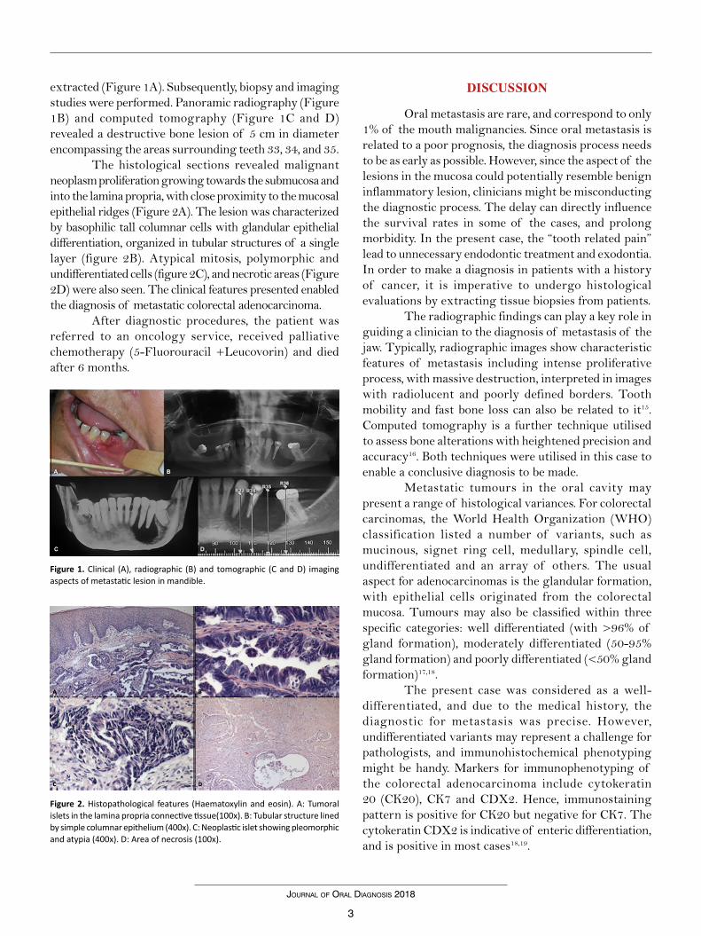
Figure 1. Clinical (A), radiographic (B) and tomographic (C and D) imaging aspects of metastatic lesion in mandible.
Figure 2. Histopathological features (Haematoxylin and eosin). A: Tumoral islets in the lamina propria connective tissue(100x). B: Tubular structure lined by simple columnar epithelium (400x). C: Neoplastic islet showing pleomorphic and atypia (400x). D: Area of necrosis (100x).
extracted (Figure 1A). Subsequently, biopsy and imaging studies were performed. Panoramic radiography (Figure 1B) and computed tomography (Figure 1C and D) revealed a destructive bone lesion of 5 cm in diameter encompassing the areas surrounding teeth 33, 34, and 35.
The histological sections revealed malignant neoplasm proliferation growing towards the submucosa and into the lamina propria, with close proximity to the mucosal epithelial ridges (Figure 2A). The lesion was characterized by basophilic tall columnar cells with glandular epithelial differentiation, organized in tubular structures of a single layer (figure 2B). Atypical mitosis, polymorphic and undifferentiated cells (figure 2C), and necrotic areas (Figure 2D) were also seen. The clinical features presented enabled the diagnosis of metastatic colorectal adenocarcinoma.
After diagnostic procedures, the patient was referred to an oncology service, received palliative chemotherapy (5-Fluorouracil +Leucovorin) and died after 6 months.
DISCUSSION
Oral metastasis are rare, and correspond to only 1% of the mouth malignancies. Since oral metastasis is related to a poor prognosis, the diagnosis process needs to be as early as possible. However, since the aspect of the lesions in the mucosa could potentially resemble benign inflammatory lesion, clinicians might be misconducting the diagnostic process. The delay can directly influence the survival rates in some of the cases, and prolong morbidity. In the present case, the “tooth related pain” lead to unnecessary endodontic treatment and exodontia. In order to make a diagnosis in patients with a history of cancer, it is imperative to undergo histological evaluations by extracting tissue biopsies from patients.
The radiographic findings can play a key role in guiding a clinician to the diagnosis of metastasis of the jaw. Typically, radiographic images show characteristic features of metastasis including intense proliferative process, with massive destruction, interpreted in images with radiolucent and poorly defined borders. Tooth mobility and fast bone loss can also be related to it15. Computed tomography is a further technique utilised to assess bone alterations with heightened precision and accuracy16. Both techniques were utilised in this case to enable a conclusive diagnosis to be made.
Metastatic tumours in the oral cavity may present a range of histological variances. For colorectal carcinomas, the World Health Organization (WHO) classification listed a number of variants, such as mucinous, signet ring cell, medullary, spindle cell, undifferentiated and an array of others. The usual aspect for adenocarcinomas is the glandular formation, with epithelial cells originated from the colorectal mucosa. Tumours may also be classified within three specific categories: well differentiated (with >96% of gland formation), moderately differentiated (50-95% gland formation) and poorly differentiated (<50% gland formation)17,18.
The present case was considered as a well-differentiated, and due to the medical history, the diagnostic for metastasis was precise. However, undifferentiated variants may represent a challenge for pathologists, and immunohistochemical phenotyping might be handy. Markers for immunophenotyping of the colorectal adenocarcinoma include cytokeratin 20 (CK20), CK7 and CDX2. Hence, immunostaining pattern is positive for CK20 but negative for CK7. The cytokeratin CDX2 is indicative of enteric differentiation, and is positive in most cases18,19.
4
Journal of oral Diagnosis 2018
In the presented case, the patient was a 64-year-old woman and the site affected was the mandible. Some similarities could be seen in this particular case with the cases searched through PUBMED data basis for metastatic colorectal adenocarcinoma in the mouth. The gender was an exception, because in most of the cases higher prevalence were males. However, for metastasis in oral cavity, regardless the origin, the disease seem to have a predilection for females2. The epidemiologic data about oral metastasis is limited, due to rarity of the cases. For this reason, the information source is based on small series evaluation and single cases1–3.
Regarding treatment, patients diagnosed with colorectal adenocarcinoma early will receive mandatory surgery. Adjuvant treatments such as radiotherapy and chemotherapy may also be utilised where necessary (20). Mortality rates across the globe are variable dependent upon the region investigated. For colorectal adenocarcinoma the mortality rates are low (8.5% of total) in developed countries, and higher in less developed regions (4). In cases of metastatic disease, there are no indications that systemic treatment might be successful, but can be considered useful for palliative treatments.
CONCLUSION
Metastasis in the jawbone and oral mucosa are considered rare and hence are rarely diagnosed or misdiagnosed, meaning patients often receive incorrect treatments. This may result in exacerbation of symptoms and increased rates of mortality as seen in the case above. It is therefore imperative to undertake thorough investigation utilising an array of techniques including, histology and radiography. This is particularly important in patients complaining of oral symptoms and with histories of malignancies.
REFERENCES
1. Hirshberg A, Shnaiderman-Shapiro A, Kaplan I, Berger R. Me-tastatic tumours to the oral cavity - pathogenesis and analysis of 673 cases. Oral Oncol. 2008;44:743–52.
2. Servato JP, de Paulo LFB, de Faria PR, Cardoso SV, Loyola AM. Metastatic tumours to the head and neck: retrospective analysis from a Brazilian tertiary referral centre. Int J Oral Maxillofac Surg. 2013;42:1391–6.
3. Hirshberg A, Berger R, Allon I, Kaplan I. Metastatic tumors to the jaws and mouth. Head Neck Pathol. 2014;8:463–74.
4. Ferlay J, Soerjomataram I, Dikshit R, Eser S, Mathers C, Rebelo M, et al. Cancer incidence and mortality worldwide: sources, methods and major patterns in GLOBOCAN 2012. Int J Cancer. 2015;136:E359-86.
5. Baranović M, Vidaković B, Sauerborn D, Perić B, Uljanić I, Mahovne I. Colorectal Adenocarcinoma Metastasizing to the Oral Mucosa of the Upper Jaw. Srp Arh Celok Lek. 2015;143:314–6.
6. Singh T, Amirtham U, Satheesh CT, Lakshmaiah KC, Suresh TM, Babu KG, et al. Floor-of-mouth metastasis in colorectal cancer. Ann Saudi Med. 2011;31:87–9.
7. Amin A, Jha M, Reddy A. Lower lip numbness in a patient with colorectal cancer. BMJ Case Rep. 2011;2011.
8. Mason AC, Azari KK, Farkas LM, Duvvuri U, Myers EN. Me-tastatic adenocarcinoma of the colon presenting as a mass in the mandible. Head Neck. 2005;27:729–32.
9. Alvarez-Alvarez C, Iglesias-Rodríguez B, Pazo-Irazu S, Delgado-Sánchez-Gracián C. Colonic adenocarcinoma with metastasis to the gingiva. Med Oral Patol Oral Cirugia Bucal. 2006;11:E85-7.
10. Kawamura M, Nakabayashi Y, Otsuka M, Sakata H, Yanaga K. Gingival metastasis from rectal cancer. J Gastrointest Surg Off J Soc Surg Aliment Tract. 2008;12:1121–2.
11. Ren Q-G, Huang T, Yang S-L, Hu J-L. Colon cancer metasta-sis to the mandibular gingiva with partial occult squamous differentiation: A case report and literature review. Mol Clin Oncol. 2017;6:189–92.
12. Watanabe M, Tada M, Satomi T, Chikazu D, Mizumoto M, Sakurai H. Metastatic rectal adenocarcinoma in the mandibular gingiva: a case report. World J Surg Oncol. 2016;14:199.
13. Grammatica L, Achille G, Montepara M, Zito A, Montemurro S. [Mandibular metastases from rectal adenocarcinoma: clinical case report and review of the literature]. Acta Otorhinolaryngol Ital Organo Uff Della Soc Ital Otorinolaringol E Chir Cerv-facc. 2001;21:115–8.
14. Mojica-Manosa P, Rigual N, Tan D, Sullivan M. An Unusual Case of a Metastatic Adenocarcinoma of the Rectum to the Mandible: A Case Report and Review of the Literature. J Oral Maxillofac Surg. 2006;64:1436–9.
15. Dunfee BL, Sakai O, Pistey R, Gohel A. Radiologic and Pa-thologic Characteristics of Benign and Malignant Lesions of the Mandible. RadioGraphics. 2006;26:1751–68.
16. Ferreira LA, Grossmann E, Januzzi E, Paula MVQ de, Carvalho ACP, Ferreira LA, et al. Diagnosis of temporomandibular joint disorders: indication of imaging exams. Braz J Otorhinolaryn-gol. 2016;82:341–52.
17. Hamilton SR, Bosman FT, Boffetta P, et al. Carcinoma of the colon and rectum. In: WHO Classification of Tumours of the Digestive System. Lynon; 2010. 134-46 p. (Bosman FT, Carneiro F, Hruban RH, Theise ND, eds.; vol. IARC Press).
18. Fleming M, Ravula S, Tatishchev SF, Wang HL. Colorec-tal carcinoma: Pathologic aspects. J Gastrointest Oncol. 2012;3:153–73.
19. Chu PG, Weiss LM. Keratin expression in human tissues and neoplasms. Histopathology. 2002;40:403–39.
20. Mendenhall WM, Rout WR, Lind DS, Zlotecki RA, Hochwald SN, Schell SR, et al. Role of radiation therapy in the treat-ment of resectable rectal adenocarcinoma. J Surg Oncol. 2002;79:107–17; discussion 118.