methotrexate, paclitaxel, and doxorubicin radiosensitize...
TRANSCRIPT

Methotrexate, Paclitaxel, and DoxorubicinRadiosensitize HER2-Amplified HumanBreast Cancer Cells to the AugerElectron–Emitting RadiotherapeuticAgent 111In-NLS-Trastuzumab
Danny L. Costantini*1,2, Daniela F. Villani*3, Katherine A. Vallis4, and Raymond M. Reilly1,5,6
1Department of Pharmaceutical Sciences, University of Toronto, Toronto, Ontario, Canada; 2Department of Diagnostic Imaging,Hospital for Sick Children, Toronto, Ontario, Canada; 3Faculty of Health Sciences, McMaster University, Hamilton, Ontario,Canada; 4Gray Institute for Radiation Oncology and Biology, University of Oxford, Oxford, United Kingdom;5Department of Medical Imaging, University of Toronto, Toronto, Ontario, Canada; and 6Toronto GeneralResearch Institute, University Health Network, Toronto, Ontario, Canada
Our goal in this study was to elucidate the mechanisms bywhich methotrexate radiosensitizes HER2-positive human breastcancer cells to the Auger electron emitter 111In-trastuzumabmodified with nuclear-localization sequence peptides (111In-NLS-trastuzumab) and to compare these mechanisms with thepotential sensitizing effects of paclitaxel and doxorubicin whencombined with this radiopharmaceutical. Methods: Experimentswere performed in MDA-MB-231 human breast cancer cells, theirHER2-transfected subclones (231-H2N), and 2 trastuzumab-resistant variants (trastuzumab-resistant-1 and -2 [TrR1 and TrR2]).Effects of coexposure of these cells to 111In-NLS-trastuzumaband low-dose, radiosensitizing methotrexate, paclitaxel, ordoxorubicin were assessed by clonogenic cell-survival assay.Quantification of residual DNA damage was measured by thegH2AX-immunofluorescence assay, and cell cycle distributionwas measured by fluorescence-activated cell sorting analysis.The radiation-enhancement ratio was calculated as the ratioof the surviving fraction (SF) of cells treated with 111In-NLS-trastuzumab alone to that of cells treated concurrently with111In-NLS-trastuzumab and methotrexate, paclitaxel, or doxoru-bicin. Results: A reduction in the SF in HER2-positive 231-H2N(55.7% 6 1.3%) and TrR1 (62.6% 6 6.5%) cells was demon-strated after exposure to 111In-NLS-trastuzumab (;0.2 MBq/mg,100 nmol/L) but not in MDA-MB-231 or TrR2 cells expressinglow levels of HER2 (SF . 90%, P . 0.05). Coadministration ofmethotrexate, paclitaxel, or doxorubicin enhanced the cyto-toxicity of 111In-NLS-trastuzumab toward 231-H2N and TrR1cells but not toward MDA-MB-231 or TrR2 cells. The radiation-enhancement ratios for methotrexate, paclitaxel, and doxorubicinfor 231-H2N or TrR1 cells were 2.0–2.2, 1.6–1.8, and 2.7–2.8,respectively. Methotrexate or doxorubicin combined with 111In-
NLS-trastuzumab, compared to treatment with 111In-NLS-trastuzumab alone, significantly increased residual gH2AX fociin 231-H2N and TrR1 cells but not in MDA-MB-231 or TrR2 cellsor in any cell line treated concurrently with paclitaxel and 111In-NLS-trastuzumab. Cells exposed to low-dose methotrexate accu-mulated in the G1/S phase of the cell cycle, whereas low-dosepaclitaxel or doxorubicin caused cells to arrest in the G2/M phase.Conclusion: Low-dose methotrexate, paclitaxel, or doxorubicinpotently sensitized HER2-overexpressing human breast cancercells, with and without acquired trastuzumab-resistance, to theAuger electron emissions from 111In-NLS-trastuzumab throughcell cycle distribution changes and in part through the inhibitory ef-fects of these agents on DNA damage repair.
Key Words: Auger electron; radiosensitizer; trastuzumab; breastcancer; radioimmunotherapy
J Nucl Med 2010; 51:477–483DOI: 10.2967/jnumed.109.069716
Despite the success of radioimmunotherapy for ad-vanced lymphohematopoietic malignancies, the treatmentof patients with solid tumors such as breast cancer has beenlimited because of the small amounts of antibody that canbe targeted to tumors. Also of concern in treatment are the ir-radiation and killing of normal cells (e.g., bone marrow stemcells) by the moderate-energy and long-range (2–10 mm)b-particles emitted by 131I and 90Y, which have been com-monly conjugated to radiotherapeutic agents (1,2). Therefore,considerable attention has been given to the combination ofradioimmunotherapy with drugs that are known radiosen-sitizers and that can amplify the lethal effects of ionizingradiation on cancer cells while having minimal or no toxicityon tissues at the low concentrations used (3).
Received Aug. 23, 2009; revision accepted Nov. 24, 2009.For correspondence or reprints contact: Raymond M. Reilly, Leslie Dan
Faculty of Pharmacy, University of Toronto, 144 College St., Toronto,Ontario M5S 3M2, Canada.
E-mail: [email protected]*Contributed equally to this work.COPYRIGHT ª 2010 by the Society of Nuclear Medicine, Inc.
111IN-NLS-TRASTUZUMAB RADIOSENSITIZATION • Costantini et al. 477
by on July 14, 2018. For personal use only. jnm.snmjournals.org Downloaded from

This strategy may be especially relevant for patientswhose tumors display amplification of the HER2 receptortyrosine kinase. HER2 overexpression occurs in 25%230%of breast cancers (4), has been correlated with resistance tohormonal therapy (5–7) and chemotherapy (8), and isdirectly associated with poor long-term survival (4). Tras-tuzumab (Herceptin; Hoffmann-La Roche) is a humanizedanti-HER2 monoclonal antibody approved for immunother-apy of HER2-amplified breast cancer (4). However, only50% of patients with tumors exhibiting moderate to high(21 to 31) immunohistochemistry scores or having greaterthan 3 copies of the HER2 gene by fluorescence in situhybridization are predicted to respond to trastuzumab whengiven in combination with chemotherapy (9,10). Moreover,the short duration of response to trastuzumab (9–12 mo)and the rapid development of drug resistance limit theeffectiveness of trastuzumab treatment in this patientpopulation (11,12). In an attempt to enhance its antineo-plastic activity, our group has been studying Auger electronradioimmunotherapy of HER2-amplified breast cancerusing 111In-trastuzumab modified with 13 mer peptides(CGYGPKKKRKVGG) harboring the nuclear-localizationsequence (NLS) of SV-40 large T antigen, which promotesits nuclear importation after HER2-mediated internalizationinto breast cancer cells (13). Auger electrons are highlydamaging to DNA when they decay near the cell nucleus,making them exquisitely selective and highly potent forkilling targeted single cancer cells (14). Indeed, we recentlyreported that 111In-NLS-trastuzumab, compared with non-radiolabeled trastuzumab (Herceptin), could slow thegrowth of HER2-positive breast cancer xenografts in mice,with minimal harm to normal tissues, and prolong survival(15). In in vitro studies, we further found that 111In-NLS-trastuzumab was able to kill breast cancer cells that areresistant to trastuzumab and that the potency of the radio-pharmaceutical could be enhanced by coexposing these cellsto low, noncytotoxic concentrations of methotrexate (16).
Understanding the mechanisms resulting in tumor cellradiosensitization caused by the interaction of chemother-apeutic drugs and Auger electron radiotherapeutics wouldbe useful in the development of rational clinical protocolsthat combine these agents. The first objective of this study,therefore, was to elucidate the mechanisms that underliemethotrexate radiosensitization of HER2-positive breastcancer cells to 111In-NLS-trastuzumab. Methotrexate is anantifolate drug that inhibits purine de novo synthesis. Thisantifolate activity of methotrexate is hypothesized toimpede DNA synthesis and repair and amplify the lethaleffects of the Auger electron emissions from 111In-NLS-trastuzumab (17,18). Therefore, we examined the effects oflow-dose methotrexate on cell cycle progression usingfluorescence-activated cell sorting and measured DNAdamage by the gH2AX fluorescence assay in breast cancercells exposed to the combination of methotrexate and 111In-NLS-trastuzumab. Other chemotherapeutic agents, such aspaclitaxel and doxorubicin, are commonly administered to
breast cancer patients in combination with trastuzumab andare also known radiosensitizers (4). Therefore, we alsosought to determine whether paclitaxel and doxorubicinwould radiosensitize HER2-postive breast cancer cells to111In-NLS-trastuzumab. The radiosensitizing effects of theseagents, when combined with 111In-NLS-trastuzumab, wereevaluated and compared with those caused by methotrexate.
MATERIALS AND METHODS
Cell CultureMDA-MB-231 human breast cancer cells were obtained from
the American Type Culture Collection, and the 231-H2N andtrastuzumab-resistant 1 and 2 (TrR1 and TrR2) cell lines werekindly provided by Dr. Robert S. Kerbel (Sunnybrook HealthSciences Centre). The 231-H2N cell line was derived from MDA-MB-231 cells that were transfected to stably overexpress c-erbB-2(HER2), whereas TrR1 and TrR2 cells were isolated from 231-H2N tumors in athymic mice with acquired trastuzumab resistance(19). 231-H2N and TrR1 cells express high levels of HER2 (5.0–6.0 · 105 receptors per cell), which is approximately 10-foldgreater than MDA-MB-231 and TrR2 cells, which express 0.4–0.6 ·105 HER2 receptors per cell (16). All cell lines were cultured inDulbecco’s minimal essential medium (Ontario Cancer Institute)supplemented with 10% fetal bovine serum (Sigma-Aldrich) con-taining 100 U of penicillin per milliliter and 100 mg of streptomycinper milliliter at 37�C in an atmosphere of 5% CO2.
Trastuzumab and RadiosensitizersTrastuzumab was reconstituted according to the package insert.
Methotrexate and doxorubicin were prepared by dissolving 1 mgof the drug (Sigma-Aldrich) in 1 mL of alkalinized (pH 11.0) orneutral (pH 7.0) distilled, deionized water, respectively. Themethotrexate stock solution was adjusted to pH 7.0 before use.Paclitaxel (Sigma-Aldrich) was prepared by dissolving 1 mg ofthe drug in 100 mL of dimethyl sulfoxide. Stock solutions werediluted in culture medium to the desired final concentration. Thefinal concentration of dimethyl sulfoxide in the culture mediumwas 0.1% (v/v) or less.
111In-Trastuzumab Modified with NLS-PeptidesTrastuzumab (Herceptin) was modified with synthetic 13 mer
NLS-peptides (CGYGPKKKRKVGG) and labeled with 111InCl3(MDS-Nordion) using diethylenetriaminepentaacetic acid (DTPA)(Sigma-Aldrich) to a specific activity of 201 6 6 MBq/mg (13).Briefly, trastuzumab (500 mg, 10 mg/mL) was reacted with a 10-fold molar excess of DTPA dianhydride for 1 h at room temperaturebefore reaction with a 15-fold molar excess of sulfo-succinimidyl-4-(N-maleimidomethyl)cyclohexane-1-carboxylate (SMCC) (2–5mmol/L). Maleimide-derivatized DTPA-trastuzumab was con-centrated to 2–5 mg/mL and reacted overnight at 4�C with a 60-fold molar excess of NLS-peptides (5–10 mmol/L diluted inphosphate-buffered saline [PBS], pH 7.0). DTPA-trastuzumabmodified with NLS-peptides (NLS-DTPA-trastuzumab) was puri-fied on a Sephadex-G50 minicolumn (GE Healthcare) eluted withPBS, pH 7.5. Under these conditions, 3–4 NLS-peptides wereconjugated to trastuzumab at an SMCC:IgG:NLS-peptide molarratio of 15:1:60 (13).
NLS-conjugated trastuzumab or unmodified DTPA-trastuzumabwas radiolabeled by incubation of 37–111 MBq of monoclonalantibodies with 111InCl3 (MDS-Nordion) for 60 min at room
478 THE JOURNAL OF NUCLEAR MEDICINE • Vol. 51 • No. 3 • March 2010
by on July 14, 2018. For personal use only. jnm.snmjournals.org Downloaded from

temperature. 111In-labeled monoclonal antibodies were purified ona Sephadex-G50 minicolumn and buffer-exchanged to PBS, pH7.5, using a Microcon YM-50 ultrafiltration device (Amicon;Millipore). The radiochemical purity was routinely greater than97% as determined by instant thin-layer chromatography–silicagel (Pall Corp.) developed in 100 mM sodium citrate, pH 5.0.All radioactivity measurements were made using an automaticg-counter (Wallac Wizard-1480; Perkin Elmer).
Clonogenic AssaysApproximately 2 · 106 breast cancer cells were incubated with
111In-NLS-trastuzumab (100 nmol/L) alone or concurrently withan inhibitory concentration of 10% (IC10) of methotrexate,paclitaxel, or doxorubicin in 1 mL of culture medium in micro-tubes for 24 h at 37�C. The 24-h time point was chosen becausenuclear accumulation of 111In-NLS-trastuzumab increases for upto 24 h in HER2-overexpressing breast cancer cells (13). Controlsconsisted of cells treated with PBS (pH 7.5) or cells treated withescalating doses of methotrexate, paclitaxel, or doxorubicin (theconcentrations of the tested chemotherapeutic drugs ranged from0.01 to 100 mmol/L). For treatments including methotrexate, cellswere serum-starved by lowering the serum concentration from10% to 1% to reduce the concentrations of thymidine, 5-methyltetrahydrofolate, and purine ribonucleosides that counteract theeffects of methotrexate (20). After treatment, the cells werecentrifuged at 1,000g for 5 min and washed twice with normalculture medium containing 10% serum. Sufficient cells were thenplated in triplicate in 12-well plates and cultured in normalmedium at 37�C. After 10–14 d, the dishes were stained withmethylene blue, and colonies of 50 cells or more were counted.The surviving fraction (SF) was calculated by dividing the numberof colonies formed for treated cells by the number for untreatedcells. Survival curves were derived for cells treated with metho-trexate, paclitaxel, or doxorubicin by plotting the SF values versusthe log molar concentration of agent used. The inhibitoryconcentrations of 50% (IC50) and IC10 were estimated with Origin6.0 (Microcal Software Inc.) using the dose-response equation y 5
A1 1 [(A2 2 A1)/(1 1 10(log EC50 – x) · p], where p is the slope(set to 21), and A1 and A2 are the amplitudes of the baseline andmaximum response, respectively. The radiation-enhancement ratio(RER) was determined by dividing the SF of cells treated with111In-NLS-trastuzumab alone by that of cells treated concurrentlywith 111In-NLS-trastuzumab and methotrexate, paclitaxel, or doxo-rubicin. An RER greater than 1 indicated radiosensitization (16,21).
Cell Cycle AnalysisCells were plated into 100-mm tissue culture plates (Sarstedt
Inc.) at a density of 1 · 106 cells per plate and cultured overnight.Control cells and cells treated with methotrexate, paclitaxel, ordoxorubicin were incubated in 10 mL of medium in 100-mm cellculture dishes for 24 h at 37�C. Treated and control cells were thensuspended in 300 mL of PBS and fixed with 700 mL of 100% ice-cold ethanol for 1 h. The cells were then washed, resuspendedwith 10 mg of RNase (Invitrogen) per milliliter, and stained withpropidium iodide (25 mg/mL; Sigma-Aldrich) in 100 mL of PBS,pH 7.5, for 1 h at 37�C. The DNA content was analyzed witha flow cytometer (BD FACSCalibur), and the distribution of cellsin the different phases of the cell cycle was determined.
Measurement of DNA DamageThe ability of 111In-NLS-trastuzumab alone or combined with
methotrexate, paclitaxel, or doxorubicin to cause DNA double-
strand breaks (DSBs) in breast cancer cells was evaluated usingthe gH2AX-assay, which detects phosphorylation of histone-H2AX at serine-139 (gH2AX) at sites of DSBs as discretenuclear foci using gH2AX-specific antibodies (22). Control cellswere treated with methotrexate, paclitaxel, doxorubicin, orunlabeled trastuzumab alone or the combination of unlabeledtrastuzumab and methotrexate, paclitaxel, or doxorubicin. Cellswere cultured overnight in 1 mL of medium containing 111In-NLS-trastuzumab (100 nmol/L) alone or combined with meth-otrexate, paclitaxel, or doxorubicin. Exactly 24 h after theincubation period, cells were fixed with 2% paraformaldehydecontaining 0.5% Triton X-100 in PBS, pH 8.2, for 15 min. Afterthree 10-min washes with PBS, pH 7.5, containing 0.5% bovineserum albumin and 0.2% polysorbate-20, the cells were per-meabilized for 15 min with PBS, pH 8.2, containing 0.5%Nonidet P-40 (Sigma-Aldrich) and blocked for 1 h in 2% bovineserum albumin and 1% donkey serum. The slides were thenincubated with antiphospho-gH2AX (1:800; Upstate Biotech-nology) in 3% bovine serum albumin overnight at 4�C and thenwith AlexaFluor-488 antimouse IgG (Molecular Probes) for 45min at room temperature. All slides were mounted with Vecta-shield medium containing 4,6-diamidino-2-phenylindole (DAPI)(Vector Laboratories) and kept at 4�C overnight. Images weretaken with an inverted LSM510 confocal microscope (Carl Zeiss)at the Advanced Optical Microscopy Facility (Princess MargaretHospital). Excitation was at 364 nm or 488 nm for visualization ofDAPI or AlexaFluor-488, using 385- to 470-nm and 505- to 550-nm emission filters, respectively. For imaging of gH2AX, 10–15z-stack images at approximately 1-mm intervals were acquiredthroughout the entire cell nucleus, merged using LSM-Viewersoftware (version, 3.5.0.376; Zeiss), and stored as .tiff files. Thenumber of gH2AX-foci present in each cell was counted manuallyusing ImageJ software (version, 1.36b; National Institutes ofHealth) as described previously (23).
Statistical MethodsData are presented as mean 6 SEM. Statistical comparisons
were made using the Student t test. P , 0.05 was consideredsignificant.
RESULTS
Effect of Methotrexate, Paclitaxel, and Doxorubicinon Survival of Breast Cancer Cells
MDA-MB-231, 231-H2N, TrR1, and TrR2 human breastcancer cells were incubated with increasing concentrationsof methotrexate, paclitaxel, and doxorubicin for 24 h todetermine a low, relatively noncytotoxic concentration ofdrug that resulted in survival of 90% or more cells (i.e.,IC10) by clonogenic assay. There was a strong dose-dependent decrease in colony formation of the differentbreast cancer cells treated with increasing amounts ofchemotherapeutic agent. Table 1 shows the IC50 and IC10
values of each agent for each cell line; IC10 values rangedbetween 0.01 and 12 mmol/L, 0.02 and 0.14 mmol/L, and0.02 and 0.19 mmol/L for methotrexate, paclitaxel, anddoxorubicin, respectively. These IC10 doses were chosen toevaluate the radiosensitizing effect of combined treatmentof low-dose chemotherapy with 111In-NLS-trastuzumab.
111IN-NLS-TRASTUZUMAB RADIOSENSITIZATION • Costantini et al. 479
by on July 14, 2018. For personal use only. jnm.snmjournals.org Downloaded from

Effect of 111In-NLS-Trastuzumab Combined withLow-Dose Methotrexate, Paclitaxel, or Doxorubicinon Clonogenic Survival
The SF of the 4 cell lines treated for 24 h with 111In-NLS-trastuzumab (100 nmol/L) alone or combined with theIC10 concentrations for methotrexate, paclitaxel, and doxo-rubicin was measured in clonogenic assays. Compared withno 111In-NLS-trastuzumab exposure, exposure to 111In-NLS-trastuzumab significantly decreased the SF for 231-H2N and TrR1 cells (55.6% 6 1.3% and 74.1% 6 5.4%,respectively) that express intermediate to high levels ofHER2 but not for MDA-MB-231 or TrR2 cells (97.9% 6
7.2% and 89.5% 6 3.8%, respectively) that express lowlevels of HER2 (16) (Fig. 1). The effect of a 24-h treatmentwith 111In-NLS-trastuzumab combined with low-dosemethotrexate, paclitaxel, or doxorubicin (i.e., the IC10 doseof drug) on the SF of cells was then measured. Thecombined treatment, compared with 111In-NLS-trastuzu-mab alone, caused an even greater reduction in the SF of231-H2N and TrR1 cells, with doxorubicin causing thegreatest decrease in the SF in both cell lines (Fig. 1). Incontrast, the combined treatment, compared with treatmentwith 111In-NLS-trastuzumab alone, had little effect on theSF of MDA-MB-231 and TrR2 cells. The RERs for low-dose chemotherapy combined with 111In-NLS-trastuzumabare shown in Table 2. The greatest RERs were observed for231-H2N and TrR1 cells when 111In-NLS-trastuzumab wascombined with doxorubicin (RER, 2.7–2.8), followed bycotreatment with methotrexate (RER, 2.0–2.2) and pacli-taxel (RER, 1.6–1.8).
DNA Damage Induced by 111In-NLS-TrastuzumabCombined with Methotrexate, Paclitaxel,or Doxorubicin
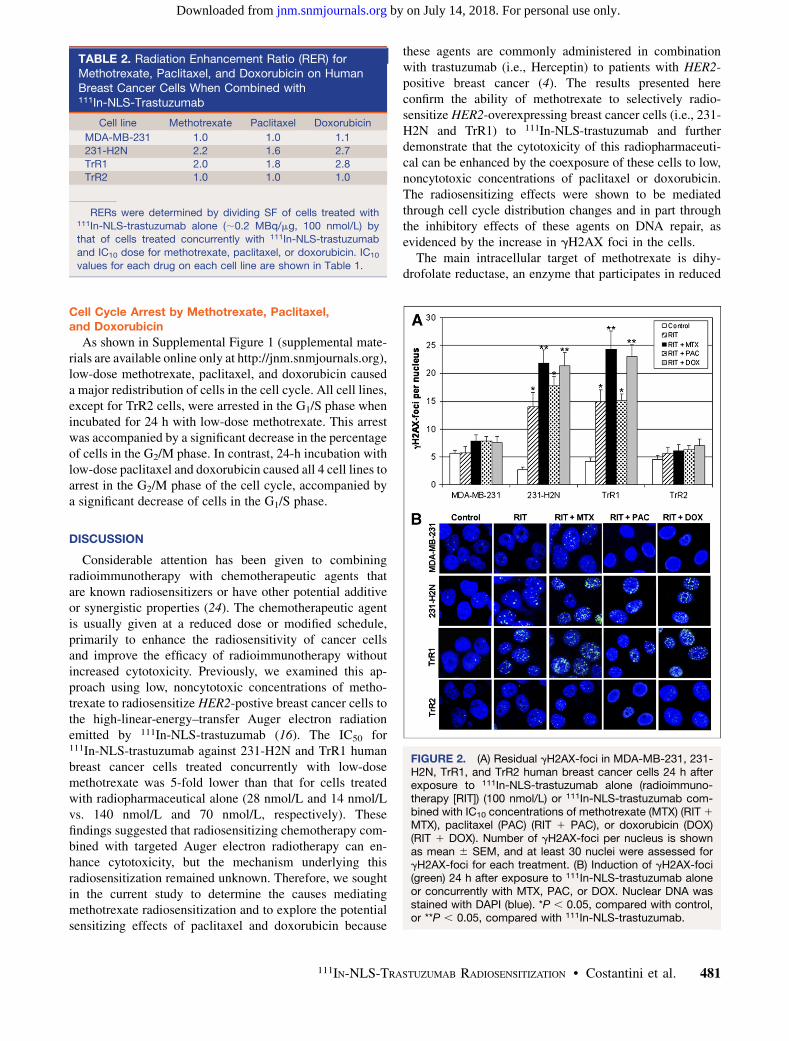
A 4- to 5-fold increase in the number of residualgH2AX-foci 24 h after incubation with 111In-NLS-trastu-
zumab (100 nmol/L), compared with after no incubation,was observed in 231-H2N (14.0 6 2.6 foci/nucleus vs. 2.7 6
0.5 foci/nucleus, P , 0.05) and TrR1 cells (15.0 6 2.1 foci/nucleus vs. 4.1 6 0.7 foci/nucleus, P , 0.05) (Fig. 2). Incontrast, treatment with 111In-NLS-trastuzumab did notsignificantly increase the formation of residual gH2AX-foci in MDA-MB-231 or TrR2 cells (5.7 6 1.1 foci/nucleusand 5.6 6 1.1 foci/nucleus, respectively), compared with231-H2N or TrR1 cells, likely because of the 10-fold lowerexpression of HER2. 231-H2N and TrR1 cells exposed toa low dose of methotrexate or doxorubicin in combinationwith 111In-NLS-trastuzumab, compared with cells exposedto either agent alone, showed a higher number of gH2AX-foci (231-H2N: 21.8 6 2.4 foci/nucleus and 21.4 6 2.4foci/nucleus, respectively; TrR1: 24.3 6 3.3 and 23.0 6
2.2, respectively) (P , 0.05). However, the combination oflow-dose paclitaxel and 111In-NLS-trastuzumab, comparedwith treatment with 111In-NLS-trastuzumab alone, did notsignificantly increase the number of residual gH2AX-fociin these cells (231-H2N: 17.8 6 1.6 foci/nucleus; TrR1:15.1 6 1.1 foci/nucleus, P . 0.05). There was a modestincrease in the formation of gH2AX-foci in MDA-MB-231and TrR2 cells exposed to 111In-NLS-trastuzumab andchemotherapy, compared with cells exposed to only 1 ofthese agents, but this increase did not reach the level ofstatistical significance. Compared with control untreatedcells, cells treated with an equivalent dose of methotrexate,paclitaxel, doxorubicin, or unlabeled trastuzumab alone, orthe combination of unlabeled trastuzumab and methotrex-ate, paclitaxel, or doxorubicin, did not show significantlyhigher gH2AX foci/nucleus (data not shown).
TABLE 1. IC10 and IC50 Values for Methotrexate,Paclitaxel, and Doxorubicin on MDA-MB-231, 231-H2N,TrR1, and TrR2 Human Breast Cancer Cells
Cell line Methotrexate* Paclitaxel Doxorubicin
MDA-MB-231
IC10 (mmol/L) 1.01 6 0.01 0.07 6 0.03 0.19 6 0.08
IC50 (mmol/L) 103.3 6 13.4 0.32 6 0.13 1.26 6 0.39231-H2N
IC10 (mmol/L) 12.0 6 0.23 0.14 6 0.10 0.02 6 0.004
IC50 (mmol/L) 120.8 6 23.3 1.36 6 0.24 0.16 6 0.04
TrR1IC10 (mmol/L) 0.01 6 0.001 0.12 6 0.03 0.03 6 0.01
IC50 (mmol/L) 0.13 6 0.01 2.01 6 0.47 0.55 6 0.25
TrR2IC10 (mmol/L) 1.13 6 0.16 0.02 6 0.01 0.02 6 0.003
IC50 (mmol/L) 102.4 6 10.7 0.27 6 0.06 0.21 6 0.08
*Values were reported previously in Costantini et al. (16).
Results are expressed as mean 6 SEM of 3 measurements
performed in duplicate.
FIGURE 1. SFs of MDA-MB-231, 231-H2N, TrR1, and TrR2human breast cancer cells after treatment with 111In-NLS-trastuzumab alone (radioimmunotherapy [RIT]) (;0.2 MBq/mg,100 nmol/L) or 111In-NLS-trastuzumab combined with IC10
concentrations of methotrexate (MTX) (RIT 1 MTX ), paclitaxel(PAC) (RIT 1 PAC), or doxorubicin (DOX) (RIT 1 DOX). IC10
values of each drug for each cell line are shown in Table 1.*P , 0.05, compared with RIT.
480 THE JOURNAL OF NUCLEAR MEDICINE • Vol. 51 • No. 3 • March 2010
by on July 14, 2018. For personal use only. jnm.snmjournals.org Downloaded from
Cell Cycle Arrest by Methotrexate, Paclitaxel,and Doxorubicin
As shown in Supplemental Figure 1 (supplemental mate-rials are available online only at http://jnm.snmjournals.org),low-dose methotrexate, paclitaxel, and doxorubicin causeda major redistribution of cells in the cell cycle. All cell lines,except for TrR2 cells, were arrested in the G1/S phase whenincubated for 24 h with low-dose methotrexate. This arrestwas accompanied by a significant decrease in the percentageof cells in the G2/M phase. In contrast, 24-h incubation withlow-dose paclitaxel and doxorubicin caused all 4 cell lines toarrest in the G2/M phase of the cell cycle, accompanied bya significant decrease of cells in the G1/S phase.
DISCUSSION
Considerable attention has been given to combiningradioimmunotherapy with chemotherapeutic agents thatare known radiosensitizers or have other potential additiveor synergistic properties (24). The chemotherapeutic agentis usually given at a reduced dose or modified schedule,primarily to enhance the radiosensitivity of cancer cellsand improve the efficacy of radioimmunotherapy withoutincreased cytotoxicity. Previously, we examined this ap-proach using low, noncytotoxic concentrations of metho-trexate to radiosensitize HER2-postive breast cancer cells tothe high-linear-energy–transfer Auger electron radiationemitted by 111In-NLS-trastuzumab (16). The IC50 for111In-NLS-trastuzumab against 231-H2N and TrR1 humanbreast cancer cells treated concurrently with low-dosemethotrexate was 5-fold lower than that for cells treatedwith radiopharmaceutical alone (28 nmol/L and 14 nmol/Lvs. 140 nmol/L and 70 nmol/L, respectively). Thesefindings suggested that radiosensitizing chemotherapy com-bined with targeted Auger electron radiotherapy can en-hance cytotoxicity, but the mechanism underlying thisradiosensitization remained unknown. Therefore, we soughtin the current study to determine the causes mediatingmethotrexate radiosensitization and to explore the potentialsensitizing effects of paclitaxel and doxorubicin because
these agents are commonly administered in combinationwith trastuzumab (i.e., Herceptin) to patients with HER2-positive breast cancer (4). The results presented hereconfirm the ability of methotrexate to selectively radio-sensitize HER2-overexpressing breast cancer cells (i.e., 231-H2N and TrR1) to 111In-NLS-trastuzumab and furtherdemonstrate that the cytotoxicity of this radiopharmaceuti-cal can be enhanced by the coexposure of these cells to low,noncytotoxic concentrations of paclitaxel or doxorubicin.The radiosensitizing effects were shown to be mediatedthrough cell cycle distribution changes and in part throughthe inhibitory effects of these agents on DNA repair, asevidenced by the increase in gH2AX foci in the cells.
The main intracellular target of methotrexate is dihy-drofolate reductase, an enzyme that participates in reduced
TABLE 2. Radiation Enhancement Ratio (RER) forMethotrexate, Paclitaxel, and Doxorubicin on HumanBreast Cancer Cells When Combined with111In-NLS-Trastuzumab
Cell line Methotrexate Paclitaxel Doxorubicin
MDA-MB-231 1.0 1.0 1.1
231-H2N 2.2 1.6 2.7
TrR1 2.0 1.8 2.8
TrR2 1.0 1.0 1.0
RERs were determined by dividing SF of cells treated with111In-NLS-trastuzumab alone (;0.2 MBq/mg, 100 nmol/L) bythat of cells treated concurrently with 111In-NLS-trastuzumab
and IC10 dose for methotrexate, paclitaxel, or doxorubicin. IC10
values for each drug on each cell line are shown in Table 1.
FIGURE 2. (A) Residual gH2AX-foci in MDA-MB-231, 231-H2N, TrR1, and TrR2 human breast cancer cells 24 h afterexposure to 111In-NLS-trastuzumab alone (radioimmuno-therapy [RIT]) (100 nmol/L) or 111In-NLS-trastuzumab com-bined with IC10 concentrations of methotrexate (MTX) (RIT 1
MTX), paclitaxel (PAC) (RIT 1 PAC), or doxorubicin (DOX)(RIT 1 DOX). Number of gH2AX-foci per nucleus is shownas mean 6 SEM, and at least 30 nuclei were assessed forgH2AX-foci for each treatment. (B) Induction of gH2AX-foci(green) 24 h after exposure to 111In-NLS-trastuzumab aloneor concurrently with MTX, PAC, or DOX. Nuclear DNA wasstained with DAPI (blue). *P , 0.05, compared with control,or **P , 0.05, compared with 111In-NLS-trastuzumab.
111IN-NLS-TRASTUZUMAB RADIOSENSITIZATION • Costantini et al. 481
by on July 14, 2018. For personal use only. jnm.snmjournals.org Downloaded from

folate metabolism (18). Methotrexate is thought to radio-sensitize cells through its effect on the depletion of reducedfolates required for the production of purine deoxyribonu-cleotides and thymidylate necessary for DNA synthesis andrepair (18,25). Indeed, the low, noncytotoxic concentrationof methotrexate that was determined in clonogenic assaysto radiosensitize 231-H2N and TrR1 breast cancer cells to111In-NLS-trastuzumab was found to cause these cells toaccumulate in the G1/S phase of the cell cycle, possiblythrough the depletion of intracellular purine nucleotidepools (Supplemental Fig. 1). Although we did not attemptto assess the distinction between cells in the G1 and early Sphases of the cell cycle, a more accurate estimate may beobtained by measuring the DNA content of bromodeox-yuridine-labeled S-phase cells using dual-parameter flowcytometry (26). Nevertheless, the extent of DNA damage(gH2AX-foci) in these cells at 24 h after exposure to 111In-NLS-trastuzumab was enhanced by coexposure to metho-trexate (Fig. 2), suggesting that nucleotide depletion bymethotrexate can indirectly affect the repair of DNAdamage in cells exposed to Auger electron radiation.Alternatively, chemotherapeutic radiosensitizers may pro-duce their effect mainly by direct action on DNA (17).Doxorubicin, for example, forms complexes with DNA byintercalation between base pairs, interfering with DNApolymerase and topoisomerase II activity, and inhibitsenzyme repair of radiation-induced DNA DSBs (27). In-deed, the addition of low-concentration doxorubicin to111In-NLS-trastuzumab, compared with cells treated witheither of these agents alone, induced more DNA damageand increased the killing of 231-H2N and TrR1 cells. Thisresult is in agreement with the observations by Supiot et al.(21), who demonstrated synergy between doxorubicin anda-particle radioimmunotherapy on induction of DNA dam-age and cell death in RPMI8226 myeloma cells. Thepowerful arrest of 231-H2N and TrR1 cells in the radiosen-sitive G2/M phase of the cell cycle induced by doxorubicin(Supplemental Fig. 1) may also account for the observedincrease in DNA damage and reduction in the SF of thesecells after coexposure with 111In-NLS-trastuzumab.
Paclitaxel also caused 231-H2N and TrR1 cells to arrestin the radiosensitive G2/M phase of the cell cycle (Supple-mental Fig. 1) (28). However, the level of paclitaxelradiosensitization, combined with 111In-NLS-trastuzumab,was moderately lower than with combinations of theradiopharmaceutical with methotrexate or doxorubicin.For example, the RER for the addition of paclitaxel to111In-NLS-trastuzumab for 231-H2N and TrR1 cells was1.621.8, versus 2.0–2.2 and 2.7–2.8 for combinations withmethotrexate and doxorubicin, respectively (Table 2). Thelower level of radiosensitization caused by paclitaxel alsocorrelated with fewer residual gH2AX-foci in 231-H2Nand TrR1 cells after cotreatment with 111In-NLS-trastuzu-mab (Fig. 2). This correlation is consistent with the fact thatpaclitaxel does not promote DNA damage but instead bindsto and promotes the polymerization of tubulin, which
interferes with normal microtubule breakdown (29). More-over, paclitaxel has been shown to trigger apoptosis,possibly by inducing the phosphorylation and inactivationof the antiapoptotic factors Bcl-2 and Bcl-xL (30). It ispossible that paclitaxel-mediated radiosensitization ofHER2-positive breast cancer cells results from mechanismsunrelated to DNA DSB induction. Other DNA-incorpo-rated, Auger electron–emitting radiopharmaceuticals, suchas 5-[125I]iodo-29-deoxyuridine, have been shown to pro-mote apoptosis through a caspase-3–mediated process (31).Thus, chemotherapeutic agents that amplify the apoptoticresponse in HER2-positive breast cancer cells exposed toAuger electron radiation may also be effective in causingradiosensitization.
Future studies examining low-dose chemotherapy radio-sensitization and 111In-NLS-trastuzumab in vivo will needto assess the dose-limiting toxicities on normal tissuesassociated with this combined treatment. Indeed, one limi-tation of this regimen is that the radiosensitization effectmight not be exclusive to tumor cells, particularly when theradiopharmaceutical is administered at high doses. How-ever, minimal toxicity toward MDA-MB-231 and TrR2cells expressing low levels of HER2 was observed from thiscombination, suggesting that radiosensitization may bespecific for cells overexpressing HER2 (i.e., HER2-ampli-fied breast cancer cells). Moreover, the feasibility ofcombining radiosensitizing chemotherapy with radioimmu-notherapy has been demonstrated previously in preclinicaland clinical studies (32). For example, DeNardo et al. (33)examined the effect of combining paclitaxel with radio-immunotherapy using 90Y-chimeric anti-L6 antibody (90Y-ChL6) in mice bearing HBT-3477 human breast cancerxenografts. Paclitaxel administered 6 or 24 h after radio-immunotherapy did not substantially increase normal-tissuetoxicity but resulted in cures or complete responses in all(46/46) treated mice, whereas only 21% (6/29) of animalswere cured when paclitaxel was given before 90Y-ChL6(33). Thus, the optimal combination and scheduling of low-dose chemotherapy and 111In-NLS-trastuzumab needs to bedetermined because the radiosensitizing effect may behighly dependent on the timing of administration of thechemotherapeutic drug relative to radioimmunotherapy.One approach may be to administer the radiotherapeuticagent, wait for 72 h for optimal tumor uptake andelimination from normal tissues (13), and then administerthese low-molecular-weight radiosensitizers, which are ex-pected to equilibrate rapidly between the plasma and thetissues (including tumor), thereby providing concurrentlyhigh concentrations of radioactivity and radiosensitizers.
The trastuzumab-resistant TrR1 cells (19) were effi-ciently killed by 111In-NLS-trastuzumab, and the toxicitywas enhanced by coexposure to radiosensitizing concen-trations of methotrexate, paclitaxel, or doxorubicin (Fig. 1).Combination therapy with low-dose chemotherapy and111In-NLS-trastuzumab may, therefore, offer a route toovercoming the challenges associated with trastuzumab
482 THE JOURNAL OF NUCLEAR MEDICINE • Vol. 51 • No. 3 • March 2010
by on July 14, 2018. For personal use only. jnm.snmjournals.org Downloaded from

resistance. Indeed, only about half of patients with HER2-overexpressing metastases respond to trastuzumab whencombined with paclitaxel or doxorubicin, and in almost allinitially responding patients, resistance to trastuzumabdevelops within a year (11,12). There have been few studiesthat have examined the combination of radiotherapeuticsthat emit high-linear-energy transfer radiation, such asAuger electron–emitting 111In-NLS-trastuzumab and radio-sensitizers (34). Achieving such a synergistic interaction bycombining radiosensitizing chemotherapy and 111In-NLS-trastuzumab, compared with either of these therapies alone,could dramatically improve the response.
CONCLUSION
Methotrexate, paclitaxel, and doxorubicin radiosensitizetrastuzumab-sensitive and trastuzumab-resistant humanbreast cancer cell lines overexpressing HER2 to 111In-NLS-trastuzumab, and the combination of radiosensitizing che-motherapy and targeted Auger electron radiotherapy withthis radiopharmaceutical, compared with either of theseagents alone, is more cytotoxic to these cells. The radio-sensitizing effects of these agents were shown to be mediatedthrough changes in cell cycle distribution and partly throughinhibition of DNA repair (methotrexate and doxorubicin)after exposure to 111In-NLS-trastuzumab. These results arepromising for the development of novel regimens for radio-immunotherapy of HER2-amplified breast cancer incorpo-rating low-dose, radiosensitizing chemotherapy.
ACKNOWLEDGMENTS
This research was supported by grants 016456 and 019513from the Canadian Breast Cancer Research Alliance, fundsfrom the Canadian Cancer Society, and a predoctoralfellowship from the Canadian Breast Cancer Foundation(Ontario Chapter).
REFERENCES
1. DeNardo SJ, Denardo GL. Targeted radionuclide therapy for solid tumors: an
overview. Int J Radiat Oncol Biol Phys. 2006;66:S89–S95.
2. Reilly RM, Sandhu J, Alvarez-Diez TM, et al. Problems of delivery of
monoclonal antibodies: pharmaceutical and pharmacokinetic solutions. Clin
Pharmacokinet. 1995;28:126–142.
3. Buchsbaum DJ. Experimental radioimmunotherapy. Semin Radiat Oncol.
2000;10:156–167.
4. Slamon DJ, Leyland-Jones B, Shak S, et al. Use of chemotherapy plus
a monoclonal antibody against HER2 for metastatic breast cancer that
overexpresses HER2. N Engl J Med. 2001;344:783–792.
5. Kolar Z, Murray PG, Zapletalova J. Expression of c-erbB-2 in node negative
breast cancer does not correlate with estrogen receptor status, predictors of
hormone responsiveness, or PCNA expression. Neoplasma. 2002;49:110–113.
6. Konecny G, Pauletti G, Pegram M, et al. Quantitative association between HER-
2/neu and steroid hormone receptors in hormone receptor-positive primary
breast cancer. J Natl Cancer Inst. 2003;95:142–153.
7. Love RR, Duc NB, Havighurst TC, et al. Her-2/neu overexpression and response
to oophorectomy plus tamoxifen adjuvant therapy in estrogen receptor-positive
premenopausal women with operable breast cancer. J Clin Oncol. 2003;21:
453–457.
8. Nahta R, Hortobagyi GN, Esteva FJ. Growth factor receptors in breast cancer:
potential for therapeutic intervention. Oncologist. 2003;8:5–17.
9. Seidman AD, Fornier MN, Esteva FJ, et al. Weekly trastuzumab and pacli-
taxel therapy for metastatic breast cancer with analysis of efficacy by
HER2 immunophenotype and gene amplification. J Clin Oncol. 2001;19:
2587–2595.
10. van de Vijver MJ. Assessment of the need and appropriate method for testing for
the human epidermal growth factor receptor-2 (HER2). Eur J Cancer. 2001;
37(suppl 1):11–17.
11. Baselga J. Clinical trials of Herceptin (trastuzumab). Eur J Cancer. 2001;
37(suppl 1):18–24.
12. Piccart M. Circumventing de novo and acquired resistance to trastuzumab: new
hope for the care of ErbB2-positive breast cancer. Clin Breast Cancer. 2008;
8(suppl 3):S100–S113.
13. Costantini DL, Chan C, Cai Z, Vallis KA, Reilly RM. 111In-labeled trastuzumab
(Herceptin) modified with nuclear localization sequences (NLS): an Auger
electron-emitting radiotherapeutic agent for HER2/neu-amplified breast cancer.
J Nucl Med. 2007;48:1357–1368.
14. Costantini DL, Hu M, Reilly RM. Peptide motifs for insertion of radiolabeled
biomolecules into cells and routing to the nucleus for cancer imaging or
radiotherapeutic applications. Cancer Biother Radiopharm. 2008;23:3–24.
15. Costantini DL, McLarty K, Lee H, Done SJ, Vallis KA, Reilly RM. The
pharmacokinetics, normal tissue toxicity and anti-tumor effects of 111In-NLS-
trastuzumab in mice bearing HER2-overexpressing breast cancer xenografts
[abstract]. J Nucl Med. 2009;50(suppl 2):571.
16. Costantini DL, Bateman K, McLarty K, Vallis KA, Reilly RM. Trastuzumab-
resistant breast cancer cells remain sensitive to the auger electron-emitting
radiotherapeutic agent 111In-NLS-trastuzumab and are radiosensitized by metho-
trexate. J Nucl Med. 2008;49:1498–1505.
17. Lawrence TS, Blackstock AW, McGinn C. The mechanism of action of
radiosensitization of conventional chemotherapeutic agents. Semin Radiat Oncol.
2003;13:13–21.
18. Spittle MF. Methotrexate and radiation. Int J Radiat Oncol Biol Phys. 1978;
4:103–107.
19. du Manoir JM, Francia G, Man S, et al. Strategies for delaying or treating in vivo
acquired resistance to trastuzumab in human breast cancer xenografts. Clin
Cancer Res. 2006;12:904–916.
20. Li JC, Kaminskas E. Accumulation of DNA strand breaks and methotrexate
cytotoxicity. Proc Natl Acad Sci USA. 1984;81:5694–5698.
21. Supiot S, Gouard S, Charrier J, et al. Mechanisms of cell sensitization to a
radioimmunotherapy by doxorubicin or paclitaxel in multiple myeloma cell
lines. Clin Cancer Res. 2005;11:7047s–7052s.
22. Peng M, Litman R, Jin Z, Fong G, Cantor SB. BACH1 is a DNA repair protein
supporting BRCA1 damage response. Oncogene. 2006;25:2245–2253.
23. Cai Z, Vallis KA, Reilly RM. Computational analysis of the number, area and
density of g-H2AX foci in breast cancer cells exposed to 111In-DTPA-hEGF or
g-rays using Image-J software. Int J Radiat Biol. 2009;85:262–271.
24. Goldenberg DM, Sharkey RM. Advances in cancer therapy with radiolabeled
monoclonal antibodies. Q J Nucl Med Mol Imaging. 2006;50:248–264.
25. Allen BG, Johnson M, Marsh AE, Dornfeld KJ. Base excision repair of both
uracil and oxidatively damaged bases contribute to thymidine deprivation-
induced radiosensitization. Int J Radiat Oncol Biol Phys. 2006;65:1544–1552.
26. Tsurusawa M, Niwa M, Katano N, Fujimoto T. Flow cytometric analysis by
bromodeoxyuridine/DNA assay of cell cycle perturbation of methotrexate-
treated mouse L1210 leukemia cells. Cancer Res. 1988;48:4288–4293.
27. Bonner JA, Lawrence TS. Doxorubicin decreases the repair of radiation-induced
DNA damage. Int J Radiat Biol. 1990;57:55–64.
28. Formenti SC, Symmans WF, Volm M, et al. Concurrent paclitaxel and radiation
therapy for breast cancer. Semin Radiat Oncol. 1999;9:34–42.
29. Liebmann J, Cook JA, Fisher J, Teague D, Mitchell JB. In vitro studies of Taxol as
a radiation sensitizer in human tumor cells. J Natl Cancer Inst. 1994;86:441–446.
30. Blagosklonny MV, Fojo T. Molecular effects of paclitaxel: myths and reality (a
critical review). Int J Cancer. 1999;83:151–156.
31. Urashima T, Nagasawa H, Wang K, Adelstein SJ, Little JB, Kassis AI. Induction
of apoptosis in human tumor cells after exposure to Auger electrons: comparison
with g-ray exposure. Nucl Med Biol. 2006;33:1055–1063.
32. Wong JY. Systemic targeted radionuclide therapy: potential new areas. Int J
Radiat Oncol Biol Phys. 2006;66:S74–S82.
33. DeNardo SJ, Kukis DL, Kroger LA, et al. Synergy of Taxol and radio-
immunotherapy with yttrium-90-labeled chimeric L6 antibody: efficacy and
toxicity in breast cancer xenografts. Proc Natl Acad Sci USA. 1997;94:4000–4004.
34. Sharkey RM, Goldenberg DM. Use of antibodies and immunoconjugates for
the therapy of more accessible cancers. Adv Drug Deliv Rev. 2008;60:
1407–1420.
111IN-NLS-TRASTUZUMAB RADIOSENSITIZATION • Costantini et al. 483
by on July 14, 2018. For personal use only. jnm.snmjournals.org Downloaded from

Doi: 10.2967/jnumed.109.069716Published online: February 11, 2010.
2010;51:477-483.J Nucl Med. Danny L. Costantini, Daniela F. Villani, Katherine A. Vallis and Raymond M. Reilly In-NLS-Trastuzumab
111Emitting Radiotherapeutic Agent −Breast Cancer Cells to the Auger Electron -Amplified HumanHER2Methotrexate, Paclitaxel, and Doxorubicin Radiosensitize
http://jnm.snmjournals.org/content/51/3/477This article and updated information are available at:
http://jnm.snmjournals.org/site/subscriptions/online.xhtml
Information about subscriptions to JNM can be found at:
http://jnm.snmjournals.org/site/misc/permission.xhtmlInformation about reproducing figures, tables, or other portions of this article can be found online at:
(Print ISSN: 0161-5505, Online ISSN: 2159-662X)1850 Samuel Morse Drive, Reston, VA 20190.SNMMI | Society of Nuclear Medicine and Molecular Imaging
is published monthly.The Journal of Nuclear Medicine
© Copyright 2010 SNMMI; all rights reserved.
by on July 14, 2018. For personal use only. jnm.snmjournals.org Downloaded from