methylation protects micrornas from an ago1- associated ... · methylation protects micrornas from...
TRANSCRIPT
Methylation protects microRNAs from an AGO1-associated activity that uridylates 5′ RNAfragments generated by AGO1 cleavageGuodong Rena,b,1, Meng Xiea,1, Shuxin Zhanga, Carissa Vinovskisa, Xuemei Chenc,d,2, and Bin Yua,2
aCenter for Plant Science Innovation and School of Biological Sciences, University of Nebraska–Lincoln, Lincoln, NE 68588-0660; bState Key Laboratoryof Genetic Engineering, Department of Biochemistry and Institute of Plant Biology, School of Life Sciences, Fudan University, Shanghai 200433, China;and cDepartment of Botany and Plant Sciences and Institute of Integrative Genome Biology and dHoward Hughes Medical Institute, University ofCalifornia, Riverside, CA 92521
Contributed by Xuemei Chen, March 21, 2014 (sent for review January 14, 2014)
In plants, methylation catalyzed by HEN1 (small RNA methyl trans-ferase) prevents microRNAs (miRNAs) from degradation triggeredby uridylation. Howmethylation antagonizes uridylation of miRNAsin vivo is not well understood. In addition, 5′ RNA fragments (5′fragments) produced by miRNA-mediated RNA cleavage can beuridylated in plants and animals. However, the biological signifi-cance of this modification is unknown, and enzymes uridylating5′ fragments remain to be identified. Here, we report that inArabidopsis, HEN1 suppressor 1 (HESO1, a miRNA nucleotidyl trans-ferase) uridylates 5′ fragments to trigger their degradation. We alsoshow that Argonaute 1 (AGO1), the effector protein of miRNAs,interacts with HESO1 through its Piwi/Argonaute/Zwille and PIWIdomains, which bind the 3′ end of miRNA and cleave the targetmRNAs, respectively. Furthermore, HESO1 is able to uridylateAGO1-bound miRNAs in vitro. miRNA uridylation in vivo requiresa functional AGO1 in hen1, in which miRNA methylation is im-paired, demonstrating that HESO1 can recognize its substrates inthe AGO1 complex. On the basis of these results, we propose thatmethylation is required to protect miRNAs from AGO1-associatedHESO1 activity that normally uridylates 5′ fragments.
Small interfering RNAs (siRNAs) and microRNAs (miRNAs),∼20–25 nucleotides (nt) in size, are important regulators
of gene expression. miRNAs and siRNAs are derived fromimperfect hairpin transcripts and perfect long double-strandedRNAs, respectively (1, 2). miRNAs and siRNAs are then asso-ciated with Argonaute (AGO) proteins to repress gene expres-sion through target cleavage and/or translational inhibition (3).The cleavage of target mRNAs usually occurs at a positionopposite the tenth and eleventh nucleotides of miRNAs,resulting in a 5′ RNA fragment (5′ fragment) and a 3′ frag-ment (4). In Arabidopsis, the major effector protein formiRNA-mediated gene silencing is AGO1, which possessesthe endonuclease activity required for target cleavage (5–7).In Drosophila, the exosome removes the 5′ fragments throughits 3′-to-5′ exoribonuclease activity (8). How 5′ fragments aredegraded in higher plants remains unknown. It has beenshown that the 5′ fragments are subject to untemplated uri-dine addition at their 3′ termini (uridylation) in both animalsand plants (9). However, the biological significance of thismodification remains unknown because of a lack of knowledgeof the enzymes targeting 5′ fragments for uridylation.Uridylation plays important roles in regulating miRNA bio-
genesis. In animals, TUT4, a terminal uridyl transferase, isrecruited by Lin-28 (an RNA binding protein) to the let-7 pre-cursor (prelet-7), resulting in uridylation of prelet-7 (10, 11).This modification impairs the stability of prelet-7, resulting inreduced levels of let-7. In addition, monouridylation has beenshown to be required for the processing of some miRNA pre-cursors (12). Deep sequencing analysis reveals that precursoruridylation is a widespread phenomenon occurring in manymiRNA families in animals (13). Uridylation also regulates thefunction and stability of mature miRNAs and siRNAs in both
animals and plants (14–16). Uridylation of miR26 in animalsreduces its activity without affecting its stability (17). In contrast,uridylation of some siRNAs in Caenorhabditis elegans restrictsthem to CSR-1 (an AGO protein) and reduces their abundance,which is required for proper chromosome segregation (18). Inthe green algae Chlamydomonas reinhardtii and the floweringplant Arabidopsis, uridylation causes the degradation of miRNAsand siRNAs (19–21). Enzymes that uridylate miRNAs andsiRNAs have been identified in both animals and plants. Inhumans and C. elegans, terminal uridyl transferases zinc finger,CCHC domain containing (ZCCHC) 6, ZCCHC11, terminaluridylyl transferase 1, and other enzymes have been shown touridylate miRNAs in a miRNA sequence-specific manner (22),whereas HESO1 acts on most miRNAs and siRNAs in Arabidopsis(20, 21). Nevertheless, it is unclear how these terminal uridyltransferases recognize their targets.Here we show that HESO1 catalyzes the uridylation of 5′
fragments produced by AGO1-mediated cleavage of miRNAtarget RNAs. Uridylation of the 5′ fragment of MYB domainprotein 33 (MYB33-5′; a target of miR159) is impaired in heso1-2,resulting in increased abundance of MYB33-5′. In addition, theproportion of MYB33-5′ with 3′ truncation is increased in heso1-2compared with in wild-type plants. These results demonstratethat HESO1-mediated uridylation triggers 5′ fragment degra-dation through a mechanism that may be different from 3′-to-5′trimming activity. Furthermore, we show that HESO1 interactswith AGO1 and is able to uridylate AGO1-bound miRNAs invitro. On the basis of these observations, we propose that HESO1
Significance
This study, for the first time to the authors’ knowledge, estab-lishes HUA1 enhancer 1 (HEN1) suppressor 1 as a 5′ fragmenturidyl transferase and shows that uridylation triggers the deg-radation of 5′ fragments. This study also demonstrates thatHEN1 suppressor 1 interacts with Argonaute (AGO1) and is ableto act on microRNA substrates in the AGO1 complex. Further-more, this study reveals that methylation protects microRNAsfrom AGO1-associated uridylation activity in plants. Becausemethylation and uridylation are conserved processes in smallRNA pathways in plants and animals, this study may havea broad effect in related fields.
Author contributions: G.R., M.X., X.C., and B.Y. designed research; G.R., M.X., S.Z., andC.V. performed research; G.R., M.X., and B.Y. analyzed data; and G.R., X.C., and B.Y. wrotethe paper.
The authors declare no conflict of interest.
Freely available online through the PNAS open access option.1G.R. and M.X. contributed equally to this work.2To whom correspondence may be addressed. E-mail: [email protected] or [email protected].
This article contains supporting information online at www.pnas.org/lookup/suppl/doi:10.1073/pnas.1405083111/-/DCSupplemental.
www.pnas.org/cgi/doi/10.1073/pnas.1405083111 PNAS | April 29, 2014 | vol. 111 | no. 17 | 6365–6370
GEN
ETICS
can uridylate AGO1-associated 5′ fragments and miRNAs, re-sulting in their degradation.
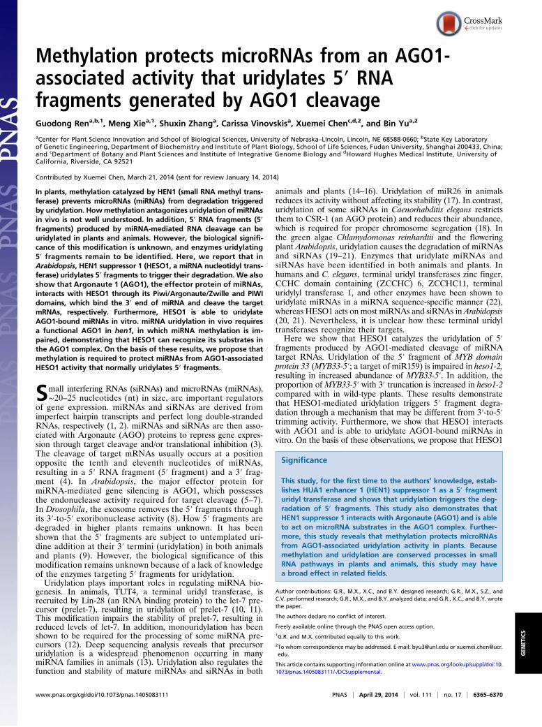
ResultsHESO1 Uridylates 5′ RNA Fragments Generated by miRNA-MediatedCleavage. HESO1 possesses terminal uridyl transferase activityon 21-nt small RNAs in vitro (20, 21). However, whether HESO1acts on other RNAs is not known. To address this question, wegenerated a [32P]-labeled single-stranded RNA (ssRNA; ∼100 nt),which corresponds to a portion of UBQ5 mRNA through in vitrotranscription. HESO1 lengthened this ssRNA in the presence ofUTP (Fig. 1A). This result suggested that HESO1 might havesubstrates other than small RNAs and, therefore, prompted us totest whether 5′ fragments are also substrates of HESO1. Wecompared 5′ fragment uridylation in the null heso1-2 mutant (20)with that in Landsberg erecta (Ler; wild-type control of heso1-2),using a 3′ adaptor-ligation mediated rapid amplification ofcDNA ends (al-RACE) approach. Total RNAs from Ler orheso1-2 were isolated, ligated to a 3′ adapter, and reverse-tran-scribed with a primer recognizing the 3′ adapter. SeminestedPCR was subsequently performed to amplify 5′ fragments gen-erated by AGO1 slicing of MYB domain protein 33 (MYB33-5′),Auxin Response Factor 10 (ARF10-5′), and Lost Meristems 1(LOM1-5′), which are targets of miR159, miR160, and miR171,respectively (23–26). PCR products of the expected sizes were
gel-purified, cloned, and sequenced (Fig. S1), and 75%, 59.1%,and 26.5% of MYB33-5′, ARF10-5′, and LOM1-5′ were uridy-lated in Ler, respectively (Fig. 1 B and C and Dataset S1). Incontrast, the proportions of uridylated MYB33-5′, ARF10-5′, andLOM1-5′ were reduced to 5.9%, 23.8%, and 12.9% in heso1-2,respectively (Fig. 1 B and C and Dataset S1). Furthermore, the 3′tail length of 5′ fragments was reduced in heso1-2 compared with
Fig. 1. HESO1 uridylates 5′ fragments. (A) HESO1 uridylates a long single-stranded RNA (ssRNA) in vitro. A 5′-end [32P]-labeled ssRNA was incubatedwith buffer, MBP, or MBP-HESO1 in the presence of UTP for 120 min, andproducts were resolved on a denaturing polyacrylamide gel. (B) Uridine ad-dition (red rectangle) at the 3′ end of the cleavage site of MYB33-5′. ▲ and▼ represent the adaptor. (C) Uridylation of 5′ fragments in Ler and heso1-2.Uridines in lowercase indicate that they can alternatively be considered asa templated addition. The numbers of clones for each modification wereshown in parentheses. Clones are the numbers of sequenced clones. Ratio isthe frequency of clones with 3′-end modifications among sequenced clones.
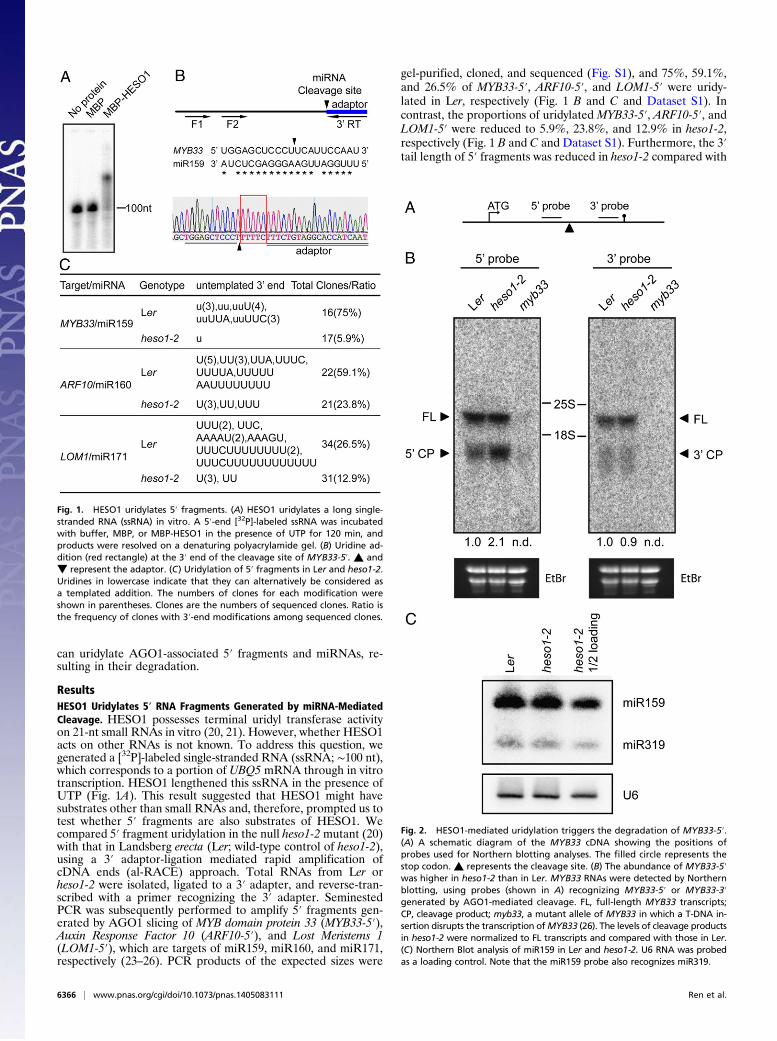
Fig. 2. HESO1-mediated uridylation triggers the degradation of MYB33-5′.(A) A schematic diagram of the MYB33 cDNA showing the positions ofprobes used for Northern blotting analyses. The filled circle represents thestop codon. ▲ represents the cleavage site. (B) The abundance of MYB33-5′was higher in heso1-2 than in Ler. MYB33 RNAs were detected by Northernblotting, using probes (shown in A) recognizing MYB33-5′ or MYB33-3′generated by AGO1-mediated cleavage. FL, full-length MYB33 transcripts;CP, cleavage product; myb33, a mutant allele of MYB33 in which a T-DNA in-sertion disrupts the transcription ofMYB33 (26). The levels of cleavage productsin heso1-2 were normalized to FL transcripts and compared with those in Ler.(C) Northern Blot analysis of miR159 in Ler and heso1-2. U6 RNA was probedas a loading control. Note that the miR159 probe also recognizes miR319.
6366 | www.pnas.org/cgi/doi/10.1073/pnas.1405083111 Ren et al.
that in Ler (1-3nt versus 1–15 nt; Fig. 1C). These results, togetherwith the in vitro activity analysis (Fig. 1 A and C), demonstratedthat HESO1 catalyzes uridylation of 5′ fragments generated bymiRNA-mediated cleavage. However, the presence of uridylated5′ fragments in the null heso1-2 mutant (Fig. 1C) indicated thatadditional HESO1 homologs might also act on 5′ fragments.
HESO1-Mediated Uridylation Triggers the Degradation of the 5′Fragment of MYB33 Generated by AGO1 Cleavage. Next, we exam-ined whether uridylation induced the degradation of 5′ frag-ments, using MYB33 as a reporter RNA. MYB33 was selectedbecause the majority of its 5′ fragments (MYB33-5′) are uridy-lated (Fig. 1C) (9). We compared the accumulation of MYB33-5′in heso1-2 with that in Ler by Northern blotting, with probesrecognizing MYB33-5′ (Fig. 2A). To determine the specificity ofprobe for MYB33-5′, we included a myb33 mutant in which atransfer (T)-DNA insertion abolished the transcription ofMYB33(26). We were able to detect MYB33-5′ in Ler and heso1-2, butnot in myb33. The levels of MYB33-5′ increased in heso1-2 rela-tive to those in Ler (Fig. 2B). This could be a result of the en-hanced cleavage ofMYB33 by AGO1 or decreased degradation ofMYB33-5′. If increased levels of MYB33-5′ were caused by en-hanced target cleavage, the abundance of MYB33-3′ would in-crease as well. Our data showed that the levels of MYB33-3′ weresimilar in heso1-2 to those in Ler (Fig. 2B), indicating that miRNA-mediated MYB33 cleavage did not increase in heso1-2. Consistentwith this observation, the levels of miR159 were not altered and theabundance ofMYB33 was only slightly elevated in heso1-2 (Fig. 2 Band C and Fig. S2A). Thus, we concluded that HESO1-mediateduridylation promotes 5′ fragment degradation.
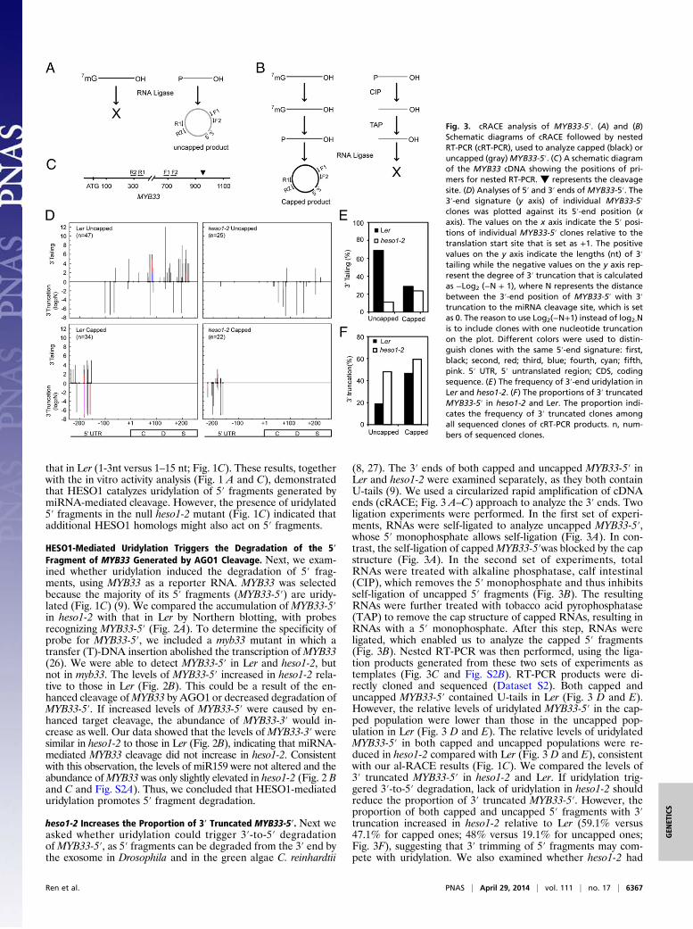
heso1-2 Increases the Proportion of 3′ Truncated MYB33-5′. Next weasked whether uridylation could trigger 3′-to-5′ degradationofMYB33-5′, as 5′ fragments can be degraded from the 3′ end bythe exosome in Drosophila and in the green algae C. reinhardtii
(8, 27). The 3′ ends of both capped and uncapped MYB33-5′ inLer and heso1-2 were examined separately, as they both containU-tails (9). We used a circularized rapid amplification of cDNAends (cRACE; Fig. 3 A–C) approach to analyze the 3′ ends. Twoligation experiments were performed. In the first set of experi-ments, RNAs were self-ligated to analyze uncapped MYB33-5′,whose 5′ monophosphate allows self-ligation (Fig. 3A). In con-trast, the self-ligation of cappedMYB33-5′was blocked by the capstructure (Fig. 3A). In the second set of experiments, totalRNAs were treated with alkaline phosphatase, calf intestinal(CIP), which removes the 5′ monophosphate and thus inhibitsself-ligation of uncapped 5′ fragments (Fig. 3B). The resultingRNAs were further treated with tobacco acid pyrophosphatase(TAP) to remove the cap structure of capped RNAs, resulting inRNAs with a 5′ monophosphate. After this step, RNAs wereligated, which enabled us to analyze the capped 5′ fragments(Fig. 3B). Nested RT-PCR was then performed, using the liga-tion products generated from these two sets of experiments astemplates (Fig. 3C and Fig. S2B). RT-PCR products were di-rectly cloned and sequenced (Dataset S2). Both capped anduncapped MYB33-5′ contained U-tails in Ler (Fig. 3 D and E).However, the relative levels of uridylated MYB33-5′ in the cap-ped population were lower than those in the uncapped pop-ulation in Ler (Fig. 3 D and E). The relative levels of uridylatedMYB33-5′ in both capped and uncapped populations were re-duced in heso1-2 compared with Ler (Fig. 3 D and E), consistentwith our al-RACE results (Fig. 1C). We compared the levels of3′ truncated MYB33-5′ in heso1-2 and Ler. If uridylation trig-gered 3′-to-5′ degradation, lack of uridylation in heso1-2 shouldreduce the proportion of 3′ truncated MYB33-5′. However, theproportion of both capped and uncapped 5′ fragments with 3′truncation increased in heso1-2 relative to Ler (59.1% versus47.1% for capped ones; 48% versus 19.1% for uncapped ones;Fig. 3F), suggesting that 3′ trimming of 5′ fragments may com-pete with uridylation. We also examined whether heso1-2 had
Fig. 3. cRACE analysis of MYB33-5′. (A) and (B)Schematic diagrams of cRACE followed by nestedRT-PCR (cRT-PCR), used to analyze capped (black) oruncapped (gray)MYB33-5′. (C) A schematic diagramof the MYB33 cDNA showing the positions of pri-mers for nested RT-PCR. ▼ represents the cleavagesite. (D) Analyses of 5′ and 3′ ends of MYB33-5′. The3′-end signature (y axis) of individual MYB33-5′clones was plotted against its 5′-end position (xaxis). The values on the x axis indicate the 5′ posi-tions of individual MYB33-5′ clones relative to thetranslation start site that is set as +1. The positivevalues on the y axis indicate the lengths (nt) of 3′tailing while the negative values on the y axis rep-resent the degree of 3′ truncation that is calculatedas −Log2 (−N + 1), where N represents the distancebetween the 3′-end position of MYB33-5′ with 3′truncation to the miRNA cleavage site, which is setas 0. The reason to use Log2(−N+1) instead of log2-Nis to include clones with one nucleotide truncationon the plot. Different colors were used to distin-guish clones with the same 5′-end signature: first,black; second, red; third, blue; fourth, cyan; fifth,pink. 5′ UTR, 5′ untranslated region; CDS, codingsequence. (E) The frequency of 3′-end uridylation inLer and heso1-2. (F) The proportions of 3′ truncatedMYB33-5′ in heso1-2 and Ler. The proportion indi-cates the frequency of 3′ truncated clones amongall sequenced clones of cRT-PCR products. n, num-bers of sequenced clones.
Ren et al. PNAS | April 29, 2014 | vol. 111 | no. 17 | 6367
GEN
ETICS
any effect on the 5′-to-3′ truncation of uncapped MYB33-5′.However, no obvious changes for the positions of 5′ truncationwere observed in heso1-2 relative to Ler (Fig. 3D).
Exoribonuclease 4 Can Degrade 5′ Fragments. Studies have shownthat exoribonucleases are involved in the degradation of RNAproducts generated by miRNA-mediated cleavage in Drosophilaand C. reinhardtii (8, 27). We therefore asked whether exoribo-nucleases have roles in degrading 5′ fragments in Arabidopsis.We examined whether exoribonuclease 4 (XRN4), which is amajor cytoplasmic 5′-to-3′ exoribonuclease in Arabidopsis (28,29), could degrade MYB33-5′. The levels of MYB33-5′ in xrn4-5,in which a T-DNA insertion completely abolished XRN4 func-tion (29), were higher than those in wild-type control (Col) byNorthern blotting. In contrast, the full-length MYB33 transcriptwas not obviously affected by xrn4-5 (Fig. S3), suggesting that the5′ fragments are subjected to 5′-to-3′ degradation in Arabidopsis.We also tested the function of the exosome components CSL4and RRP6L in MYB33-5′ degradation. Northern blotting showedthat the levels ofMYB33-5′ in csl4-1 and rrp6l1-1 rrp6l2-1 rrp6l3-1were comparable with those in Col (Fig. S3), suggesting that CSL4and RRP6L may not be involved in 5′ fragment degradation.
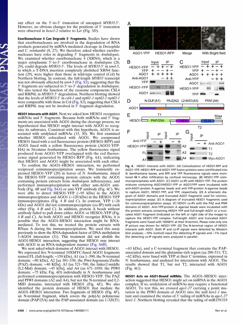
HESO1 Interacts with AGO1.Next we asked how HESO1 recognizesmiRNAs and 5′ fragments. Because both miRNAs and 5′ frag-ments are associated with AGO1 during the cleavage process, wehypothesized that HESO1 might interact with AGO1 to recog-nize its substrates. Consistent with this hypothesis, AGO1 is as-sociated with uridylated miRNAs (15, 30). We first examinedwhether HESO1 colocalized with AGO1. We coexpressedHESO1 fused with a red fluorescence protein (HESO1-RFP) andAGO1 fused with a yellow fluorescence protein (AGO1-YFP-HA) in Nicotiana benthamiana. The yellow fluorescence signalproduced from AGO1-YFP overlapped with the red fluores-cence signal generated by HESO1-RFP (Fig. 4A), indicatingthat HESO1 and AGO1 might be associated with each other.To confirm the AGO1–HESO1 interaction, we performed
reciprocal coimmunoprecipitation assays. We transiently ex-pressed HESO1-YFP (20) in leaves of N. benthamiana, mixedthe HESO1-YFP containing protein extracts with the AGO1containing protein extracts from Arabidopsis inflorescence, andperformed immunoprecipitation with either anti-AGO1 anti-body (Fig. 4B and Fig. S4A) or anti-YFP antibody (Fig. 4C). Wewere able to detect HESO1-YFP (∼95 kDa) in the AGO1immunoprecipitates and AGO1 (∼120 kDa) in the HESO1-YFPimmunoprecipitates (Fig. 4 B and C). In contrast, YFP (∼26kDa) and AGO1 did not coimmunoprecipitate (co-IP) with eachother (Fig. 4 B and C). In addition, protein A beads withoutantibody failed to pull down either AGO1 or HESO1-YFP (Fig.4 B and C). As both AGO1 and HESO1 recognize RNAs, it ispossible that the AGO1-HESO1 interaction might be RNA-mediated. To test this, we treated the protein extracts withRNase A during the immunoprecipitation. We used this assaypreviously to show the RNA-dependent factor of DNA methylation1-AGO4 interaction (31). This treatment did not abolish theAGO1-HESO1 interaction, suggesting that HESO1 may interactwith AGO1 in an RNA-independent manner (Fig. S4B).We next asked which domains of AGO1 interact with HESO1.
We expressed five N-terminal 10XMYC-fused AGO1 fragmentsnamed FL (full-length; ∼150 kDa), A1 (aa 1–390; the N-terminaldomain; ∼80 kDa), A2 [aa 381–530; the Piwi/Argonaute/Zwille(PAZ) domain; ∼40 KDa], A3 [aa 521–700; the linker2-middle(L2-Mid) domain; ∼45 kDa], and A4 (aa 671–1050; the PIWIdomain; ∼75 kDa; Fig. 4D) individually in N. benthamiana andperformed coimmunoprecipitation with HESO1-YFP. The PAZand PIWI domains (A2 and A4), but not the N-terminal and L2-MID domains, interacted with HESO1 (Fig. 4E). We alsoidentified the protein domains of HESO1 that mediate theAGO1–HESO1 interaction. Two fragments of HESO1 (Fig. 4F),an N-terminal fragment, which covers the poly(A) polymerasedomain (PAP/25A) and the PAP-associated domain (aa 1–320;T1;
∼63 kDa), and a C-terminal fragment that contains the PAP-associated domain and the glutamine-rich region (aa 200–511; T2;∼62 kDa), were fused with YFP at their C terminus, expressed inN. benthamiana, and analyzed for interactions with AGO1. Theresults showed that T1, but not T2, interacted with AGO1(Fig. 4G).
HESO1 Acts on AGO1-Bound miRNAs. The AGO1–HESO1 inter-action suggested that HESO1 might act on miRNA in the AGO1complex. If so, uridylation of miRNAs may require a functionalAGO1. To test this, we crossed ago1-27 carrying a point mu-tation in the PIWI domain of AGO1 into the null hen1-1 mu-tant and examined the status of 3′ tailing of miRNAs in ago1-27hen1-1. Northern blotting revealed that the tailing of miR159/319
Fig. 4. HESO1 interacts with AGO1. (A) Colocalization of HESO1-RFP andAGO1-YFP. HESO1-RFP and AGO1-YFP fusion proteins were coinfiltrated intoN. benthamiana leaves, and RFP and YFP fluorescence signals were moni-tored 48 h after infiltration by confocal microscopy. (B) HESO1-YFP coim-munoprecipitates with AGO1. (C) AGO1 co-IPs with HESO1-YFP. The proteinmixtures containing AGO1/HESO1-YFP or AGO1/YFP were incubated withanti-AGO1-protein A-agarose beads and anti-YFP-protein A-agarose beadsto capture AGO1, HESO1-YFP, and YFP, respectively. (D) A schematic dia-gram of AGO1 domains and truncated AGO1 fragments used for coimmu-noprecipitation assays. (E) A diagram of truncated HESO1 fragments usedfor coimmunoprecipitation assays. (F) HESO1 co-IPs with the PAZ and PIWIdomains of AGO1. Anti-YFP-protein A agarose beads were incubated withthe protein extracts containing HESO1-YFP and full-length AGO1 or a trun-cated AGO1 fragment (indicated on the left or right side of the image) tocapture the HESO1-YFP complex. Full-length AGO1 and truncated AGO1fragments were fused with 10XMYC at their N-termini. Please note only oneIP picture was shown for HESO1-YFP. (G) The N-terminal region of HESO1interacts with AGO1. Both IP and co-IP signals were detected by Westernblot analyses; ∼10% (vol/vol) input (for detecting IP signals) and ∼1% input(for detecting co-IP signals) were analyzed in parallel.
6368 | www.pnas.org/cgi/doi/10.1073/pnas.1405083111 Ren et al.
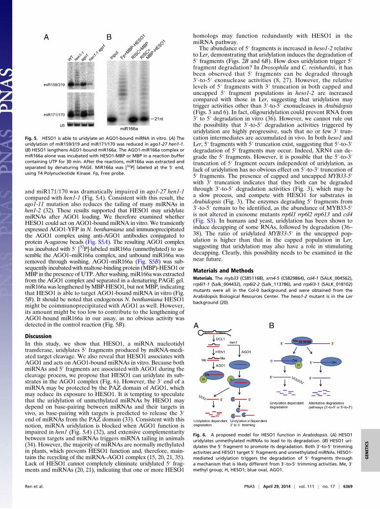
and miR171/170 was dramatically impaired in ago1-27 hen1-1compared with hen1-1 (Fig. 5A). Consistent with this result, theago1-11 mutation also reduces the tailing of many miRNAs inhen1-2 (32). These results supported that HESO1 may uridylatemiRNAs after AGO1 loading. We therefore examined whetherHESO1 could act on AGO1-bound miRNA in vitro. We transientlyexpressed AGO1-YFP in N. benthamiana and immunoprecipitatedthe AGO1 complex using anti-AGO1 antibodies conjugated toprotein A-agarose beads (Fig. S5A). The resulting AGO1 complexwas incubated with 5′ [32P]-labeled miR166a (unmethylated) to as-semble the AGO1–miR166a complex, and unbound miR166a wasremoved through washing. AGO1–miR166a (Fig. S5B) was sub-sequently incubated withmaltose-binding protein (MBP)-HESO1 orMBP in the presence of UTP. After washing, miR166a was extractedfrom the AGO1 complex and separated in a denaturing PAGE gel.miR166a was lengthened byMBP-HESO1, but notMBP, indicatingthat HESO1 is able to target AGO1-bound miRNA in vitro (Fig.5B). It should be noted that endogenous N. benthamiana HESO1might be coimmunoprecipitated with AGO1 as well. However,its amount might be too low to contribute to the lengthening ofAGO1-bound miR166a in our assay, as no obvious activity wasdetected in the control reaction (Fig. 5B).
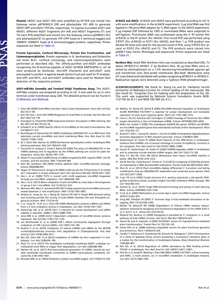
DiscussionIn this study, we show that HESO1, a miRNA nucleotidyltransferase, uridylates 5′ fragments produced by miRNA-medi-ated target cleavage. We also reveal that HESO1 associates withAGO1 and acts on AGO1-bound miRNAs in vitro. Because bothmiRNAs and 5′ fragments are associated with AGO1 during thecleavage process, we propose that HESO1 can uridylate its sub-strates in the AGO1 complex (Fig. 6). However, the 3′ end of amiRNA may be protected by the PAZ domain of AGO1, whichmay reduce its exposure to HESO1. It is tempting to speculatethat the uridylation of unmethylated miRNAs by HESO1 maydepend on base-pairing between miRNAs and their targets invivo, as base-pairing with targets is predicted to release the 3′end of miRNAs from the PAZ domain (33). Consistent with thisnotion, miRNA uridylation is blocked when AGO1 function isimpaired in hen1 (Fig. 5A) (32), and extensive complementaritybetween targets and miRNAs triggers miRNA tailing in animals(34). However, the majority of miRNAs are normally methylatedin plants, which prevents HESO1 function and, therefore, main-tains the recycling of the miRNA–AGO1 complex (15, 20, 21, 35).Lack of HESO1 cannot completely eliminate uridylated 5′ frag-ments and miRNAs (20, 21), indicating that one or more HESO1
homologs may function redundantly with HESO1 in themiRNA pathway.The abundance of 5′ fragments is increased in heso1-2 relative
to Ler, demonstrating that uridylation induces the degradation of5′ fragments (Figs. 2B and 6B). How does uridylation trigger 5′fragment degradation? In Drosophila and C. reinhardtii, it hasbeen observed that 5′ fragments can be degraded through3′-to-5′ exonuclease activities (8, 27). However, the relativelevels of 5′ fragments with 3′ truncation in both capped anduncapped 5′ fragment populations in heso1-2 are increasedcompared with those in Ler, suggesting that uridylation maytrigger activities other than 3′-to-5′ exonucleases in Arabidopsis(Figs. 3 and 6). In fact, oligouridylation could prevent RNA from3′ to 5′ degradation in vitro (36). However, we cannot rule outthe possibility that 3′-to-5′ degradation activities triggered byuridylation are highly progressive, such that no or few 3′ trun-cation intermediates are accumulated in vivo. In both heso1 andLer, 5′ fragments with 5′ truncation exist, suggesting that 5′-to-3′degradation of 5′ fragments may occur. Indeed, XRN4 can de-grade the 5′ fragments. However, it is possible that the 5′-to-3′truncation of 5′ fragment occurs independent of uridylation, aslack of uridylation has no obvious effect on 5′-to-3′ truncation of5′ fragments. The presence of capped and uncapped MYB33-5′with 3′ truncation indicates that they both can be degradedthrough 3′-to-5′ degradation activities (Fig. 3), which may bea slow process, and compete with HESO1 for substrates inArabidopsis (Fig. 3). The enzymes degrading 5′ fragments from3′-to-5′ remain to be identified, as the abundance of MYB33-5′is not altered in exosome mutants rrp6l1 rrp6l2 rrp613 and csl4(Fig. S3). In humans and yeast, uridylation has been shown toinduce decapping of some RNAs, followed by degradation (36–38). The ratio of uridylated MYB33-5′ in the uncapped pop-ulation is higher than that in the capped population in Ler,suggesting that uridylation may also have a role in stimulatingdecapping. Clearly, this possibility needs to be examined in thenear future.
Materials and MethodsMaterials. The myb33 (CS851168), xrn4-5 (CS829864), csl4-1 (SALK_004562),rrp6l1-1 (Salk_004432), rrp6l2-2 (Salk_113786), and rrp6l3-1 (SALK_018102)mutants were all in the Col-0 background and were obtained from theArabidopsis Biological Resources Center. The heso1-2 mutant is in the Lerbackground (20).
Fig. 5. HESO1 is able to uridylate an AGO1-bound miRNA in vitro. (A) Theuridylation of miR159/319 and miR171/170 was reduced in ago1-27 hen1-1.(B) HESO1 lengthens AGO1-bound miR166a. The AGO1-miR166a complex ormiR166a alone was incubated with HESO1-MBP or MBP in a reaction buffercontaining UTP for 30 min. After the reactions, miR166a was extracted andseparated by denaturing PAGE. MiR166a was [32P] labeled at the 5′ end,using T4 Polynucleotide Kinase. Fp, Free probe.
Fig. 6. A proposed model for HESO1 function in Arabidopsis. (A) HESO1uridylates unmethylated miRNAs to lead to its degradation. (B) HESO1 uri-dylates the 5′ fragment to promote its degradation. Both 3′-to-5′ trimmingactivities and HESO1 target 5′ fragments and unmethylated miRNAs. HESO1-mediated uridylation triggers the degradation of 5′ fragments througha mechanism that is likely different from 3′-to-5′ trimming activities. Me, 3′methyl group; H, HESO1; blue oval, AGO1.
Ren et al. PNAS | April 29, 2014 | vol. 111 | no. 17 | 6369
GEN
ETICS
Plasmid. HESO1 and AGO1 CDS were amplified by RT-PCR and cloned intoGateway vector pB7WGR2,0 (39) and pEarleyGate 101 (40) to generateHESO1-RFP and AGO1-YFP-HA, respectively. To express truncated AGO1 andHESO1, different AGO1 fragments (A1–A4) and HESO1 fragments (T1 andT2) were PCR-amplified and cloned into the Gateway vectors pGWB521 (41)and pEarleyGate101 to generate YFP (YFP fused at C terminus)-tagged and10XMYC (10XMYC fused at N terminus)-tagged proteins, respectively. Primersequences are listed in Table S1.
Protein Expression, Confocal Microscopy, Protein Size Fractionation, andCoimmunoprecipitation. Protein expression in N. benthamiana and Escherichiacoli strain BL21, confocal microscopy, and coimmunoprecipitation wereperformed as described (42). The affinity-purified anti-AGO1 antibodiesrecognizing the N-terminal peptide of AGO1 (N-MVR KRRTDAPSC-C; ref. 6)were produced by GenScript. Anti-GFP (Clontech) and anti-AGO1 wereprecoupled to protein A agarose beads (Santa Cruz) and used for IP analyses.Anti-GFP, anti-MYC, and anti-AGO1 antibodies were used for Western blotdetection of the respective proteins.
AGO1-miR166a Assembly and Terminal Uridyl Transferase Assay. The AGO1-miR166a complex was prepared according to ref. 5 and used for an in vitroterminal uridyl transferase assay (20). The detailed protocol can be found inSI Materials and Methods.
Al-RACE and cRACE. Al-RACE and cRACE were performed according to ref. 9,with some modifications. In the al-RACE experiment, 5 μg total RNA was firstligated to 100 pmol RNA adaptor by T4 RNA ligase. In the cRACE experiment,5 μg treated (CIP followed by TAP) or nontreated RNAs were subjected toself-ligation. First-strand cDNA was synthesized using the 3′ RT primer (foral-RACE) or the R1 primer (for cRACE). First-round PCR was performed using3′RT/F1 (for al-RACE) or R1/F1 (for cRACE), and then 1 μL PCR product wasdiluted 50 times and used for the second round of PCR, using 3′RT/F2 (For al-race) or R2/F2 (for cRACE) and F2. The PCR products were cloned intopGEM-T Easy Vector (Promega) and sequenced. Primer sequences are listedin Table S1.
Northern Blot. Small RNA Northern blot was conducted as described (43). Todetect MYB33-5′or MYB33- 3′ by Northern blot, 30 μg total RNAs were re-solved by electrophoresis on a 1.2% denaturing-formaldehyde agarose geland transferred onto Zeta-probe membranes (Bio-Rad). Membranes wereUV cross-linked and hybridized with probes recognizingMYB33-5′ orMYB33-3′.Radioactive signals were detected using a Typhoon 9500 phosphorimager.
ACKNOWLEDGMENTS. We thank Dr. Xiang Liu and Dr. Heriberto Cerutti(University of Nebraska–Lincoln) for critical reading of the manuscript. Wealso thank Dr. Yongchao Dou for helping with the preparation of Fig. 3D.This work was supported by National Science Foundation Grants MCB-1121193 (to B.Y.) and MCB-1021465 (to X.C.).
1. Chen XM (2009) Small RNAs and their roles in plant development. Annu Rev Cell DevBiol 25:21–44.
2. Kim VN, Han J, Siomi MC (2009) Biogenesis of small RNAs in animals. Nat Rev Mol CellBiol 10(2):126–139.
3. Hutvagner G, Simard MJ (2008) Argonaute proteins: Key players in RNA silencing. NatRev Mol Cell Biol 9(1):22–32.
4. Schwab R, et al. (2005) Specific effects of microRNAs on the plant transcriptome. DevCell 8(4):517–527.
5. Baumberger N, Baulcombe DC (2005) Arabidopsis ARGONAUTE1 is an RNA Slicer thatselectively recruits microRNAs and short interfering RNAs. Proc Natl Acad Sci USA102(33):11928–11933.
6. Qi Y, Denli AM, Hannon GJ (2005) Biochemical specialization within Arabidopsis RNAsilencing pathways. Mol Cell 19(3):421–428.
7. Vaucheret H, Vazquez F, Crété P, Bartel DP (2004) The action of ARGONAUTE1 in themiRNA pathway and its regulation by the miRNA pathway are crucial for plant de-velopment. Genes Dev 18(10):1187–1197.
8. Orban TI, Izaurralde E (2005) Decay of mRNAs targeted by RISC requires XRN1, the Skicomplex, and the exosome. RNA 11(4):459–469.
9. Shen BZ, Goodman HM (2004) Uridine addition after microRNA-directed cleavage.Science 306(5698):997.
10. Hagan JP, Piskounova E, Gregory RI (2009) Lin28 recruits the TUTase Zcchc11 to inhibitlet-7 maturation in mouse embryonic stem cells. Nat Struct Mol Biol 16(10):1021–1025.
11. Heo I, et al. (2009) TUT4 in concert with Lin28 suppresses microRNA biogenesisthrough pre-microRNA uridylation. Cell 138(4):696–708.
12. Heo I, et al. (2012) Mono-uridylation of pre-microRNA as a key step in the biogenesisof group II let-7 microRNAs. Cell 151(3):521–532.
13. Newman MA, Mani V, Hammond SM (2011) Deep sequencing of microRNA precursorsreveals extensive 3′ end modification. RNA 17(10):1795–1803.
14. Ameres SL, Hung JH, Xu J, Weng ZP, Zamore PD (2011) Target RNA-directed tailingand trimming purifies the sorting of endo-siRNAs between the two Drosophila Ar-gonaute proteins. RNA 17(1):54–63.
15. Li JJ, Yang ZY, Yu B, Liu J, Chen XM (2005) Methylation protects miRNAs and siRNAsfrom a 3′-end uridylation activity in Arabidopsis. Curr Biol 15(16):1501–1507.
16. Kamminga LM, et al. (2010) Hen1 is required for oocyte development and piRNAstability in zebrafish. EMBO J 29(21):3688–3700.
17. Jones MR, et al. (2009) Zcchc11-dependent uridylation of microRNA directs cytokineexpression. Nat Cell Biol 11(9):1157–1163.
18. van Wolfswinkel JC, et al. (2009) CDE-1 affects chromosome segregation throughuridylation of CSR-1-bound siRNAs. Cell 139(1):135–148.
19. Ibrahim F, et al. (2010) Uridylation of mature miRNAs and siRNAs by the MUT68nucleotidyltransferase promotes their degradation in Chlamydomonas. Proc NatlAcad Sci USA 107(8):3906–3911.
20. Ren GD, Chen XM, Yu B (2012) Uridylation of miRNAs by hen1 suppressor1 in Ara-bidopsis. Curr Biol 22(8):695–700.
21. Zhao YY, et al. (2012) The Arabidopsis nucleotidyl transferase HESO1 uridylates un-methylated small RNAs to trigger their degradation. Curr Biol 22(8):689–694.
22. Wyman SK, et al. (2011) Post-transcriptional generation of miRNA variants by mul-tiple nucleotidyl transferases contributes to miRNA transcriptome complexity. Ge-nome Res 21(9):1450–1461.
23. Rhoades MW, et al. (2002) Prediction of plant microRNA targets. Cell 110(4):513–520.
24. Mallory AC, Bartel DP, Bartel B (2005) MicroRNA-directed regulation of ArabidopsisAUXIN RESPONSE FACTOR17 is essential for proper development and modulatesexpression of early auxin response genes. Plant Cell 17(5):1360–1375.
25. Llave C, Xie ZX, Kasschau KD, Carrington JC (2002) Cleavage of Scarecrow-like mRNAtargets directed by a class of Arabidopsis miRNA. Science 297(5589):2053–2056.
26. Millar AA, Gubler F (2005) The Arabidopsis GAMYB-like genes, MYB33 and MYB65,are microRNA-regulated genes that redundantly facilitate anther development. PlantCell 17(3):705–721.
27. Ibrahim F, Rohr J, Jeong WJ, Hesson J, Cerutti H (2006) Untemplated oligoadenylationpromotes degradation of RISC-cleaved transcripts. Science 314(5807):1893.
28. Kastenmayer JP, Green PJ (2000) Novel features of the XRN-family in Arabidopsis:Evidence that AtXRN4, one of several orthologs of nuclear Xrn2p/Rat1p, functions inthe cytoplasm. Proc Natl Acad Sci USA 97(25):13985–13990.
29. Souret FF, Kastenmayer JP, Green PJ (2004) AtXRN4 degrades mRNA in Arabidopsisand its substrates include selected miRNA targets. Mol Cell 15(2):173–183.
30. Zhao YY, Mo BX, Chen XM (2012) Mechanisms that impact microRNA stability inplants. RNA Biol 9(10):1218–1223.
31. Xie M, Ren GD, Costa-Nunes P, Pontes O, Yu B (2012) A subgroup of SGS3-like proteinsact redundantly in RNA-directed DNAmethylation. Nucleic Acids Res 40(10):4422–4431.
32. Zhai JX, et al. (2013) Plant microRNAs display differential 3′ truncation and tailingmodifications that are ARGONAUTE1 dependent and conserved across species. PlantCell 25(7):2417–2428.
33. Yuan YR, et al. (2005) Crystal structure of A. aeolicus argonaute, a site-specific DNA-guided endoribonuclease, provides insights into RISC-mediated mRNA cleavage. MolCell 19(3):405–419.
34. Ameres SL, et al. (2010) Target RNA-directed trimming and tailing of small silencingRNAs. Science 328(5985):1534–1539.
35. Yu B, et al. (2005) Methylation as a crucial step in plant microRNA biogenesis. Science307(5711):932–935.
36. Song MG, Kiledjian M (2007) 3′ Terminal oligo U-tract-mediated stimulation of de-capping. RNA 13(12):2356–2365.
37. Mullen TE, Marzluff WF (2008) Degradation of histone mRNA requires oligour-idylation followed by decapping and simultaneous degradation of the mRNA both 5′to 3′ and 3′ to 5′. Genes Dev 22(1):50–65.
38. Rissland OS, Norbury CJ (2009) Decapping is preceded by 3′ uridylation in a novelpathway of bulk mRNA turnover. Nat Struct Mol Biol 16(6):616–623.
39. Karimi M, Inzé D, Depicker A (2002) GATEWAY vectors for Agrobacterium-mediatedplant transformation. Trends Plant Sci 7(5):193–195.
40. Earley KW, et al. (2006) Gateway-compatible vectors for plant functional genomicsand proteomics. Plant J 45(4):616–629.
41. Tanaka Y, Nakamura S, Kawamukai M, Koizumi N, Nakagawa T (2011) Developmentof a series of gateway binary vectors possessing a tunicamycin resistance gene asa marker for the transformation of Arabidopsis thaliana. Biosci Biotechnol Biochem75(4):804–807.
42. Ren GD, et al. (2012) Regulation of miRNA abundance by RNA binding proteinTOUGH in Arabidopsis. Proc Natl Acad Sci USA 109(31):12817–12821.
43. Park W, Li JJ, Song RT, Messing J, Chen XM (2002) CARPEL FACTORY, a Dicer homolog,and HEN1, a novel protein, act in microRNA metabolism in Arabidopsis thaliana.Curr Biol 12(17):1484–1495.
6370 | www.pnas.org/cgi/doi/10.1073/pnas.1405083111 Ren et al.