methylation-specific pcr: a novel pcr assay for methylation status
TRANSCRIPT

Proc. Natl. Acad. Sci. USAVol. 93, pp. 9821-9826, September 1996Medical Sciences
Methylation-specific PCR: A novel PCR assay for methylationstatus of CpG islands
(DNA methylation/tumor suppressor genes/pl6/p15)
JAMES G. HERMAN*t, JEREMY R. GRAFF*, SANNA MYOHANEN*, BARRY D. NELKIN*, AND STEPHEN B. BAYLIN*t*Oncology Center and :Department of Medicine, The Johns Hopkins Medical Institutions, 424 North Bond Street, Baltimore, MD 21231
Communicated by Victor A. McKusick, Johns Hopkins Hospital, Baltimore, MD, June 3, 1996 (received for review April 3, 1996)
ABSTRACT Precise mapping of DNA methylation pat-terns in CpG islands has become essential for understandingdiverse biological processes such as the regulation of im-printed genes, X chromosome inactivation, and tumor sup-pressor gene silencing in human cancer. We describe a newmethod, MSP (methylation-specific PCR), which can rapidlyassess the methylation status of virtually any group of CpGsites within a CpG island, independent of the use of methyl-ation-sensitive restriction enzymes. This assay entails initialmodification of DNA by sodium bisulfite, converting allunmethylated, but not methylated, cytosines to uracil, andsubsequent amplification with primers specific for methylatedversus unmethylated DNA. MSP requires only small quanti-ties of DNA, is sensitive to 0.1% methylated alleles of a givenCpG island locus, and can be performed on DNA extractedfrom paraffin-embedded samples. MSP eliminates the falsepositive results inherent to previous PCR-based approacheswhich relied on differential restriction enzyme cleavage todistinguish methylated from unmethylated DNA. In thisstudy, we demonstrate the use of MSP to identify promoterregion hypermethylation changes associated with transcrip-tional inactivation in four important tumor suppressor genes(p16, p15, E-cadherin, and von Hippel-Lindau) in humancancer.
In higher order eukaryotes, DNA is methylated only at cy-tosines located 5' to guanosine in the CpG dinucleotide (1).This modification has important regulatory effects on geneexpression, especially when involving CpG-rich areas known asCpG islands, located in the promoter regions of many genes (2,3). While almost all gene-associated islands are protected frommethylation on autosomal chromosomes (3), extensive meth-ylation of CpG islands has been associated with transcriptionalinactivation of selected imprinted genes (4, 5) and genes on theinactive X-chromosome of females (6, 7). Aberrant methyl-ation of normally unmethylated CpG islands has been docu-mented as a relatively frequent event in immortalized andtransformed cells (8) and has been associated with transcrip-tional inactivation of defined tumor suppressor genes in humancancers (9-12). In this last situation, promoter region hyper-methylation stands as an alternative to coding region muta-tions in eliminating tumor suppressor gene function (9, 10).Therefore, mapping of methylation patterns in CpG islandshas become an important tool for understanding both normaland pathologic gene expression events.Mapping of methylated regions in DNA has relied primarily
on Southern hybridization approaches, based on the inabilityof methylation-sensitive restriction enzymes to cleave se-quences that contain one or more methylated CpG sites. Thismethod provides an assessment of the overall methylationstatus of CpG islands, including some quantitative analysis(13), but requires large amounts of high molecular weight
DNA (generally 5 ,tg or more), can detect methylation only ifpresent in greater than a few percent of the alleles and can onlyprovide information about those CpG sites found withinsequences recognized by methylation-sensitive restriction en-zymes. A more sensitive method of methylation detectioncombines the use of methylation-sensitive enzymes and PCR(14). After digestion of DNA with the enzyme, PCR willamplify from primers flanking the restriction site only if DNAcleavage has been prevented by methylation (15, 16). LikeSouthern-based approaches, this method can only monitorCpG methylation in methylation-sensitive restriction sites.Moreover, the restriction of unmethylated DNA must becomplete, since any uncleaved DNA will be amplified by PCRyielding a false positive result for methylation. This approachhas been useful in studying samples where a high percentageof alleles of interest are methylated, such as the study ofimprinted genes (5, 15, 16) and X chromosome-inactivatedgenes (14). However, difficulties in distinguishing betweenincomplete restriction and low numbers of methylated allelesmake this approach unreliable for detection of tumor suppres-sor gene hypermethylation in small samples or in sampleswhere methylated alleles represent a small fraction of thepopulation.The chemical modification of cytosine to uracil by bisulfite
treatment has provided another method for the study of DNAmethylation that avoids the use of restriction enzymes (17). Inthis reaction, all cytosines are converted to uracil, but thosethat are methylated (5-methylcytosine) are resistant to thismodification and remain as cytosine (18). This altered DNAcan then be amplified and sequenced, providing detailedinformation within the amplified region of the methylationstatus of all CpG sites (17). However, this method is technicallyrather difficult and labor-intensive, and, without cloning of theamplified products, the technique is less sensitive than South-ern analysis, requiring - 25% of the alleles to be methylated fordetection (19).We now report a novel PCR method, methylation-specific
PCR (MSP), which is sensitive and specific for methylation ofvirtually any block of CpG sites in a CpG island. We designedprimers to distinguish methylated from unmethylated DNA inbisulfite-modified DNA, taking advantage of the sequencedifferences resulting from bisulfite modification. UnmodifiedDNA or DNA incompletely reacted with bisulfite can also bedistinguished, since marked sequence differences exist be-tween these DNAs. The frequency of CpG sites in CpG islandsrenders this technique uniquely useful and extremely sensitivefor such regions. Herein, we detail the MSP procedure andshow its use for detecting the aberrant methylation of fourtumor suppressor genes in human neoplasia.
MATERIALS AND METHODSDNA and Cell Lines. Genomic DNA was obtained from cell
lines, primary tumors, and normal tissue as described (10-12).
Abbreviations: MSP, methylation-specific PCR; VHL, von Hippel-Lindau.tTo whom reprint requests should be addressed.
The publication costs of this article were defrayed in part by page chargepayment. This article must therefore be hereby marked "advertisement" inaccordance with 18 U.S.C. §1734 solely to indicate this fact.
9821

9822 Medical Sciences: Herman et al.
The renal carcinoma cell line was kindly provided by MichaelLerman (National Cancer Institute, Frederick, MD).
Bisulfite Modification. DNA (1 ,ug) in a volume of 50 gl wasdenatured by NaOH (final concentration, 0.2 M) for 10 min at37°C. For samples with nanogram quantities of human DNA,1 ,ug of salmon sperm DNA (Sigma) was added as carrierbefore modification. Thirty microliters of 10 mM hydroqui-none (Sigma) and 520 ,ul of 3 M sodium bisulfite (Sigma) at pH5, both freshly prepared, were added and mixed, and sampleswere incubated under mineral oil at 50°C for 16 hr. ModifiedDNA was purified using the Wizard DNA purification resinaccording to the manufacturer (Promega) and eluted into 50ptl of water. Modification was completed by NaOH (finalconcentration, 0.3 M) treatment for 5 min at room tempera-ture, followed by ethanol precipitation. DNA was resuspendedin water and used immediately or stored at -20°C.Genomic Sequencing. Genomic sequencing of bisulfite-
modified DNA (17) was accomplished using the solid-phaseDNA sequencing approach (19). Bisulfite modified DNA (100ng) was amplified with p16 gene-specific primers 5'-TTTTTAGAGGATTTGAGGGATAGG (sense) and 5'-CTACCTAATTCCAATTCCCCTACA (anti-sense). PCRconditions were as follows: 96°C for 3 min and 80°C for 3 min,after which 1 unit of Taq polymerase (BRL) was added; then35 cycles of 96°C for 20 sec, 56°C for 20 sec, 72°C for 90 sec;and finally 5 min at 72°C. The PCR mixture contained lx
buffer (BRL) with 1.5 mM MgCl2, 20 pmol of each primer, and0.2 mM dNTPs. To obtain products for sequencing, a secondround of PCR was performed with 5 pmol of nested primers.In this reaction, the sense primer, 5'-GTTTTCCCAGTCAC-GACAGTATTAGGAGGAAGAAAGAGGAG, containsM13-40 sequence (underlined) introduced as a site to initiatesequencing, and the anti-sense primer 5'-TCCAATTC-CCCTACAAACTTC is biotinylated to facilitate purificationof the product before sequencing. PCR was performed asabove, for 32 cycles with 2.5 mM MgC42. All primers forgenomic sequencing were designed to avoid any CpGs in thesequence. Biotinylated PCR products were purified usingstreptavidin-coated magnetic beads (Dynal, Oslo), and se-quencing reactions were performed with Sequenase andM13-40 sequencing primer under conditions specified by themanufacturer (United States Biochemical).PCR Amplification. Primer pairs described in Table 1
(20-24) were purchased from Life Technologies. The PCRmixture contained lx PCR buffer (16.6 mM ammoniumsulfate/67 mM Tris, pH 8.8/6.7 mM MgCl2/10 mM 2-mer-
captoethanol), dNTPs (each at 1.25 mM), primers (300 ng eachper reaction), and bisulfite-modified DNA ('50 ng) or un-
modified DNA (50-100 ng) in a final volume of 50 Al. PCRspecific for unmodified DNA also included 5% dimethylsulfoxide. Reactions were hot-started at 95°C for 5 min beforethe addition of 1.25 units of Taq polymerase (BRL). Ampli-fication was carried out in a HIybaid OmniGene temperaturecycler for 35 cycles (30 sec at 95°C, 30 sec at the annealingtemperature listed in Table 1, and 30 sec at 72°C), followed bya final 4-min extension at 72°C. Controls without DNA were
performed for each set of PCRs. Each PCR (10 ,l) was directlyloaded onto nondenaturing 6-8% polyacrylamide gels, stainedwith ethidium bromide, and directly visualized under UVillumination.
Restriction Analysis. Of the 50 ,lI of PCR mixture, 10 Al wasdigested with 10 units of BstUI (New England Biolabs) for 4hr, according to conditions specified by the manufacturer.Restriction digests were ethanol precipitated before gel analysis.
RESULTS
Validating the Design Strategy for MSP: Genomic Sequenc-ing ofpl6. An initial study was required to validate whether ourstrategy for MSP would prove feasible for assessing themethylation status of CpG islands. We needed to determinewhether the density of methylation, in key regions to be tested,was great enough to facilitate our primer design. We chose totest this for thepl6 tumor suppressor gene in which we (10, 25)and others (26, 27) have documented that hypermethylation ofa 5' CpG island is associated with complete loss of geneexpression in many cancer types. However, other than for CpGsites located in recognition sequences for methylation-sensitive enzymes, the density of methylation and its correla-tion to transcriptional silencing has not been established. Wethus employed the genomic sequencing technique to explorethis relationship. As has been found for other CpG islandsexamined in this manner (19,28,29), the CpG island ofp16 hadno methylation at any CpG site in those cell lines and normaltissues previously found to be unmethylated by Southernanalysis (Fig. 1; refs. 10 and 25). However, it was extensivelymethylated in cancer cell lines shown to be methylated bySouthern analysis (Fig. 1). In fact, all cytosines within CpGdinucleotides in this region were completely methylated in thecancers lacking pi6 transcription. This marked difference insequence following bisulfite treatment suggested that our
Table 1. PCR primers used for MSP
AnnealPrimer temp., Genomic
set Sense primer,* 5' 3' Antisense primer,* 5' > 3' Size, bp °C positiontp16-Wt CAGAGGGTGGGGCGGACCGC CGGGCCGCGGCCGTGG 140 65 +171p16-M TTATTAGAGGGTGGGGCGGATCGC GACCCCGAACCGCGACCGTAA 150 65 +167p16-U TTATTAGAGGGTGGGGTGGATTGT CAACCCCAAACCACAACCATAA 151 60 +167p16-M2 TTATTAGAGGGTGGGGCGGATCGC CCACCTAAATCGACCTCCGACCG 234 65 +167p16-U2 TTATTAGAGGGTGGGGTGGATTGT CCACCTAAATCAACCTCCAACCA 234 60 +167p15-W CGCACCCTGCGGCCAGA AGTGGCCGAGCGGCCGG 137 65 +46plS-M GCGTTCGTATTTTGCGGTT CGTACAATAACCGAACGACCGA 148 60 +40p15-U TGTGATGTGTTTGTATTTTGTGGTT CCATACAATAACCAAACAACCAA 154 60 +34VHL-M TGGAGGATTTTTTTGCGTACGC GAACCGAACGCCGCGAA 158 60 -116VHL-U GTTGGAGGATTTTTTTGTGTATGT CCCAAACCAAACACCACAAA 165 60 -118Ecad-M TTAGGTTAGAGGGTTATCGCGT TAACTAAAAATTCACCTACCGAC 116 57 -205Ecad-U TAATTTTAGGTTAGAGGGTTATTGT CACAACCAATCAACAACACA 97 53 -210
*Sequence differences between modified primers and unmodified DNA are in boldface type and differences between methylated/modified andummethylated/modified are underlined.tPrimers were placed near the transcriptional start site. Genomic position is the location of the 5' nucleotide of the sense primer in relation tothe major transcriptional start site defined in the following references and Genbank accession numbers:p16 (most 3' site), X94154 (20);plS, S75756(22); VHL, U19763 (23); and E-cadherin, L34545 (24).*W represents unmodified or wild-type primers. M, methylated-specific primers; and U, unmethylated-specific primers.
Proc. Natl. Acad. Sci. USA 93 (1996)

Proc. Natl. Acad. Sci. USA 93 (1996) 9823
H157
.......
H249G A T C
:488:
p#:swwRas sR
_i10!:
... .: ..
.: ':: _t._. .........
.._'
: ttS
; t....... ,. ,.:e=__
........... : .' '
.'..'b".'!.:.
._- ,.... : :: : .t_:_ .:
*_gi*-,_ ..*?ti. :. ....
._. ... .i
4_ft..:; :.
i::d.:i:.
::
:: :::
_@ .
.:t..::+/4t:
I..
iw-la
,Mii-
*0:.:
41 w4~
FIG. 1. Genomic sequencing of p16. The sequence shown has themost 5' region at the bottom of the gel, beginning at + 175 in relationto a major transcriptional start site (20). All cytosines in the unmethy-lated cell line H249 have been converted to thymidine, while all Cs inCpG dinucleotides in the methylated cell H157 remain as C, indicatingmethylation. Designated by the bracket (]) is a BstUI site, which is at-59 in relation to the translational start site in GenBank sequenceU12818 (21), but which is incorrectly identified as CGAG in sequenceX94154 (20). This CGCG site represents the 3' location of the sense
primer used for p16 MSP.
strategy for specific amplification of either methylated or
unmethylated alleles was feasible.Primer Design for MSP. Primers were designed to discrim-
inate between methylated and unmethylated alleles followingbisulfite treatment and to discriminate between DNA modifiedby bisulfite and that which had not been modified. To accom-
plish this, primer sequences were chosen for regions contain-ing frequent cytosines (to distinguish unmodified from mod-ified DNA), and CpG pairs near the 3' end of the primers (toprovide maximal discrimination in the PCR between methyl-ated and unmethylated DNA). Since the two strands of DNAare no longer complementary after bisulfite treatment, prim-ers can be designed for either modified strand. For conve-
nience, we have designed primers for the sense strand. Thefragment of DNA to be amplified was intentionally small, toallow the assessment of methylation patterns in a limitedregion and to facilitate the application of this technique tosamples, such as paraffin blocks, where amplification of largerfragments is not possible. In Table 1, primer sequences are
shown for all genes tested, emphasizing the differences insequence between the three types of DNA that are exploitedfor the specificity of MSP. The multiple mismatches in these
primers, which are specific for these different types of DNA,suggest that each primer set should provide amplification onlyfrom the intended template.MSP Analysis of p16. We first tested the primers designed
for p16 on DNA from cancer cell lines and normal tissues forwhich the methylation status had previously been defined bySouthern analysis (10, 25). In all cases, the primer set usedconfirmed the methylation status determined by Southernanalysis. For example, lung cancer cell lines U1752 and H157,as well as other cell lines with methylatedpl6 alleles, amplifiedonly with the methylated primers (Fig. 2A). DNA from normaltissues (lymphocytes, lung, kidney, breast, and colon) and thelung cancer cell lines H209 and H249, having only unmethy-lated p16 alleles, amplified only with unmethylated primers(examples in Fig. 2A). PCR with these primers could beperformed with or without 5% dimethyl sulfoxide. DNA nottreated with bisulfite (unmodified) failed to amplify with eitherset of methylated or unmethylated specific primers, but readilyamplified with primers specific for the sequence before mod-ification (Fig. 2A). DNA from the cell line H157 after bisulfitetreatment also produced a weak amplification with unmodifiedprimers, suggesting an incomplete bisulfite reaction. We haveoccasionally observed this in other samples. However, thisunmodified DNA, unlike partially restricted DNA in previousPCR assays relying on methylation-sensitive restriction en-zymes, is not recognized by the primers specific for modifiedDNA. It therefore does not provide a false positive result orinterfere with the ability to distinguish methylated from un-methylated alleles.We next sought to define the sensitivity of MSP for detection
of methylatedpl6 alleles. DNA from cell lines with methylatedp16 alleles was mixed with DNA with unmethylatedpl6 allelesbefore bisulfite treatment. We could consistently detect 0.1%of methylated DNA ("50 pg) present in an otherwise un-methylated sample (Fig. 2B). We have also determined thesensitivity limit for the amount of input DNA. As little as 1 ngof human DNA, mixed with salmon sperm DNA as a carrier,was detectable by MSP (data not shown).
Fresh human tumor samples often contain normal andtumor tissue, making the detection of changes specific for thetumor difficult. However, the sensitivity of MSP suggests itwould be useful for primary tumors as well, allowing fordetection of aberrantly methylated alleles even if they con-tribute relatively little to the overall DNA in a sample. In eachcase, while normal tissues (lymphocytes, lung, kidney, andcolon) were unmethylated at thepl6 locus, tumors found to bemethylated at the p16 CpG island by Southern analysis alsocontained methylated DNA detected by MSP, in addition tosome unmethylated alleles (examples in Fig. 2B). Analysis ofDNA from paraffin-embedded tumors revealed methylatedand unmethylated alleles (example in Fig. 2B), as shown for thesame primary lung cancer in Fig. 2B. To confirm that theseresults were not unique to this primer set, we used a seconddownstream primer forpl6 that would amplify a slightly largerfragment (Table 1). This second set of primers reproduced theresults described above (Fig. 2C), confirming the methylationstatus defined by Southern blot analysis.To verify further the specificity of the primers for the
methylated alleles and to check specific cytosines for methyl-ation within the region amplified, we took advantage of thedifferences in sequence at a methylation-sensitive restrictionsite between methylated/modified DNA and unmethylated/modified DNA. Specifically, the BstUI recognition site,CGCG, will remain CGCG if both Cs are methylated afterbisulfite treatment and amplification but will become TGTGif unmethylated. Digestion of the amplified products withBstUI will then distinguish these two products, as restriction ofp16 amplified products illustrates. Only unmodified productsand methylated/modified products, both of which retain the
Medical Sciences: Herman et al.

9824 Medical Sciences: Herman et al.
A Untreated NormnalUl1752 H157 DNA lymphocytes
* lJLuMM W tJUMMW LJ M W U U M M W
B
10 lUlnig Normtal1 100 1 500 1 1000 cancer Ilung
* W M U M 1 PAm MAm IM
Fixed 10Ijilig canicer
IIf A
H209 H249
Li U M M W L lJ M M W
PrimaryD H2?49i S1?57 ltnig cance-r
U tL M M Li UJ M MBstUl: * - + + - + +
C H249 HI 57 canicer E* tL M LJ M Li M
11209 1249 H15? LJ1 /52
* U M LJ M U M Li M
404bp307bp _240bp
FIG. 2. MSP ofp16. Primer sets used for amplification are designated as unmethylated (U), methylated (M), or unmodified/wild-type (W). *,Molecular weight marker pBR322-MspI digest. (A) Amplification of bisulfite-treated DNA from cancer cell lines and normal lymphocytes, anduntreated DNA (from cell line H249). (B) Mixing of various amounts of H157 DNA with 1 ,ug of H249 DNA before bisulfite treatment to assessthe detection sensitivity of MSP for methylated alleles. Modified DNA from a primary lung cancer sample, normal lung, and the paraffin-embedded(fixed) tissue block of this primary lung cancer are also shown. (C) Amplification with the p16-U2 (U) primers, and p16-M2 (M) described in Table1. (D) The amplified p16 products of C were restricted with BstUI (+) or were not restricted (-). (E) Testing for regional methylation of CpGislands with MSP, using sense primers p16-U2 (U) and p16-M2 (M), which are methylation-specific, and an antisense primer that is notmethylation-specific.
CGCG site, are cleaved by BstUI. Products amplified withunmethylated/modified primers failed to be cleaved (Fig. 2D).The primer sets discussed above were designed to discrim-
inate heavily methylated CpG islands from unmethylatedalleles. To do this, both the upper (sense) and lower (antisense)primers contained CpG sites that could produce methylation-dependent sequence differences after bisulfite treatment. MSPmight be employed to examine more regional aspects of CpGisland methylation. To examine this, we tested whether methyl-ation-dependent differences in the sequence ofjust one primerwould still allow the discrimination between unmethylated andmethylatedpl6 alleles. The antisense primer used for genomicsequencing, 5'-CTACCTAATTCCAATTCCCCTACA, wasused as the antisense primer, which contains no CpG sites, andwas paired with either a methylated or unmethylated senseprimer (Table 1). Amplification of the predicted 313-bp PCRproduct only occurred with the unmethylated sense primer inH209 and H249 (unmethylated by Southern) and only with themethylated sense primer in H157 and U1752 (methylated bySouthern), indicating that methylation of CpG sites within adefined region can be recognized by specific primers and distin-guish between methylated and unmethylated alleles (Fig. 2E).The Use of MSP for the Analysis of Other Genes. We
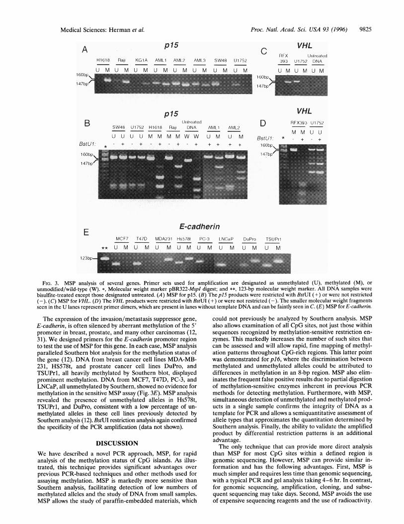
extended our study to include three other genes transcription-ally silenced in human cancers by aberrant hypermethylationof 5' CpG islands. The cyclin-dependent kinase inhibitor p15is aberrantly methylated in many leukemic cell lines andprimary leukemias (11). For p15, MSP again verified themethylation status determined by Southern analysis. Thus,normal lymphocytes and cancer cell lines SW48 and U1752,containing only unmethylated alleles ofp15 alleles by Southernanalysis (11), amplified only with the unmethylated set ofprimers, while the lung cancer cell line H1618 and leukemiacell line KG1A amplified only with the methylated set ofprimers (Fig. 3A), consistent with previous Southern analysis
results (11). DNA from the cell line Raji produced a strongPCR product with methylated primers and a weaker band withunmethylated primers. This was the same result for methyl-ation obtained previously by Southern analysis (11). Noncul-tured leukemia samples, like the primary tumors studied forp16, had amplification with the methylated primer set as wellas the unmethylated set. This heterogeneity also matchedSouthern analysis (11). Again, as for pl6, differential modifi-cation of BstUI restriction sites in the amplified product ofp15was used to verify the specific amplification by MSP (Fig. 3B).Amplified products using methylated primer sets from celllines H1618 and Raji or unmodified primer sets, were com-pletely cleaved by BstUI, while amplified products from theunmethylated primer set did not cleave. The smaller sizes ofproducts observed in the unmodified product reflect the 11-bpdifference in size of the original PCR product. Primary acutemyelogenous leukemia samples also demonstrated cleavageonly in the methylated product but had less complete cleavage.This suggests a heterogeneity in methylation, where methyl-ation is extensive in the region underlying the methylation-specific primers, allowing amplification by MSP but is notinclusive of all CpG sites between the primers for each allele.
Aberrant CpG island promoter region methylation is asso-ciated with inactivation of the von Hippel-Lindau (VHL)tumor suppressor gene in "20% of clear cell renal carcinomas(9). This event, like mutations for VHL (30), is restricted toclear cell renal cancers (9). Primers designed for the VHLsequence were used to study DNA from the renal cell cancercell line RFX393, which is methylated at VEL by Southernanalysis (data not shown), and DNA from the lung cancer cellline U1752, which is unmethylated at this locus (9). In eachcase, the methylation status of VHL determined by MSPconfirmed that found by Southern analysis (Fig. 3C), andBstUI restriction site analysis validated the PCR productspecificity (Fig. 3D).
1 DObpl47bp
Proc. Natl. Acad. Sci. USA 93 (1996)
Proc. Natl. Acad. Sci. USA 93 (1996) 9825
p15C
H1618 Rali KG1A AML1 AML2 AML3 SW48 U1 752
U M U M U M U M U M U M U M U M1GObp\
147bp/
p15Unitreated
SW48 U1752 H1618 Ralp DNA AML 1
U U U U M M M M W W U M
AML2
AAU lV
+ + 1GObp\
_ l 147bp/
RFX393
U M
E-cadherinMCF1 T47D MDA231 Hs578t PC-3 l NCaP
** U M U M U M U M U M U M
DuJPro TSUPrI
U M U M
FIG. 3. MSP analysis of several genes. Primer sets used for amplification are designated as unmethylated (U), methylated (M), or
unmodified/wild-type (W). *, Molecular weight marker pBR322-MspI digest; and **, 123-bp molecular weight marker. All DNA samples were
bisulfite-treated except those designated untreated. (A) MSP for p15. (B) The p15 products were restricted with BstUI (+) or were not restricted(-). (C) MSP for VHL. (D) The VHL products were restricted with BstUI (+) or were not restricted (-). The smaller molecular weight fragmentsseen in the U lanes represent primer dimers, which are present in lanes without template DNA and can be faintly seen in C. (E) MSP for E-cadherin.
The expression of the invasion/metastasis suppressor gene,
E-cadherin, is often silenced by aberrant methylation of the 5'promoter in breast, prostate, and many other carcinomas (12,31). We designed primers for the E-cadherin promoter regionto test the use of MSP for this gene. In each case, MSP analysisparalleled Southern blot analysis for the methylation status ofthe gene (12). DNA from breast cancer cell lines MDA-MB-231, HS578t, and prostate cancer cell lines DuPro, andTSUPrl, all heavily methylated by Southern blot, displayedprominent methylation. DNA from MCF7, T47D, PC-3, andLNCaP, all unmethylated by Southern, showed no evidence formethylation in the sensitive MSP assay (Fig. 3E). MSP analysisrevealed the presence of unmethylated alleles in Hs578t,TSUPrl, and DuPro, consistent with a low percentage of un-
methylated alleles in these cell lines previously detected bySouthern analysis (12). BstUI restriction analysis again confirmedthe specificity of the PCR amplification (data not shown).
DISCUSSION
We have described a novel PCR approach, MSP, for rapidanalysis of the methylation status of CpG islands. As illus-trated, this technique provides significant advantages over
previous PCR-based techniques and other methods used forassaying methylation. MSP is markedly more sensitive thanSouthern analysis, facilitating detection of low numbers ofmethylated alleles and the study of DNA from small samples.MSP allows the study of paraffin-embedded materials, which
could not previously be analyzed by Southern analysis. MSPalso allows examination of all CpG sites, not just those withinsequences recognized by methylation-sensitive restriction en-
zymes. This markedly increases the number of such sites thatcan be assessed and will allow rapid, fine mapping of methyl-ation patterns throughout CpG-rich regions. This latter pointwas demonstrated forp16, where the discrimination betweenmethylated and unmethylated alleles could be attributed todifferences in methylation in an 8-bp region. MSP also elim-inates the frequent false positive results due to partial digestionof methylation-sensitive enzymes inherent in previous PCRmethods for detecting methylation. Furthermore, with MSP,simultaneous detection of unmethylated and methylated prod-ucts in a single sample confirms the integrity of DNA as a
template for PCR and allows a semiquantitative assessment ofallele types that approximates the quantitation determined bySouthern analysis. Finally, the ability to validate the amplifiedproduct by differential restriction patterns is an additionaladvantage.The only technique that can provide more direct analysis
than MSP for most CpG sites within a defined region isgenomic sequencing. However, MSP can provide similar in-formation and has the following advantages. First, MSP ismuch simpler and requires less time than genomic sequencing,with a typical PCR and gel analysis taking 4-6 hr. In contrast,for genomic sequencing, amplification, cloning, and subse-quent sequencing may take days. Second, MSP avoids the use
of expensive sequencing reagents and the use of radioactivity.
A VHLUrnlreate(d
U1752 DNA
U M U M1GObp
147bp]
B
BstUl:
1 GObP\
147bp'
E
123bp-
VHL
D RFX393 U1752
M M U UBstU1: * - + - +
Medical Sciences: Herman et al.

9826 Medical Sciences: Herman et al.
Both of these factors make MSP better suited for the analysisof large numbers of samples. Third, the use of PCR as the stepto distinguish methylated from unmethylated DNA in MSPallows for a significant increase in the sensitivity of methylationdetection. For example, if cloning is not used before genomicsequencing of the DNA, <10% methylated DNA in a back-ground of unmethylated DNA cannot be seen (19). The use ofPCR and cloning does allow sensitive detection of methylationpatterns in very small amounts ofDNA by genomic sequencing(17, 32). However, in practice, this would require sequencinganalysis of 10 clones to detect 10% methylation, 100 clones todetect 1% methylation, and, to reach the level of sensitivity wehave demonstrated with MSP (1:1000), one would have tosequence 1000 individual clones.
In summary, MSP is a simple, sensitive, and specific methodfor determining the methylation status of virtually any CpG-rich region. In addition to detecting aberrant CpG islandmethylation of tumor suppressor genes, MSP will be useful formonitoring CpG islands important in other biological pro-cesses. For example, MSP should facilitate monitoring patternsof methylation in imprinted genes at key stages of embryo-genesis. Assays used to define clonality of cell populations, asassessed by detecting methylation patterns of X chromosome-inactivated genes in female cells, should be readily adaptableto the MSP approach. Finally, MSP should prove extraordi-narily valuable clinically for the detection of methylationpatterns in small DNA samples associated with disease statessuch as the fragile X syndrome, altered gene imprint states, andcancer.
We would like to thank Dr. Paula Vertino for helpful discussion andDr. Ed Gabrielson for primary tumor DNA. S.M. is the recipient of anaward from the Academy of Finland. This work was supported by NIHGrants SP5-CA58184-03S1 and SRO1 CA43318-10.
1. Holliday, R. & Grigg, G. W. (1993) Mutat. Res. 285, 61-67.2. Bird, A. (1992) Cell 70, 5-8.3. Bird, A. P. (1986) Nature (London) 321, 209-213.4. Li, E., Beard, C. & Jaenisch, R. (1993) Nature (London) 366,
362-365.5. Tremblay, K. D., Saam, J. R., Ingram, R. S., Tilghman, S. M. &
Bartolomei, M. S. (1995) Nat. Genet. 9, 407-413.6. Pfeifer, G. P., Steigerwald, S. D., Mueller, P. R., Wold, B. &
Riggs, A. D. (1989) Science 246, 810-813.7. Riggs, A. D. & Pfeifer, G. P. (1992) Trends Genet. 8, 169-174.8. Antequera, F., Boyes, J. & Bird, A. (1990) Cell 62, 503-514.9. Herman, J. G., Latif, F., Weng, Y., Lerman, M. I., Zbar, B., Liu,
S., Samid, D., Duan, D. S., Gnarra, J. R., Linehan, W. M. &Baylin, S. B. (1994) Proc. Natl. Acad. Sci. USA 91, 9700-9704.
10. Merlo, A., Herman, J. G., Mao, L., Lee, D. J., Gabrielson, E.,Burger, P. C., Baylin, S. B. & Sidransky, D. (1995) Nat. Med. 1,686-692.
11. Herman, J. G., Jen, J., Merlo, A. & Baylin, S. B. (1996) CancerRes. 56, 722-727.
12. Graff, J. R., Herman, J. G., Lapidus, R. G., Chopra, H., Xu, R.,Jarrard, D. F., Isaacs, WB, Pitha, P. M., Davidson, N. E. &Baylin, S. B. (1995) Cancer Res. 55, 5195-5199.
13. Issa, J. P., Ottaviano, Y. L., Celano, P., Hamilton, S. R., David-son, N. E. & Baylin, S. B. (1994) Nat. Genet. 7, 536-540.
14. Singer-Sam, J., Grant, M., LeBon, J. M., Okuyama, K., Chapman,V., Monk, M. & Riggs, A. D. (1990) Mol. Cell. Biol. 10, 4987-4989.
15. Razin, A. & Cedar, H. (1991) Microbiol. Rev. 55, 451-458.16. Stoger, R., Kubicka, P., Liu, C. G., Kafri, T., Razin, A., Cedar, H.
& Barlow, D. P. (1993) Cell 73, 61-71.17. Frommer, M., McDonald, L. E., Millar, D. S., Collis, C. M., Watt,
F., Grigg, G. W., Molloy, P. L. & Paul, C. L. (1992) Proc. Natl.Acad. Sci. USA 89, 1827-1831.
18. Wang, R. Y.-H., Gehrke, C. W. & Ehrlich, M. (1980) NucleicAcids Res. 8, 4777-4790.
19. Myohanen, S., Wahlfors, J. & Janne, J. (1994) DNA Sequence 5,1-8.
20. Hara, E., Smith, R., Parry, D., Tahara, H., Steven, S. & Peters,G. (1996) Mol. Cell. Biol. 16, 859-867.
21. Hussussian, C. J., Struewing, J. P., Goldstein, A. M., Higgins,P. A., Ally, D. S., Sheahan, M. D., Clark, W. H., Jr., Tucker,M. A. & Dracopoli, N. C. (1994) Nat. Genet. 8, 15-21.
22. Jen, J., Harper, J. W., Bigner, S. H., Bigner, D. D., Papadopou-los, N., Markowitz, S., Willson, J. K., Kinzler, K. W. & Vo-gelstein, B. (1994) Cancer Res. 54, 6353-6358.
23. Kuzmin, I., Duh, F. M., Latif, F., Geil, L., Zbar, B. & Lerman,M. I. (1995) Oncogene 10, 2185-2194.
24. Bussemakers, M. J., Giroldi, L. A., van Bokhoven, A. &Schalken, J. A. (1994) Biochem. Biophys. Res. Commun. 203,1284-1290.
25. Herman, J. G., Merlo, A., Mao, L., Lapidus, R. G., Issa, J. P. J.,Davidson, N. E., Sidransky, D. & Baylin, S. B. (1995) Cancer Res.55, 4525-4530.
26. Gonzalez-Zulueta, M., Bender, C. M., Yang, A. S., Nguyen, T.,Beart, R. W., Van Tornout, J. M. & Jones, P. A. (1995) CancerRes. 55, 4531-4535.
27. Otterson, G. A., Khleif, S. N., Chen, W., Coxon, A. B. & Kaye,F. J. (1995) Oncogene 11, 1211-1216.
28. Park, J. G. & Chapman, V. M. (1994) Mol. Cell. Biol. 14, 7975-7983.
29. Reeben, M., Myohanen, S., Saarma, M. & Prydz, H. (1995) Gene157, 325-329.
30. Gnarra, J. R., Tory, K., Weng, Y., Schmidt, L., Wei, M. H., et al.(1994) Nat. Genet. 7, 85-90.
31. Yoshiura, K., Kanai, Y., Ochiai, A., Shimoyama, Y., Sugimura, T.& Hirohashi, S. (1995) Proc. Natl. Acad. Sci. USA 92, 7416-7419.
32. Clark, S. J., Harrison, J., Paul, C. L. & Frommer, M. (1994)Nucleic Acids Res. 22, 2990-2997.
Proc. Natl. Acad. Sci. USA 93 (1996)