methyltransferase 5 (prmt5) to mediate histone arginine ... · methyltransferase 5 (prmt5) to...
TRANSCRIPT
1
Transcription Factor Positive Regulatory Domain 4 (PRDM4) recruits Protein Arginine
Methyltransferase 5 (PRMT5) to mediate histone arginine methylation and control neural stem
cell proliferation and differentiation.
Alexandra Chittka1, Justyna Nitarska2, Ursula Grazini1 and William D Richardson1
1Wolfson Institute for Biomedical Research and Research Department of Cell and Developmental
Biology, University College London, Gower St, London, WC1E 6BT, UK
2MRC Laboratory for Molecular Cell Biology, University College London, Gower St, London, WC1E
6BT, UK
*Running Title: PRDM4 controls NSC differentiation
Corresponding Author: Alexandra Chittka, Wolfson Institute for Biomedical Research and Research
Department of Cell and Developmental Biology, University College London, Gower St, London,
WC1E 6BT, UK; Tel: +44 (0)207 679 6744 Fax: +44 (0) 207 209 0470; E-mail:
Keywords: histone arginine methylation; neural stem cells; PRDM4; PRMT5; SC1
Background: Neural stem cells generate all the
cell types of the central nervous system.
Results: Transcription factor, PRDM4, recruits
protein arginine methyltransferase 5 (PRMT5)
to control the timing of neurogenesis.
Conclusions: PRDM4 and PRMT5-mediated
histone arginine methylation controls neura l
stem cell proliferation and differentiation.
Significance: Histone arginine methylation is a
novel epigenetic mechanism which regulates
neural stem cell reprogramming.
SUMMARY
During development of the cerebral cortex,
neural stem cells (NSCs) undergo a temporal
switch from proliferative (symmetric) to
neuron-generating (asymmetric) divisions.
We investigated the role of Schwann cell
factor 1 (SC1/PRDM4), a member of the
PRDM family of transcription factors, in this
critical transition. We discovered that SC1
recruits the chromatin modifier PRMT5, an
arginine methyltransferase that catalyzes
symmetric dimethylation of histone H4
arginine 3 (H4R3me2s), and that this
modification is preferentially associated with
undifferentiated cortical NSCs. Over-
expressing SC1 in embryonic NSCs led to an
increase in the number of Nestin-expressing
precursors; mutational analysis of SC1
showed that this was dependent on
recruitment of PRMT5. We found that SC1
protein levels are down-regulated at the onset
of neurogenesis and that experimental knock-
down of SC1 in primary NSCs triggers
precocious neuronal differentiation. We
propose that SC1 and PRMT5 are
components of an epigenetic regulatory
complex that maintains the “stem-like”
cellular state of the NSC by preserving their
proliferative capacity and modulating their
cell cycle progression. Our findings provide
evidence that histone arginine methylation
regulates NSC differentiation.
INTRODUCTION
During central nervous system (CNS)
development, embryonic neural stem cells
(NSCs) in the ventricular zone (VZ) of the brain
and spinal cord first proliferate symmetrically to
increase NSC numbers and expand the VZ, then
they switch to an asymmetric mode of division
to generate post-mitotic neurons while
maintaining the NSC pool (1-4). After
neurogenesis is complete, the NSCs switch to
production of glial cells (astrocytes and
oligodendrocytes). The mechanisms that contro l
the temporal transition from proliferation to
differentiation are poorly understood.
Aberrations in the timing of this transition can
lead to abnormalities in CNS development (5,6)
http://www.jbc.org/cgi/doi/10.1074/jbc.M112.392746The latest version is at JBC Papers in Press. Published on October 9, 2012 as Manuscript M112.392746
Copyright 2012 by The American Society for Biochemistry and Molecular Biology, Inc.
by guest on June 2, 2018http://w
ww
.jbc.org/D
ownloaded from
2
In the developing cerebral cortex NSCs generate
a series of neuronal subtypes that populate
different cortical layers in sequence, then switch
to glial cell production (7,8). This program of
neurogenesis can be recapitulated by individua l
mouse cortical NSCs isolated at embryonic day
10 (E10) and cultured at low density in vitro
(2,3). This suggests that the temporal program
of NSC division and cell fate specification is
cell-intrinsic, but the molecular nature of the
program is not known. The transitions in NSC
fate are likely to be governed by cell lineage-
specific transcription factors acting in concert
with epigenetic mechanisms (9-14). The latter
include post-translational modifications of
histones associated with regulatory elements of
genes as well as DNA methylation at CpG
dinucleotides, which together affect the
accessibility of chromatin to the genera l
transcriptional machinery. The details of the
epigenetic regulation of NSC differentiation are
still poorly understood. In addition to cell
lineage-specific transcription factors, cell cycle
parameters such as the length of specific cell
cycle stages play an important role in controlling
NSC proliferation and differentiation (5,6,15-17)
and these parameters change during cortica l
development (18). Lineage-specific
transcription factors (5) can fine-tune the
expression of cell cycle genes and in this way
influence the cell fates and division modes of
NSCs and consequently their decision either to
proliferate or differentiate (16,19,20).
Schwann cell factor 1 (SC1) is a protein that was
first identified as a binding partner of the p75
neurotrophin receptor (p75NTR) (21). SC1,
also known as PRDM4, belongs to the PRDM
family of proteins of which 17 members have
been identified in the human genome (22). All
PRDM family members are characterized by the
presence of a PR (positive regulatory) domain
and multiple zinc finger (ZnFg) domains. The
PR domains are similar to, but distinct from, the
SET domains found in many histone lysine
methyltransferases (MTases) (23). PRDM
proteins are either epigenetic modifiers in their
own right or else they recruit third party
chromatin modifiers - e.g. histone deacetylases
(HDACs), histone lysine MTases or histone
arginine MTases - to regulate cell type-specific
gene expression in various tissues (24-33). Our
previous work identified SC1/PRDM4 as an
HDAC-associated transcriptional repressor that
modulates cell cycle progression (33). SC1 is
highly expressed in the developing mouse
cerebral cortex (34) so we set out to understand
its role in the development of cortical NSCs as
they switch from proliferative to neuron-
generating divisions. We report that SC1
recruits a type II arginine MTase, PRMT5, that
catalyzes histone H4R3 symmetric
dimethylation (H4R3me2s) - a modification that
we recently showed to be present in
undifferentiated NSCs in the murine cortex prior
to the onset of neurogenesis (35). We now show
that both SC1 and PRMT5 are highly expressed
in the pre-neurogenic cortex and provide
evidence that the interaction between SC1 and
PRMT5 regulates the proliferative capacity of
cultured cortical NSCs. Our findings suggest an
important role for histone arginine methylation
in epigenetic programming of NSCs during
cortical development.
EXPERIMENTAL PROCEDURES
Cell culture, transfections - HEK293T cells
were cultured in Dulbecco’s modified Eagle’s
medium (DMEM, Invitrogen) supplemented
with 10% (v/v) fetal calf serum (FCS) and
glutamine; P19 cells were cultured in alpha-
MEM (Invitrogen) supplemented with 5% FCS
and glutamine; PC12 cells were cultured in
DMEM supplemented with 5% FCS, 10% horse
serum (HS) and glutamine. Transfections were
performed using Lipofectamine 2000
(Invitrogen) according to the manufacturer’s
instructions. The cells were harvested and
processed 48 hours post-transfection unless
indicated otherwise.
Immunoprecipitation, methylation assays -
HEK293T cells were transfected with described
plasmids and harvested in immunoprecipitation
(IP) buffer (50 mM TrisHCl pH7.4, 0.5% [v/v]
NP-40, 300 mM NaCl) supplemented with a
protease inhibitor cocktail (Sigma) and
phosphatase inhibitor cocktails 1 and 2 (Sigma).
Cells were lysed for 20 minutes on ice in IP
buffer and the insoluble material sonicated for
10 s on ice. Lysates were centrifuged and the
supernatants pre-cleared using protein A/G
beads (Santa Cruz), then immunoprecipitated
overnight at 4oC using anti-Myc (Upstate), anti-
PRMT5 (Upstate) or anti-HA antibodies
(Covance). The complexes were collected on
protein A/G beads and washed 5 times with IP
buffer, followed by a wash with cold phosphate-
buffered saline (PBS) at 4oC. Proteins were
by guest on June 2, 2018http://w
ww
.jbc.org/D
ownloaded from

3
boiled at 95oC for 5 min in Laemmli buffer (60
mM Tis-HCl, pH6.8, 10% [v/v] glycerol, 2%
[w/v] sodium dodecyl sulphate, 5% [v/v] -
mercaptoethanol, 0.01% [w/v] bromophenol
blue) and separated by SDS-polyacrylamide ge l
electrophoresis (PAGE). After separation,
proteins were transferred onto polyvinylidene
difluoride (PVDF) membranes (Millipore) and
Western blots were performed with specified
antibodies in TBST buffer (100mM Tris-HCl,
pH7.5, 150mM NaCl, 0.1% [v/v] Tween-20)
containing 5% (w/v) skimmed milk powder
(Tesco) and detected using ECL (GE
Healthcare). IP’s from E10.5 mouse cortices
were performed using the same buffers as above.
At least 20 embryos were used/IP experiment.
For methylation assays the immunoprecipitates
on the beads were washed as described above
and then rinsed twice in methylation buffer
(50 mM Tris-HCl pH8.5, 5 mM MgCl2, 4 mM
dithiothreitol [DTT], 2 l of S-adenosyl-L-
([3H]methyl) methionine (3H-SAM) (Amersham,
GE Healthcare) and 1 g of histone mixture
(Roche) were added to the reaction in a tota l
volume of 30 l. Methylation reaction was
allowed to proceed for 2 hours at 30oC and
stopped by adding Laemmli buffer and boiling
the samples for 5 minutes. Products of the
methylation reactions were separated using 15%
SDS PAGE, transferred onto PVDF membranes
and visualized by Coommassie brilliant blue
staining and fluorography. To examine H4-
specific methylation, histones were incubated
with immunoprecipitated mycSC1 complex and 3H-SAM as described above and analyzed on
Western blots using anti-H4R3me2s antibodies
(Abcam).
Primary neural stem cell cultures - Primary
neural stem cells were isolated from mouse
E10.5 cerebral cortices according to published
procedures (36). Briefly, cortices were harvested
in EBSS (Invitorgen) and the meninges
removed. The cells were dissociated by
incubation in trypsin at 37oC for 40-50 minutes.
Trypsinization was stopped by adding
DMEM/10% FCS. Cells were then further
dissociated using pre-separation filters
(Milteneyi Biotec), centrifuged gently,
resuspended in a small volume of
DMEM/10% FCS and plated at a density of
2.5x10⁵ cells/13 mm poly-D-lysine coated
coverslip. The cells were cultured in DMEM
supplemented with 10 ng/ml bFGF (PeproTech),
0.25% FCS, B27 supplement, Na pyruvate and
glutamine (all from Invitrogen) (2). Cultures
were routinely immunolabelled to monitor their
ability to generate neurons, astrocytes and
oligodendrocytes.
Antibodies and immunofluorescence microscopy
- Cultured cells on coverslips were fixed in 4%
(w/v) paraformaldehyde (PFA) for 10 minutes at
20-25oC and permeabilized with cold methanol
for 2-3 minutes at -20oC. They were incubated
for one hour at 20-25oC in blocking solution
(10% normal goat serum, 0.1% [v/v] Triton X-
100 in PBS). The following antibodies were
used: anti-TuJ1 (Sigma, 1:500), anti-GFAP
(Sigma, 1:1000), anti-Nestin (Santa Cruz,
1:400), anti-O4 (kind gift from Nigel Pringle,
1:10), anti-SC1/PRDM4 (our own antibody,
1:100, Abcam 1:100 and a gift from P Perez and
MV Chao, 1:40 (33)), anti-PRMT5 (Upstate
Biotech, 1:100), anti-H4R3me2s (Abcam,
1:1000), anti-EGFP (Fine Chemical Products
Ltd., 1:3000), Anti-Flag (Sigma, 1:1000), anti-
BrdU (American Type Culture Collection,
Manassas, VA, 1:10), anti-cycB1 (GNS1, Santa
Cruz Biotechnology, Inc, 1:500), anti-Pan
methyl Lysine (Abcam, 1:1000). For
immunolabelling with antibody O4, methanol
treatment and Triton X-100 were not used.
When staining for BrdU and another antigen, the
cells were stained sequentially, first for an
antigen of interest other than BrdU, then rinsed
and treated as follows to visualise BrdU. First,
the cells were fixed with 70% ethanol/ 20%
glacial acetic acid mixture at RT, then in 70%
ethanol at -20oC. The cells were then rinsed in
PBS/1% Triton X-100 at RT and denatured in
PBS/1% Triton X-100/2M HCl for 30 minutes at
37oC. After washing, anti-BrdU antibody was
added overnight at 4oC. The rest of the staining
was as described above. Coverslips were
mounted in DAKO mounting medium (DAKO).
The following secondary antibodies were used:
goat anti-mouse Alexa 488, goat anti-rabbit
Alexa 568, goat anti-mouse Alexa 647
(Invitrogen, 1:1000), goat anti-rat Alexa 488
(Invitrogen, 1:500). Fluorescent images were
taken with a Leica Microsystems SPE confoca l
microscope.
E10.5 embryos were collected from timed-mated
C57B/6 mice (Harlan), rinsed in PBS and fixed
in 4% PFA at 4°C for 1–2 hours. Embryos were
cryoprotected in 30% (w/v) sucrose in PBS and
by guest on June 2, 2018http://w
ww
.jbc.org/D
ownloaded from
4
subsequently mounted in OCT (Tissue-Tek) on
dry ice. Mounted embryos were sectioned at
10 µm using a Leica cryostat and air-dried for at
least 1 hour. Sections were permeabilized for 3
minutes with −20°C methanol, rinsed three
times in PBS, incubated in sodium citrate buffer
(10mM sodium citrate, 0.05% Tween-20, pH
6.0) at 95oC for 30 minutes for antigen retrieval,
allowed to cool to room temperature, rinsed
three times in PBS, then incubated for 1 hour at
20-25°C in blocking solution, following
incubation with the primary antibody in
blocking solution overnight at 4°C. Sections
were washed three times in PBS at 20-25°C and
incubated with fluorescent secondary antibodies
and Hoechst dye (to visualize cell nuclei) for
1 hour at 20-25°C, rinsed three times in PBS,
once in water and mounted using DAKO
mounting medium. Fluorescence images were
made using a Leica Microsystems SPE confocal
microscope.
siRNA Transfections - siRNA oligonucleotides
against rat SC1 were synthesized by Thermo
Scientific and assessed either by applying
siRNA to rat NSCs or PC12 cells and
monitoring the expression of endogenous SC1
protein by Western blotting and RT-PCR or by
co-transfecting SC1-specific or scrambled-
sequence siRNA with an EGFP expression
vector into cultured rat NSCs followed by
immunolabelling with anti-EGFP and anti-SC1.
We used DharmaFECT Duo Transfection
reagent (Thermo Scientific) to introduce siRNA
along with DNA according to the
manufacturer’s instructions. The cells were
transfected with the siRNA oligonucleotides on
two consecutive days 24 hours after plating and
processed for immunocytochemistry and
Western blot analysis or semi-quantitative RT-
PCR 48 hours after the second application of the
siRNA reagent. The following siRNA
oligonucleotides were used: 1) siRNA-1 Pool:
ACAAUUUGGUGCACGCUUU and
GGAUGAUGUUUGUGCGCAA, 2) siRNA-2
Pool: UAAUGAUGGCCACGAAGUA and
GUUCCUAUUCAGAGUUCAA. Scrambled-
sequence siRNAs were used as controls.
Reverse transcription-PCR (RT-PCR) - RNA
was isolated from P19 cells or PC12 cells using
Trizol reagent (Invitrogen) according to the
manufacturer’s instructions. 1 g of total RNA
was used for RT-PCR. Total RNA was treated
with RNase-free DNase (Ambion) and cDNA
synthesized using random hexamer primers
(Invitrogen) and MMLV reverse transcriptase
(USB). After incubation at 42oC for 1.5 hours,
the enzyme was inactivated at 75oC and the
cDNA used for PCR. The following gene-
specific primers were used: 1) Rat SC1: Fwd 5´-
AAAGCCAGGAACCGTGAA-3´; Rev 5´ -
ATGACCCATAAAGTGAACGTG-3´; 2)
Mouse cycB: Fwd 5′-
TCCCTCTCCAAGCCCGATGG -3′; Rev 5′-
TGGCCGTTACACCGACCAGC-3′; 3) Mouse
Bub1b: Fwd 5′-
AAGGGATTGAACGCAAGGCTG-3′; Rev 5′-
CATCAAAAACGGTGATCCTGCG-3′; 4)
Mouse and rat GAPDH: Fwd 5´-
ACAACTTTGGCATTGTGGAA-3’; Rev 5´-
GATGCAGGGATGATGTTCTG-3’; 5) Rat
cycB: Fwd 5'-
TGGACAAGGTGCCAGTGTGCG-3'; Rev 5'-
GGTCTCCTGCAGCAGCCGAAA-3' 6) Rat
Bub1b: Fwd 5'-
GCCAGGCCCGTGGAACACAG-3'; Rev 5'-
CAGGACGGAGGCACTCCCGA-3'.
RESULTS
Expression of SC1 in embryonic cortical NSCs -
SC1 mRNA is highly expressed in the
developing cortex (34). We investigated the
expression of SC1 protein in dissociated primary
mouse cortical NSCs isolated at embryonic day
10.5 (E10.5) and cultured for up to 10 days in
vitro (10DIV). We identified cells in these
cultures by immunolabelling for Nestin (NSCs),
TuJ1 (neurons), GFAP (astrocytes) or O4
(oligodendrocyte precursors, OLPs). The
different cell types were generated in the
appropriate temporal order (2,37): Nestin+
precursors were present from the outset,
followed by TuJ1+ neurons, GFAP+ astrocytes
and O4+ OLPs at progressively longer culture
periods (A.C., unpublished observations). We
found that immediately after plating, Nestin+
NSCs could be characterized as either strongly
or weakly SC1-positive (Fig. 1A, arrowhead
and arrow, respectively, quantified in Fig. 1D).
Co-immunolabelling with anti-TuJ1 and anti-
SC1 at 3DIV revealed that TuJ1 negative NSCs
co-expressed SC1 strongly, whereas more
mature neurons with high levels of TuJ1
expression, expressed low levels of SC1
(Fig. 1B). After the onset of glial differentiation
at 10DIV we detected high levels of SC1
expression in all proliferating O4-positive OLPs
by guest on June 2, 2018http://w
ww
.jbc.org/D
ownloaded from
5
(Fig. 1C), but not in GFAP-expressing
differentiated astrocytes (A.C., unpublished).
Thus, high levels of SC1 appeared to be
expressed preferentially in mitotically active
cells (NSCs and OLPs) and down-regulated in
differentiated neurons and astroglia. This
suggested that down-regulation of SC1 might be
involved in, and possibly required for, the
switch from cell proliferation to differentiation.
Knock-down of SC1 in NSCs leads to precocious
neuronal differentiation - To test whether SC1
down-regulation is sufficient to trigger NSC
differentiation we examined the effects of SC1
knock-down in primary rat NSCs isolated from
E11.5 cortex. We used rat NSCs in our
experiment since we could knock down rat SC1
protein expression very efficiently. Transfecting
two independent sets of rat SC1-specific siRNA
oligonucleotides (SC1siRNA-1 and
SC1siRNA-2) into cultured rat NSCs markedly
decreased the expression of endogenous SC1
protein, as detected on Western blots (Fig. 2A).
Moreover, SC1 immunoreactivity was low or
undetectable in siRNA-transfected NSCs,
compared to control siRNA-transfected NSCs
(Fig. 2B). Successfully transfected cells were
identified in this experiment by co-transfection
of an enhanced green fluorescent protein
(EGFP) expression vector. Both SC1siRNA-1
and SC1siRNA-2 gave similar results and are
referred to below simply as SC1siRNA (Fig. 2
and unpublished observations). To investigate
the effect of SC1 depletion on NSC
differentiation, SC1-specific or scrambled-
sequence siRNAs were applied twice to cultured
E11.5 rat NSCs, 24 hours and 48 hours after
plating. At 96 hours after plating the treated
NSCs were fixed and immunolabelled with anti-
EGFP to identify transfected cells and anti-TuJ1
to visualize differentiating neurons. NSCs
treated with SC1siRNA consistently gave rise to
10% of TuJ1-expressing cells with the process-
bearing morphology of neurons (Fig. 2C, top
panels). We did not detect a change in the
number of astrocytes or oligodendrocytes after
the application of SC1siRNA in these
experiments within the time frame of
investigation (unpublished observations). Very
few TuJ1-expressing cells were observed when
NSCs were transfected with control, scrambled-
sequence siRNA (Fig. 2C, bottom panels). To
confirm the specificity of these knock-down
experiments we transfected rat NSCs with
SC1siRNA together with a human SC1 cDNA
that was insensitive to inhibition by the rat-
specific SC1siRNAs (Fig. 2D). This “rescued”
SC1 expression in the presence of rat
SC1siRNA and resulted in a reduction in
numbers of TuJ1-positive neurons to contro l
levels (i.e. as observed in NSC cultures without
added siRNA or with scrambled-sequence
SC1siRNA) (Fig. 2E). Consistent with the
observation that a fraction of NSCs treated with
SC1siRNA induced precocious neurogenesis,
we found that a similar fraction of NSCs treated
as above showed reduced levels of BrdU
incorporation (Fig. 2F). Thus, we concluded
that knock-down of SC1 in NSCs leads to
precocious neuronal differentiation of a subset
of the NSCs and controls their proliferative
capacity, consistent with our observation that
newly differentiating neurons express low levels
of SC1 protein.
SC1 is associated with histone methyltransferase
activity - Like other PRDM proteins, SC1
possesses a PR/SET domain - a hallmark of
lysine histone MTases (HMTases) - which can
regulate gene expression by modifying histones
in chromatin (23). Therefore we tested the
possibility that SC1 methylates histones as part
of its transcriptional repressor function. We
transiently expressed Myc-tagged SC1
(mycSC1) in HEK293T cells and
immunoprecipitated mycSC1 protein from cell
lysates using an anti-Myc antibody. Purified
histones from calf thymus were incubated with
immunoprecipitated mycSC1 and subjected to
an in vitro radioactive HMTase assay (38). We
detected methylated histones, the preferred
substrate being histone H4 (Fig. 3A). As a
positive control, we immunoprecipitated myc-
Su(var)3-9, an H3K9 HMTase, and showed that
this preferentially methylated histone H3
(Fig. 3A). As negative controls, we
immunoprecipitated mycCREB or empty Myc
vector and showed that neither of these
expressed histone methylase activity (Fig. 3A).
Immunoprecipitated mycSC1 also methylated
recombinant histone H4 (Fig. 3B). We
concluded that SC1 exhibits an HMTase
activity, preferentially towards histone H4.
Histone methylation can occur on a variety of
lysine and arginine residues leading to
repression or activation of gene transcription,
depending on the precise modification. To
identify the histone modification mediated by
mycSC1, the products of in vitro methylation
by guest on June 2, 2018http://w
ww
.jbc.org/D
ownloaded from
6
reactions were analyzed by Western blotting
using antibodies directed against different H4
modifications. We detected an increased leve l
of H4R3me2s in the sample containing
immunoprecipitated mycSC1 (Fig. 3C,
highlighted by an asterisk), while no change in
overall lysine methylation was observed using
an antibody directed against pan-methyl Lysine
(Fig.3C, panel 3). We concluded that mycSC1
mediates symmetric arginine dimethylation on
H4R3.
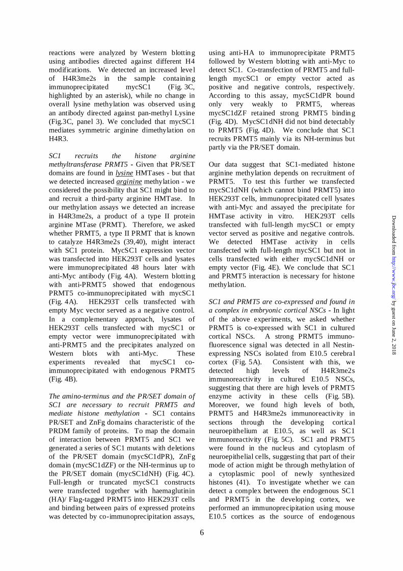
SC1 recruits the histone arginine
methyltransferase PRMT5 - Given that PR/SET
domains are found in lysine HMTases - but that
we detected increased arginine methylation - we
considered the possibility that SC1 might bind to
and recruit a third-party arginine HMTase. In
our methylation assays we detected an increase
in H4R3me2s, a product of a type II protein
arginine MTase (PRMT). Therefore, we asked
whether PRMT5, a type II PRMT that is known
to catalyze H4R3me2s (39,40), might interact
with SC1 protein. MycSC1 expression vector
was transfected into HEK293T cells and lysates
were immunoprecipitated 48 hours later with
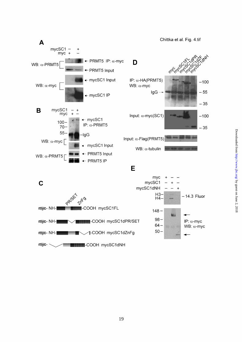
anti-Myc antibody (Fig. 4A). Western blotting
with anti-PRMT5 showed that endogenous
PRMT5 co-immunoprecipitated with mycSC1
(Fig. 4A). HEK293T cells transfected with
empty Myc vector served as a negative control.
In a complementary approach, lysates of
HEK293T cells transfected with mycSC1 or
empty vector were immunoprecipitated with
anti-PRMT5 and the precipitates analyzed on
Western blots with anti-Myc. These
experiments revealed that mycSC1 co-
immunoprecipitated with endogenous PRMT5
(Fig. 4B).
The amino-terminus and the PR/SET domain of
SC1 are necessary to recruit PRMT5 and
mediate histone methylation - SC1 contains
PR/SET and ZnFg domains characteristic of the
PRDM family of proteins. To map the domain
of interaction between PRMT5 and SC1 we
generated a series of SC1 mutants with deletions
of the PR/SET domain (mycSC1dPR), ZnFg
domain (mycSC1dZF) or the NH-terminus up to
the PR/SET domain (mycSC1dNH) (Fig. 4C).
Full-length or truncated mycSC1 constructs
were transfected together with haemaglutinin
(HA)/ Flag-tagged PRMT5 into HEK293T cells
and binding between pairs of expressed proteins
was detected by co-immunoprecipitation assays,
using anti-HA to immunoprecipitate PRMT5
followed by Western blotting with anti-Myc to
detect SC1. Co-transfection of PRMT5 and full-
length mycSC1 or empty vector acted as
positive and negative controls, respectively.
According to this assay, mycSC1dPR bound
only very weakly to PRMT5, whereas
mycSC1dZF retained strong PRMT5 binding
(Fig. 4D). MycSC1dNH did not bind detectably
to PRMT5 (Fig. 4D). We conclude that SC1
recruits PRMT5 mainly via its NH-terminus but
partly via the PR/SET domain.
Our data suggest that SC1-mediated histone
arginine methylation depends on recruitment of
PRMT5. To test this further we transfected
mycSC1dNH (which cannot bind PRMT5) into
HEK293T cells, immunoprecipitated cell lysates
with anti-Myc and assayed the precipitate for
HMTase activity in vitro. HEK293T cells
transfected with full-length mycSC1 or empty
vector served as positive and negative controls.
We detected HMTase activity in cells
transfected with full-length mycSC1 but not in
cells transfected with either mycSC1dNH or
empty vector (Fig. 4E). We conclude that SC1
and PRMT5 interaction is necessary for histone
methylation.
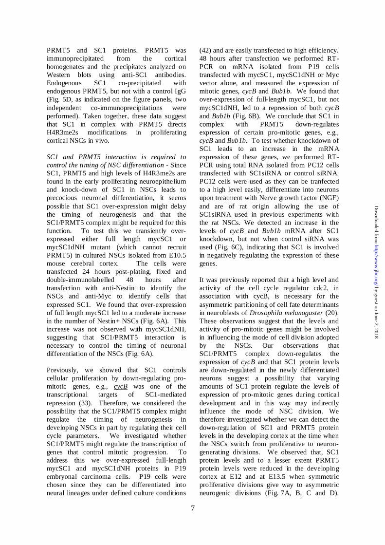
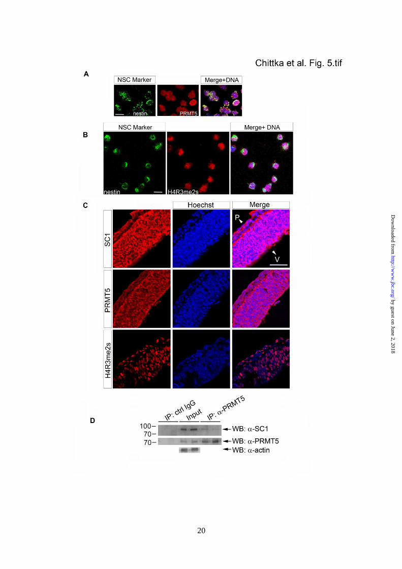
SC1 and PRMT5 are co-expressed and found in
a complex in embryonic cortical NSCs - In light
of the above experiments, we asked whether
PRMT5 is co-expressed with SC1 in cultured
cortical NSCs. A strong PRMT5 immuno-
fluorescence signal was detected in all Nestin-
expressing NSCs isolated from E10.5 cerebra l
cortex (Fig. 5A). Consistent with this, we
detected high levels of H4R3me2s
immunoreactivity in cultured E10.5 NSCs,
suggesting that there are high levels of PRMT5
enzyme activity in these cells (Fig. 5B).
Moreover, we found high levels of both,
PRMT5 and H4R3me2s immunoreactivity in
sections through the developing cortica l
neuroepithelium at E10.5, as well as SC1
immunoreactivity (Fig. 5C). SC1 and PRMT5
were found in the nucleus and cytoplasm of
neuroepithelial cells, suggesting that part of their
mode of action might be through methylation of
a cytoplasmic pool of newly synthesized
histones (41). To investigate whether we can
detect a complex between the endogenous SC1
and PRMT5 in the developing cortex, we
performed an immunoprecipitation using mouse
E10.5 cortices as the source of endogenous
by guest on June 2, 2018http://w
ww
.jbc.org/D
ownloaded from
7
PRMT5 and SC1 proteins. PRMT5 was
immunoprecipitated from the cortica l
homogenates and the precipitates analyzed on
Western blots using anti-SC1 antibodies.
Endogenous SC1 co-precipitated with
endogenous PRMT5, but not with a control IgG
(Fig. 5D, as indicated on the figure panels, two
independent co-immunoprecipitations were
performed). Taken together, these data suggest
that SC1 in complex with PRMT5 directs
H4R3me2s modifications in proliferating
cortical NSCs in vivo.
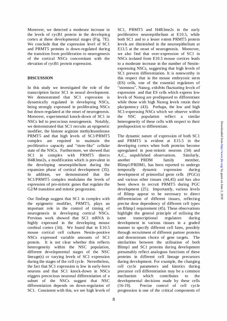
SC1 and PRMT5 interaction is required to
control the timing of NSC differentiation - Since
SC1, PRMT5 and high levels of H4R3me2s are
found in the early proliferating neuroepithelium
and knock-down of SC1 in NSCs leads to
precocious neuronal differentiation, it seems
possible that SC1 over-expression might delay
the timing of neurogenesis and that the
SC1/PRMT5 complex might be required for this
function. To test this we transiently over-
expressed either full length mycSC1 or
mycSC1dNH mutant (which cannot recruit
PRMT5) in cultured NSCs isolated from E10.5
mouse cerebral cortex. The cells were
transfected 24 hours post-plating, fixed and
double-immunolabelled 48 hours after
transfection with anti-Nestin to identify the
NSCs and anti-Myc to identify cells that
expressed SC1. We found that over-expression
of full length mycSC1 led to a moderate increase
in the number of Nestin+ NSCs (Fig. 6A). This
increase was not observed with mycSC1dNH,
suggesting that SC1/PRMT5 interaction is
necessary to control the timing of neurona l
differentiation of the NSCs (Fig. 6A).
Previously, we showed that SC1 controls
cellular proliferation by down-regulating pro-
mitotic genes, e.g., cycB was one of the
transcriptional targets of SC1-mediated
repression (33). Therefore, we considered the
possibility that the SC1/PRMT5 complex might
regulate the timing of neurogenesis in
developing NSCs in part by regulating their cell
cycle parameters. We investigated whether
SC1/PRMT5 might regulate the transcription of
genes that control mitotic progression. To
address this we over-expressed full-length
mycSC1 and mycSC1dNH proteins in P19
embryonal carcinoma cells. P19 cells were
chosen since they can be differentiated into
neural lineages under defined culture conditions
(42) and are easily transfected to high efficiency.
48 hours after transfection we performed RT-
PCR on mRNA isolated from P19 cells
transfected with mycSC1, mycSC1dNH or Myc
vector alone, and measured the expression of
mitotic genes, cycB and Bub1b. We found that
over-expression of full-length mycSC1, but not
mycSC1dNH, led to a repression of both cycB
and Bub1b (Fig. 6B). We conclude that SC1 in
complex with PRMT5 down-regulates
expression of certain pro-mitotic genes, e.g.,
cycB and Bub1b. To test whether knockdown of
SC1 leads to an increase in the mRNA
expression of these genes, we performed RT-
PCR using total RNA isolated from PC12 cells
transfected with SC1siRNA or control siRNA.
PC12 cells were used as they can be tranfected
to a high level easily, differentiate into neurons
upon treatment with Nerve growth factor (NGF)
and are of rat origin allowing the use of
SC1siRNA used in previous experiments with
the rat NSCs. We detected an increase in the
levels of cycB and Bub1b mRNA after SC1
knockdown, but not when control siRNA was
used (Fig. 6C), indicating that SC1 is involved
in negatively regulating the expression of these
genes.
It was previously reported that a high level and
activity of the cell cycle regulator cdc2, in
association with cycB, is necessary for the
asymmetric partitioning of cell fate determinants
in neuroblasts of Drosophila melanogaster (20).
These observations suggest that the levels and
activity of pro-mitotic genes might be involved
in influencing the mode of cell division adopted
by the NSCs. Our observations that
SC1/PRMT5 complex down-regulates the
expression of cycB and that SC1 protein levels
are down-regulated in the newly differentiated
neurons suggest a possibility that varying
amounts of SC1 protein regulate the levels of
expression of pro-mitotic genes during cortica l
development and in this way may indirectly
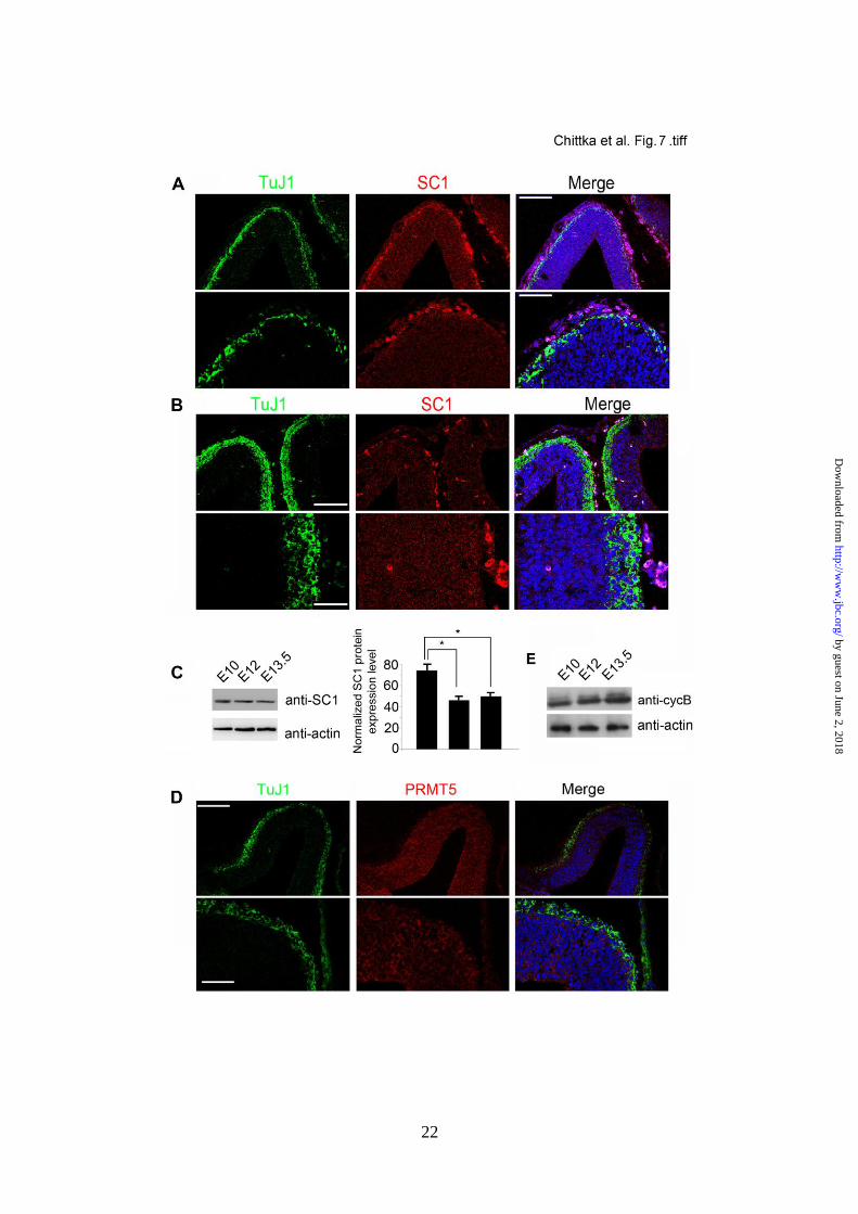
influence the mode of NSC division. We
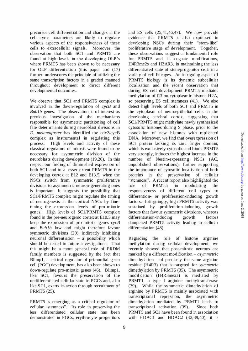
therefore investigated whether we can detect the
down-regulation of SC1 and PRMT5 protein
levels in the developing cortex at the time when
the NSCs switch from proliferative to neuron-
generating divisions. We observed that, SC1
protein levels and to a lesser extent PRMT5
protein levels were reduced in the developing
cortex at E12 and at E13.5 when symmetric
proliferative divisions give way to asymmetric
neurogenic divisions (Fig. 7A, B, C and D).
by guest on June 2, 2018http://w
ww
.jbc.org/D
ownloaded from
8
Moreover, we detected a moderate increase in
the levels of cycB1 protein in the developin g
cortex at these developmental stages (Fig. 7E).
We conclude that the expression level of SC1
and PRMT5 proteins is down-regulated during
the transition from proliferation to neurogenesis
of the cortical NSCs concomitant with the
elevation of cycB1 protein expression.
DISCUSSION
In this study we investigated the role of the
transcription factor SC1 in neural development.
We demonstrated that SC1 expression is
dynamically regulated in developing NSCs,
being strongly expressed in proliferating NSCs
but down-regulated at the onset of neurogenesis.
Moreover, experimental knock-down of SC1 in
NSCs led to precocious neurogenesis. Notably,
we demonstrated that SC1 recruits an epigenetic
modifier, the histone arginine methyltransferase
PRMT5 and that high levels of SC1/PRMT5
complex are required to maintain the
proliferative capacity and “stem-like” cellular
state of the NSCs. Furthermore, we showed that
SC1 in complex with PRMT5 directs
H4R3me2s, a modification which is prevalent in
the developing neuroepithelium during the
expansion phase of cortical development (35).
In addition, we demonstrated that the
SC1/PRMT5 complex modulates the levels of
expression of pro-mitotic genes that regulate the
G2/M transition and mitotic progression.
Our findings suggest that SC1 in complex with
the epigenetic modifier, PRMT5, plays an
important role in the control of timing of
neurogenesis in developing cortical NSCs.
Previous work showed that SC1 mRNA is
highly expressed in the developing mouse
cerebral cortex (34). We found that in E10.5
mouse cortical cell cultures Nestin-positive
NSCs expressed variable amounts of SC1
protein. It is not clear whether this reflects
heterogeneity within the NSC population,
different developmental stages of the NSC
lineage(s) or varying levels of SC1 expression
during the stages of the cell cycle. Nevertheless,
the fact that SC1 expression is low in early-born
neurons and that SC1 knock-down in NSCs
triggers precocious neuronal differentiation of a
subset of the NSCs suggest that NSC
differentiation depends on down-regulation of
SC1. Consistent with this, we see high levels of
SC1, PRMT5 and H4R3me2s in the early
proliferative neuroepithelium at E10.5, while
both SC1 and to a lesser extent PRMT5 protein
levels are diminished in the neuroepithelium at
E13.5 at the onset of neurogenesis. Moreover,
we also find that over-expression of SC1 in
NSCs isolated from E10.5 mouse cortices leads
to a moderate increase in the number of Nestin-
expressing NSCs, suggesting that high levels of
SC1 prevent differentiation. It is noteworthy in
this respect that in the mouse embryonic stem
(ES) cells, one of the essential regulators of
“stemness”, Nanog, exhibits fluctuating levels of
expression and that ES cells which express low
levels of Nanog are predisposed to differentiate
while those with high Nanog levels retain their
pluripotency (43). Perhaps, the low and high
SC1-expressing NSCs which we observe within
the NSC population reflect a similar
heterogeneity of these cells with respect to their
predisposition to differentiate.
The dynamic nature of expression of both SC1
and PRMT5 is evident at E15.5 in the
developing cortex when both proteins become
upregulated in post-mitotic neurons (34) and
A.C., unpublished observations. Similarly,
another PRDM family member,
Blimp1/PRDM1, has been reported to undergo
temporally dynamic expression during
development of primordial germ cells (PGCs)
and various other tissues (44,45) and has also
been shown to recruit PRMT5 during PGC
development (25). Importantly, various levels
of Blimp appear to be necessary to direct
differentiation of different tissues, reflecting
precise dose dependency of different cell types
on Blimp1 requirement (45). These observations
highlight the general principle of utilising the
same transcriptional regulators during
development in various tissues in a graded
manner to specify different cell fates, possibly
through recruitment of different partner proteins
and downstream choice of gene targets. The
similarities between the utilisation of both
Blimp1 and SC1 proteins during development
presumably reflect analogous functions of these
proteins in different cell lineage precursors
during development. For example, the changing
cell cycle parameters and kinetics during
precursor cell differentiation may be a common
mechanism which contributes to the
developmental decisions made by these cells
(16-19). Precise control of cell cycle
progression is one of the critical components of
by guest on June 2, 2018http://w
ww
.jbc.org/D
ownloaded from
9
precursor cell differentiation and changes in the
cell cycle parameters are likely to regulate
various aspects of the responsiveness of these
cells to extracellular signals. Moreover, the
observation that both SC1 and PRMT5 are
found at high levels in the developing OLP’s
where PRMT5 has been shown to be necessary
for OLP differentiation (this paper and (17)
further underscores the principle of utilizing the
same transcription factors in a graded manned
throughout development to direct different
developmental outcomes.
We observe that SC1 and PRMT5 complex is
involved in the down-regulation of cycB and
Bub1b genes. The observation is of interest as
previous investigation of the mechanisms
responsible for asymmetric partitioning of cell
fate determinants during neuroblast divisions in
D. melanogaster has identified the cdc2/cycB
complex as instrumental in regulating this
process. High levels and activity of these
classical regulators of mitosis were found to be
necessary for asymmetric division of the
neuroblasts during development (19,20). In this
respect our finding of diminished expression of
both SC1 and to a lesser extent PRMT5 in the
developing cortex at E12 and E13.5, when the
NSCs switch from symmetric proliferative
divisions to asymmetric neuron-generating ones
is important. It suggests the possibility that
SC1/PRMT5 complex might regulate the timing
of neurogenesis in the cortical NSCs by fine-
tuning the expression levels of pro-mitotic
genes. High levels of SC1/PRMT5 complex
found in the pre-neurogenic cortex at E10.5 may
keep the expression of pro-mitotic genes cycB
and Bub1b low and might therefore favour
symmetric divisions (20), indirectly inhibiting
neuronal differentiation – a possibility which
should be tested in future investigations. That
this might be a more general role of PRDM
family members is suggested by the fact that
Blimp1, a critical regulator of primordial germ
cell (PGC) development, has also been shown to
down-regulate pro-mitotic genes (46). Blimp1,
like SC1, favours the preservation of the
undifferentiated cellular state in PGCs and, also
like SC1, exerts its action through recruitment of
PRMT5 (25).
PRMT5 is emerging as a critical regulator of
cellular “stemness”. Its role in preserving the
less differentiated cellular state has been
demonstrated in PGCs, erythrocyte progenitors
and ES cells (25,41,46,47). We now provide
evidence that PRMT5 is also expressed in
developing NSCs during their “stem-like”
proliferative stage of development. Together,
these observations suggest a fundamental role
for PRMT5 and its cognate modifications,
H4R3me2s and H2AR3, in maintaining the less
differentiated state of stem/progenitor cells in a
variety of cell lineages. An intriguing aspect of
PRMT5 biology is its dynamic subcellular
localization and the recent observation that
during ES cell development PRMT5 mediates
methylation of R3 on cytoplasmic histone H2A,
so preserving ES cell stemness (41). We also
detect high levels of both SC1 and PRMT5 in
the cytoplasm of neuroepithelial cells in the
developing cerebral cortex, suggesting that
SC1/PRMT5 might methylate newly synthesized
cytosolic histones during S phase, prior to the
association of new histones with replicated
DNA. Moreover, we find that overexpression of
SC1 protein lacking its zinc finger domain,
which is exclusively cytosolic and binds PRMT5
very strongly, induces the highest increase in the
number of Nestin-expressing NSCs (AC,
unpublished observations), further supporting
the importance of cytosolic localisation of both
proteins in the preservation of cellular
“stemness”. A recent report also highlighted the
role of PRMT5 in modulating the
responsiveness of different cell types to
differention- or proliferation-inducing growth
factors. Intirguingly, high PRMT5 activity was
sustained by proliferation-inducing growth
factors that favour symmetric divisions, whereas
differentiation-inducing growth factors
dampened PRMT5 activity leading to cellular
differentiation (48).
Regarding the role of histone arginine
methylation during cellular development, we
recently showed that post-mitotic neurons are
marked by a different modification – asymmetric
dimethylation - of precisely the same arginine
residue (H4R3) that is targeted for symmetric
dimethylation by PRMT5 (35). The asymmetric
modification (H4R3me2a) is mediated by
PRMT1, a type I arginine methyltransferase
(39). While the symmetric dimethylation of
arginine by PRMT5 is mainly associated with
transcriptional repression, the asymmetric
dimethylation mediated by PRMT1 leads to
transcriptional activation (39). Since both
PRMT5 and SC1 have been found in association
with HDAC1 and HDAC2 (33,39,40), it is
by guest on June 2, 2018http://w
ww
.jbc.org/D
ownloaded from
10
possible that SC1 might be a common
component of the repressive chromatin
remodelling complexes during early neura l
development, and that the principal role of SC1
in such complexes might be to provide targeting
specificity via its sequence-specific DNA
binding properties. It is therefore conceivable
that down-regulation of SC1 at the onset of
neurogenesis has the effect of vacating sites in
chromatin that were previously targets of
symmetric methylation (repression) by
SC1/PRMT5, making them accessible for
asymmetric methylation (activation) by PRMT1.
This is consistent with our observation that
knockdown of SC1 in developing NSCs induces
precocious neurogenesis, as it might allow
PRMT1-mediated deposition of H4R3me2a
modifications leading to the activation of genes
necessary for neuronal differentiation. It is also
noteworthy in this respect that previous work
has identified a protein, Tis21/Btg2 - a known
stimulator of PRMT1 activity - as a marker of
NSCs that are undergoing their final mitosis on
their way to becoming post-mitotic neurons
(18). Moreover, it was previously shown that in
PC12 cells, which can respond to NGF by
differentiating into sympathetic-like neurons,
application of NGF increases asymmetric
arginine dimethylation of proteins mediated by
PRMT1 (49,50). Taken together, these
observations suggest that NSC division and
neurogenesis is at least partly regulated by the
sequential activation of PRMT5 and PRMT1;
high levels of SC1/PRMT5 protein complex
during the proliferative stage of cortica l
development might control the onset of
neurogenesis by controlling the cell cycle
parameters of the developing NSCs, possibly by
maintaining symmetric proliferative divisions of
the NSCs during the early phase of cortica l
development, whereas the progression to
asymmetric division and neuronal differentiation
depends on PRMT1.
In conclusion, our study identifies SC1 as a
modulator of the NSC developmenta l
programme that acts through recruitment of a
histone arginine methyltransferase, PRMT5.
Given that SC1 is a p75NTR interacting protein
(Chittka et al., 2004), it will be important to
determine whether Neurotrophins or other
signalling molecules can trigger modifications
of SC1 that regulate its ability to recruit PRMT5
and thereby transmit extracellular information to
the nuclear interior. Perhaps such
differentiation-inducing factors as the
Neurotrophins regulate which epigenetic
modifiers will be recruited by SC1 at different
stages of cortical development and regulate the
activity of PRMTs involved in the process of
neuronal differentiation. Together, our findings
uncover a novel role for histone arginine
methylation in the control of cortical NSC
proliferation and differentiation.
by guest on June 2, 2018http://w
ww
.jbc.org/D
ownloaded from

11
REFERENCES
1. Okano, H., and Temple, S. (2009) Current Opinion in Neurobiology 19, 112-119
2. Qian, X. M., Shen, Q., Goderie, S. K., He, W. L., Capela, A., Davis, A. A., and
Temple, S. (2000) Neuron 28, 69-80
3. Shen, Q., Wang, Y., Dimos, J. T., Fasano, C. A., Phoenix, T. N., Lemischka, I. R.,
Ivanova, N. B., Stifani, S., Morrisey, E. E., and Temple, S. (2006) Nature
Neuroscience 9, 743-751
4. Guillemot, F. (2007) Progress in Neurobiology 83, 37-52
5. Ohnuma, S., Philpott, A., and Harris, W. A. (2001) Current Opinion in Neurobiology
11, 66-73
6. Ohnuma, S., and Harris, W. A. (2003) Neuron 40, 199-208
7. Hirabayashi, Y., and Gotoh, Y. (2005) Neuroscience Research 51, 331-336
8. Miller, F. D., and Gauthier, A. S. (2007) Neuron 54, 357-369
9. Hsieh, J., and Gage, F. H. (2004) Current Opinion in Genetics & Development 14,
461-469
10. Jepsen, K., Solum, D., Zhou, T. Y., McEvilly, R. J., Kim, H. J., Glass, C. K.,
Hermanson, O., and Rosenfeld, M. G. (2007) Nature 450, 415-U418
11. Song, M. R., and Ghosh, A. (2004) Nature Neuroscience 7, 229-235
12. Matsumoto, S., Banine, F., Struve, J., Xing, R. B., Adams, C., Liu, Y., Metzger, D.,
Chambon, P., Rao, M. S., and Sherman, L. S. (2006) Developmental Biology 289,
372-383
13. Ballas, N., Grunseich, C., Lu, D. D., Speh, J. C., and Mandel, G. (2005) Cell 121,
645-657
14. Hermanson, O., Jepsen, K., and Rosenfeld, M. G. (2002) Nature 419, 934-939
15. Lukaszewicz, A., Savatier, P., Cortay, V., Giroud, P., Huissoud, C., Berland, M.,
Kennedy, H., and Dehay, C. (2005) Neuron 47, 353-364
16. Dirks, P. B. (2010) Molecular Oncology 4, 420-430
17. Huang, J. H., Vogel, G., Yu, Z. B., Almazan, G., and Richard, S. (2011) Journal of
Biological Chemistry 286, 44424-44432
18. Iacopetti, P., Michelini, M., Stuckmann, I., Oback, B., Aaku-Saraste, E., and Huttner,
W. B. (1999) Proceedings of the National Academy of Sciences of the United States of
America 96, 4639-4644
19. Tedeschi, A., and Di Giovanni, S. (2009) Embo Reports 10, 576-583
20. Tio, M., Udolph, G., Yang, X. H., and Chia, W. (2001) Nature 409, 1063-1067
21. Chittka, A., and Chao, M. V. (1999) Proceedings of the National Academy of
Sciences of the United States of America 96, 10705-10710
22. Fumasoni, I., Meani, N., Rambaldi, D., Scafetta, G., Alcalay, M., and Ciccarelli, F. D.
(2007) Bmc Evolutionary Biology 7
23. Schneider, R., Bannister, A. J., and Kouzarides, T. (2002) Trends in Biochemical
Sciences 27, 396-402
24. Yu, J., Angelin-Duclos, C., Greenwood, J., Liao, J., and Calame, K. (2000) Molecular
and Cellular Biology 20, 2592-2603
25. Ancelin, K., Lange, U. C., Hajkova, P., Schneider, R., Bannister, A. J., Kouzarides,
T., and Surani, M. A. (2006) Nature Cell Biology 8, 623-630
26. Bikoff, E. K., Morgan, M. A., and Robertson, E. J. (2009) Current Opinion in
Genetics & Development 19, 379-385
27. Hayashi, K., and Matsui, Y. (2006) Cell Cycle 5, 615-620
by guest on June 2, 2018http://w
ww
.jbc.org/D
ownloaded from
12
28. Eom, G., Kim, K., Kim, S., Kee, H., Kim, J., Jin, H., Kim, J., Kim, J., Choe, N., Kim,
K., Lee, J., Kook, H., Kim, N., and Seo, S. (2009) Biochemical and Biophysical
Research Communications 388, 131-136
29. Derunes, C., Briknarova, K., Geng, L. Q., Li, S., Gessner, C. R., Hewitt, K., Wu, S.
D., Huang, S., Woods, V. I., and Ely, K. R. (2005) Biochemical and Biophysical
Research Communications 333, 925-934
30. John, S. A., and Garrett-Sinha, L. A. (2009) Experimental Cell Research 315, 1077-
1084
31. Gyory, I., Wu, J., Fejer, G., Seto, E., and Wright, K. L. (2004) Nature Immunology 5,
299-308
32. Davis, C. A., Haberland, M., Arnold, M. A., Sutherland, L. B., McDonald, O. G.,
Richardson, J. A., Childs, G., Harris, S., Owens, G. K., and Olson, E. N. (2006)
Molecular and Cellular Biology 26, 2626-2636
33. Chittka, A., Arevalo, J. C., Rodriguez-Guzman, M., Perez, P., Chao, M. V., and
Sendtner, M. (2004) Journal of Cell Biology 164, 985-996
34. Kendall, S. E., Ryczko, M. C., Mehan, M., and Verdi, J. M. (2003) Developmental
Brain Research 144, 151-158
35. Chittka, A. (2010) Plos One 5
36. Kessaris, N., Jamen, F., Rubin, L. L., and Richardson, W. D. (2004) Development
131, 1289-1298
37. Qian, X. M., Davis, A. A., Goderie, S. K., and Temple, S. (1997) Neuron 18, 81-93
38. Nishioka, K., and Reinberg, D. (2003) Methods 31, 49-58
39. Bedford, M. T., and Clarke, S. G. (2009) Molecular Cell 33, 1-13
40. Pal, S., and Sif, S. (2007) Journal of Cellular Physiology 213, 306-315
41. Xu, X. J., Hoang, S., Mayo, M. W., and Bekiranov, S. (2010) Bmc Bioinformatics 11
42. McBurney, M. W. (1993) International Journal of Developmental Biology 37, 135-
140
43. Chambers, I., Silva, J., Colby, D., Nichols, J., Nijmeijer, B., Robertson, M., Vrana, J.,
Jones, K., Grotewold, L., and Smith, A. (2007) Nature 450, 1230-U1238
44. Hayashi, K., Lopes, S., and Surani, M. A. (2007) Science 316, 394-396
45. Robertson, E. J., Charatsi, I., Joyner, C. J., Koonce, C. H., Morgan, M., Islam, A.,
Paterson, C., Lejsek, E., Arnold, S. J., Kallies, A., Nutt, S. L., and Bikoff, E. K.
(2007) Development 134, 4335-4345
46. Saitou, M. (2009) Current Opinion in Genetics & Development 19, 386-395
47. Zhao, Q., Rank, G., Tan, Y. T., Li, H. T., Moritz, R. L., Simpson, R. J., Cerruti, L.,
Curtis, D. J., Patel, D. J., Allis, C. D., Cunningham, J. M., and Jane, S. M. (2009)
Nature Structural & Molecular Biology 16, 304-311
48. Andreu-Perez, P., Esteve-Puig, R., de Torre-Minguela, C., Lopez-Fauqued, M., Bech-
Serra, J. J., Tenbaum, S., Garcia-Trevijano, E. R., Canals, F., Merlino, G., Avila, M.
A., and Recio, J. A. (2011) Science Signaling 4
49. Cimato, T. R., Ettinger, M. J., Zhou, X. B., and Aletta, J. M. (1997) Journal of Cell
Biology 138, 1089-1103
50. Cimato, T. R., Tang, J., Xu, Y., Guarnaccia, C., Herschman, H. R., Pongor, S., and
Aletta, J. M. (2002) Journal of Neuroscience Research 67, 435-442
by guest on June 2, 2018http://w
ww
.jbc.org/D
ownloaded from
13
ACKNOWLEDGEMENTS: We thank our colleagues in the WIBR, especially Joana Paes de Faria,
Ingvar Ferby, Sarah Hopkins, Huiliang Li, Andrei Okorokov, Nigel Pringle and Kaylene Young for
discussions and help with NSC cultures and staining procedures. This work was supported by a
Wellcome Trust Career Re-entry Fellowship WT076656MA to AC and the UK Medical Research
Council to WDR.
ABBREVIATIONS: BrdU – bromodeoxyuridine, PRMT – protein arginine methyltransferase, SC1 –
Schwann cell factor 1, NSC – neural stem cell
FIGURE LEGENDS
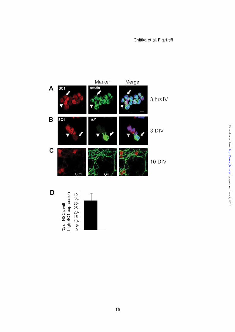
FIGURE 1. Dynamic expression of SC1 in developing NSCs. (A-C) NSC cultures immunolabelled
with anti-SC1 (left), anti-Nestin, anti-TuJ1 or O4 (centre), and merged with Hoechst DNA stain
(right). (A) 3 hours after plating NSCs show different levels of expression of SC1 in Nestin-
expressing precursor cells: arrowheads, cells with high SC1 expression and arrows, cells with low
SC1 expression. (B) At 3 DIV differentiating neurons activate TuJ1 expression and down-regulate
SC1 (arrowhead, high SC1-expressing cell; arrow, Tuj1+ neuron with low-level SC1 expression).
(C) After 10 DIV high levels of SC1 expression are detected in O4+ oligodendrocyte precursors. (D)
Quantification of the percentage of NSCs with high SC1 expression levels. Scale bar, 10 m.
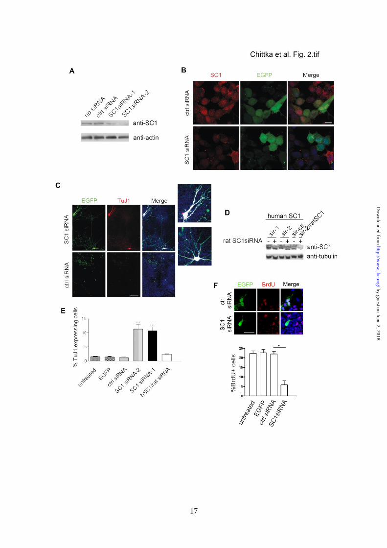
FIGURE 2. siRNA knock-down of SC1 leads to precocious differentiation of NSCs into neurons. (A)
Western blots of protein samples from rat NSCs transfected with: 1) no siRNA, 2) control (scrambled
sequence) siRNA, 3) SC1 siRNA-1, 4) SC1 siRNA-2, probed for expression of endogenous SC1.
Bottom panel – same blots probed for actin demonstrate similar protein levels in the designated lanes.
(B) NSCs co-transfected with an EGFP expression vector together with either control siRNA (above)
or SC1-specific siRNA (below) were immunolabelled with anti-SC1 and anti-EGFP to identify
transfected cells. (C) Expression of TuJ1 in NSCs transfected with EGFP and siRNA-1 or EGFP and
control siRNA. (Right) Merged images with Hoechst-stained DNA. Two of the neurite-bearing
EGFP+ cells from the siRNA-1 treated cultures are magnified to visualise the extensive arborisation.
(D) Human or rat SC1 was overexpressed in HEK293T cells with or without rat-specific SC1siRNA-1
or SC1siRNA-2 (sir-1 and sir-2, respectively) and its expression monitored by probing Western blots
of transfected cell lysates with anti-SC1. Neither of the applied siRNAs affected the expression of
human SC1 (top panel), attesting to the target specificity of the siRNAs. Blots were also probed with
anti-tubulin antibodies to control for gel loadings (bottom panel). Co-transfection of ratSC1 cDNA
with rat SC1siRNA-2 resulted in the expected reduction in the levels of rat SC1 protein (sir-2/ratSC1
lane). (E) Quantification of TuJ1 expression in NSCs transfected with either control siRNA, siRNA-1
or siRNA-2 or indicated control siRNA [mean ± s.d., n=3, t-test, P<0.0005(*)]. At least 300
transfected cells were counted per coverslip. F) Quantification of BrdU incorporation into rat NSCs
after transfection with control siRNA, siRNA-2, EGFP alone or nothing (bottom panel). Cells
expressing EGFP and indicated siRNA’s were immunolabelled to detect BrdU incorporation and
EGFP expression (top panel). Scale bars: 100 m (C), 10 m (B and F).
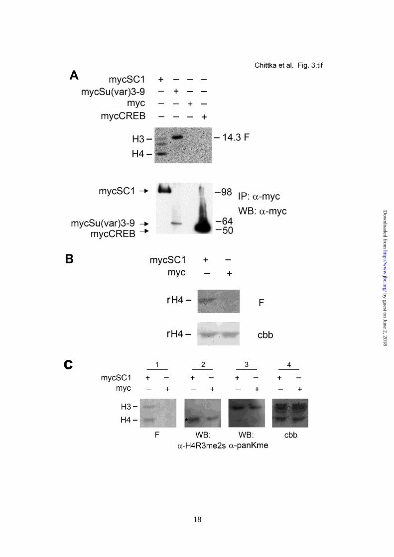
FIGURE 3. Immunoprecipitated SC1 complex exhibits an H4 HMTase activity. (A) Myc-tagged SC1
or indicated controls were expressed in HEK293T cells and immunoprecipitated (IPed) using an anti-
Myc antibody (below). The same IPs were used for an in vitro HMTase assay with purified calf
thymus histone mix; a fluorogram of the in vitro methylation reaction is shown (above, F). (B) Cells
were transfected with mycSC1 expression vector or empty Myc vector and cell lysates were
immunoprecipitated with anti-Myc antibody. The mycSC1 immunoprecipitate methylates
recombinant H4 in an in vitro methylation reaction. F – fluorogram of the methylation reaction
products, cbb – coommassie brilliant blue stained membrane showing the histones used for the in
vitro methylation reaction. (C) mycSC1 mediated histone methylation increases the levels of
H4R3me2s. Same procedure was carried out as in (A) and the blots of methylated histones were
probed with antibodies against lysine and arginine modifications. (Panel 1) fluorogram of methylated
histones in the indicated IPs, (Panel 2) the same blot probed with anti-H4R3me2s antibody, (Panel 3)
by guest on June 2, 2018http://w
ww
.jbc.org/D
ownloaded from
14
the same blot probed with anti-pan Lysine antibody and (Panel 4) the same membrane stained with
Coomassie brilliant blue (cbb).
FIGURE 4. SC1 recruits PRMT5 to direct H4 methylation. (A) mycSC1 or empty vector was
expressed in HEK293T cells as indicated on the panel. Anti-Myc IPs from transfected cells were
analyzed using anti-PRMT5 antibody. Endogenous PRMT5 is found in the complex (arrow, top)
with IPed mycSC1. Total protein inputs and Iped mycSC1 are shown (two middle and bottom panels,
respectively). (B) Endogenous PRMT5 was IPed from HEK293T cells transfected with mycSC1 or
empty vector and the IPs were analyzed by Western blot with anti-Myc (arrow, MycSC1 protein);
two middle and bottom panels show the input mycSC1 and PRMT5 and IPed PRMT5, respectively.
(C) Diagram of the deletion constructs of Myc-tagged SC1 used to map the domains of interaction
with PRMT5. (D) The NH-terminus and to a lesser extent PR/SET domain of SC1 bind PRMT5.
Indicated Myc-tagged SC1 full length or deleted constructs and HA/Flag-tagged PRMT5 were co-
expressed in HEK293T cells. Anti-HA IPs (PRMT5) were probed on Western blots with anti-Myc
antibody to detect co-IPed myc-tagged SC1 constructs. Inputs were analyzed using anti-Flag
antibody for PRMT5 and anti-Myc antibody for SC1. Anti-tubulin antibody was used as a loading
control. mycSC1 proteins that co-IP with PRMT5 are highlighted by asterisks on the top panel of the
Western blot. (E) mycSC1FL, mycSC1dNH or empty vector were transfected into HEK293T cells.
Anti-Myc IPs were used for in vitro HMTase assays. (Above) fluorogram (F) of histones methylated
by the indicated IPed complexes, (below) Western blot of IPed proteins used for the in vitro
methylation reactions, probed with anti-Myc.
FIGURE 5. PRMT5, SC1 and H4R3me2s are expressed in developing NSCs and cortical
neuroepithelium and can be co-immuneprecipitated from E10.5 cortex. (A) Expression of PRMT5 in
E10.5 Nestin+ NSCs, 3 hours after plating, was detected by immunolabelling with PRMT5-specific
antibodies. Right panels, merged images with Hoechst DNA stain. (B) H4R3me2s modification is
detected in Nestin+ NSCs 3 hours after plating. (C) Both SC1 and PRMT5 are expressed in the
developing mouse cortex at E10.5 and high levels of H4R3me2s modifications are detected in the
cortical neuroepithelium at this stage. Expression of the relevant proteins or modification in panels
(A, B and C) was detected by immunolabelling with anti-SC1, anti-PRMT5 or anti-H4R3me2s. (D)
Endogenous PRMT5 was IPed from E10.5 cortices and the presence of endogenous SC1 in the co-
IPed complex was analyzed by using anti-SC1 antibodies. Non-specific IgG was used to control for
the specificity of IP reactions. Input and IPed PRMT5 is shown on the middle panel and anti-actin
antibody was used as a loading control. Scale bars: 10 m (A, B), 50 m (C). V, ventricular zone; P,
pial surface.
FIGURE 6. SC1 and PRMT5 complex increases the number of Nestin-expressing neural precursors
and regulates expression of pro-mitotic genes. A) Over-expressed mycSC1 increases the number of
Nestin+ NSCs. mycSC1FL and mycSC1dNH proteins were detected by immunolabelling with anti-
myc antibodies and NSCs by the presence of Nestin immunoreactivity. Quantification of
Nestin+/mycSC1 expressing NSCs is shown in the graph on the right At least 300 cells were counted
per transfection and data are shown as mean ± s.d [n=3 p<0.05(*)]. (B) Semi-quantitative RT-PCR
was used to estimate the relative levels of cycB and Bub1b mRNA in P19 cells transfected with
mycSC1FL, mycSC1dNH or empty vector. (C) Semi-quantitative RT-PCR was used to estimate the
relative levels of SC1, cycB and Bub1b mRNA in PC12 cells transfected with SC1siRNA, control
siRNA or no siRNA. Levels of mRNA were normalized to GAPDH mRNA. Scale bar: 5 m(A).
FIGURE 7. SC1 and PRMT5 protein levels are reduced in the developing cortex at E12 and E13.5.
Mouse cortices from E12 (A) and E13.5 (B) embryos were immunolabelled for TuJ1 and SC1. SC1
protein levels are reduced compared to those detected at E10.5 prior to the onset of neurogenesis (see
Fig. 5). Bright red signal in the tissue represents non-specific labelling of blood vessels after antigen
retrieval by heating with citrate buffer. (C) Western blot analysis of SC1 protein expression in the
developing cortex. Protein homogenates from embryonic cortices of indicated ages were analysed by
probing with anti-SC1 antibodies (top panel) and anti-actin (bottom panel) antibodies to control for
by guest on June 2, 2018http://w
ww
.jbc.org/D
ownloaded from
15
protein loading. Normalised protein levels of SC1 are shown in the graph. (D) Mouse cortex from
E13.5 embryos was immunolabelled for TuJ1 and PRMT5. Moderate levels of PRMT5 protein were
detected in the cortex at E13.5. TuJ1 staining is towards the pial surface in all panels. (E) Western blot
analysis of cycB1 protein expression in the developing cortex. Protein homogenates from embryonic
cortices of indicated ages were analysed by probing with anti-cycB1 antibodies (top panel) and anti-
actin (bottom panel) antibodies to control for protein loading. Data (in C) are shown as mean ± s.d
from three independent western blot quantifications [n=3 p<0.05(*)]. Scale bar, 75m (top panels),
25 m (bottom panels).
by guest on June 2, 2018http://w
ww
.jbc.org/D
ownloaded from

Alexandra Chittka, Justyna Nitarska, Ursula Grazini and William D. Richardsonand control neural stem cell proliferation and differentiation.
Arginine Methyltransferase 5 (PRMT5) to mediate histone arginine methylation Transcription Factor Positive Regulatory Domain 4 (PRDM4) recruits Protein
published online October 9, 2012J. Biol. Chem.
10.1074/jbc.M112.392746Access the most updated version of this article at doi:
Alerts:
When a correction for this article is posted•
When this article is cited•
to choose from all of JBC's e-mail alertsClick here
by guest on June 2, 2018http://w
ww
.jbc.org/D
ownloaded from