microbial arms race: ballistic nematocysts in ... · in aquatic environments ( 2) and considering...
TRANSCRIPT

SC I ENCE ADVANCES | R E S EARCH ART I C L E
CELL B IOLOGY
1Department of Botany, University of British Columbia, Vancouver, Canada. 2De-partment of Zoology, University of British Columbia, Vancouver, Canada. 3Officeof International Affairs, Hokkaido University, Kita 10, Nishi 8, Sapporo 060-0810,Japan. 4Alfred Wegener Institute for Polar and Marine Research, Bremerhaven, Ger-many. 5Faculty of Science, Hokkaido University, Kita 10, Nishi 8, Sapporo 060-0810,Japan. 6Marine Biophysics Unit, Okinawa Institute of Science and Technology, Oki-nawa, Japan. 7Centre for Organismal Studies, University of Heidelberg, Heidelberg,Germany.*Present address: School of Life Sciences, Arizona State University, Tempe, AZ85257, USA.†Corresponding author. Email: [email protected]
Gavelis et al., Sci. Adv. 2017;3 : e1602552 31 March 2017
2017 © The Authors,
some rights reserved;
exclusive licensee
American Association
for the Advancement
of Science. Distributed
under a Creative
Commons Attribution
NonCommercial
License 4.0 (CC BY-NC).
Microbial arms race: Ballistic “nematocysts”in dinoflagellates represent a new extreme inorganelle complexity
Gregory S. Gavelis,1,2*† Kevin C. Wakeman,3,4 Urban Tillmann,5 Christina Ripken,6Satoshi Mitarai,6 Maria Herranz,1 Suat Özbek,7 Thomas Holstein,7
Patrick J. Keeling,1 Brian S. Leander1,2
Dow
nloade
We examine the origin of harpoon-like secretory organelles (nematocysts) in dinoflagellate protists. These ballisticorganelles have been hypothesized to be homologous to similarly complex structures in animals (cnidarians); butwe show, using structural, functional, and phylogenomic data, that nematocysts evolved independently in bothlineages. We also recorded the first high-resolution videos of nematocyst discharge in dinoflagellates. Unexpectedly,our data suggest that different types of dinoflagellate nematocysts use two fundamentally different types of ballisticmechanisms: one type relies on a single pressurized capsule for propulsion, whereas the other type launches 11 to 15projectiles fromanarrangement similar to aGatlinggun.Despite their radical structural differences, these nematocystsshare a single origin within dinoflagellates and both potentially use a contraction-based mechanism to generateballistic force. The diversity of traits in dinoflagellate nematocysts demonstrates a stepwise route by which simplesecretory structures diversified to yield elaborate subcellular weaponry.
d fr
on May 8, 2020http://advances.sciencem
ag.org/om
INTRODUCTIONPlanktonic microbes are often viewed as passive food items for larger life-forms. In reality, eukaryoticmicrobes (for example, dinoflagellates and cili-ates) have evolved a range of weapons, from armor and toxins to projectileorganelles or “extrusomes” (1). The ballistic action of extrusomes has beenknown since early light microscopy, when predatory ciliates were seen tocapture prey using syringe-like toxicysts. In turn, prey ciliates sometimesbroke free of toxicysts by firing volleys of defensive extrusomes (mucocystsand trichocysts). These “arms races” are likely widespread in nature, giventhat ciliates and dinoflagellates consume up to 60% of primary productionin aquatic environments (2) and considering the diversity of extrusomesfound throughout the phytoplankton (in groups including cryptophytes,chrysophytes, raphidophytes, and euglenozoans) (1). However, extru-somes are perhaps the least studied of major organelle types.Here, we used single-cell microscopic and genomic approaches tostudy ballistic organelles in cultivable (Polykrikos kofoidii; Figs. 1, C toE, and 2) and wild-caught dinoflagellates (Nematodinium sp.; Figs. 1, Fto H, and 3). Dinoflagellates have a large arsenal of extrusomes, rangingfrom simple, ostensibly defensive organelles (for example, mucocystsand trichocysts) to elaborate organelles, for predation (for example, tae-niocysts andnematocysts) (1). Cells of the genusPolykrikos capture oth-er dinoflagellates using harpoon-like “nematocysts” (Fig. 1B and movieS1). Polykrikos can ensnare other dinoflagellates and tow them in foringestion using a long filament (3, 4). The nematocyst capsule is sealedwith a hatch-like operculum and contains a coiled tubule and a pointedstylet (5, 6). This constellation of features is shared by the nematocysts
in the phylum Cnidaria (7, 8), which is among the earliest divergingpredatory animal phyla. Dinoflagellates and cnidarians are very distant-ly related, so whether the ballistic similarities of these organelles reflecthomology or convergent evolution has been amatter of some debate. Inparticular, it has been suggested that the origin of nematocysts in cni-darians was spurred by the acquisition of genes from dinoflagellates—specifically from groups, such as Symbiodinium, which live endosym-biotically within corals (9–11). To test this hypothesis, we used nemato-genic genes identified from the Hydra nematocyst proteome (12) tosearch a collection of 30 eukaryotic genomes [including two genomesof Symbiodinium (13, 14)] and 120 dinoflagellate transcriptomes, plus atranscriptome that we previously obtained from the nematocyst-bearingdinoflagellate Polykrikos lebouriae (15).
RESULTSContrary to the symbiogenic hypothesis, we found that core nematogenicproteins were restricted to cnidarians. Critically, the enzyme responsiblefor the osmotic propellant of cnidarian nematocysts [poly-g-glutamate syn-thase, which is encoded by PgsAA (16)] was not found in any extrusome-bearing microbes, suggesting that dinoflagellates and other groups usedifferent propellants to generate ballistic force. Animal PgsAA is derivedfrombacteria via lateral gene transfer (17).Other components of cnidariannematocysts [for example, minicollagens (18), spinalin (19), and cnidoin(20)] appear to have arisen after multicellularity, with no evident gene do-nors or recipients among 130 transcriptomic and genomic data sets fromdinoflagellates. This suggests that the similarities between the nemato-cysts of dinoflagellates and cnidarians reflect convergent evolution.Moreover, no homology was detected between dinoflagellate extrusomeproteins and the known extrusome proteins fromothermicrobial eukar-yotes (for example, ciliates and cryptophytes). How then did dinoflagel-lates evolve these sophisticated ballistic organelles?
Because our current understanding of dinoflagellate nematocysts islimited to two-dimensional (2D) imagery, we assembled 3D reconstruc-tions via focused ion beam scanning electronmicroscopy (FIB-SEM) onsingle cells of P. kofoidii. FIB-SEM allowed us to reconstruct nearly an
1 of 7

SC I ENCE ADVANCES | R E S EARCH ART I C L E
on May 8, 2020
http://advances.sciencemag.org/
Dow
nloaded from
entire cell of P. kofoidii in 3D, which contained four nematocysts, eachconnected to another organelle known as a taeniocyst (Fig. 2 and movieS2).Our renderings show that the similarity of dinoflagellate and cnidariannematocysts has been overstated. While the capsule in cnidarians istopped simply by ahatch-like opening (theoperculum) that becomes “un-corked” to allow firing (fig. S1), we found an additional—and far moreelaborate—structure in Polykrikos, which lies beneath the operculum. Thisstructure is composed of three concentric rings and appears to have anozzle-like function.
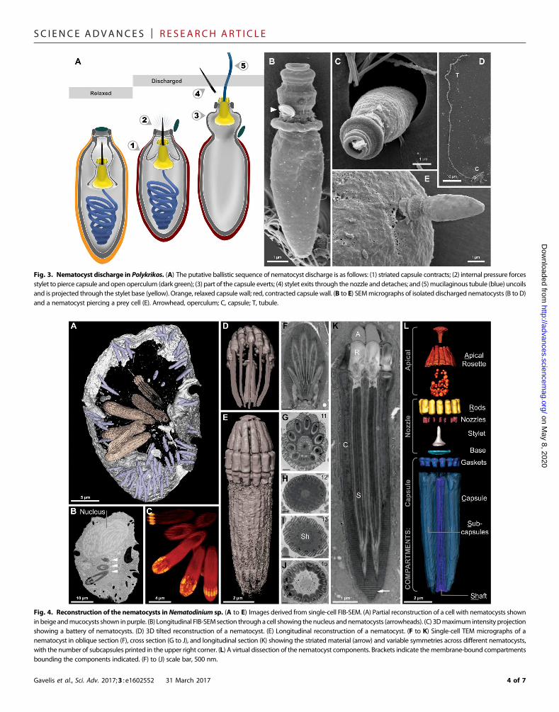
By capturing the first high-resolution videos of nematocyst discharge(movies S2 andS3), aswell as the SEMmicrographsof nematocysts arrestedat different stages of firing (fig. S2), we were able to make functional infer-ences about the ballistic mechanism of these complex organelles (Fig. 3).First, the coiled tubule must exit the capsule, which (unlike in cnidarians)has no openings (Figs. 2F and 3). Therefore, the role of the stylet is not onlyto pierce the prey but also to puncture first the capsule fromwithin, there-by liberating the coiled tubule (Fig. 3). The tubulemust pass through twoconcentric rings (in the novel “stylet base”), then through the center of thenozzle. As the tubule exits through this passage, it forces the operculumopen and then uncoils. Once fired, the ballistic tubule gradually dissolves(fig. S2,M toQ). Interactions between Polykrikos and prey dinoflagellatesreveal that the tubule does not function as the tow filament, as we initiallysuspected. Rather, the tubule discharges distally—toward theprey—and is
Gavelis et al., Sci. Adv. 2017;3 : e1602552 31 March 2017
perhaps intended to puncture it (Figs. 1B and 3E and movie S1). Thetow filament, by contrast, is on the proximal end of the nematocystand likely originates from the posterior vesicle.
Positioned distally to each nematocyst is a previously describedorganelle—the taeniocyst—that emerges from a finger-like projectionnear the top of each Polykrikos cell (Fig. 2A). Although the taeniocystmakes first contact with the prey, its functions are unclear. We provideevidence that taeniocysts are also ballistic structures. On five occasions,we observed that taeniocysts violently discharge when isolated from thecell (movie S5) by launching their contents through an apical channel(Fig. 2B). The taeniocyst and nematocyst seem to work in tandem, with(i) the taeniocyst intially adhering to the prey, followed by (ii) dischargeof the nematocyst, which punctures the prey, and last (iii), the prey isretrieved using a tow filament. According to observations by Westfalland Bradbury (5), each mature nematocyst-taeniocyst complex resideswithin a single membrane-bound compartment (for example, the“chute”) (5). Our observations were largely in agreement with the pre-vious study, although we did not observe any vesicular structures in thechute between the nematocyst and taeniocysts and instead found a novel“linker” organelle. This cylindrical structure connected the taeniocyst andthe nematocyst (Fig. 2), further indicating that these two extrusomestypes are deployed in succession, in a coordinated ballistic response.No comparable arrangement is found in cnidarians.
Fig. 1. Diversity and independent origins of extrusomes. (A to E) Nematocysts in the dinoflagellate P. kofoidii, including a live whole cell (A) and a cell that was preserved inLugol’s iodide solutionwhile capturing a prey cell of A. tamarense (B). (C) Enhanced contrast image shows the defensive trichocysts deployed byA. tamarense (arrows) in responseto attack by P. kofoidii. (D and E) Isolated nematocysts from P. kofoidii, seen as unfired (D) and discharged (E). (F to H) Nematocysts in the dinoflagellate Nematodinium sp., whichhave an eye-like ocelloid (F). A battery of nematocysts is visible in the live cell (G) and remains intact after cell lysis (H). (I) Genomic distribution of known extrusome proteins(vertical labels) across eukaryotes based on best reciprocal Basic Local Alignment Search Tool (BLAST) hits (black squares), which is a commonpredictor of protein homology. Taxaare listed within an established phylogenetic framework (38, 39).
2 of 7

SC I ENCE ADVANCES | R E S EARCH ART I C L E
on May 8, 2020
http://advances.sciencemag.org/
Dow
nloaded from
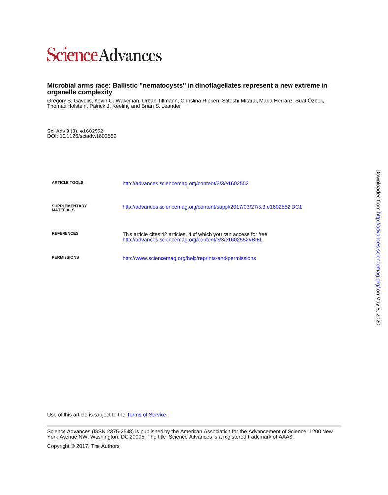
In summary, the structure and function of nematocysts inPolykrikosappear fundamentally different from—and more complex than—thosein cnidarians. Therefore, to understand how these ballistics evolved, weexamined the diversity of nematocysts in other dinoflagellates. We per-formed FIB-SEM on a second dinoflagellate, Nematodinium sp., a raregenus known fromother ultrastructural studies to have a different nem-atocyst arrangement than in polykrikoids (21, 22). Extrusome featuresinNematodinium are so divergent as to question whether dinoflagellatenematocysts are monophyletic. Nematodinium lacks taeniocysts andcoiled ballistic tubules—instead, each nematocyst consists of a ring ofparallel subcapsules reminiscent of a Gatling gun (21, 22).
We performed FIB-SEM on a single high-pressure frozen Nema-todinium cell containing eight nematocysts, of which we fully recon-structed three (Fig. 4C). Our 3D reconstructions confirmed the intricacyof nematocysts in Nematodinium sp. and revealed several novel features(Fig. 4). Although nematocysts of Nematodinium were thought to becapped by an “operculum” made of homogeneous material (21–23),we showed that this was not the case. Instead, the nematocyst is toppedby an elegant rosette-like structure,within amembrane-boundapical com-partment (Fig. 4L). Beneath this, we found structures previously imaged
Gavelis et al., Sci. Adv. 2017;3 : e1602552 31 March 2017
only by Greuet (which he did not name) (22) and noticed that they con-tained concentric rings similar to the nozzle in the nematocysts ofPolykrikos. Although these putative nozzles were too small for us tointernally reconstruct using FIB-SEM, their concentric ringswere evidentin our transmission electron microscope (TEM) sections (fig. S4C). Asin Polykrikos, each nozzle inserts into a capsule that is positioned direct-ly below it and is held in place by gasket-like rings (Fig. 4, K and L).
Themost compelling case for homology between the nematocysts ofPolykrikos andNematodinium is the identical pattern of striatedmaterialwithin their capsules (Figs. 4K and 5, D to F, and fig. S2, B and C). Thisshared trait and their development in tiered compartments (figs. S2 toS4), plus the placement of nematocyst-bearing dinoflagellates within asingle Gymnodiniales (9) clade (fig. S6), suggest that these nematocystshad a single origin within unarmored dinoflagellates. Therefore, nozzlesand stylet bases represent shared derived characters in at least the mostrecent ancestor of Polykrikos and Nematodinium.
What drives the ballistics in dinoflagellate nematocysts? In cnidar-ians, an osmotic propellant is synthesized by PgsAA, which was absentin our 130 dinoflagellate data sets. It is possible that dinoflagellates usean as-yet uncharacterized osmotic propellant. An alternate possibility is
Fig. 2. Reconstruction of the nematocysts in the dinoflagellate P. kofoidii. (A) SEM micrograph of a cell of P. kofoidii, including an armed taeniocyst (arrow) in the apicalregion that first contacts prey. (B) SEMmicrograph of an isolated taeniocyst that has discharged its amorphous contents. (C) SEMmicrograph of an isolated nematocyst that hasbecome arrested very early in discharge; arrowhead, operculum. (D) FIB-SEM section of taeniocyst and nematocyst enclosed by amembranous chute (arrowhead) and (E) maximumintensity projection of the same region seen slightly from above and below (F). (G) Virtual dissection of the nematocyst-taeniocyst complex. Brackets indicate themembrane-boundcompartments in which those components are grouped during early development. Later in development, compartments fuse to form the chute.
3 of 7
SC I ENCE ADVANCES | R E S EARCH ART I C L E
htD
ownloaded from
Fig. 3. Nematocyst discharge in Polykrikos. (A) The putative ballistic sequence of nematocyst discharge is as follows: (1) striated capsule contracts; (2) internal pressure forcesstylet to pierce capsule and open operculum (dark green); (3) part of the capsule everts; (4) stylet exits through the nozzle and detaches; and (5)mucilaginous tubule (blue) uncoilsand is projected through the stylet base (yellow). Orange, relaxed capsule wall; red, contracted capsule wall. (B to E) SEMmicrographs of isolated discharged nematocysts (B to D)and a nematocyst piercing a prey cell (E). Arrowhead, operculum; C, capsule; T, tubule.
on May 8, 2020
tp://advances.sciencemag.org/
Fig. 4. Reconstruction of the nematocysts inNematodinium sp. (A to E) Images derived from single-cell FIB-SEM. (A) Partial reconstruction of a cell with nematocysts showninbeigeandmucocysts shown inpurple. (B) Longitudinal FIB-SEMsection througha cell showing thenucleus andnematocysts (arrowheads). (C) 3Dmaximum intensity projectionshowing a battery of nematocysts. (D) 3D tilted reconstruction of a nematocyst. (E) Longitudinal reconstruction of a nematocyst. (F to K) Single-cell TEM micrographs of anematocyst in oblique section (F), cross section (G to J), and longitudinal section (K) showing the striated material (arrow) and variable symmetries across different nematocysts,with the number of subcapsules printed in the upper right corner. (L) A virtual dissection of the nematocyst components. Brackets indicate themembrane-bound compartmentsbounding the components indicated. (F) to (J) scale bar, 500 nm.
Gavelis et al., Sci. Adv. 2017;3 : e1602552 31 March 2017 4 of 7

SC I ENCE ADVANCES | R E S EARCH ART I C L E
Do
that they generate force by contracting fibers in the capsulewall to createpressure within the capsule. Our ultrastructural interpretations providesome support for this, because the striated material was found with twodifferent periodicities, which potentially represent a contracted and anuncontracted state (fig. S5, D to F). These are unlikely to be fixationartifacts because they were consistent across specimens prepared bystandard chemical fixation and freeze-substitution.
Previously, it was also unclear which part of the nematocyst, if any,in Nematodinium is the actual projectile, because these nematocystswere resistant to induced firing, unlike in Polykrikos. However, severalminutes after cell lysis, these nematocysts spontaneously ruptured andexpanded, providing insight into their ballistic mechanism. Whereasother nematocyst components remained static, we observed rapid elon-gation of the subcapsules; within 1 s, the subcapsules extended up totwice their initial lengths (fig. S5, K to N). In TEM, subcapsules appearto release an accordion-like membrane with a diffuse lattice (fig. S5I).This is similar to mucocysts—defensive organelles that share the samesize, shape, and lattice-like ultrastructure (fig. S5J)—but any homologybetween nematocysts and mucocysts remains to be demonstrated.
on May 8, 2020
http://advances.sciencemag.org/
wnloaded from
DISCUSSIONThese findings illustrate a new level of hierarchical complexity in organ-elles, given that each nematocyst is essentially a bouquet of smallerballistic organelles. The shared similarities between single projectilenematocysts and the multibarreled ones (which are variable, with 11 to15 barrels; Fig. 4, G to J) suggest how complex ballistics could have arisenfrom simpler preexisting secretory structures (fig. S7). We suspect thatderived features, such as stylets [capable of piercing armor (Fig. 3E)],injectable tubules (potentially to deliver toxins), and pressurized ballis-tics (seemingly to increase speed), may be counteradaptations to preyarmor and defensive ballistics (for example, mucocysts and trichocysts),resulting from an evolutionary arms race. We previously showed thatwild-caught cells ofNematodinium sp. had eaten other dinoflagellates—including their discharged trichocysts—having evidently overcomethese defenses (1). Illuminating the interplay between dinoflagellate pred-ators and prey will be ecologically relevant, because Polykrikosmodulatesthe populations of important planktonic organisms (24–26), includingthe armored dinoflagellate Alexandrium tamarense, which is amongthe most prevalent agents of toxic algal blooms (27, 28). We successfullycultivated P. kofoidii on A. tamarense, as well as a second armored dino-flagellate,Lingulodiniumpolyedra. In the process,we observed at least oneP. kofoidii cell ensnare its prey (Fig. 1B) despite a counterassault fromthe prey’s trichocysts (Fig. 1C).
Although our hypothesis that a cellular arms race drove the elabo-ration of extrusomes has yet to be tested, it is clear that obligate preda-tion has become a successful strategy for these dinoflagellates [that is,polykrikoids have lost photosynthesis multiple times (15)]. Despite themisconception that phytoplankton are passive cells, eukaryotic algaehave given rise to (and arose from) multiple predatory lineages and,in the process, have independently evolved sophisticated ballistic organ-elles that exceed those of animals in complexity.
MATERIALS AND METHODSProteome mining for nematogenic proteinsOur database consisted of published protein predictions from across arepresentative group of 30 eukaryotes (Fig. 1I). This included a novelproteome that we predicted from the dinoflagellate P. lebouriae (15),
Gavelis et al., Sci. Adv. 2017;3 : e1602552 31 March 2017
which bears nematocysts. Among the other representatives were publicgenomes sequenced from extrusome-bearing eukaryotes [Symbiodinium(13, 14), Paramecium, Tetrahymena, Cryptomonas,Hydra,Nematostella,and Thelohanellus], as well as two genomes from taxa with complex se-cretory structures (Toxoplasma andCryptosporidium), and two genomesfrom parasites that invade hosts via ballistic spore cells (Helicosporidiumand Encephalitozoon). To compensate for the low coverage of the onlyavailable myxozoan genome (Thelohanellus kitauei), we also used apublished proteome predicted from a transcriptome of the myxozoanTetracapsuloides bryosalmonae.
Only 4 of these 31 data sets were from dinoflagellates (few dinoflag-ellates have been sequenced owing to their massive genome sizes), so toensure that dinoflagellate proteins were thoroughly searched, we alsoqueried against public predicted proteomes from the Marine MicrobialEukaryote Transcriptome Sequencing Project (MMETSP)—to which wecontributed three dinoflagellate transcriptomes (Togula jolla, Protoceratiumreticulatum, and Polarella glacialis). In total, the MMETSP contains 120dinoflagellate transcriptomes from across 44 species and 27 ciliate tran-scriptomes from across 19 species, many of which have extrusomes (29).
CollectionCells ofNematodinium sp. andActiniscus pentasteriaswere collected offa seaplane station dock in Sidney, BritishColumbia,Canada (48.652545°N,123.447200°W) in June 2014, and cells of P. kofoidii and Gymnodiniumfasciculatum were collected off a pier in Vancouver, British Columbia,Canada (49.272704°N, 123.187827°W), in July 2015, and off the dock ofFridayHarborMarine Labs,Washington,USA (48.545755,−123.012741)in June 2016 by towing a 20-mm-mesh plankton net through the surfacewater. Contents were immediately passed through a 150-mm-meshplankton net to exclude larger organisms, leaving in a fraction that con-sisted mostly of predatory dinoflagellates. Within 4 hours of collection,cells were visually identified using an inverted light microscope and indi-vidually picked and transferred by pulled glass micropipettes into dishesof filtered seawater. Cells of Nematodinium sp. were identified by thepresence of both nematocysts and an eye-like ocelloid (Fig. 1F). CellsofP. kofoidiiwere discerned as binucleate “pseudocolonies”with four gir-dle flagella, which lacked plastids and have nematocysts (Fig. 1A). Cells ofA. pentasterias were identified by the pair of five-pointed silica starsaround their nuclei and their apical “docidosome” extrusomes (30). Cellslikely belonging toG. fasciculatumwere identified by the presence of doc-idosomes, lack of plastids, and gymnodinoid shape (round, unarmoredcells with equatorial girdle).
CulturesBehavioral observations of P. kofoidii and its prey—L. polyedra and anonlytic strain of A. tamarense—were made between 2009 and 2010 onthepolyclonal culture previously establishedbyTillmannandHoppenrath(31). Interactions between predators and prey in well culture plates orculture flasks were viewed through differential interference contrast opticsusing an Axiovert 200 M inverted light microscope (Zeiss). For higher-resolution imagery, individual cells were transferred using a micropipetteonto glass slides and imaged with Zeiss Axioskop 2. Under these con-ditions, discharge of nematocysts was video recorded using a Sony DSP3-CCD camera (Sony Deutschland).
Standard chemical fixation of single cells for TEMEach isolated cell of Nematodinium sp. and P. kofoidii was micropi-petted onto a poly-L-lysine–coated slide. Cells were fixed with 2% glu-taraldehyde in filtered seawater for 30 min on ice. After two washes in
5 of 7

SC I ENCE ADVANCES | R E S EARCH ART I C L E
on May 8, 2020
http://advances.sciencemag.org/
Dow
nloaded from
filtered seawater, cells were postfixed in 1%OsO4 for 30min. Cells weredehydrated through a graded series of ethanol (50, 70, 85, 90, 95, 100,and 100%), infiltrated with acetone-resin mixtures (acetone, 2:1, 1:1,and 1:2; Epon 812 resin), and embedded in Epon 812 resin. Polymeri-zation at 60°C produced a resin-embedded cell affixed to the glass slide.Using a power drill, resin was shaved to a 1-mm block (3), which wasremoved from the glass slide with a fine razor. The block, containing asingle cell, was super glued to a resin stub in the desired orientation forsectioning. Thin sections were produced with a diamond knife, post-stained with uranyl acetate and lead citrate, and viewed under a HitachiH7600 TEM.
High-pressure freezing and freeze substitution of singlecells for TEM and FIB-SEMUsing a micropipette, cells of Nematodinium sp. and P. kofoidii wereindividually transferred into a droplet of filtered seawater. Cells werefrozen immediately to minimize fixation artifacts, using a Leica EMHPM100 high-pressure freezer (Leica). Subsequently, freeze substitu-tion was used to remove the aqueous content of the cells and replaceit with an acetone solution containing 5% water, 1% osmium tetroxide,and 0.1% uranyl acetate, at −80°C for 48 hours, −20°C for 6 hours, thengraded back to 4°C for 13 hours. The prepared samples were washedtwice in 100% acetone. Two cells were recovered by micropipette. Eachcell was placed on a separate ThermoNox coverslip, where it adhered toa patch of poly-L-lysine. In preparation for FIB-SEM, cells were infiltratedwith a 1:1 mix of acetone and Embed 812 resin for 2 hours, then 100%resin overnight. A second ThermoNox coverslip was applied, sandwich-ing each cell in a thin layer of resin between the coverslips. Resin waspolymerized at 65°C for 24 hours. Afterward, the top coverslip was re-moved with a razorblade to expose the resin face overlying the cell.
Focused ion beam scanning electron microscopyOne cell each of Nematodinium sp. and P. kofoidii was imaged by anFEI Helios NanoLab 650 DualBeam FIB-SEM. The ion beam milledthrough the cell in 250-nm increments, yielding 169 image slices forNematodinium and 946 for Polykrikos. Images were aligned as a z stackin Amira 5.5. Features of interest, including the nozzle, stylet, and tubule,were semiautomatically segmented, that is, manually traced in approxi-mately one of every three slices, before automatic interpolation filled inthe volumes between the slices, following the manufacter’s instructions.Surfaces of these structures were generated, then smoothed and colorizedto produce 3D models of nematocysts and their components. We alsoproduced 3Dmodels without segmentation, as maximum intensity pro-jections of the image stack. Replicates of each organelle were imaged, withsix nematocysts present inNematodinium and four in Polykrikos, acrossvarious stages of development.
Confocal microscopyCells ofP. kofoidiiwere fixed in 4%paraformaldehyde in filtered seawaterfor 10 min, then rinsed three times in 0.1 M phosphate-buffered saline(PBS) solutionbefore storage inPBSwith0.05%NaN3 (sodiumazide, as apreservative) at 4°C. Fixed cells of P. kofoidii were washed from PBS:NaN3 solution with 3 × 15 min exchanges of 0.1 M PBS, followed bypermeabilization in PBT (0.1 M PBS + 0.1% Triton X-100) for 30 minat 4°C. For antibody staining, the cellswere incubated inblocking solution(PBT + 1% bovine serum albumin) at 4°C for 30 min and posteriorlyincubated at 4°C for 12 hours with a primary mouse anti-tubulin acety-lated antibody (Sigma-Aldrich) at a 1:100 concentration in blockingsolution. Primary antibody solution was then removed with multiple
Gavelis et al., Sci. Adv. 2017;3 : e1602552 31 March 2017
exchanges of PBT. Specimens were then incubated with a secondaryanti-mouse Alexa Fluor 647 antibody (Molecular Probes) at a concen-tration of 1:100 in blocking solution, at 4°C for 12 hours. Secondaryantibodies were removed with multiple exchanges of PBT. Filamen-tous actin fibers were posteriorly labeled by incubating the cells in a1:100 dilution of Alexa Fluor 488–conjugated phalloidin (MolecularProbes) in PBT for 1 hour followed by 3 × 15 min exchanges ofPBS before imaging by confocal laser-scanning microscopy. Incuba-tions were always performed in the dark while rocking at 4°C in glasswell plates.
Molecular phylogenetic analysesA single cell each of A. pentasterias and G. fasciculatum was individuallylysed in a polymerase chain reaction tube and amplified with primersdescribed in Gomez et al. (32) (Nematodinium sp. and polykrikoidswere not sequenced because populations from this area have alreadybeen barcoded). The 18S and 28S ribosomal DNA (rDNA) sequenceswere short fragments, but they were included for lack of any othersequence data from these species.
A dinoflagellate phylogeny was estimated using 18S and 28S rDNAsequences, concatenated as 2389 nucleotide alignment, across 50 repre-sentative dinoflagellate taxa, including all nematocyst-bearing dinoflag-ellates. Nucleotides were aligned with MUSCLE (33), and fast-evolvingand ambiguously aligned regions were removed using Gblocks 0.91b(34). The nucleotide substitutionmodel (GTRGAMMA)was estimatedusing the Models package in Mega 6.0.5 (35). A maximum likelihoodphylogeny was run with 500 bootstraps in RaxML (36). A second,Bayesian analysis was run for 10,000 generations in MrBayes 3.2 (37),using the high-heating setting of (Nchains = 3).
SUPPLEMENTARY MATERIALSSupplementary material for this article is available at http://advances.sciencemag.org/cgi/content/full/3/3/e1602552/DC1fig. S1. A synthesis of fundamental differences between the nematocysts in cnidarians anddinoflagellates.fig. S2. Ultrastructure and discharge of nematocysts in P. kofoidii.fig. S3. Nematocyst development in P. kofoidii.fig. S4. Nematocyst development in Nematodinium sp.fig. S5. Contractile and projectile traits in the nematocysts of Nematodinium sp.fig. S6. Molecular phylogeny of dinoflagellates with complex extrusomes.fig. S7. Model of nematocyst homology and evolution in dinoflagellates.fig. S8. Cytoskeletal associations with the nematocyst-taeniocyst complex in P. kofoidii.movie S1. FIB-SEM reconstruction of the nematocyst-taeniocyst complex in P. kofoidii.movie S2. Discharge of nematocyst isolated from P. kofoidii.movie S3. Discharge of nematocyst isolated from P. kofoidii.movie S4. P. kofoidii hunting L. polyedra.movie S5. Discharge of a taeniocyst isolated from P. kofoidii.References (40–44)
REFERENCES AND NOTES1. K. Hausmann, Extrusive organelles in protists. Int. Rev. Cytol. 52, 197–276 (1978).2. C. Schmoker, S. Hernández-León, A. Calbet, Microzooplankton grazing in the
oceans: Impacts, data variability, knowledge gaps and future directions. J. Plankton Res.35, 691–706 (2013).
3. K. Matsuoka, H.-J. Cho, D. M. Jacobson, Observations of the feeding behavior andgrowth rates of the heterotrophic dinoflagellate Polykrikos kofoidii (Polykrikaceae,Dinophyceae). Phys. Chem. Chem. Phys. 39, 82–86 (2000).
4. M. J. Lee, H. J. Jeong, K. H. Lee, S. H. Jang, J. H. Kim, K. Y. Kim, Mixotrophy in thenematocyst–taeniocyst complex-bearing phototrophic dinoflagellate Polykrikoshartmannii. Harmful Algae 49, 124–134 (2015).
5. J. A. Westfall, P. C. Bradbury, The fine structure of the nematocyst taeniocyst complex inpolykrikos-kofoidi. J. Protozool. 29, 474–475 (1982).
6 of 7

SC I ENCE ADVANCES | R E S EARCH ART I C L E
on May 8, 2020
http://advances.sciencemag.org/
Dow
nloaded from
6. M. Hoppenrath, N. Yubuki, T. R. Bachvaroff, B. S. Leander, Re-classification ofPheopolykrikos hartmannii as Polykrikos (Dinophyceae) based partly on the ultrastructureof complex extrusomes. Eur. J. Protistol. 46, 29–37 (2010).
7. S. Öezbek, The cnidarian nematocyst: A miniature extracellular matrix within a secretoryvesicle. Protoplasma 248, 635–640 (2011).
8. C. N. David, S. Özbek, P. Adamczyk, S. Meier, B. Pauly, J. Chapman, J. S. Hwang, T. Gojobori,T. W. Holstein, Evolution of complex structures: Minicollagens shape the cnidariannematocyst. Trends Genet. 24, 431–438 (2008).
9. M. Hoppenrath, T. R. Bachvaroff, S. M. Handy, C. F. Delwiche, B. S. Leander, Molecularphylogeny of ocelloid-bearing dinoflagellates (Warnowiaceae) as inferred from SSU andLSU rDNA sequences. BMC Evol. Biol. 9, 116 (2009).
10. J. S. Hwang, S. Nagai, S. Hayakawa, Y. Takaku, T. Gojobori, The search for the origin ofcnidarian nematocysts in dinoflagellates, in Evolutionary Biology from Concept toApplication, P. Pontarotti, Ed. (Springer, 2008), pp. 135–152.
11. S. Shostak, V. Kolluri, Symbiogenetic origins of cnidarian cnidocysts. Symbiosis 19, 1–29 (1995).12. P. G. Balasubramanian, A. Beckmann, U. Warnken, M. Schnölzer, A. Schüler,
E. Bornberg-Bauer, T. W. Holstein, S. Özbek, Proteome of Hydra nematocyst. J. Biol. Chem.287, 9672–9681 (2012).
13. S. Lin, S. Cheng, B. Song, X. Zhong, X. Lin, W. Li, L. Li, Y. Zhang, H. Zhang, Z. Ji, M. Cai,Y. Zhuang, X. Shi, L. Lin, L. Wang, Z. Wang, X. Liu, S. Yu, P. Zeng, H. Hao, Q. Zou, C. Chen,Y. Li, Y. Wang, C. Xu, S. Meng, X. Xu, J. Wang, H. Yang, D. A. Campbell, N. R. Sturm,S. Dagenais-Bellefeuille, D. Morse, The Symbiodinium kawagutii genome illuminatesdinoflagellate gene expression and coral symbiosis. Science 350, 691–694 (2015).
14. E. Shoguchi, C. Shinzato, T. Kawashima, F. Gyoja, S. Mungpakdee, R. Koyanagi, T. Takeuchi,K. Hisata, M. Tanaka, M. Fujiwara, M. Hamada, A. Seidi, M. Fujie, T. Usami, H. Goto,S. Yamasaki, N. Arakaki, Y. Suzuki, S. Sugano, A. Toyoda, Y. Kuroki, A. Fujiyama,M. Medina, M. A. Coffroth, D. Bhattacharya, N. Satoh, Draft assembly of the Symbiodiniumminutum nuclear genome reveals dinoflagellate gene structure. Curr. Biol. 23,1399–1408 (2013).
15. G. S. Gavelis, R. A. White III, C. A. Suttle, P. J. Keeling, B. S. Leander, Single-celltranscriptomics using spliced leader PCR: Evidence for multiple losses of photosynthesisin polykrikoid dinoflagellates. BMC Genomics 16, 528 (2015).
16. J. Weber, Nematocysts (stinging capsules of Cnidaria) as Donnan-potential-dominatedosmotic systems. Eur. J. Biochem. 184, 465–476 (1989).
17. E. Denker, E. Bapteste, H. Le Guyader, M. Manuel, N. Rabet, Horizontal gene transfer andthe evolution of cnidarian stinging cells. Curr. Biol. 18, R858–R859 (2008).
18. P. Adamczyk, S. Meier, T. Gross, B. Hobmayer, S. Grzesiek, H. P. Bächinger, T. W. Holstein,S. Özbek, Minicollagen-15, a novel minicollagen isolated from Hydra, forms tubulestructures in nematocysts. J. Mol. Biol. 376, 1008–1020 (2008).
19. A. W. Koch, T. W. Holstein, C. Mala, E. Kurz, J. Engel, C. N. David, Spinalin, a new glycine-and histidine-rich protein in spines of Hydra nematocysts. J. Cell Sci. 111, 1545–1554(1998).
20. A. Beckmann, S. Xiao, J. P. Müller, D. Mercadante, T. Nüchter, N. Kröger, F. Langhojer,W. Petrich, T. W. Holstein, M. Benoit, F. Gräter, S. Özbek, A fast recoiling silk-like elastomerfacilitates nanosecond nematocyst discharge. BMC Biol. 13, 3 (2015).
21. L. Mornin, D. Francis, Fine structure of Nematodinium armatum a naked dinoflagellate.J. Microsc. 6, 759 (1967).
22. C. Greuet, Étude ultrastructurale et évolution des cnidocystes de Nematodinium,Péridinien Warnowiidae Lindemann. Proc. Natl. Acad. Sci. U.S.A. 7, 345–355 (1971).
23. C. Greuet, R. Hovasse, About genesis of nematocysts of Polykrikos-schwartzi-Butschli.Proc. Natl. Acad. Sci. U.S.A. 13, 145–149 (1977).
24. U. Tillmann, Interactions between planktonic microalgae and protozoan grazers.J. Eukaryot. Microbiol. 51, 156–168 (2004).
25. Y. Matsuyama, M. Miyamoto, Y. Kotani, Grazing impacts of the heterotrophicdinoflagellate Polykrikos kofoidii on a bloom of Gymnodinium catenatum. Aquat. Microb.Ecol. 17, 91–98 (1999).
26. H. J. Jeong, K. H. Park, J. S. Kim, H. Kang, C. H. Kim, H.-J. Choi, Y. S. Kim, J. Y. Park,M. G. Park, Reduction in the toxicity of the dinoflagellate Gymnodinium catenatum whenfed on by the heterotrophic dinoflagellate Polykrikos kofoidii. Aquat. Microb. Ecol. 31,307–312 (2003).
27. U. John, U. Tillmann, J. Hülskötter, T. J. Alpermann, S. Wohlrab, D. B. Van de Waal,Intraspecific facilitation by allelochemical mediated grazing protection within a toxigenicdinoflagellate population. Proc. Biol. Sci. 282, 20141268 (2015).
28. S. Wohlrab, U. Tillmann, A. Cembella, U. John, Trait changes induced by speciesinteractions in two phenotypically distinct strains of a marine dinoflagellate. ISME J. 10,2658–2668 (2016).
29. X. Chen, X. Zhao, X. Liu, A. Warren, F. Zhao, M. Miao, Phylogenomics of non-model ciliatesbased on transcriptomic analyses. Protein Cell 6, 373–385 (2015).
30. G. Hansen, Light and electron microscopical observations of the dinoflagellate Actiniscuspentasterias (Dinophyceae). J. Phycol. 29, 486–499 (1993).
Gavelis et al., Sci. Adv. 2017;3 : e1602552 31 March 2017
31. U. Tillmann, M. Hoppenrath, Life cycle of the pseudocolonial dinoflagellate Polykrikoskofoidii (Gymnodiniales, Dinoflagellata). J. Phycol. 49, 298–317 (2013).
32. F. Gómez, D. Moreira, P. López-García, Molecular phylogeny of noctilucoid dinoflagellates(Noctilucales, Dinophyceae). Protist 161, 466–478 (2010).
33. R. C. Edgar, MUSCLE: A multiple sequence alignment method with reduced time andspace complexity. BMC Bioinf. 5, 113 (2004).
34. J. Castresana, Selection of conserved blocks from multiple alignments for their use inphylogenetic analysis. Mol. Biol. Evol. 17, 540–552 (2000).
35. K. Tamura, G. Stecher, D. Peterson, A. Filipski, S. Kumar, MEGA6: Molecular evolutionarygenetics analysis version 6.0. Mol. Biol. Evol. 30, 2725–2729 (2013).
36. A. Stamatakis, RAxML-VI-HPC: Maximum likelihood-based phylogenetic analyses withthousands of taxa and mixed models. Bioinformatics 22, 2688–2690 (2006).
37. F. Ronquist, J. P. Huelsenbeck, MrBayes 3: Bayesian phylogenetic inference under mixedmodels. Bioinformatics 19, 1572–1574 (2003).
38. F. Burki, Y. Inagaki, J. Bråte, J. M. Archibald, P. J. Keeling, T. Cavalier-Smith, M. Sakaguchi,T. Hashimoto, A. Horak, S. Kumar, D. Klaveness, K. S. Jakobsen, J. Pawlowski,K. Shalchian-Tabrizi, Large-scale phylogenomic analyses reveal that two enigmaticprotist lineages, Telonemia and Centroheliozoa, are related to photosyntheticchromalveolates. Genome Biol. Evol. 1, 231–238 (2009).
39. J. Janouskovec, D. V. Tikhonenkov, F. Burki, A. T. Howe, M. Kolísko, A. P. Mylnikov,P. J. Keeling, Factors mediating plastid dependency and the origins of parasitism inapicomplexans and their close relatives. Proc. Natl. Acad. Sci. U.S.A. 112, 10200–10207 (2015).
40. T. Holstein, The morphogenesis of nematocytes in Hydra and Forskalia: An ultrastructural-study.J. Ultrastruct. Res. 75, 276–290 (1981).
41. J. Lom, P. Depuytor, Observations sur l’ ultrastructure des trophozoites de myxosporidies.C. R. Hebd. Seances Acad. Sci. 260, 2588–2598 (1965).
42. M. Vesk, I. A. N. Lucas, The rhabdosome: A new type of organelle in the dinoflagellateDinophysis. Protoplasma 134, 62–64 (1986).
43. N. S. Kang, H. J. Jeong, Ø. Moestrup, W. SHIN, S. W. NAM, J. Y. Park, M. F. De Salas,K. W. Kim, J. H. Noh, Description of a new planktonic mixotrophic dinoflagellateParagymnodinium shiwhaense n. gen., n. sp from the coastal waters off western Korea:Morphology, pigments, and ribosomal DNA gene sequence. J. Eukaryotic Microbiol. 57,121–144 (2010).
44. N. S. Kang, H. J. Jeong, Ø. Moestrup, T. G. Park, Gyrodiniellum shiwhaense n. gen., n. sp., anew planktonic heterotrophic dinoflagellate from the coastal waters of western Korea:Morphology and ribosomal DNA gene sequence. J. Eukaryotic Microbiol. 58, 284–309(2011).
Acknowledgments: This work was made possible by the University of BritishColumbia technicians, G. Martens, D. Horne, and B. Ross, who carried out high-pressurefreezing of the specimens and G. Owen who operated the FIB-SEM imaging of Nematodinium.G.S.G would like to thank E. Gavelis for carrying out plankton tows as well as thefaculty and staff at Friday Harbor Laboratories for accommodating us while we collected cellsfrom the plankton. Funding: S.Ö. was funded by the DFG (Oe 416/4-1). Author contributions:G.S.G., P.J.K., and B.S.L. conceived the project and wrote the paper. G.S.G. performedphylogenomic analysis of nematogenic proteins, collected uncultivated dinoflagellates,chemically fixed and imaged cells under TEM, reconstructed FIB-SEM data in 3D, producedillustrations for the paper, and induced nematocyst firing in Polykrikos and Nematodinium,which was imaged through SEM and light microscopy. U.T. cultured Polykrikos along with prey,which he recorded through light microscopic images, film, and SEM. K.C.W. and C.R.imaged the Polykrikos through FIB-SEM, which was funded by S.M. T.H. and S.Ö. provideda proteomic data set, which their laboratory groups obtained from Hydra. All authorsparticipated in the drafting process. Competing interests: The authors declare that they haveno competing interests. Data and materials availability: Proteomic data from Hydra isavailable upon request from T.H. and S.U., and transcriptomic data from Polykrikos lebouriaeis available upon request from G.S.G. Other data sets used in phylogenomic analysis werefrom public genomes, and transcriptomes were from the Marine Microbial EukaryoteTranscriptome Sequencing project, which can be freely downloaded from iMicrobe interactivedata commons. All other data needed to evaluate the conclusions in the paper are presentin the paper and/or the Supplementary Materials. Additional data related to this paper may berequested from the authors.
Submitted 17 October 2016Accepted 10 February 2017Published 31 March 201710.1126/sciadv.1602552
Citation: G. S. Gavelis, K. C. Wakeman, U. Tillmann, C. Ripken, S. Mitarai, M. Herranz, S. Özbek,T. Holstein, P. J. Keeling, B. S. Leander, Microbial arms race: Ballistic “nematocysts” indinoflagellates represent a new extreme in organelle complexity. Sci. Adv. 3, e1602552 (2017).
7 of 7
organelle complexityMicrobial arms race: Ballistic ''nematocysts'' in dinoflagellates represent a new extreme in
Thomas Holstein, Patrick J. Keeling and Brian S. LeanderGregory S. Gavelis, Kevin C. Wakeman, Urban Tillmann, Christina Ripken, Satoshi Mitarai, Maria Herranz, Suat Özbek,
DOI: 10.1126/sciadv.1602552 (3), e1602552.3Sci Adv
ARTICLE TOOLS http://advances.sciencemag.org/content/3/3/e1602552
MATERIALSSUPPLEMENTARY http://advances.sciencemag.org/content/suppl/2017/03/27/3.3.e1602552.DC1
REFERENCES
http://advances.sciencemag.org/content/3/3/e1602552#BIBLThis article cites 42 articles, 4 of which you can access for free
PERMISSIONS http://www.sciencemag.org/help/reprints-and-permissions
Terms of ServiceUse of this article is subject to the
is a registered trademark of AAAS.Science AdvancesYork Avenue NW, Washington, DC 20005. The title (ISSN 2375-2548) is published by the American Association for the Advancement of Science, 1200 NewScience Advances
Copyright © 2017, The Authors
on May 8, 2020
http://advances.sciencemag.org/
Dow
nloaded from