microbiologic analysis of peri-pancreatic fluid collected
TRANSCRIPT

ORIGINAL ARTICLE: Clinical Endoscopy
Abbrcolle
DISCto th
*Drs
Copy0016http:
Rece
www
Microbiologic analysis of peri-pancreatic fluid collected during EUSin patients with pancreatitis: impact on antibiotic therapy
eviatioction.
LOSURis publi
Negm
right ª-5107/$//dx.do
ived De
.giejo
Ahmed A. Negm, MD,*,1 Hendrik Poos, MD,*,1 Elmar Kruck, MD,2 Ralf-Peter Vonberg, MD,3
Dirk Domagk, MD,4 Ahmed Madisch, MD,2 Torsten Voigtländer, MD,1 Michael P. Manns, MD,1
Jochen Wedemeyer, MD,1,5 Tim O. Lankisch, MD1
Hannover, Germany
Background: Pancreatitis is a potentially life-threatening condition frequently accompanied by peri-pancreaticfluid collections (PPFC), such as pseudocysts or pancreatic necrosis. Aspiration of PPFCs during EUS interventionsfor microbiologic analysis is still rarely performed in clinical routine.
Objective: To evaluate the role of routine microbiologic analysis of PPFCs and its impact on antibiotic therapy inpatients with pancreatitis.
Design: Prospective, observational, multicenter study.
Setting: Four treatment centers.
Patients: A total of 44 consecutive patients who presented for endoscopic treatment of PPFCs were included.
Intervention: Concomitantly, PPFC during intervention and concomitant blood cultures were obtained.
Main Outcome Measurements: Microbiologic examination of PPFCs and blood samples.
Results: Colonization of PPFCs was found in 59% of PPFC cultures, whereas all but 2 concomitant blood culturesshowed no microbial growth. Risk factors for a colonization were the presence of necrosis (P Z .006), acute pan-creatitis (PZ .033), leukocytosis (PZ .001), elevated C-reactive protein levels (PZ .003), fever (PZ .02), turbidmaterial (P Z .031), and longer hospital stay (P Z .003). In 23 patients with fluid colonization despite empiricantibiotic therapy, the treatment had to be adjusted in 18 patients (78%) according to the observed antibioticsusceptibility profile.
Limitations: Contamination cannot be totally excluded.
Conclusion: The microbiologic colonization of PPFCs in patients with pancreatitis is common. Only the directmicrobiologic analysis of PPFCs, but not of blood cultures, is useful to optimize an effective antibiotic therapyin patients with pancreatitis. (Gastrointest Endosc 2013;78:303-11.)
Mortality dramatically increases if peri-pancreatic fluid investigation has shown a clear benefit up to now.4,5 Fur-
collections (PPFC), such as pseudocysts or ne-crosis, become infected.1,2 The secondary infection ofPPFC remains the leading cause of mortality in patientswith pancreatitis.3 Prophylactic antibiotic therapy appearsreasonable but remains controversial, and no largerns: EUS-FNA, EUS-guided FNA; PPFC, peri-pancreatic fluid
E: All authors disclosed no financial relationships relevantcation.
and Poos contributed equally to this article.
2013 by the American Society for Gastrointestinal Endoscopy36.00i.org/10.1016/j.gie.2013.03.001
cember 20, 2012. Accepted March 1, 2013.
urnal.org
thermore, the differentiation between sterile and infectedPPFCs in pancreatitis according to the clinical appearanceand laboratory parameters remains difficult because bothmay present with fever, leukocytosis, and severe abdomi-nal pain.6 If an infection is suspected, empiric antibiotic
Current affiliations: Department of Gastroenterology, Hepatology andEndocrinology, Medical School Hannover, Hannover (1), Department ofGastroenterology, KRH Klinikum Siloah, Hannover (2), Institute forMedical Microbiology and Hospital Epidemiology, Medical SchoolHannover, Hannover (3), Department of Medicine B, University ofMuenster, Muenster (4), Department of Medicine 1, Klinikum RegionHannover, KRH Klinikum Robert Koch Gehrden, Gehrden, Germany (5).
Reprint requests: Tim O. Lankisch, MD, Department of Gastroenterology,Hepatology and Endocrinology, Hannover Medical School, Carl-Neuberg-Str. 1, 30625 Hannover, Germany.
Volume 78, No. 2 : 2013 GASTROINTESTINAL ENDOSCOPY 303

Microbiologic analysis of peri-pancreatic fluid Negm et al
therapy with a broad-spectrum antibiotic like carbapenemshas been suggested.6,7 However, empiric non-targeted–based therapy carries the risk of selecting antibiotic-resistant strains and treatment failure. Samples from PPFCsfor microbiologic analysis may be obtained percutaneously,during surgery, or by EUS-guided FNA (EUS-FNA).
EUS is now a standard technique for the diagnosis andmanagement of PPFCs, including the chance to obtainsamples for microbiologic analysis and transgastric ortransduodenal drainage.8 Although guidelines recommendmicrobiologic analysis of PPFCs, the importance ofobtaining aspirates for microbiologic analysis during EUSin clinical routine has never been validated.
Therefore, our aim was to evaluate the value of themicrobiologic analysis of aspirates from PPFCs in compari-son with concomitant blood cultures and its impact on theantibiotic management in a multicenter study.
PATIENTS AND METHODS
PatientsThis prospective study was conducted between April
2010 and October 2011 at the endoscopic unit of 2 univer-sity hospitals and 2 municipal hospitals. We included all pa-tients with PPFCs who presented for endoscopicintervention. Informed written consent was obtainedfrom all patients, and the trial was approved by the localethics committee. The exclusion criteria were age under18 years or absence of the written informed consent beforeintervention. Acute pancreatitis was defined as the pres-ence of at least 2 of 3 of the following features: abdominalpain, serum lipase activity (3 times the upper limit of nor-mal), and characteristic findings of contrast-enhanced CT.6
Chronic pancreatitis was classified as the presence ofpancreatic calcifications, dilatation of pancreatic ducts,and chronic abdominal pain.
Endoscopic procedureIndications for endoscopic intervention were either
drainage of large or symptomatic pseudocysts or diagnosticpuncture of symptomatic PPFCs (abdominal pain and/orfever). All EUS procedures were performed by using Olym-pus endoscopes (Olympus, Hamburg, Germany), whichwere disinfected according to the guidelines of the RobertKoch Institute, and contamination was excluded by a regularsmear test.9 Peri-pancreatic fluid collections were aspiratedthrough a puncture set or the biopsy needle. Approximately0.5 to 10 mL of fluid (mean 2 mL) was collected and trans-ferred into a sterile tube. Concomitantly, blood cultureswere obtained directly or within 12 hours after intervention.At least 1 single dose of antibiotic was documented as an an-tibiotic treatment before intervention.
Microbiologic analysisAspirate samples were cultured under aerobic condi-
tions on 5% Columbia sheep blood agar (Becton Dickinson
304 GASTROINTESTINAL ENDOSCOPY Volume 78, No. 2 : 2013
Take-home message
� Aspiration of peri-pancreatic fluid for microbiologicanalysis during endoscopic intervention is a valuablediagnostic tool because it might lead to more adequatetherapy and might help to establish a local antibioticguideline for the management of peri-pancreatic fluidcollection in patients with pancreatitis.
GmbH, Heidelberg, Germany), MacConkey agar (OxoidGmbH, Wesel, Germany), and yeast extract agar for 48hours, with the first reading after 24 hours. Anaerobicgrowth was observed by the use of Schaedler agar (BectonDickinson GmbH) for up to 96 hours. Incubation of bloodcultures (BD BACTEC standard aerobic and anaerobicmedia; Becton Dickinson GmbH, Heidelberg, Germany)was terminated after 7 days if no microbial growth hadbe registered. Species differentiation was then performedaccording to German laboratory practice guideline DINEN ISO 15189. Species identification and antibiotic suscep-tibility testing were performed by using the VITEK-2-XL(bioMérieux, Nuertingen, Germany) system and MerlinMICRONAUT Sprint Dispenser automated broth micro-titer system (Genzyme Viro-Tec, Russelsheim, Germany).Micro-titer plates of 384 wells (No. EG-009) were used asrecommended by the German Network for the Antimicro-bial Resistance Surveillance.10 Microorganisms present inconcentrations O10,000/mL were considered as infection;lower concentrations were judged as contamination only.
Management of data and statistical analysisData collection and storage were performed by using
a specially designed data bank (Microsoft Access 2003,Unterschleißheim, Germany). Data were expressed asnumber/percent or mean� standard deviation. All collectedparameters of patients with positive aspirate cultures werecompared with those of sterile culture results. Noncontinu-ous parameters were analyzed by c2 test or Fisher exact testas appropriate, and continuous parameters were analyzedby using the Mann-Whitney U test. P values! .05 were con-sidered statistically significant. Parameters with significantstatistical difference as well as parameters with differences! .100 were further included in a multivariate analysis(logistic regression by using stepwise backward elimination)to detect independent risk factors. All calculations weredone by using the SPSS Statistical Package (version 19.0,SPSS Inc, Chicago, Ill). All authors had access to the studydata and had reviewed and approved the final manuscript.
RESULTS
Patients and clinical characteristicsDuring the study period, 44 consecutive patients were
prospectively recruited from 2 university hospitals and 2peripheral hospitals. Reasons for PPFCs were mainly acute
www.giejournal.org

TABLE 1. Demographic, clinical, and laboratory testcharacteristics of study patients (n [ 51)
Parameter
Patients (n [ 44)
No. ormean ± SD
% orrange
Demographic data
Male 30/44 68%
Female 14/44 32%
Age, y 52 � 13.7 32-90
Clinical presentation
Hospital stay beforeintervention, d
35 � 35 2-154
Patients in ICU 10/44 23%
Fever 19/44 43%
Abdominal pain 31/44 70%
Background of peri-pancreaticfluid collection
Acute pancreatitis 32/44 73%
Chronic pancreatitis 12/44 27%
Cause of pancreatitis(n Z 44)
Biliary 15/44 34%
Alcoholic 7/44 16%
Post ERCP 4/44 9%
Postoperative 3/44 7%
Other* 4/44 9%
Unknown 11/44 25%
Endosonographiccharacteristic
Pure PPFC withoutnecrosis
22/44 50%
PPFC with necrosis 22/44 50%
Size, cm 7.7 � 3.5 2-17
Turbid aspirate 32/44 73%
First intervention 24/44 55%
EUS-guided drainage 27/44 61%
Diagnostic puncture 17/44 39%
Laboratory parametersbefore intervention
Leukocyte count(normal 4.4-11.3 � 103/mL)
12.9 � 8.5 3.4-43.4
CRP (up to 8 mg/L) 151 � 117 1-375
TABLE 1. Continued
Parameter
Patients (n [ 44)
No. ormean ± SD
% orrange
Amylase (up to 100 U/L) 126 � 179 12-594
Lipase (13-60 U/L) 180 � 312 5-1612
SD, Standard deviation; ICU, intensive care unit; PPFC,peri-pancreatic fluid collection; CRP, C-reactive protein.*Hyperlipidemia (n Z 1), autoimmune pancreatitis (n Z 1),traumatic (n Z 1), and drug related (n Z 1).
www.giejournal.org
Negm et al Microbiologic analysis of peri-pancreatic fluid
pancreatitis (73%) and chronic pancreatitis (37%). Pancre-atitis was mainly related to biliary obstruction (34%), alco-hol abuse (16%), or after ERCP (9%). In 25% of patients,the cause of pancreatitis remained unknown. Detailed clin-ical and laboratory characteristics are given in Table 1. Mainindications for intervention of PPFCs were pain (n Z 31;70%) and/or fever (n Z 19; 43%). Criteria of transgastric ortransduodenal drainage of the pseudocyst were dependentmainly on size (mean cyst size of 7.7 cm) and on theclinical symptoms of patients with abdominal pain and/orfever. Another factor that supported our decision was theEUS appearance of the fluid collection (turbid fluid on EUSin 73% of cases). Clear aspirates were found in 12 of 44PPFCs. Fifty percent of patients presented with PPFCswithout signs of necrosis.
The patients were hospitalized for an average of 1 monthbefore intervention; from these patients, 23% were treatedin the intensive care unit. The mean delay of onset ofsymptoms for acute pancreatitis was 25 days (5-91 days).The endosonographic puncture of PPFCs was performedon therapeutic interventions (eg, transgastric drainage) in27 cases (61%) or for diagnostic reasons in 17 cases(39%). Twenty patients (45%) had already received at least1 EUS intervention at the pancreas before the indexintervention; in 6 patients, transgastric drainage wasplaced before the index intervention. Three patientsdeveloped fever after EUS intervention despite receivingantibiotic prophylaxis, whereas only 1 patient without pre-interventional antibiotics developed postprocedural fever.Otherwise, no adverse events were observed during or af-ter EUS intervention, especially no signs of bleeding orperforation.
General microbiological characteristicsAspiration of peri-pancreatic fluid collections was
successful in all examinations. Twenty-six of the aspiratescultures (59%) showed microbial growth. Only 2 concom-itant blood cultures (13%) of those positive aspirate cul-tures showed microbial growth of the same organisms asfound in the aspirate culture. On the other hand, wheneveraspirate culture showed microorganisms present in con-centrations !10,000/mL (in 18 aspirate cultures), the
Volume 78, No. 2 : 2013 GASTROINTESTINAL ENDOSCOPY 305

Figure 1. Results of microbiologic analysis of peri-pancreatic fluid collection aspirates versus blood cultures and its effect on modification of antibiotictreatment. *Same organisms found in aspirate and blood culture. **Patients continued antibiotics despite negative growth because of high fever (n Z 2)or C-reactive protein levels (n Z 6).
Microbiologic analysis of peri-pancreatic fluid Negm et al
concomitant blood culture also remained sterile (Fig. 1).Polymicrobial growth (Table 2) was more common (n Z17/26; 65%) in comparison to monomicrobial cultures(n Z 9/26; 35%). Gram-positive bacteria were more preva-lent than gram-negative bacteria and Candida species(61%, 18%, and 20%, respectively). Only 1 bacterial species(2%) showed multiple-drug resistance to antibiotics (meth-icillin-resistant Staphylococcus aureus). A total of 51 organ-isms were isolated, comprising 17 different species. Themost frequently encountered organisms were Enterococcusspecies (37%), Candida species (20%), and Streptococcusspecies (16%).
Risk factors for colonization of PPFCA comparison between the group of patients in whom the
aspirate cultures showed a positive growth (nZ 26) and the
306 GASTROINTESTINAL ENDOSCOPY Volume 78, No. 2 : 2013
group with sterile aspirate cultures (nZ 18) was performedin all collected preinterventional parameters. The univariateanalysis revealed that fever before intervention, acutepancreatitis, the presence of pancreatic necrosis, turbidmaterial aspirated during intervention, and elevatedleukocyte or C-reactive protein levels are risk factors forbacterial growth in peri-pancreatic fluid (Table 3). Incontrast, patient sex, age, intensive care unit admission,the cause of pancreatitis, the presence of echogenicmaterial on EUS, previous interventions before the indexintervention, or stenting before the index interventionwere not significantly associated with microbial growthof PPFCs later on. Even when we stratified the patientsaccording to previous interventions in relation to thepresence of microbial growth there were no significantdifferences (P Z .946).
www.giejournal.org
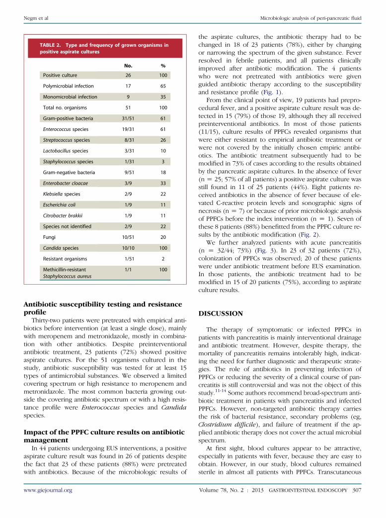
TABLE 2. Type and frequency of grown organisms inpositive aspirate cultures
No. %
Positive culture 26 100
Polymicrobial infection 17 65
Monomicrobial infection 9 35
Total no. organisms 51 100
Gram-positive bacteria 31/51 61
Enterococcus species 19/31 61
Streptococcus species 8/31 26
Lactobacillus species 3/31 10
Staphylococcus species 1/31 3
Gram-negative bacteria 9/51 18
Enterobacter cloacae 3/9 33
Klebsiella species 2/9 22
Escherichia coli 1/9 11
Citrobacter brakkii 1/9 11
Species not identified 2/9 22
Fungi 10/51 20
Candida species 10/10 100
Resistant organisms 1/51 2
Methicillin-resistantStaphylococcus aureus
1/1 100
Negm et al Microbiologic analysis of peri-pancreatic fluid
Antibiotic susceptibility testing and resistanceprofile
Thirty-two patients were pretreated with empirical anti-biotics before intervention (at least a single dose), mainlywith meropenem and metronidazole, mostly in combina-tion with other antibiotics. Despite preinterventionalantibiotic treatment, 23 patients (72%) showed positiveaspirate cultures. For the 51 organisms cultured in thestudy, antibiotic susceptibility was tested for at least 15types of antimicrobial substances. We observed a limitedcovering spectrum or high resistance to meropenem andmetronidazole. The most common bacteria growing out-side the covering antibiotic spectrum or with a high resis-tance profile were Enterococcus species and Candidaspecies.
Impact of the PPFC culture results on antibioticmanagement
In 44 patients undergoing EUS interventions, a positiveaspirate culture result was found in 26 of patients despitethe fact that 23 of these patients (88%) were pretreatedwith antibiotics. Because of the microbiologic results of
www.giejournal.org
the aspirate cultures, the antibiotic therapy had to bechanged in 18 of 23 patients (78%), either by changingor narrowing the spectrum of the given substance. Feverresolved in febrile patients, and all patients clinicallyimproved after antibiotic modification. The 4 patientswho were not pretreated with antibiotics were givenguided antibiotic therapy according to the susceptibilityand resistance profile (Fig. 1).
From the clinical point of view, 19 patients had prepro-cedural fever, and a positive aspirate culture result was de-tected in 15 (79%) of those 19, although they all receivedpreinterventional antibiotics. In most of those patients(11/15), culture results of PPFCs revealed organisms thatwere either resistant to empirical antibiotic treatment orwere not covered by the initially chosen empiric antibi-otics. The antibiotic treatment subsequently had to bemodified in 73% of cases according to the results obtainedby the pancreatic aspirate cultures. In the absence of fever(n Z 25; 57% of all patients) a positive aspirate culture wasstill found in 11 of 25 patients (44%). Eight patients re-ceived antibiotics in the absence of fever because of ele-vated C-reactive protein levels and sonographic signs ofnecrosis (n Z 7) or because of prior microbiologic analysisof PPFCs before the index intervention (n Z 1). Seven ofthese 8 patients (88%) benefitted from the PPFC culture re-sults by the antibiotic modification (Fig. 2).
We further analyzed patients with acute pancreatitis(n Z 32/44; 73%) (Fig. 3). In 23 of 32 patients (72%),colonization of PPFCs was observed; 20 of these patientswere under antibiotic treatment before EUS examination.In those patients, the antibiotic treatment had to bemodified in 15 of 20 patients (75%), according to aspirateculture results.
DISCUSSION
The therapy of symptomatic or infected PPFCs inpatients with pancreatitis is mainly interventional drainageand antibiotic treatment. However, despite therapy, themortality of pancreatitis remains intolerably high, indicat-ing the need for further diagnostic and therapeutic strate-gies. The role of antibiotics in preventing infection ofPPFCs or reducing the severity of a clinical course of pan-creatitis is still controversial and was not the object of thisstudy.11-14 Some authors recommend broad-spectrum anti-biotic treatment in patients with pancreatitis and infectedPPFCs. However, non-targeted antibiotic therapy carriesthe risk of bacterial resistance, secondary problems (eg,Clostridium difficile), and failure of treatment if the ap-plied antibiotic therapy does not cover the actual microbialspectrum.
At first sight, blood cultures appear to be attractive,especially in patients with fever, because they are easy toobtain. However, in our study, blood cultures remainedsterile in almost all patients with PPFCs. Transcutaneous
Volume 78, No. 2 : 2013 GASTROINTESTINAL ENDOSCOPY 307

TABLE 3. Risk factors of positive culture growth in peri-pancreatic aspirates
Parameter
No growth (n [ 18) Growth (n [ 26)
P valueNo. or mean ± SD % or range No. or mean ± SD % or range
Male 11 61% 19 73% .402
Female 7 39% 7 27%
Age, y 56 � 14.6 40-90 54 � 13.3 32-83 .756
Hospital stay, d 21.5 � 29.9 2-127 44.4 � 34.9 7-154 .003
ICU admission 3 17% 7 27% .425
Acute pancreatitis 10 56% 22 85% .033
First intervention 10 56% 14 54% .911
Previous intervention 8 44% 12 46% .946
Stent before intervention 1 6% 5 19 .194
Fever 4 22% 15 58% .020
CRP (mg/L) 93.4 � 106 (normal,!8) 1-311 189.3 � 109 2-375 .003
Leukocyte count (� 103/mL) 8.9 � 4.1 (normal, 4.4-11.3) 3.4-20.2 15.6 � 9.7 4.9-43.4 .001
Lipase (U/L) 192.7 � 277 (normal, 60) 5-895 172.3 � 338 8-1612 .611
Turbid aspirate 9 50% 23 88% .031
PPFC with necrosis 6 33% 16 62% .066
SD, Standard deviation; ICU, intensive care unit; CRP, C-reactive protein; PPFC, peri-pancreatic fluid collection.
Microbiologic analysis of peri-pancreatic fluid Negm et al
puncture of PPFCs represents another option but is diffi-cult to perform, especially when PPFCs are difficult toreach. Obtaining samples for microbiologic analysis duringsurgery has been described in different studies and is per-formed routinely once surgery is necessary in patients withpancreatitis.15
Since the last decade, EUS plays an important roleof diagnosis and treatment of PPFCs. EUS-guided transgas-tric drainage has proven to be efficient in the treatmentof pseudocysts and pancreatic necrosis.8 During EUSintervention, aspiration of fluids from PPFCs can beobtained easily for microbiologic analysis. Main indicationsfor intervention of PPFCs were pain and/or fever. Thisstudy did not focus on asymptomatic PPFCs, which oftenresolve spontaneously with resolution of the pancreatitis.EUS-FNA outside the clinical setting of this study has notbeen studied and is not being advocated. However, therole of microbiologic analysis of fluids from PPFCs, andespecially the impact on antibiotic management, havenever been studied.
Our data showed that polymicrobial growth was fre-quently found with a relatively high percentage of Candidaspecies, which is consistent with other studies thatexamined samples obtained during surgery.15 In thisstudy, we mainly detected Enterococcus species, Candidaspecies, and Streptococcus species, whereas other studiesfound Staphylococcus species more often.15 The presence
308 GASTROINTESTINAL ENDOSCOPY Volume 78, No. 2 : 2013
of Candida species must be taken into account if drugtherapy of PPFCs is an option.16,17
Risk factors for colonization of PPFCs included fever,acute pancreatitis, pancreatic necrosis, duration of hospitalstay, turbid material aspirated during intervention, andelevated leukocyte and C-reactive protein levels. Our dataindicated that obtaining samples in patients with thoserisk factors is required. Interestingly, with the exceptionof pancreatic necrosis, we did not identify a specific endo-sonographic sign as a risk factor for microbial infection. Infact, previous EUS intervention and even manipulationbefore the index intervention such as stenting was notsignificantly associated with the observed presence ofmicrobial growth in this study. These results are differentfrom those regarding other infectious diseases, such ascholangitis, where multiple interventions in the biliary sys-tem are a risk factor for bacteriobilia.18 However, priormanipulation by endoscopic procedures may predisposeto colonization. In addition, our patients were in generalhospitalized longer and were potentially at risk forsecondary infection. However, the hospital stay was notindependently associated with microbial growth of PPFCsin this cohort. Further studies have to be performed toclarify these findings.
Available blood cultures drawn during episodes of fevershowed only a very low sensitivity. These results are consis-tent with previous data showing a low sensitivity of blood
www.giejournal.org

Figure 2. Prevalence of positive aspirate culture and its effect on modification of antibiotic treatment in patients with fever versus without fever beforethe intervention.
Negm et al Microbiologic analysis of peri-pancreatic fluid
cultures in patients with infection.19 The high sensitivity ofcultures from PPFCs is physiologically plausible becausethe material for microbiologic analysis is obtained directlyfrom the place where the inflammation occurs. It isnoteworthy that microorganisms found in blood culturesalso were found in PPFC samples, indicating that culturesfrom PPFC results are as effective as positive bloodcultures. These data strongly support the need for fluidaspiration in patients with PPFCs presenting for EUS.However, this evaluation of the effect of antibioticadjustment will be the subject of a future case-controlstudy. The clinical improvement of patients in which theantibiotic regimen was modified may be due to a combina-tion of antibiotic modification and endoscopic procedure(eg, transgastric drainage). We believe that it would have
www.giejournal.org
been unethical not to adjust antibiotic treatment accordingto microbiologic results in patients with pancreatitis andPPFCs. Randomized controlled trials are needed to investi-gate the independent role of guided antibiotic manage-ment of PPFCs in an improvement of outcome.
We cannot totally exclude contamination of the endo-scope while passing the oral cavity, oropharynx, andesophagus before reaching the stomach and duodenum.For example, the presence of Staphylococcus aureus in asingle patient may be from contamination. However, atthepresent time there is noknownprocedure to totally avoidcontamination during the passage. To avoid contaminationvia cross-transmission between different patients, the duo-denoscopes were vigilantly disinfected according to theguidelines of the Robert Koch Institute, the national institute
Volume 78, No. 2 : 2013 GASTROINTESTINAL ENDOSCOPY 309

Figure 3. Prevalence of positive aspirate culture and its effect on modification of antibiotic treatment in patients with acute pancreatitis versus otherpancreatic disorders.
Microbiologic analysis of peri-pancreatic fluid Negm et al
for prevention of infections in Germany.9 Therecommendations of this institute are binding onGerman scientists. The detection of microorganisms inconcentrations O10,000/mL of PPFC was consideredas infection; lower concentrations were judged ascontamination or colonization only. A potential accidentalcontamination of samples may have occurred during theprocedure. Finally, the clinical success after changes in theantibiotic regimen was able to distinguish betweencontaminants and true infectious agents.
In this study, we found a colonization of PPFCs despitethe use of meropenem, ampicillin with sulbactam, levoflox-acin, or piperacillin. In contrast, tazobactam showed thelowest resistance and best covering spectrum. Tazobactammay be the drug of choice in our patient cohort because
310 GASTROINTESTINAL ENDOSCOPY Volume 78, No. 2 : 2013
the resistance profile is rather low, and this antibiotic rea-ches a high concentration in PPFCs, a necessary featurethat should always be considered for the choice of antimi-crobial drug.12
We are well aware that antibiotic susceptibility profilesdepend on local antibiotic usage policy and the prior anti-biotic treatment as well as the underlying diseases ofpatients. Our results indicate that a general guideline formanagement of PPFCs cannot be applied to all centers.However, microbiologic analysis of PPFCs should beperformed to determine local guidelines to suit differentpopulations and variations in clinical practices. This studysuggests the use of tazobactam as an empiric, first-linetreatment of our patients with pancreatitis and PPFCs, ifantibiotic treatment is necessary.
www.giejournal.org

Negm et al Microbiologic analysis of peri-pancreatic fluid
Recent guidelines recommend the routine use of anti-biotic prophylaxis when pancreatic cystic lesions arepunctured.20 However, in a retrospective study by Guarner-Argente et al,21 no statistically significant differenceswere detected in postinterventional infection after EUS-FNA of pancreatic cystic lesions with or without empiricantibiotic prophylaxis. Their findings are consistent withours, because 3 patients with antibiotic prophylaxisdeveloped postprocedural fever, whereas only 1 patientwithout preinterventional antibiotics developed fever afterintervention. However, a larger prospective study is neededto clarify the role of antibiotic prophylaxis prior EUSintervention.
In conclusion, our results indicate that cultures of PPFCsseem to be more valuable than blood cultures in identify-ing microorganisms in patients with pancreatitis. A sampleof PPFCs for microbiologic analysis will be a valuable diag-nostic tool because it leads to a more adequate therapyand helps to establish a local antibiotic guideline for themanagement of PPFCs in patients with pancreatitis.
REFERENCES
1. Petrov MS, Shanbhag S, Chakraborty M, et al. Organ failure and infec-tion of pancreatic necrosis as determinants of mortality in patientswith acute pancreatitis. Gastroenterology 2010;139:813-20.
2. van Santvoort HC, Bakker OJ, Bollen TL, et al. A conservative andminimally invasive approach to necrotizing pancreatitis improvesoutcome. Gastroenterology 2011;141:1254-63.
3. Isenmann R, Kron M, Kahl S, et al. Prophylactic antibiotic treatment inpatients with predicted severe acute pancreatitis: a placebo-controlled,double-blind trial. Gastroenterology 2004;126:997-1004.
4. Larvin M. Management of infected pancreatic necrosis. Curr Gastroen-terol Rep 2008;10:107-14.
5. Villatoro E, Mulla M, Larvin M. Antibiotic therapy for prophylaxisagainst infection of pancreatic necrosis in acute pancreatitis. CochraneDatabase Syst Rev. 2010 May 12;5:CD002941.
6. Baron TH, Morgan DE. Acute necrotizing pancreatitis. New Engl J Med1999;340:1412-7.
www.giejournal.org
7. Pezzilli R, Zerbi A, Di Carlo V, et al. Practical guidelines for acutepancreatitis. Pancreatology 2010;10:523-35.
8. Sadik R, Kalaitzakis E, Thune A, et al. EUS-guided drainage is moresuccessful in pancreatic pseudocysts compared with abscesses. WorldJ Gastroenterol 2011;17:499-505.
9. Infection control requirements when reprocessing flexible endoscopesand related devices. Recommendations of the Infection ControlCommittee of the Robert Koch Institute. Bundesgesundheitsbl-Gesundheitsforsch-Gesundheitsschutz 2002;45:395-411.
10. Bitter-Suermann D, Marre R, Ullmann U. GENARS (German Networkon Antimicrobial Resistance Surveillance). Chemother J 1998;7:155-6.
11. Villatoro E, Bassi C, Larvin M. Antibiotic therapy for prophylaxis againstinfection of pancreatic necrosis in acute pancreatitis (review).Cochrane Database Syst Rev 2006;(4):CD002941.
12. Bai Y, Gao J, Zou D-W, et al. Antibiotics prophylaxis in acute necrotizingpancreatitis: an update. Am J Gastroenterol 2010;105:705-7.
13. Rebours V, Lévy P, Bretagne J-F, et al. Do guidelines influence medicalpractice? Changes in management of acute pancreatitis 7 years afterthe publication of the French guidelines. Eur J Gastroenterol Hepatol2012;24:143-8.
14. Loveday BPT, Srinivasa S, Vather R, et al. High quantity and variablequality of guidelines for acute pancreatitis: a systematic review. Am JGastroenterol 2010;105:1466-76.
15. Büchler MW, Gloor B, Müller CA, et al. Acute necrotizing pancreatitis:treatment strategy according to the status of infection. Ann Surg2000;232:619-26.
16. Banks PA, Gerzof SG, Langevin RE, et al. CT-guided aspiration of sus-pected pancreatic infection: bacteriology and clinical outcome. Int JPancreatol 1995;18:265-70.
17. Chakrabarti A, Rao P, Tarai B, et al. Candida in acute pancreatitis. SurgToday 2007;37:207-11.
18. Negm AA, Schott A, Vonberg RP, et al. Routine bile collectionfor microbiological analysis during cholangiography and its impacton the management of cholangitis. Gastrointest Endosc 2010;72:284-91.
19. Melzer M, Toner R, Lacey S, et al. Biliary tract infection and bacterae-mia: presentation, structural abnormalities, causative organisms andclinical outcomes. Postgrad Med J 2007;83:773-6.
20. Adler DG, Jacobson BC, Davila RE, et al. ASGE guideline: complicationsof EUS. Gastrointest Endosc 2005;61:8-12.
21. Guarner-Argente C, Shah P, Buchner A. Use of antimicrobials for EUS-guided FNA of pancreatic cysts: a retrospective, comparative analysis.Gastrointest Endosc 2011;74:81-6.
Volume 78, No. 2 : 2013 GASTROINTESTINAL ENDOSCOPY 311