microbiology the study of life
TRANSCRIPT

The Study of
LifeOn a cellular level
BiologyMICRO

Cell Theory & Microscopes1. How did microscopes help change the way we see life
(literally)?

MICROSCOPES“You may owe your life to the development of the microscope”

• Dutch lens makers Hans & Zacharias Janssen invent the first microscope.
• It was called a compound microscope because it had 2 lenses
Janssen bros. – 1595

• Hooke improved the design of the microscope by adding a third lens
• Studied cork and called the empty spaces “cells”, from the Latin “cellula”, meaning “small compartment”
Robert Hooke – 1665

• was the first person to see living cells• called the organisms he saw “animalcules”
Antoni van Leeuwenhoek– 1665
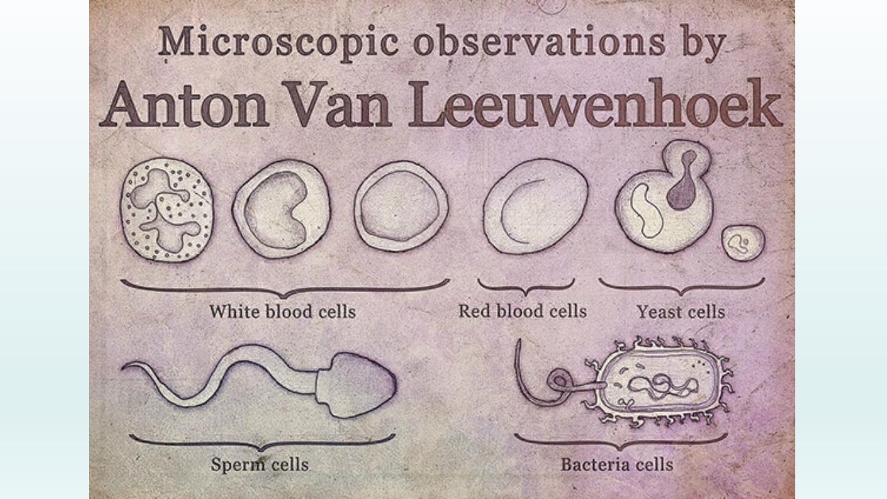

They knew sperm was necessary to make babies, but didn’t know how one lead to the other…
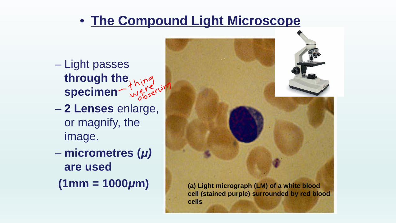
• The Compound Light Microscope
– Light passes through the specimen
– 2 Lenses enlarge, or magnify, the image.
– micrometres (μ)are used
(1mm = 1000μm) (a) Light micrograph (LM) of a white blood cell (stained purple) surrounded by red blood cells

Before we knew all life came from cells…

Before we knew all life came from cells…
…spontaneous generation was the commonly held belief that living things could arise from non-living matter.

Spontaneous Generation: The recipe for mice!
This idea persisted for approximately 2,000 years!

Spontaneous Generation
Without the sophisticated technology we have today, how would you disprove spontaneous generation?

How did they do it?
What experiments were done to “prove” or disprove spontaneous generation?
Read pg 247 – 248

Francesco Redi
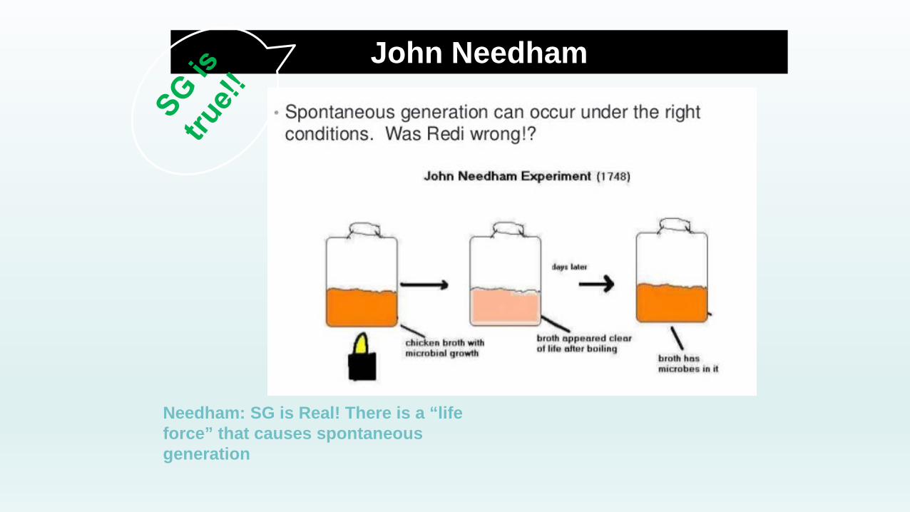
John Needham
Needham: SG is Real! There is a “life force” that causes spontaneous generation

Lazzaro Spallanzani
Critics: No! All you’ve shown is that air is needed for spontaneous generation

Louis Pasteur
…. and the scientific method lived happily ever after

• Do you think there could be any current widely-held “scientific” ideas that could be equally as wrong as spontaneous generation?

Intro to Microscopes Lab!!Learning Goals:1. I can properly handle a microscope2. I can label the parts of a microscope3. I can manipulate the microscope to view a specimen with clarity4. I can make proper scientific drawings (pg. 481) Hand in 2 drawings
Groups of 3-4

How to make a Scientific Drawing!

Inquiry Lab pg. 245-246
Goals:1. determine the diameter of your field of view on low and high
power 2. determine the scale of your drawing 3. estimate the size of objects in your field of view
Hand in your work
First: by how much does the microscope magnify your specimen?

Magnification
•To calculate the total magnification:
magnification = ocular lens x objective lens
•An increase in the specimen’s apparent size.
PowerOcular Lens
MagnificationObjective
MagnificationTotal
Magnification
Low
Medium
High

Steps 3 & 4 Measuring Field Diameter
• Slide a clear ruler onto the stage & observe under low power.
• Measure the diameter of your field of view to the nearest 0.5 mm
•Record the measurement below & convert to μm (1 mm = 1000 μm)
•The diameter of your field of view
Field Magnification Field Diameter (mm)
Field diameter (μm)
Low
High
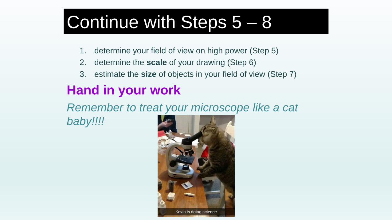
Continue with Steps 5 – 81. determine your field of view on high power (Step 5)2. determine the scale of your drawing (Step 6)3. estimate the size of objects in your field of view (Step 7)
Hand in your workRemember to treat your microscope like a cat baby!!!!

Outcomes 1, 2, & 11
• Independent assignment tomorrow– Parts of the microscope– Using a microscope (magnification)– Spontaneous generation– Scientific drawings & scale of drawings– Cell theory (today)

CELL THEORYAfter spontaneous generation was finally disproved for good, a new theory emerged

• Botanist; looked at a lot of plants under the ‘scope
• Proposed that all plants are made up of cells.
• Plant cells are self-contained living “units”, which work together to support the needs of the plant.
Matthias Schleiden – 1830s
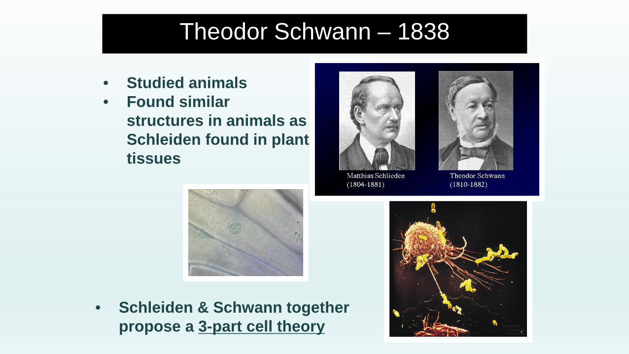
• Studied animals• Found similar
structures in animals as Schleiden found in plant tissues
• Schleiden & Schwann together propose a 3-part cell theory
Theodor Schwann – 1838

Cell Theory Part 1
All living things are made up of one or more cells

Cells are the smallest functional units of organisms
- Take in nutrients- Use energy to do work (life processes)- Get rid of wastes- Maintain certain temperatures and chemical conditions (e.g. acidity)
Multi-cellular organisms are just cells working together to accomplish these basic tasks
Cell Tissue Organ System Body
Cell Theory Part 2
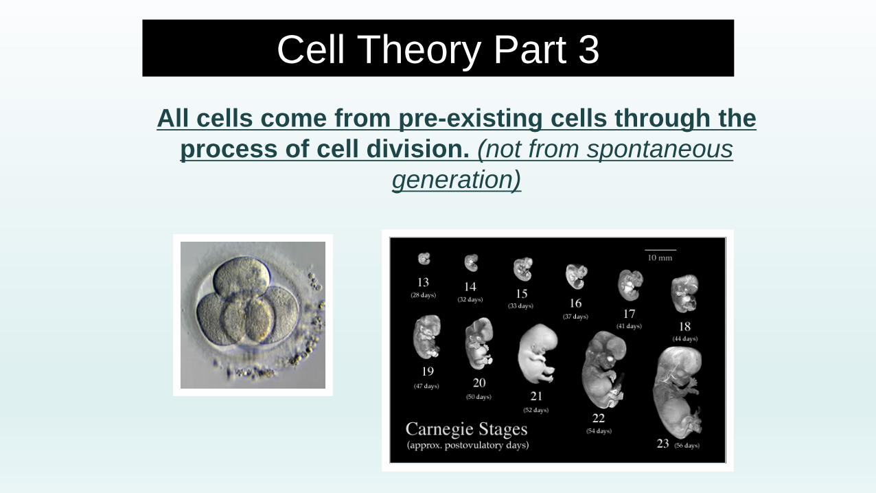
All cells come from pre-existing cells through the process of cell division. (not from spontaneous
generation)
Cell Theory Part 3