microbiome lactobacillus plantarum strain maintains growth
TRANSCRIPT

17. M. Fuccillo, A. L. Joyner, G. Fishell, Nat. Rev. Neurosci. 7,772–783 (2006).
18. J. Briscoe, EMBO J. 28, 457–465 (2009).19. T. Mori et al., Glia 54, 21–34 (2006).20. F. Long, X. M. Zhang, S. Karp, Y. Yang, A. P. McMahon,
Development 128, 5099–5108 (2001).21. B. Djukic, K. B. Casper, B. D. Philpot, L.-S. Chin, K. D. McCarthy,
J. Neurosci. 27, 11354–11365 (2007).22. R. V. Pearse 2nd, K. J. Vogan, C. J. Tabin, Dev. Biol. 239, 15–29
(2001).23. J. Jeong, J. Mao, T. Tenzen, A. H. Kottmann, A. P. McMahon,
Genes Dev. 18, 937–951 (2004).24. A. D. R. Garcia, R. Petrova, L. Eng, A. L. Joyner, J. Neurosci. 30,
13597–13608 (2010).25. C. C. Harwell et al., Neuron 73, 1116–1126 (2012).26. L. E. Gonzalez-Reyes et al., Neuron 75, 306–319 (2012).27. M. K. Cooper, J. A. Porter, K. E. Young, P. A. Beachy, Science
280, 1603–1607 (1998).28. A. Rohner et al., Mol. Cancer Ther. 11, 57–65 (2012).
29. A. V. Molofsky et al., Nature 509, 189–194(2014).
30. X. Tong et al., Nat. Neurosci. 17, 694–703(2014).
ACKNOWLEDGMENTS
Glast-CreERT2 mice are available from M. Götz under a materialtransfer agreement with Helmholtz Zentrum München–DeutschesForschungszentrum für Gesundheit und Umwelt. This work wassupported by the Canadian Institutes of Health Research (FDN143337 to C.W.B., MOP 126137/NIA 288936 to P.J.S., and MOP111152/MOP 123390 to K.K.M.); Natural Sciences and EngineeringResearch Council of Canada (DG 418546-2 to P.J.S. and 408044-2011 to K.K.M.); Canada Research Chairs Program (F.C., C.E., andK.K.M.); Brain Canada/W. Garfield Weston Foundation (F.C. andK.K.M.); James McGill Chair Program (C.W.B.); and CanadianFoundation for Innovation (LOF 28331 to P.J.S.). W.T.F wassupported by a postdoctoral fellowship from the Research Instituteof the McGill University Health Centre. J.P. was supported by a
Vanier fellowship. We thank M. Götz for GLAST CreERT2 mice;G. Quesseveur for help with tissue processing; L. Li for mousetechnical assistance; S. Scales (Genentech) for Shh antibody;T. Alves-Ferreira for ImageJ support; A. Montpetit, A. Staffa, andstaff at Genome Quebec and the Research Institute of McGillUniversity Hospital Centre Molecular Imaging Platform for supportand use of instrumentation; and E. Ruthazer and D. van Meyelfor helpful feedback on the manuscript. The authors declare noconflicts of interest.
SUPPLEMENTARY MATERIALS
www.sciencemag.org/content/351/6275/849/suppl/DC1Materials and MethodsFigs. S1 to S25Tables S1 to S5References (31–40)
21 April 2015; accepted 7 January 201610.1126/science.aab3103
MICROBIOME
Lactobacillus plantarum strainmaintains growth of infant miceduring chronic undernutritionMartin Schwarzer,1,2* Kassem Makki,1,3 Gilles Storelli,1 Irma Machuca-Gayet,1†Dagmar Srutkova,2 Petra Hermanova,2 Maria Elena Martino,1 Severine Balmand,4
Tomas Hudcovic,2 Abdelaziz Heddi,4 Jennifer Rieusset,3 Hana Kozakova,2
Hubert Vidal,3 François Leulier1*
In most animal species, juvenile growth is marked by an exponential gain in body weightand size. Here we show that the microbiota of infant mice sustains both weight gain andlongitudinal growth when mice are fed a standard laboratory mouse diet or a nutritionallydepleted diet. We found that the intestinal microbiota interacts with the somatotropichormone axis to drive systemic growth. Using monocolonized mouse models, we showedthat selected lactobacilli promoted juvenile growth in a strain-dependent manner thatrecapitulated the microbiota's effect on growth and the somatotropic axis. These findingsshow that the host's microbiota supports juvenile growth. Moreover, we discovered thatlactobacilli strains buffered the adverse effects of chronic undernutrition on the postnatalgrowth of germ-free mice.
During the juvenile growth period, the gainin animal body size varies widely as a re-sult of the interactions between nutrition-al input and the organism’s hormonal cues.Inmammals, postnatal growth is controlled
by the activity of the somatotropic axis (fig. S1), in
which growth hormone (GH) instructs the liverand peripheral tissues to produce insulin-likegrowth factor–1 (IGF-1), to promote organ andsystemic growth (1–3). Chronic undernutritiontriggers a state of GH resistance (4, 5) that leadsto stunting, and juveniles become small and thin(6). Acutemalnutrition, in contrast, causeswasting,defined as severe weight loss and mediated inpart through the disruption of the gutmicrobiota(7). However, the contribution of the gut micro-biota to normal postnatal growth and its influ-ence on the activity of the somatotropic axisduring chronic undernutrition remain unknown.To address this question, we first compared
the growth parameters of wild-type (WT) andgerm-free (GF) infant male mice fed a standardbreeding diet (25% proteins, 9% fats; table S1)until young adulthood (8 weeks old, Fig. 1 andfig. S2). After weaning, the GF andWT animalsingested similar amounts of food relative to bodyweight (fig. S3), yet at 8 weeks of age, GF miceweighed 14.5% less and were 4% shorter than
WT mice (Fig. 1, A and C; fig. S2, A and B; andtable S2). These growth differences were mostpronounced after weaning (Fig. 1, A toD, and fig.S2, C andD). Thus, with a standard breeding diet,the gut microbiota ensures optimal weight gainand longitudinal growth, especially aroundwean-ing. Remarkably, the 17%weight gain seen inWTanimals (fig. S2A and table S2) was not a con-sequence of increased adiposity. The epididymalfat pads and adipocyte size of WT and GF malesremained similar (fig. S4, A toD). Likewise, levelsof leptin, a circulating marker of fat stores (8),were similar in the sera of WT and GF animals(fig. S4E). However, theweight gain of the organsof WT animals was greater than that of GF mice(fig. S2E and table S2), confirming that a WTmi-crobiota is associated with optimal systemic so-matic growth. This contrasts with the increasedadiposity that results from subtherapeutic anti-biotic treatments in infantmice that is apparentlycaused by disrupting the gut microbiota commu-nity (9, 10). WT animals were 4% longer (fig. S2Band table S2), indicating that the microbiota alsoinfluences skeletal growth. Bone growth param-eters, including femur length, cortical thickness,cortical bone fraction, and the trabecular fractionof the femur (Fig. 1, E and F; fig. S2, F to I; andtable S2)were all reduced inGF animals, althoughcortical bone mineral density (BMD) was un-affected (fig. S2J). Previously, Sjögren et al. showedthat trabecular BMD was increased in GF ani-mals relative to their WT siblings (11). However,that study was conducted on females of a differ-ent genetic background than ours. Nevertheless,taken together, our results show that the gut mi-crobiota sustains postnatal somatic tissue growth,leading to increased mass gain and enhancedlongitudinal growth.Postnatal systemic growth is mainly driven by
the activity of the somatotropic axis (1–3), wherethe pituitary glandproducesGH,which induces theproduction of IGF-1. The liver is themajor source ofcirculating IGF-1 and together with IGF-1 bindingprotein-3 (IGFBP-3) serves as an endocrine deter-minant of somatic growth (3, 12, 13) (fig. S1). Inaddition, IGF-1 is produced by peripheral tis-sues, including muscles, and acts to promote tis-sue growth in an autocrine/paracrine manner
854 19 FEBRUARY 2016 • VOL 351 ISSUE 6275 sciencemag.org SCIENCE
1Institut de Génomique Fonctionnelle de Lyon, Université deLyon, Ecole Normale Supérieure de Lyon, Centre National de laRecherche Scientifique, Université Claude Bernard Lyon 1, UnitéMixte de Recherche 5242, 46 Allée d’Italie, 69364 Lyon Cedex07, France. 2Laboratory of Gnotobiology, Institute ofMicrobiology of the Czech Academy of Sciences, v. v. i., NovyHradek, Czech Republic. 3Laboratoire CarMeN, Université Lyon1, Unité Mixte de Recherche INSERM U-1060 et INRA U-1397,Faculté de Médecine Lyon-Sud, Chemin du Grand Revoyet,69600 Oullins, France. 4UMR203 BF2I, Biologie FonctionnelleInsectes et Interactions, Université de Lyon, INRA, INSA-Lyon,F-69621 Villeurbanne, France.*Corresponding author. E-mail: [email protected](F.L.); [email protected] (M.S.) †Present address:Institut National de la Santé et de la Recherche Médicale,Université Claude Bernard Lyon 1, Unité Mixte de Recherche 1033,Faculté de Médecine Lyon-Est, Rue Guillaume Paradin, 69372 LyonCedex 08, France.
RESEARCH | REPORTSon O
ctober 19, 2017
http://science.sciencemag.org/
Dow
nloaded from

(14) (fig. S1). We therefore measured circulatinglevels of GH and IGF-1, the major components ofthis axis (3, 15, 16), in the sera of WT and GFanimals. GH levels peaked around birth andgradually declined during postnatal growth inboth WT and GF animals (Fig. 2A). IGF-1 titerswere significantly reduced in GF animals (Fig.2B), as were the circulating levels of IGFBP-3 insera (17) (Fig. 2C). In addition, Igf1 expressionwas reduced in muscles of GF animals at both28 and 56 days (fig. S5, A and B). In the liver,both Igf1 and Igfbp3 expression were reduced inGF mice at 28 days (Fig. 2, D and E), at the sametime that IGF-1 circulating titers peaked in WTanimals (Fig. 2B). Likewise, the phosphorylationof Akt at Ser 473 (phospho-S473-Akt), a markerof IGF-1 receptor (IGF-1R) signaling activity (18)(fig. S1), was reduced in the liver of GF animals ascompared to WT animals both at 28 (Fig. 2F)and 56 days (fig. S5C).
To assess the importance of IGF-1 levels inmediating postnatal growth dynamics, we re-peatedly injected GF and WT animals with re-combinant IGF-1 (rIGF-1) for 10 days after weaningand analyzed their growth parameters. GF animalssignificantly increased their weight, as well as bodyand femur length, over the treatment period (fig.S5, D to G). Injections of rIGF-1 into WT animalsdid not promote growth (fig. S5, D to F), eventhough they modulated two established markersof IGF-1 activity: reduced glycemia and increasedphospho-S473-Akt signals in the liver (fig. S5, Hand I). In GF mice, IGF-1 levels in sera were re-duced compared to those in WT mice despitenormal circulating GH levels, and an exogenoussupply of rIGF-1 was sufficient to enhance growthto levels seen inWT animals (fig. S5, D, E, and G).We thus concluded that the microbiota promotedgrowth by facilitating IGF-1 production and ac-tivity. To further test this hypothesis, we treated
WT animals with the cyclolignan compound pic-ropodophyllin (PPP), a specific noncompetitiveinhibitor of IGF-1R (19, 20), for 10 days. The PPPtreatment significantly retarded the growth gainsexpected of WT animals (Fig. 4, D to F) and de-creased the rIGF-1–mediated impact on phospho-S473-Akt signals in the liver andglycemia dynamics(fig. S8, J and K). These results established thatIGF-1 activity is necessary for growth in WTanimals. Together, our data show that the gutmicrobiota influences the production and activ-ity of IGF-1 required for postnatal growth.We then tested the effects of chronic under-
nutrition on, and the contribution of the gutmicrobiota to, postnatal growth. To this end,we weaned GF and WT juveniles onto a nutri-tionally depleted diet low in proteins (8.6%), fats(2.4%), and vitamins (table S1) and monitoredtheir growth until 8 weeks of age (Fig. 3). Duringtheweek of adaptation to solid food, bothGF and
SCIENCE sciencemag.org 19 FEBRUARY 2016 • VOL 351 ISSUE 6275 855
Fig. 1. The microbiota maintains mouse juvenile growth. (A to D) Weight (A) and body length (C) growth curves during the juvenile period (day 7 to day 56after birth) and daily weight (B) and body size (D) gains after weaning (day 21 to day 56) of WT (gray) (n = 16) and GF (green) (n = 12) infant male mice bred withtheir mothers from birth until day 21, weaned, and then fed a breeding diet from 21 to 56 days old. (E) Photograph of representative femur bones at day56. (F) Three-dimensional reconstructions of representative distal parts of femur bones at day 56. Error bars indicate SEM. *P < 0.05, **P < 0.01, ***P <0.001, ****P < 0.0001.
Fig. 2. The microbiota maintains systemic somatotropic axis activity. (A to C) Levels of GH (A), IGF-1 (B), and IGFBP-3 (C) in sera of WT (gray) and GF(green)mice at given time points after birth, n ≥ 5mice per group. (D to F) Expression levels of Igf1 (D) and Igfbp3 in liver (E) at day 28 after birth (n = 5 or 6miceper group). (F) Western blots and quantification of phospho-S473-Akt in liver ofWTand GFmice (n = 5 or 6mice per group) at day 28 after birth . Representativeblots of three mice per group are shown. Data are presented as means ± SEM. *P < 0.05, ***P < 0.001, ****P < 0.0001.
RESEARCH | REPORTSon O
ctober 19, 2017
http://science.sciencemag.org/
Dow
nloaded from

WT animals lost weight, yet weight loss wasmore extensive in GF animals (fig. S6C). Afteradaptation to solid food, the growth of GF ani-mals was arrested, whereas WT animals resumedgrowth and recovered weight and longitudinalgrowth (Fig. 3, A to C, and fig. S6, A to E), albeit toa lesser extent than those on the breeding diet.The stunted phenotype of GF animals was not theresult of an alteration of the GF animals’ foodintake relative to their bodyweight (fig. S7, A toD)nor of an altered capacity to absorb energy fromthe diet (fig. S7E). We conclude therefore that thegut microbiota contributes to maintaining mousejuvenile growth during chronic undernutrition.We then analyzed how the gut microbiota in-
fluences the somatotropic axis during under-nutrition (Fig. 4). We quantified GH, IGF-1, andIGFBP-3 levels in GF and WT animals afterweaning onto the depleted diet. At 28 days, GHlevels were elevated in GF animals as comparedwith WT animals, whereas at 56 days, GF ani-mals displayed reduced circulating levels of GH,similar to WT animals (Fig. 4A). In contrast toWT animals, both IGF-1 and IGFBP-3 circulatinglevels failed to peak at 56 days in GF animals(Fig. 4, B and C). In muscle at 28 days (7 daysafter weaning onto the depleted diet), the ex-pression of both GH-receptor (Ghr) and Igf1 inWT animals was elevated compared to that in GFanimals (fig. S8, A and B). Further, Socs3, atranscriptional target of GHR signaling (21) (fig.S1), was also increased in WT muscles (fig. S8C).In the liver at 28 days, Ghr expression was in-creased in WT animals as compared with GFcounterparts (fig. S8D). At 56 days, circulatingtiters of IGF-1 (Fig. 4B), along with Igf1 expres-sion in the muscles and liver (fig. S8, E and F),increased; Igfbp3 and Socs3 expression (fig. S8, Gand H) and the phospho-S473-Akt signal werealso increased in the liver of WT animals (fig.S8I). Collectively, these results indicate a reducedactivity of the somatotropic axis in GF animals.Next, we repeatedly injected 28-day-old WT ani-mals weaned on the depleted diet with the PPPcompound for 10 days and monitored theirgrowth. We observed that PPP-treated animalshad reduced weight, body length, and femurlength gains over the treatment period as com-pared with untreatedmice (Fig. 4, D to G). Takentogether, these results indicate that during under-nutrition, the gut microbiota helps to maintainsystemic somatotropic axis activity and that thisactivity does result in some postnatal growth.We have previously showed that gutmicrobiota
promoteDrosophila juvenile growthduringunder-nutrition (22). Furthermore, monoassociation ofGF Drosophila with selected lactobacilli strainsrecapitulates the growth promotion seen when amore complex microbiota is present (22). Lacto-bacilli strains are commensal in a variety of ani-mals, including Drosophila and mammals (23, 24).These taxa also share evolutionarily conservednutrient-sensing endocrine pathways that regu-late juvenile growth (i.e, Drosophila insulin/IGF-like peptides and mammalian IGFs) (25). Wethus tested the functional potential of specificlactobacilli strains onmurine juvenile growth and
the somatotropic axis during undernutrition. TwoLactobacillus plantarum strains that display dif-ferent growth-promoting capacities inDrosophilamonocolonized models were selected for mono-association experiments in GFmice. Using mono-colonizedDrosophila, we identified L. plantarumWJL
(LpWJL) as a potent growth promoter, whereasL. plantarumNIZO2877 (LpNIZO2877) showed a sta-tistically less pronounced effect on Drosophilagrowth (fig. S9). We then monitored postnatalgrowth of monocolonized infant male mice withLpWJL and LpNIZO2877 strains. These mice wereobtained after monocolonizing GF adult mice ofboth sexes with the LpWJL or LpNIZO2877 strains;monocolonized adults were mated 20 days aftercolonization, and groups of six offspring werenursed by their monocolonized dam and natu-rally colonized by the respective Lactobacillusstrain acquired from the dam. Lactating miceand their pups were maintained on the breedingdiet. Twenty-one days after birth, the juvenileswere weaned on either the breeding or the de-pleted diet (fig. S10). On the depleted diet, al-though both LpWJL and LpNIZO2877 juveniles gainedmore weight (+52% and +27% respectively; Fig.3, A and B; fig. S6, A to D; and table S2) and bodylength (+14% and +8% respectively; Fig. 3, B andC; fig. S6E; and table S2) as compared with GF
animals, the LpWJL-associated animals grew better(+25% weight and +6% length) than LpNIZO2877-associated animals (Fig. 3, A to C, and fig. S6, Ato E). The quantitative difference between thetwo strains was not a result of differences in foodintake relative to body weight (fig. S7, A to D),differential capacity to absorb energy from thediet (fig. S7E), or a marked difference in the ef-ficiency of bacterial colonization of the intestinaltract (fig. S7, F to H). LpWJL-colonized animalsshowed a 14% body length gain when comparedto GF animals, whereas LpNIZO2877-associated ani-mals gained only 8% (fig. S6E and table S2). Sim-ilarly, bone growth such as femur length wasdifferentially affected (Fig. 3D, fig. S6F, andtable S2), indicating that the growth benefit de-pends on the strain of L. plantarum. In termsof weight gain, on the depleted diet, WT animalswere 53% heavier than GF animals (fig. S6D andtable S2). LpWJL-colonized animals showed a52% weight gain, whereas LpNIZO2877-associatedanimals only showed 27% weight gain (fig. S6Dand table S2). A similar effect was observed in allorgans tested (fig. S6, G to J, and table S2). Col-lectively, the data show that during postnatalgrowth, selected lactobacilli strains can recapit-ulate the effect of a WT microbiota on mousejuvenile growth that would otherwise be stunted
856 19 FEBRUARY 2016 • VOL 351 ISSUE 6275 sciencemag.org SCIENCE
Fig. 3.The microbiota and a L. plantarum strain maintain mouse juvenile growth upon undernutrition.(A toC) Weight (A) and body length (C) growth curves of WT (n = 12 and 15, black lines),GF (n = 12 and20, gray lines), LpWJL (n = 8 and 28, red line), and LpNIZO2877 (n = 12 and 15, blue line) monocolonizedmalemice on breeding (triangles and dashed lines) or nutritionally depleted (circles and solid lines) diets. (B)Representative photographs of mice on depleted diet at day 56 after birth. (D). Photographs of repre-sentative femur bones at day 56 after birth of GF,WT, LpWJL-, and LpNIZO2877-associatedmice raised on thedepleted diet.
RESEARCH | REPORTSon O
ctober 19, 2017
http://science.sciencemag.org/
Dow
nloaded from
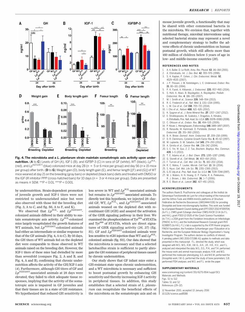
by undernutrition. Strain-dependent promotionof juvenile growth and IGF-1 titers were notrestricted to undernourished mice but werealso observed with those fed the breeding diet(Fig. 3, A to C, and fig. S6, A to F, and K).We observed that LpWJL- and LpNIZO2877-
colonized animals differed in their ability to sus-tain somatotropic axis activity. LpWJL-colonizedmice largely recapitulated the growth features ofWT animals, but LpNIZO2877-colonized animalshad either an intermediate or similar response tothat of the GF animals (Fig. 4, A to C). By 56 days,the GH titers of WT animals fed on the depleteddiet were comparable to those observed in WTanimals raised on the breeding diet. However, theIGF-1 titers of these mice had dwindled by morethan sevenfold (compare Fig. 2, A and B, andFig. 4, A and B), confirming that chronic under-nutrition affects the activity of the GH/IGF-1 axis(4). Furthermore, although GH titers of GF andLpNIZO2877-associated animals at 28 days wereelevated, they failed to elicit adequate tissue re-sponses, implying that the activity of the soma-totropic axis is impaired in GF juveniles andthat their tissues are in a state of GH resistance.We hypothesized that reduced GH sensitivity is
less severe in WT and LpWJL-associated animalsbut remains in LpNIZO2877-associated animals. Todirectly test this hypothesis, we injected 28–day-old GF, WT, LpWJL-, and LpNIZO2877-associatedanimals weaned on the depleted diet with re-combinant GH (rGH) and assayed the activationof the GHR signaling pathway in their liver. Weexamined thephosphorylation of Tyr694 of STAT5aand Tyr699 of STAT5b, which are direct signa-tures of GHR signaling activity (26, 27) (fig.S1). GF and LpNIZO2877-colonized animals wereless sensitive to rGH injection thanWT and LpWJL-colonized animals (fig. S11). Our data showed thatthe microbiota is necessary and that a selectedlactobacillus strain is sufficient to partly abro-gate theGH resistance of peripheral tissues causedby chronic undernutrition.Our study shows that GF infant mice enter a
GH-resistant state upon chronic undernutrition,and a WT microbiota is necessary and sufficientto boost postnatal growth by enhancing GHsensitivity and thereby increasing IGF-1 activityin peripheral tissues. In addition, this studyestablishes that a selected strain of L. planta-rum can recapitulate the beneficial effects ofthe microbiota on the somatotropic axis and on
mouse juvenile growth, a functionality that maybe shared with other commensal bacteria inthe microbiota. We envision that, together withnutritional therapy, microbial interventions usingselected bacterial strains may represent a noveland complementary strategy to buffer the ad-verse effects of chronic undernutrition on humanpostnatal growth, which still affects more than160 million of children below 5 years of age inlow- and middle-income countries (28).
REFERENCES AND NOTES
1. A. A. Butler, D. Le Roith, Annu. Rev. Physiol. 63, 141–164 (2001).2. A. Efstratiadis, Int. J. Dev. Biol. 42, 955–976 (1998).3. S. A. Kaplan, P. Cohen, J. Clin. Endocrinol. Metab. 92,
4529–4535 (2007).4. J. P. Thissen, J. M. Ketelslegers, L. E. Underwood, Endocr. Rev.
15, 80–101 (1994).5. P. K. Fazeli, A. Klibanski, J. Endocrinol. 220, R57–R65 (2014).6. S. Hizli, A. Abaci, B. Büyükgebiz, A. Büyükgebiz, Pediatr.
Endocrinol. Rev. 4, 186–195 (2007).7. M. I. Smith et al., Science 339, 548–554 (2013).8. R. C. Frederich et al., Nat. Med. 1, 1311–1314 (1995).9. L. M. Cox et al., Cell 158, 705–721 (2014).10. I. Cho et al., Nature 488, 621–626 (2012).11. K. Sjogren et al., J. Bone Mineral Res. 27, 1357–1367 (2012).12. E. Stratikopoulos, M. Szabolcs, I. Dragatsis, A. Klinakis,
A. Efstratiadis, Proc. Natl. Acad. Sci. U.S.A. 105, 19378–19383 (2008).13. C. Ohlsson et al., Endocr. Rev. 30, 494–535 (2009).14. P. Klover, L. Hennighausen, Endocrinology 148, 1489–1497 (2007).15. R. Renaville, M. Hammadi, D. Portetelle, Domest. Anim.
Endocrinol. 23, 351–360 (2002).16. B. H. Breier, Domest. Anim. Endocrinol. 17, 209–218 (1999).17. D. R. Clemmons, Cytokine Growth Factor Rev. 8, 45–62 (1997).18. M. Laplante, D. M. Sabatini, Cell 149, 274–293 (2012).19. A. Girnita et al., Cancer Res. 64, 236–242 (2004).20. S. C. Yin, W. Guo, Z. Z. Tao, Biochem. Biophys. Res. Commun.
439, 1–5 (2013).21. T. E. Adams et al., J. Biol. Chem. 273, 1285–1287 (1998).22. G. Storelli et al., Cell Metab. 14, 403–414 (2011).23. F. Turroni et al., Cell. Mol. Life Sci. 71, 183–203 (2014).24. R. C. Matos, F. Leulier, Microb. Cell Fact. 13, S6 (2014).25. S. Hyun, Cell. Mol. Life Sci. 70, 2351–2365 (2013).26. G. B. Udy et al., Proc. Natl. Acad. Sci. U.S.A. 94, 7239–7244 (1997).27. M. J. Waters, H. N. Hoang, D. P. Fairlie, R. A. Pelekanos,
R. J. Brown, J. Mol. Endocrinol. 36, 1–7 (2006).28. R. E. Black et al., Lancet 382, 427–451 (2013).
ACKNOWLEDGMENTS
The authors thank B. Prud'homme and colleagues at the Institut deGénomique Fonctionnelle de Lyon for critical reading of the manuscriptand the Arthro-Tools and ANIRA-ImmOs platforms of StructureFédérative de Recherche Biosciences (UMS3444/US8) for providingDrosophila and bone microtomography facilities. This work was fundedby a European Research Council starting grant (FP7/2007-2013-N°309704)(to F.L.), CNRS grant Echange chercheur CNRS/AVCR 13-14 (to F.L.and H.K.), grant P303-12-0535 of the Czech Science Foundation(to T.H.), a 2014 grant from the Fondation Innovations en Infectiologie(FINOVI, to H.V.), and the Institutional Research Concept grant RVO61388971 (to H.K.). F.L.’s lab is supported by the ATIP/Avenir program,FINOVI foundation, the Fondation Schlumberger pour l'Education et laRecherche, and the European Molecular Biology Organization’s YoungInvestigator Program. The authors declare no conflicts of interest.A pending patent (WO 2015/173386 A1) applies to methods and datapresented in this manuscript. F.L. directed the study, which wasdesigned with M.S.; M.S., K.M., I.M.-G., A.H., J.R., H.K., H.V., and F.L.analyzed and interpreted the data; M.S., D.S., P.H., and T.H. performedthe mouse work and animal macroscopic analysis; K.M. and M.S.performed the molecular phenotyping; G.S. and M.E.M. performed theDrosophila work; I.M.-G. performed the study of bone parameters; S.B.performed FISH analyses; and M.S. and F.L. wrote the paper.
SUPPLEMENTARY MATERIALS
www.sciencemag.org/content/351/6275/854/suppl/DC1Materials and MethodsFigs. S1 to S12Tables S1 and S2References (29–32)
12 November 2015; accepted 13 January 201610.1126/science.aad8588
SCIENCE sciencemag.org 19 FEBRUARY 2016 • VOL 351 ISSUE 6275 857
Fig. 4. The microbiota and a L. plantarum strain maintain somatotropic axis activity upon under-nutrition. (A to C) Levels of GH (A), IGF-1 (B), and IGFBP-3 (C) in sera of GF (white),WT (black), LpWJL-(red), and LpNIZO2877-(blue) colonizedmice at day 28 (n = 5 or 6mice per group) and day 56 (n ≥ 15miceper group) after birth. (D toG) Weight gain (D), body length gain (E), and femur length [(F) and (G)] of WTmiceweaned at day 21 on thebreeding (gray bars) ordepleted (black bars) diets and treatedwithDMSOorthe IGF-1R inhibitor PPP (cross-hatched bars) for 10 days (n = 3 or 4mice per group). Data are presentedas means ± SEM. **P < 0.01, ***P < 0.001.
RESEARCH | REPORTSon O
ctober 19, 2017
http://science.sciencemag.org/
Dow
nloaded from

strain maintains growth of infant mice during chronic undernutritionLactobacillus plantarum
LeulierMartino, Severine Balmand, Tomas Hudcovic, Abdelaziz Heddi, Jennifer Rieusset, Hana Kozakova, Hubert Vidal and François Martin Schwarzer, Kassem Makki, Gilles Storelli, Irma Machuca-Gayet, Dagmar Srutkova, Petra Hermanova, Maria Elena
DOI: 10.1126/science.aad8588 (6275), 854-857.351Science
, this issue p. 10.1126/science.aad3311, p. 854Sciencethat specific beneficial microbes could potentially be exploited to resolve undernutrition syndromes.activity via signaling pathways in the liver, thus overcoming growth hormone resistance. Together these studies reveal
in the gut microbiota sustained growth hormoneLactobacillus plantarum showed that strains of et al.mice, Schwarzer malnourished children. In infant mammals, chronic undernutrition results in growth hormone resistance and stunting. In
found that the microbiota of healthy children relieved the harmful effects on growth caused by the microbiota ofal.etbe transplanted effectively into germ-free mice to recapitulate their associated phenotypes. Using this model, Blanton
because a characteristic community of gut microbes seems to mediate some of the pathology. Human gut microbes can Malnutrition in children is a persistent challenge that is not always remedied by improvements in nutrition. This is
Microbiota and infant development
ARTICLE TOOLS http://science.sciencemag.org/content/351/6275/854
MATERIALSSUPPLEMENTARY http://science.sciencemag.org/content/suppl/2016/02/17/351.6275.854.DC1
CONTENTRELATED
http://stm.sciencemag.org/content/scitransmed/3/106/106ra106.fullhttp://stm.sciencemag.org/content/scitransmed/5/180/180fs11.fullhttp://stm.sciencemag.org/content/scitransmed/5/189/189sr4.fullhttp://stm.sciencemag.org/content/scitransmed/5/190/190ps10.fullhttp://science.sciencemag.org/content/sci/351/6275/802.full
REFERENCES
http://science.sciencemag.org/content/351/6275/854#BIBLThis article cites 31 articles, 7 of which you can access for free
PERMISSIONS http://www.sciencemag.org/help/reprints-and-permissions
Terms of ServiceUse of this article is subject to the
is a registered trademark of AAAS.Sciencelicensee American Association for the Advancement of Science. No claim to original U.S. Government Works. The title Science, 1200 New York Avenue NW, Washington, DC 20005. 2017 © The Authors, some rights reserved; exclusive
(print ISSN 0036-8075; online ISSN 1095-9203) is published by the American Association for the Advancement ofScience
on October 19, 2017
http://science.sciencem
ag.org/D
ownloaded from