microcantilever biosensors for chemicals and bioorganismshj56/pdffiles/2011/anlyst 1539.pdf ·...
TRANSCRIPT

Dynamic Article LinksC<Analyst
Cite this: Analyst, 2011, 136, 1539
www.rsc.org/analyst MINIREVIEW
Microcantilever biosensors for chemicals and bioorganisms
Koutilya R. Buchapudi,a Xin Huang,†b Xin Yang,a Hai-Feng Ji*b and Thomas Thundatc
Received 16th December 2010, Accepted 1st February 2011
DOI: 10.1039/c0an01007c
In the last fifteen years, microcantilevers (MCLs) have been emerging as a sensitive tool for the
detection of chemicals and bioorganisms. Because of their small size, lightweight, and high surface-to-
volume ratio, MCL-based sensors improve our capability to detect and identify biological agents by
orders of magnitude. A biosensor is a device for the detection of an analyte that combines a biological
component with a physicochemical detector component. The MCL biosensors have recently been
reviewed in several papers. All of these papers were organized based on the sensing biological elements
(antibody, enzyme, proteins, etc.) for recognition of analytes. In this review, we intend to summarize
the microcantilever biosensors in a format of each specific chemical and bioorganism species to make
information on individual biosensors easily accessible. We did this to aid researchers to locate relevant
references.
1. Introduction
Recently, micro-electromechanical systems (MEMS) have been
emerging as a platform for the development of miniature sensors
with extremely high sensitivity. It is estimated by 2010 about 1000
papers on microcantilevers sensors had been published. Micro-
machined silicon cantilevers are the simplest MEMS sensors that
can be micromachined and mass-produced. Microcantilever
aInstitute for Micromanufacturing, Louisiana Tech University, Ruston,LA, 71272, USAbDepartment of Chemistry, Drexel University, Philadelphia, PA, 19104.E-mail: [email protected]; Fax: +1 215 895 1265; Tel: +1 215 895 2562cChemical and Materials Engineering Department, University of Alberta,Edmonton, Canada, AB, T6G2V4
† Current address: Institute of Animal and Plant Quarantine, ChineseAcademy of Inspection and Quarantine, Beijing, 100029, P. R. China.
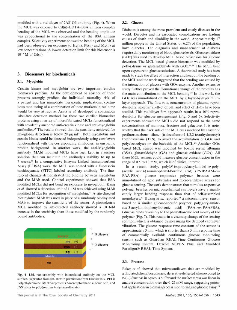
Koutilya R: Buchapudi
Mr Koutilya R. Buchapudi
obtained his master degree in
Molecular Science and Nano-
technology from Louisiana Tech
University in 2008. He is
currently pursuing his PhD
degree in biomedical engineering
at Louisiana Tech University.
He is presently working on
developing fluorescent sensors
for real-time measurement of
cell metabolism.
This journal is ª The Royal Society of Chemistry 2011
(MCL) sensor technology is an upcoming sensing technique with
extremely high sensitivity and with broad applications in chem-
ical, physical, and biological detection.1,2 With their compactness
and potential low cost, silicon-based MCLs provide a clear path
for the development of miniaturized sensors for detection of
chemical and biological agents. In liquid phase applications, the
MCL technology has the potential for biodetection applications,
such as detection of toxins and selective detection of pathogens via
immunological techniques. Because of their small size, light-
weight, and high surface-to-volume ratio, MCL-based sensors
have unprecedented sensitivity for detection of biological analytes
(potentially detecting as little as a single entity of an agent).
A SEM picture of a MCL is shown in Fig. 1. The MCL sensors
have several advantages over the other sensor technologies, including
faster response time, lower cost of fabrication, the possibility of
sensor arrays with small overall dimensions, the ability to explore
Xin Huang
Dr Xin Huang received his PhD
in Physical Chemistry from
Tsinghua University, China, in
2007. He has published more
than 20 peer-reviewed journal
papers and he is currently an
associate professor at Chinese
Academy of Inspection and
Quarantine, Institute of Animal
and Plant Quarantine. His
research interest is rapid and
sensitive detection of Exit-Entry
agriculture products.
Analyst, 2011, 136, 1539–1556 | 1539
microenvironments, and improved portability for field applications.
The MCL biological sensors are summarized briefly in this review.
The MCL sensor responses, such as resonance frequency,
deflection, Q-factor, and amplitude, undergo changes due to
adsorption of molecules on the cantilever surface or extreme
changes in the cantilever environment, for example, density and
viscosity. In theory, the MCLs could be modified and optimized
for sensitive and interference-free detection of chemicals and
physical quantities.
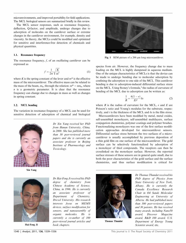
Fig. 1 SEM picture of a 200 mm long microcantilever.
1.1. Resonance frequencyThe resonance frequency, f, of an oscillating cantilever can be
expressed as
f ¼ 1
2p
ffiffiffiffiffiffiK
m*
r(1)
where K is the spring constant of the lever and m* is the effective
mass of the microcantilever. The effective mass can be related to
the mass of the beam, mb, through the relation: m* ¼ nmb, where
n is a geometric parameter. It is clear that the resonance
frequency can change due to changes in mass as well as changes
in spring constant.
1.2. MCL bending
The variation in resonance frequency of a MCL can be used for
sensitive detection of adsorption of chemical and biological
Xin Yang
Dr Xin Yang received her PhD
from Hunan University, China,
in 2000. She has published more
than 30 peer-reviewed journal
papers and she is currently an
associate professor in Beijing
Institute of Pharmacology and
Toxicology.
Hai-Feng Ji
Dr Hai-Feng Ji received his PhD
degree of chemistry from
Chinese Academy of Science,
China, in 1996. He is currently
an associate professor of
Department of Chemistry,
Drexel University. His research
interests focus on MEMS
devices, surface modification for
sensors, and nanoassembly of
organic molecules. He is
currently a co-author of 100
peer-viewed journal articles and
book chapters.
1540 | Analyst, 2011, 136, 1539–1556
species from air. However, the frequency change due to mass
loading on the MCL is highly dampened in aqueous medium.
One of the unique characteristics of MCLs is that the device can
be made to undergo bending due to molecular adsorption by
confining the adsorption to one side of the MCL. This cantilever
bending is due to adsorption-induced differential surface stress
on the MCL. Using Stoney’s formula,3 the radius of curvature of
bending of the MCL due to adsorption can be written as:
1
R¼ 6ð1� nÞ
Et2ds (2)
where R is the radius of curvature for the MCL, n and E are
Poisson’s ratio and Young’s modulus for the substrate, respec-
tively, and t is the thickness of the MCL and ds is the film stress.
Microcantilevers have been modified by metal, metal oxides,
self-assembled monolayers, self-assembled multilayers, surface
conjugation chemistries, polymer coatings and polymer brushes.
Functionalizing monolayers was one of the first surface modifi-
cation approaches developed for microcantilever sensors.
Differential surface stress between the two surfaces of a micro-
cantilever is usually accomplished by previous deposition of
a thin gold film on one surface of the microcantilever. The gold
surface can be selectively functionalized by adsorption of
a monolayer of thiol compounds. The receptors can then be
crosslinked on the monolayer surface. However, the reported
surface stresses of these sensors are in general quite small, due to
both the poor characteristics of the gold surface and the surface
chemistries, and thus surface modification is critical for
Thomas Thundat
Dr Thomas Thundat received his
PhD degree of Physics from
State University of New York,
Albany. He is currently the
Canada Excellence Research
Chair in Oil Sands Molecular
Engineering at University of
Alberta. He had published more
than 300 peer-reviewed papers
and 30 patents. He has received
many awards, including Nano50
award, Discover Magazine
award, R&D 100 award, U.S.
Department of Energy Young
Scientist award, etc.
This journal is ª The Royal Society of Chemistry 2011

developing sensitive and reliable microcantilever sensors. The
multilayer approach is one of the newer approaches for modi-
fying microcantilever surfaces. The surface of the cantilever is
modified through a positive and negative charge distribution
technique. Microcantilevers that are modified with poly-
electrolyte multilayers generate more surface stress than the
corresponding monolayer films. There is a tremendous advan-
tage in the use of multilayer structures in environmental reme-
diation, and the LbL fabrication method affords a marvelous
versatility of composition, size, and shape. However, so far, only
enzymes have been used for the fabrication of microcantilever
biosensors. Receptor-containing polyelectrolytes are not
commercially available so far. Polymer brushes have the poten-
tial to provide an even higher signal-to-noise ratio. Polymer-
brush-modified microcantilevers have recently been reported.
They exhibited significant bending and unbending in response to
alternate exposures to pure water and analytes. The amplified
bending increased the signal-to-noise ratio, thereby increased the
sensitivity of the microcantilever sensor. However, a nontrivial
synthetic work is needed in order to prepare brushes that contain
receptors for selectivity. These have been summarized in a recent
review paper.4
A biosensor is an analytical tool consisting of biochemical
recognition or active components (receptor) used in close
conjunction with a platform (transducer) that convert the
biochemical recognition into an electric signal. Biosensors can be
classified by the receptors, such as antibodies (as in immuno-
sensors), enzymes, DNA, membrane, or microorganisms, and
transducers, such as electrochemical, optical, acoustic, or
mechanical biosensors. The MCL biosensors have been reviewed
in several papers.5–9 All of these papers were classified by the
receptors. This review describes individual microcantilever
biosensors, ordered according to the analyte for which they have
been developed. This review is intended to document all of these
biosensors up to 2010, which makes it possible for the interested
reader to quickly find references to specific sensors. For this
purpose we have searched the literature published between 1994
and 2010 using the Science Citation Index. Physical sensors and
chemical sensors without biological components are not a topic
of this review. Some of the summaries are short to save space for
a mini review.
Fig. 2 Left: three replicates (dashed line) of bending responses as
a function of time for a (OPH/PSS)3 multilayer modified MCL to a 10�3
M paraoxon. Right: bending responses as a function of time for a (OPH/
PSS)3 modified MCL upon exposure to 10�3 M paraoxon, parathion, and
DFP. Reprinted from ref. 11 with permission from Elsevier B.V.
2. Biosensors for toxic chemicals and heavy metalions
2.1. Organophosphorus (OP) compounds
All nerve agents belong to the family of organophosphorus (OP)
compounds, which are among the most toxic of known
substances. The toxicity of these compounds arises from their
irreversible binding to acetylcholinesterase (AChE) that is
essential to nerve impulse responses.10 Besides nerve gases, many
pesticides also belong to the organophosphorus compound
group. In an effort to feed the growing world population, the
agriculture industry has increasingly taken the assistance of
pesticides to increase the crop yield by fending off pest infesta-
tion. Their widespread use in agriculture may contaminate
drinking water. Accordingly, there are considerable interests in
This journal is ª The Royal Society of Chemistry 2011
the development of reliable devices for the sensitive detection of
organophosphates.
Enzymes have been used for the detection of organophos-
phorus compounds. An AChE modified MCL was firstly used
for such a purpose.11 The gold surface of a MCL was modified
first with a thiol layer and then exposed to glutaraldehyde, which
acts as a crosslinker to link the AChE on the MCL surface. For
different concentrations of paraoxon the bending of the MCLs
varied. The MCL underwent a maximum of 7 nm bending due to
the inhibition of AChE by paraoxon that slightly changed the
conformation of AChE. The detection limit was
approximately 10�7 M.
In another work, Chandana et al. modified MCLs with
organophosphorus hydrolase (OPH)12 by using a layer-by-layer
technique for the detection of OP compounds. OPH is an enzyme
that can be used for continuous monitoring of OPs in the envi-
ronment. The MCL bending amplitude at equilibrium was
a function of the concentration of paraoxon with the dynamic
range extending from 10�7 to 10�3 M. The lower detection limit of
approximately 10�7 M for paraoxon was an order of magnitude
lower than the OPH-based potentiometric and optical biosensors
based on a pH modulation. There was a good measurement-by-
measurement (Fig. 2 left) and an acceptable MCL-by-MCL
reproducibility as evidenced by the standard errors of 5% and
15%, respectively. OPs measured using this technique included
parathion and diisopropyl fluorophosphate (DFP) in the order
of sensitivity, paraoxon > DFP > parathion (Fig. 2 right). The
conformational change of the OPH was most likely the main
origin of MCL bending.
2.2. Dichlorodiphenyltrichloroethane (DDT)
DDT is a chlorinated compound with insecticidal properties that
has been used worldwide for controlling insect pests. However, it
is highly hydrophobic with great stability to physical, chemical,
and biological degradation, which has resulted in the accumu-
lation of its residues in animal and human tissues, as well as in the
environment. The surface modification of a MCL was carried out
in a flow-cell (Fig. 3) by exposing to cystamine and glutaralde-
hyde solutions, followed by a DDT hapten derivative (DDT5-
BSA).13 A bending of the MCL was observed on exposure to
DDT showing the interaction between DDT and DDT5-BSA.
Analyst, 2011, 136, 1539–1556 | 1541

Fig. 3 Scheme of the experimental set-up. Reprinted from ref. 12 with
permission from Elsevier B.V.
A concentration of 4 mg ml�1 of DDT has been detected using this
method.
2.3. Endocrine disrupting chemicals (EDCs)
EDC can adversely affect the health of human, domestic, and
wildlife species by altering or inhibiting the function of the
endocrine system. Since a wide range of biological processes can
be influenced and impaired by EDCs, it is crucial to screen and
detect them. EDCs include a wide range of naturally occurring
and synthetic chemicals. These chemicals and/or their byprod-
ucts include but are not limited to pesticides, plasticizers, deter-
gents, pharmaceuticals, and biological compounds excreted by
animals and plants. MCL sensors have been reported for the
detection of EDC by using estrogen receptor and monoclonal
antibodies.14 In this study estrogen receptor alpha (ER-a),
estrogen receptor beta (ER-b), and monoclonal antibodies (Ab)
have been used to modify the MCLs for the detection of
a particular EDC. Various EDCs have been used as below. The
binding strength of EDCs with ER-b is in the order of diethyl-
stilbestrol (DES) > 17-b-estradiol > 17-a-estradiol > 2-OH-
estrone > bisphenol A > p,p0 9-dichlorodiphenyldichloroethylene
(p,p0 9-DDE). A comparison of responses of three EDCs, which
include 17-b-estradiol, 17-a-estradiol, and 2-OH-estrone, with
ER-b and ER-a illustrates which estrogen receptor subtype
provides the greatest sensitivity. Calibration plots for a MC
functionalized with anti-17-b-estradiol Ab show responses in the
range of 1 � 10�11 through 1 � 10�7 M for 17-b-estradiol with
a linear portion extending over two orders of magnitude in
concentration.
2.4. 2-Chloro-4-ethylamino-6-isopropylamino-s-triazine
(atrazine)
The use of atrazine, an agricultural based pesticide, has been
increasing at an alarming rate and a detection method using
MCL has been reported by Suri et al. at very low concen-
tration.15An antibody–antigen based detection method was used
1542 | Analyst, 2011, 136, 1539–1556
and a thiolated anti-atrazine antibody was immobilized on the
MCL surface. This MCL was exposed to atrazine at various
concentrations and it was found that the deflection of the MCL
was proportional to the concentration of atrazine. The detection
limit for atrazine is 4.65 pM, which is lower than the level
required for agricultural products. 2,4-Dichloroacetic acid was
used for a control experiment to verify the specificity of the MCL
sensor and no deflection of the MCL was observed upon expo-
sure to 2,4-dichloroacetic acid.
2.5. Trimethylamine (TMA)
A piezoresistive MCL has been used for the detection of TMA in
both gaseous and liquid phases.16 The Au side of the MCL was
functionalized with a self-assembled monolayer of 11-mercap-
toundecanoic acid (11-MUA). TMA binds to 11-MUA on the
surface of the MCL through hydrogen bonding. The deflection
of the microcantilever depends on the concentration of TMA,
and the lowest detection limit is 10 mg l�1 for liquid TMA and
1.65 g l�1 for gas TMA. The selectivity of TMA with respect to
ethanol, acetone, and butanone is also investigated.
2.6. Hydroflouric acid (HF)
HF is a strong acid used in applications including the petroleum
industry, semiconductor processing, pharmaceutical, glass,
hospital, and nuclear industries. Humans exposed to HF
undergo extreme burns of the skin, although the accompanying
high levels of pain may not be felt for up to 24 hours. Timothy
et al. coated keratin on piezoresistive MCL sensors for the
detection of poisonous HF gas in air.17 The sensing material is
thiolated gold nanoparticles in a keratin matrix. The whole
experiment was carried out in a cell where there was no external
force and liquid HF was introduced to vaporize naturally in the
hydrogel. The bending of the MCLs was induced from the
keratin disulfide bond broken. 2300 ppm of HF could be detected
using this method.
2.7. Hg2+ and Zn2+
Pollution of ground water and soil with toxic heavy metals like
mercury, cadmium, lead, etc. poses a serious health risk. Because
metals are non-degradable, they tend to bioaccumulate as they
move up the food chain. MCLs modified by a metal-binding
protein, AgNt84-6, have been used to detect a variety of heavy
metals like Hg2+ and Zn2+.18 The modified MCLs bent on expo-
sure to HgCl2 or ZnCl2 solutions. The MCLs did not respond to
Mn2+. A SDS-PAGE experiment was carried out to confirm the
interaction between AgNt84-6 and Hg2+ and Zn2+ but not with
Mn2+ ions. The detection limit was not reported.
2.8. Cd2+
Heavy metal ions pose a major threat to nature and mankind.
Cadmium, lead, and mercury metal ions are foremost among the
highly dangerous environmental and occupational hazards.
Cadmium is believed to have a biological half-life of greater than
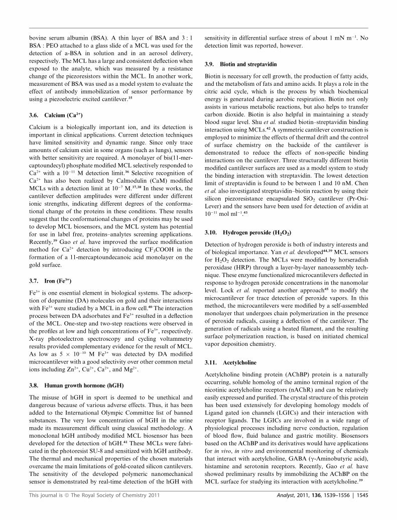
10 years in the human body. Sreepriya et al. reported a MCL
based biosensor for the detection of Cd(II) using an antigen–
antibody based method.19 The gold surface of the MCL was
This journal is ª The Royal Society of Chemistry 2011
modified with a multilayer of 2A81G5 antibody (Fig. 4). When
the MCL was exposed to Cd(II)–EDTA–BSA antigen complex
bending of the MCL was observed and the bending amplitude
was proportional to the concentration of the BSA antigen
complex. Selectivity experiments showed no bending of the MCL
had been observed on exposure to Hg(II), Pb(II) and Mg(II) at
low concentrations. A lowest detection limit for this biosensor is
10�9 M of Cd(II).
3. Biosensors for biochemicals
3.1. Myoglobin
Creatin kinase and myoglobin are two important cardiac
biomarker proteins. As the development or absence of these
proteins strongly predicts the individual mortality risk of
a patient and has immediate therapeutic implications, contin-
uous monitoring of a combination of these markers in real time
would be very attractive. Arntz et al. developed a continuous
label-free detection method for these two cardiac biomarker
proteins using an array of microfabricated MCLs functionalized
with covalently anchored anti-creatin kinase and anti-myoglobin
antibodies.20 The results showed that the sensitivity achieved for
myoglobin detection is below 20 mg ml�1. Both myoglobin and
creatin kinase could be detected independently using cantilevers
functionalized with the corresponding antibodies, in unspecific
protein background. In another work, the anti-Myoglobin
antibody (MAb) modified MCLs have been kept in a sucrose
solution that can maintain the antibody’s stability to up to
7 weeks.21 In a comparative Enzyme Linked Immunosorbent
Assay (ELISA) work, the MCL was coated with a fluorescein
isothiocyanate (FITC) labeled secondary antibody. The fluo-
rescent changes demonstrated the binding between myoglobin
and the MAb used. Control experiments showed that BSA
modified MCLs did not bend on exposure to myoglobin. Kang
et al. showed a detection limit of 1 mM was achieved using MAb
modified MCLs for recognition of myoglobin.22 A site-directed
biotinylated MAb was used in place of a randomly biotinylated
MAb to improve the sensitivity of the sensor. A piezoelectric
MCL modified by site-directed antibody showed a 10 fold
increase in the sensitivity than those modified by the randomly
bound antibodies.
Fig. 4 LbL nanoassembly with intercalated antibody on the MCL
surface. Reprinted from ref. 18 with permission from Elsevier B.V. PEI is
Polyethylenimine, MCES represents 2-mercaptoethane sulfonic acid, and
PSS refers to poly(sodium 4-styrenesulfonate).
This journal is ª The Royal Society of Chemistry 2011
3.2. Glucose
Diabetes is among the most prevalent and costly diseases in the
world. Diabetes and its associated complications are leading
causes of death and disability in the world. Approximately 17
million people in the United States, or 6.2% of the population,
have diabetes. The diagnosis and management of diabetes
require daily monitoring of blood glucose levels. Glucose oxidase
(GOx) was used to develop MCL based biosensors for glucose
detection. The MCL-based glucose biosensor was modified by
poly-L-lysine or glutaraldehyde with GOx.23,24 The MCL bent
upon exposure to glucose solutions. A theoretical study has been
made to study the effect of interaction and heat on the bending of
the MCL and the work suggested that the bending was caused by
the interaction of glucose with GOx enzyme. Another extensive
study further proved the formational change of the proteins has
the main contribution to the MCL bending.25 In this work, the
GOx was immobilized on the MCL by a layer-by-layer multi-
layer approach. The flow rate, concentration of glucose, repro-
ducibility, selectivity, effect of pH, and effect of H2O2 have been
studied. This multilayer film approach results in a 10% repro-
ducibility for glucose measurement (Fig. 5 and 6). Selectivity
experiments showed the MCLs did not respond to the same
concentrations of mannose, fructose and galactose. It is note-
worthy that the back side of the MCL was modified by a layer of
perfluorocarbons silane (tridecafluoro-1,1,2,2-tetrahydrooctyl)
triethoxysilane (TTS), to avoid the accumulation of GOx and
polyelectrolytes on the backside of the MCL.26 Another GOx
based MCL sensor was modified by bovine serum albumin
(BSA), glutaraldehyde (GA) and glucose oxidase (GOx). All
these MCL sensors could measure glucose concentration in the
range of 0.5 to 10 mM, which is of clinical interest.
In a recent study, poly(N-isopropylacrylamide)-co-poly-
(acrylic acid)-(3-aminophenyl-boronic acid) (PNIPAAM-co-
PAA-PBA), glucose responsive polymer brushes were
immobilized on gold substrates and microcantilever arrays for
glucose sensing. The work demonstrates that stimulus-responsive
polymer brushes on micromechanical cantilevers have a signifi-
cantly larger bending response than that of self-assembled
monolayers.27 Huang et al. reported28 a microcantilever sensor
based on a similar glucose-specific polymer, poly(acrylamide-
ran-3-acrylamidophenylboronic acid) (PAA-ran-PAAPBA).
Glucose binds reversibly to the phenylboronic acid moiety of the
polymer (Fig. 7). This results in a viscosity change of the sensing
solution, which is obtained by measuring the damped cantilever
vibration. The glucose response time constant of the sensor is
approximately 3 min, which is shorter than a 5 min response time
of commercially available continuous glucose monitoring
sensors such as Guardian REAL-Time Continuous Glucose
Monitoring System, Dexcom SEVEN Plus, and MiniMed
Paradigm� REAL-Time System.
3.3. Fructose
Baker et al. showed that microcantilevers that are modified by
a thiolated phenylboronic acid derivative deflected when exposed to
D-(�)-fructose in aqueous buffer and the surface stress was linear in
analyte concentration over the 0–25 mM range, suggesting poten-
tial applications in biomass process monitoring and glucose assay.29
Analyst, 2011, 136, 1539–1556 | 1543
Fig. 5 Bending response of a (PEI/GOx)3-modified MCL to various concentrations of glucose in a 0.01 M NaCl solution. Reprinted from ref. 24 with
permission from American Chemical Society.
Fig. 6 Ten replications of bending responses as a function of time for
a (GOx/PEI)3 multilayer-modified MCL following injection of a 10 mM
glucose concentration in 0.01 M NaCl solution (the injection point is
indicated with arrows). Reprinted from ref. 24 with permission from
American Chemical Society.
Fig. 7 Biocompatible, glucose-sensitive polymer poly(acrylamide-ran-3-
acrylamidophenylboronic acid) (PAA-ran-PAAPBA). (a) The polymer
composition and mechanism of interaction with glucose. (b) Glucose-
induced viscosity change of a 1.9% PAA-ran-PAAPBA solution in PBS
buffer (pH 7.4). Reprinted from ref. 27 with permission from Elsevier
B.V.
1544 | Analyst, 2011, 136, 1539–1556
3.4. C-reactive protein (CRP)
A highly sensitive CRP biosensor could provide a powerful
method to predict risk of heart attack. All the developed MCL
biosensors for CRP are based on antibody–antigen interaction.
Lee et al. fabricated the CRP biosensors based on monolithic
SiO2/Ta/Pt/PZT/Pt/SiO2 or SiO2/Th/Pt/PZT/Pt/SiO2 MCLs
showing nanogram level per millilitre sensitivity by measuring
the resonant frequency shift.30,31 The resonance frequency change
of the piezoelectric MCLs was due to a combination of mass
loading and spring constant variation arisen from antigen–anti-
body interaction of CRP. The experimentally measured resonant
frequency shift was larger than that of theoretically calculated
resonant frequency by two orders of magnitude due to
a compressive stress arising from CRP antigen–antibody inter-
action. In several other reports, Kwon et al. reported a CRP
biosensor by using a piezoelectric thick film MCL, which exhibits
the high quality factor (Q ¼ 15) in a viscous liquid at a viscosity
that mimics that of the human blood serum.32 Wee et al. used
a piezoresistive approach to detect CRP.33 Their work demon-
strated the bending and resistance change of the MCL upon its
exposure to CRP solutions. Besides CRP for cardiac disease,
their MCL biosensors modified by other antibodies were used for
the detection of prostate specific antigen (PSA), which is
a specific marker of prostate cancer and cardiac disease.
3.5. Bovine serum albumin (BSA)
Serum albumin, often referred to simply as albumin, is the most
abundant plasma protein in humans and other mammals.
Albumin is essential for maintaining the osmotic pressure needed
for proper distribution of body fluids between intravascular
compartments and body tissues. It also acts as a plasma carrier
by non-specifically binding several hydrophobic steroid
hormones and as a transport protein for hemin and fatty acids.
Adsorption of IgG (one type of immunoglobulins produced by
plasma cells) and Bovine Serum albumin (BSA) on the MCL
surface was studied Moulin et al.34 A very slow microcantilever
bending response upon antibodies injection occurs over more
than 10 h for both antibodies. This slow process was found to be
not associated with adsorption of additional proteins. Two
explanations included the expansion of the protein after surface
adsorption and the proteins rearrangement caused by attractive
(hydrophobic) protein–protein interactions. Piezoresistive and
piezoelectric approaches have also been applied to study the
interaction of anti-bovine serum albumin antibody (a-BSA) with
This journal is ª The Royal Society of Chemistry 2011
bovine serum albumin (BSA). A thin layer of BSA and 3 : 1
BSA : PEO attached to a glass slide of a MCL was used for the
detection of a-BSA in solution and in an aerosol delivery,
respectively. The MCL has a large and consistent deflection when
exposed to the analyte, which was measured by a resistance
change of the piezoresistors within the MCL. In another work,
measurement of BSA was used as a model system to evaluate the
effect of antibody immobilization of sensor performance by
using a piezoelectric excited cantilever.35
3.6. Calcium (Ca2+)
Calcium is a biologically important ion, and its detection is
important in clinical applications. Current detection techniques
have limited sensitivity and dynamic range. Since only trace
amounts of calcium exist in some organs (such as lungs), sensors
with better sensitivity are required. A monolayer of bis(11-mer-
captoundecyl) phosphate modified MCL selectively responded to
Ca2+ with a 10�11 M detection limit.36 Selective recognition of
Ca2+ has also been realized by Calmodulin (CaM) modified
MCLs with a detection limit at 10�7 M.37,38 In these works, the
cantilever deflection amplitudes were different under different
ionic strengths, indicating different degrees of the conforma-
tional change of the proteins in these conditions. These results
suggest that the conformational changes of proteins may be used
to develop MCL biosensors, and the MCL system has potential
for use in label free, proteins–analytes screening applications.
Recently,39 Gao et al. have improved the surface modification
method for Ca2+ detection by introducing CF3COOH in the
formation of a 11-mercaptoundecanoic acid monolayer on the
gold surface.
3.7. Iron (Fe3+)
Fe3+ is one essential element in biological systems. The adsorp-
tion of dopamine (DA) molecules on gold and their interactions
with Fe3+ were studied by a MCL in a flow cell.40 The interaction
process between DA adsorbates and Fe3+ resulted in a deflection
of the MCL. One-step and two-step reactions were observed in
the profiles at low and high concentrations of Fe3+, respectively.
X-ray photoelectron spectroscopy and cycling voltammetry
results provided complementary evidence for the result of MCL.
As low as 5 � 10�10 M Fe3+ was detected by DA modified
microcantilever with a good selectivity over other common metal
ions including Zn2+, Cu2+, Ca2+, and Mg2+.
3.8. Human growth hormone (hGH)
The misuse of hGH in sport is deemed to be unethical and
dangerous because of various adverse effects. Thus, it has been
added to the International Olympic Committee list of banned
substances. The very low concentration of hGH in the urine
made its measurement difficult using classical methodology. A
monoclonal hGH antibody modified MCL biosensor has been
developed for the detection of hGH.41 These MCLs were fabri-
cated in the photoresist SU-8 and sensitized with hGH antibody.
The thermal and mechanical properties of the chosen materials
overcame the main limitations of gold-coated silicon cantilevers.
The sensitivity of the developed polymeric nanomechanical
sensor is demonstrated by real-time detection of the hGH with
This journal is ª The Royal Society of Chemistry 2011
sensitivity in differential surface stress of about 1 mN m�1. No
detection limit was reported, however.
3.9. Biotin and streptavidin
Biotin is necessary for cell growth, the production of fatty acids,
and the metabolism of fats and amino acids. It plays a role in the
citric acid cycle, which is the process by which biochemical
energy is generated during aerobic respiration. Biotin not only
assists in various metabolic reactions, but also helps to transfer
carbon dioxide. Biotin is also helpful in maintaining a steady
blood sugar level. Shu et al. studied biotin–streptavidin binding
interaction using MCLs.42 A symmetric cantilever construction is
employed to minimize the effects of thermal drift and the control
of surface chemistry on the backside of the cantilever is
demonstrated to reduce the effects of non-specific binding
interactions on the cantilever. Three structurally different biotin
modified cantilever surfaces are used as a model system to study
the binding interaction with streptavidin. The lowest detection
limit of streptavidin is found to be between 1 and 10 nM. Chen
et al. also investigated streptavidin–biotin reaction by using their
silicon piezoresistance encapsulated SiO2 cantilever (Pr-Oxi-
Lever) and the sensors have been used for detection of avidin at
10�11 mol ml�1.43
3.10. Hydrogen peroxide (H2O2)
Detection of hydrogen peroxide is both of industry interests and
of biological importance. Yan et al. developed44,39 MCL sensors
for H2O2 detection. The MCLs were modified by horseradish
peroxidase (HRP) through a layer-by-layer nanoassembly tech-
nique. These enzyme functionalized microcantilevers deflected in
response to hydrogen peroxide concentrations in the nanomolar
level. Lock et al. reported another approach45 to modify the
microcantilever for trace detection of peroxide vapors. In this
method, the microcantilevers were modified by a self-assembled
monolayer that undergoes chain polymerization in the presence
of peroxide radicals, causing a deflection of the cantilever. The
generation of radicals using a heated filament, and the resulting
surface polymerization reaction, is based on initiated chemical
vapor deposition chemistry.
3.11. Acetylcholine
Acetylcholine binding protein (AChBP) protein is a naturally
occurring, soluble homolog of the amino terminal region of the
nicotinic acetylcholine receptors (nAChR) and can be relatively
easily expressed and purified. The crystal structure of this protein
has been used extensively for developing homology models of
Ligand gated ion channels (LGICs) and their interaction with
receptor ligands. The LGICs are involved in a wide range of
physiological processes including nerve conduction, regulation
of blood flow, fluid balance and gastric motility. Biosensors
based on the AChBP and its derivatives would have applications
for in vivo, in vitro and environmental monitoring of chemicals
that interact with acetylcholine, GABA (g-Aminobutyric acid),
histamine and serotonin receptors. Recently, Gao et al. have
showed preliminary results by immobilizing the AChBP on the
MCL surface for studying its interaction with acetylcholine.39
Analyst, 2011, 136, 1539–1556 | 1545

3.12. Protein kinase (PKA)
PKA is a protein kinase that modifies other proteins by chemi-
cally adding phosphate groups to them. Assessment of activated
PKA holds a great promise in analytical applications and clinical
medicine. A highly sensitive MCL biosensor assay based on an
electrical measurement has been developed for detecting acti-
vated PKA.46 The MCL surface was modified by a heat-stable
protein kinase inhibitor, PKI-(5–24) peptide, for capturing PKA
(Fig. 8). An increase in the resonant frequency shift was observed
when the PKA binds to PKI-(5–24). Synergistic interactions of
adenosine triphosphate (ATP) and the peptide inhibitor with the
kinase were also investigated by a solution phase capillary elec-
trophoretic assay, and by surface plasmon resonance technology.
The detection limit can be as low as 6.6 pM, exhibiting much
higher sensitivity and wider dynamic range than the conventional
activity assay.
3.13. Prostate specific antigen (PSA)
PSA that is detectable in serum has proved to be a useful marker
for early detection of prostate cancer and in monitoring patients
for disease progression and the effects of treatment. Wu et al.
have used a polyclonal anti-PSA antibody on the MCL47 to
detect free PSA (fPSA) concentrations from 0.2 ng ml�1 to 60 mg
ml�1, which includes the clinically relevant diagnostic PSA
concentration range. The sensor could be able to detect fPSA
even against the simulated background ‘‘noise’’ of unrelated
human serum proteins such as HP and HSA or nonhuman serum
protein such as bovine serum albumin (BSA), which was present
at concentrations as high as 1 mg ml�1 (Fig. 9 and 10). In another
work, a resonant cantilever sensor system for liquid-phase
applications is presented.48 The monolithic system consists of an
Fig. 8 Nanomechanical detection of PKA catalytic subunit on a functiona
surface of the PZT cantilever functionalized with biotin–PKI-(5–24) via the b
after incubation with varying concentrations of PKA in the presence (-) or a
from the frequency of the negative control cantilever was plotted as a functio
cantilever devices (5 cm � 5 cm) with 12 arrays of 50 mm � 150 mm were use
1546 | Analyst, 2011, 136, 1539–1556
array of four electromagnetically actuated cantilevers with
transistor-based readout, an analog feedback circuit, and
a digital interface. A package, which protects the electrical
components and the associated circuitry against liquid exposure,
allows for a stable operation of the resonant cantilevers in liquid
environments. The device is operated at the fundamental canti-
lever resonance frequency of�200 kHz in water with a frequency
stability better than 3 Hz. The use of the integrated CMOS
resonant cantilever system as a biosensor for the detection of
biomarkers, such as PSA, is demonstrated. By functionalizing
the MCLs with anti-PSA, the PSA has been detected at
concentration levels as low as 10 ng ml�1 in a sample fluid.
In a recent work, Lee et al. enhanced the sensitivity by
applying PSA polyclonal antibody (PSA pAb) as a surface stress
inducer and PSA polyclonal antibody-conjugated silica nano-
particles (pAb-SiNPs) as mass inducers have been applied to the
PSA-captured microcantilevers.49 They have confirmed the
sensitivity enhancement effects (2–4 times enhanced at the same
concentrations) enough to detect PSA at low picogram levels
(LOD of 1 pg ml�1 or below).
3.14. Human serum albumin (HSA)
Measurement of HSA has been used as model systems to study
antibody–antigen or protein-surface interactions. Stolyarova
et al. demonstrated that composite porous silicon-crystalline
MCLs provided an excellent biocompatible material for immo-
bilization of a wide variety of biological materials, resulting in
enhanced sensitivity as demonstrated on the covalently immo-
bilized antibody binding its complementary antigen.50 In another
work, Campbell and Mutharasan demonstrated the HSA
adsorption decreased in the order of CH3 > COOH > OH on
these surfaces by using resonating MCLs.51
lized cantilever. (A) Schematic representation of the binding on the Au
iotin–streptavidin interaction. (B) The resonant frequency was measured
bsence of 100 mM ATP (C), and the resonant frequency shift subtracted
n of the concentration of PKA catalytic subunit. Nanomechanical PZT
d. Reprinted from ref. 45 with permission from Elsevier B.V.
This journal is ª The Royal Society of Chemistry 2011

Fig. 9 Diagram of interactions between target and probe molecules on
a MCL. Specific biomolecular interactions between target and probe
molecules alter the intermolecular nanomechanical interactions within
a self-assembled monolayer on one side of a MCL. This can produce
a sufficiently large force to bend the cantilever beam and generate
motion. Reprinted from ref. 46 with permission from Nature Publishing
Group.
3.15. Single-chain Fv (scFv)
Single-chain Fv (scFv) is a fragment of an antibody with
a molecular mass of 28 kDa and is the smallest antibody entity
comprising of an intact antigen-binding site. Backmann et al.
reported a MCL based immunosensor for the detection of
different antigens using single-chain Fv (scFv) antibody frag-
ments as a receptor.52 The MCL was treated in such a way that
one side of the MCL is a protein repellant and the other side is
coated with the antibody. AR-GCN4, an antigen created by the
genetic fusion of the antigenic peptide GCN4(7P14P) to an
ankyrin (AR) MBP13-6, was used as a antigen to bind to the
scFv on the MCL surface. The sensitivity of the sensor is�1 nM.
3.16. Antibodies
Typically, the MCL surfaces were modified by antibodies to
recognize antigens of interest. It is obvious that antigens
Fig. 10 Detection of free PSA (fPSA). (A) Cantilever deflection versus time
using 200 mm long and 0.5 mm thick silicon nitride microcantilevers. fPSA dete
this cantilever geometry. (B) Specificity of fPSA detection against a high back
and human plasminogen (HP). Reprinted from ref. 46 with permission from
This journal is ª The Royal Society of Chemistry 2011
modified MCLs can be used to detect antibodies as well, which is
an indirect way to detect substances on a surface. One such
example is the interaction of monoclonal antibody (mAb) in
solution with a herbicide 2,4-dichlorophenoxyacetic acid (2,4-D)
immobilized on a MCL surface.53 The sensitivity obtained was
5 mg ml�1.
3.17. Cytokine
Cytokines are a category of signaling proteins and glycoproteins
that, like hormones and neurotransmitters, are used extensively
in cellular communication. Dutta et al. reported cytokine
detection using nanostructured MCLs. The MCLs were func-
tionalized using anti-human interleukin-1 beta (anti HIL 1-b)
and then exposed to different concentrations of cytokine HIL 1-b
in the ppb to ppm ranges.54 A detection limit of 0.5 ppm mole-
cules of HIL 1-b was achieved. This type of MCLs arrays func-
tionalized with antibodies can be used in the label-free analysis of
multiple proteins in a single step.
3.18. Human immunodeficiency virus (HIV)
HIV-1 is a lentivirus (a member of the retrovirus family) that can
lead to acquired immunodeficiency syndrome (AIDS), a condi-
tion in humans in which the immune system begins to fail,
leading to life-threatening opportunistic infections. Lam et al.
reported monoclonal antibodies (mAbs) A32 or T8 modified
MCL biosensors for detecting human immunodeficiency virus
type 1 (HIV-1) envelope glycoprotein (Env) gp120 from solu-
tion.55 Subsequent exposure to mAb 17b, a known substrate to
bind on gp120, further increased deflection of A32- but not
T8-presenting MCLs. The detection limits were 8 mg ml�1 gp120
and 0.17 mg ml�1 17b.
3.19. Human oestrogen receptor protein
Human oestrogen receptor beta protein was found in a well
defined set of breast cancers. The significance of oestrogen
receptor beta protein expression in breast cancers to therapy
remains to be determined but the availability of detecting oes-
trogen receptor beta in archive material will facilitate the process.
A piezoresistive MCL was reported for the detection of specific
for fPSA detection sensitivity against a background of 1 mg ml�1 of BSA
ction was feasible over a concentration range 6 ng ml�1 to 60 mg ml�1 using
ground of human serum proteins, namely, human serum albumin (HSA)
Nature Publishing Group.
Analyst, 2011, 136, 1539–1556 | 1547

protein conformations.56 In this work, oestradiol (E2) was pre-
bound on a human oestrogen receptor (EPa-LBD) protein,
which changes the conformation of the protein. The EPa-LBD
and EPa-LBD-E2 complex were distinguished by the MCLs
modified by conformation-specific peptides a/bI (Ser-Ser-Asn-
His-Gln-Ser-Ser-Arg-Leu-Ile-Glu-Leu-Leu-Ser-Arg), which
recognizes EPa-LBD-E2 complex and a/bII (Ser-Ala-Pro-Arg-
Ala-Thr-Ile-Ser-His-Tyr-Leu-Met-Gly-Gly), which recognizes
EPa-LBD (Fig. 11). This is an indirect and complementary
approach for studying conformational change in proteins. It
could be used when the conformational change of the protein is
too small and does not produce detectable bending of the MCL.
A sensitivity of 2.5 nM has been obtained using this method.
3.20. Lipid bilayer
Pera et al. demonstrated that MCLs can sense the formation of
supported phospholipid bilayers on a surface and can monitor
changes in mechanical property of lipid bilayers.57 The formation
of bilayers led to a bending of the MCLs of 70–590 nm compa-
rable to a surface stress of 27–224 mN m�1. Physisorption of
bilayers of 1,2-dioleoyl-sn-glycero-3-phospho-choline (DOPC)
on the silicon oxide surface of cantilevers led to a tensile bending
of about 70 nm. The formation of chemisorbed bilayers of mixed
thiolated 1,2-dipalmitoyl-sn-glycero-3-phosphothioethanol
(DPPTE) and DOPC on the gold side of cantilevers led to
a compressive bending of nearly 600 nm depending on the ratio
of DPPTE to DOPC. The results demonstrate that MCL sensors
Fig. 11 (a) Schematic drawing showing a two-cantilever configuration,
the a/bI attached on one cantilever and the a/bII on the other, and the
preferential binding of ERa-LBD, E2-bound or free, onto the a/bI and
a/bII, respectively. (b) A cartoon showing the sensor layer on top of the
surface and the blocking layer at the bottom surface of a cantilever and its
bending upon target binding onto the top sensing surface. Reprinted
from ref. 55 with permission from American Chemical Society.
1548 | Analyst, 2011, 136, 1539–1556
with immobilized bilayers can be used as model systems to
investigate mechanical properties of cellular membranes and may
be used for screening of membrane processes involving modifi-
cation, lateral expansion, or contraction of membranes.
3.21. Low density lipoproteins (LDL)
It is of clinic interest to detect and differentiate between low-
density lipoproteins (LDL) and their oxidized form (oxLDL).
This is because their uptake from plasma, principally favored to
the oxidized form, is believed to be responsible for the accumu-
lation of cholesterol in the aortic intima and is associated with
the first stage of coronary heart disease. A LDL and oxLDL
differentiation by MCL sensors as shown in Fig. 12.58
3.22. a-Amino acid and peptides
The a-amino acids represent one of the most important classes of
substances in nature that incorporate a stereogenic center and,
therefore, exemplify an excellent model system to demonstrate
chiral discrimination. Enantioselective antibodies modified
cantilevers have been investigated for their stereoselective
detection of trace amounts of an important class of chiral ana-
lytes, the a-amino acids.59 This is the first demonstration of chiral
discrimination using highly scalable microelectromechanical
systems. The antibodies used were raised in such a way that they
selectively bind to either D- or L-R-amino acids. The temporal
response of the cantilever (Ddeflection/Dtime) is linearly
proportional to the analyte concentration and allows the quan-
titative determination of enantiomeric purity up to an enantio-
meric excess of 99.8%. Besides amino acid, the interaction of
peptides with antibodies was also studied.60
3.23. Cyclin-dependent protein kinase (CDK2)
CDK2 is an important indicator for the cellular decision making
for proliferation, during cancer. A MCL based sensor was
developed for the detection of human CDK2.61 The Au surface
of the MCL was modified with Stefin A Triple Mutant (STM)
which is known to interact with CDK2. The cell lysate that
constitutes CDK2 was directly exposed to the MCL surface and
a lowest concentration of 80 nM CDK2 was detected.
3.24. Clenbuterol and chloramphenicol
Tan et al. developed a MCL sensor for the detection of
b-adrenergic agonist clenbuterol and the antibiotic chloram-
phenicol.62 The Au side of the MCL was modified with protein A
and antibodies for clenbuterol and chloramphenicol. These
MCLs were exposed to various concentrations of clenbuterol
and chloramphenicol and a detection limit of 0.1 ng ml�1 for
clenbuterol and 0.2 ng ml�1 for chloramphenicol was observed.
Such LODs were better than that of the corresponding direct
competitive enzyme-linked immunosorbent assay (dcELISA).
The results suggest that microcantilever immunosensors are
suitable for detection of small molecules, and the assay sensitivity
is mainly related to the quality and activities of the antibodies.
This journal is ª The Royal Society of Chemistry 2011

Fig. 12 A) Schematic of the functionalization of the cantilever top surface with heparin. (B) Comparison of the effects triggered on the cantilever by an
10 ml injection of (a) 3.5 mg ml�1 LDL and (b) 0.3 mg ml�1 oxLDL. Reprinted from ref. 57 with permission from Elsevier B.V.
3.25. Human epidermal growth factor receptor 2 (Her2)
Her2 is an antigen that is over expressed in 20–25% of invasive
breast cancers. The Her2 concentration is 2–15 ng ml�1 in normal
mammalians and 15–75 ng ml�1 in breast cancer patients.
Capobianco et al. reported63 the detection of human epidermal
growth factor receptor 2 (Her2) with piezoelectric MCL sensors
in a background of 1 mg ml�1 bovine serum albumin. The MCLs
were modified with single-chain variable fragment (scFv). A
detection limit of 5 ng ml�1 was obtained.
3.26. Immunoglobulin G (IgG)
IgG is a one type of immunoglobulins produced by plasma cells.
In the early study of MCL sensors, Moulin et al. investigated the
MCL responses on the adsorption of IgG on MCL.64 Hill et al.
reported the effect of concentration, reaction time, and pH for
these reagents on the magnitude of the MCL responses using an
anti-immunoglobulin G (anti-IgG) receptor. They also report the
application of optimum and non-optimum conditions to detect
thyroid disrupting chemicals (TDCs) using MCLs functionalized
with the transport protein thyroxine-binding globulin. Selectivity
patterns are reported for several TDCs and sensitive detection of
thyroxin at sub-nM levels is demonstrated.65 Recently, detection
of IgG was used as a model system to show the enhancement of
Fig. 13 (a) In-plane electric field from the nickel electrodes to the gold layer
case (a), as an electric potential is applied between an outer ITO electrode and a
from Elsevier B.V.
This journal is ª The Royal Society of Chemistry 2011
capture antibody immobilization under an electric field
(Fig. 13)66 and biofunctional polymer coatings.67
Characterization and control of proteolysis of peptides by
specific cellular protease are a priori requisite for effective drug
discovery. Kwon et al. reported the in situ monitoring of prote-
olysis of the peptide chain attributed to protease (Cathepsin B).68
Specifically, a peptide chain polyethylene glycol-tetrapeptide
GlyPheLysGly (PEG-GFLG) was immobilized on the surface of
the MCL (Fig. 14) using the cross-linker EDC-NHS and the
detection is based on the measurement of resonant frequency
shift arising from proteolysis of peptides. Cathepsin B Protein
(CTSB) catalytic sites Cys25 and His159 cleave PEG-GLFG
peptide chain, leading to proteolysis of GFLG. This reduces the
mass on the MCL surface and in turn a change in the resonant
frequency. Different concentrations of CTSB were used to check
for detection limit and it was found to be 0.28 mm. The author
mentioned that this implies that the nanomechanical biosensor
enables the characterization of specific cellular protease such as
its kinetics.
3.27. Alpha-fetoprotein (AFP)
AFP is a marker for hepatocellular carcinoma (HCC). AFP
levels of 10–1000 ng ml�1 in the blood serum can be related to
early stage of HCC cancer. Liu et al. developed a sensor for early
stage prognosis and disease diagnosis of HCC based on
of the sensing microcantilever. (b) An additional external electric field in
gold layer of the microcantilever. Reprinted from ref. 65 with permission
Analyst, 2011, 136, 1539–1556 | 1549
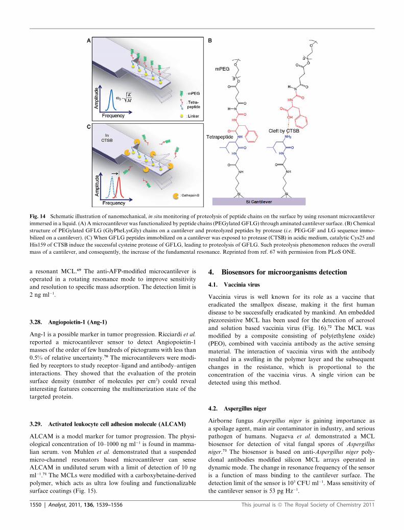
Fig. 14 Schematic illustration of nanomechanical, in situ monitoring of proteolysis of peptide chains on the surface by using resonant microcantilever
immersed in a liquid. (A) A microcantilever was functionalized by peptide chains (PEGylated GFLG) through aminated cantilever surface. (B) Chemical
structure of PEGylated GFLG (GlyPheLysGly) chains on a cantilever and proteolyzed peptides by protease (i.e. PEG-GF and LG sequence immo-
bilized on a cantilever). (C) When GFLG peptides immobilized on a cantilever was exposed to protease (CTSB) in acidic medium, catalytic Cys25 and
His159 of CTSB induce the successful cysteine protease of GFLG, leading to proteolysis of GFLG. Such proteolysis phenomenon reduces the overall
mass of a cantilever, and consequently, the increase of the fundamental resonance. Reprinted from ref. 67 with permission from PLoS ONE.
a resonant MCL.69 The anti-AFP-modified microcantilever is
operated in a rotating resonance mode to improve sensitivity
and resolution to specific mass adsorption. The detection limit is
2 ng ml�1.
3.28. Angiopoietin-1 (Ang-1)
Ang-1 is a possible marker in tumor progression. Ricciardi et al.
reported a microcantilever sensor to detect Angiopoietin-1
masses of the order of few hundreds of pictograms with less than
0.5% of relative uncertainty.70 The microcantilevers were modi-
fied by receptors to study receptor–ligand and antibody–antigen
interactions. They showed that the evaluation of the protein
surface density (number of molecules per cm2) could reveal
interesting features concerning the multimerization state of the
targeted protein.
3.29. Activated leukocyte cell adhesion molecule (ALCAM)
ALCAM is a model marker for tumor progression. The physi-
ological concentration of 10–1000 ng ml�1 is found in mamma-
lian serum. von Muhlen et al. demonstrated that a suspended
micro-channel resonators based microcantilever can sense
ALCAM in undiluted serum with a limit of detection of 10 ng
ml�1.71 The MCLs were modified with a carboxybetaine-derived
polymer, which acts as ultra low fouling and functionalizable
surface coatings (Fig. 15).
1550 | Analyst, 2011, 136, 1539–1556
4. Biosensors for microorganisms detection
4.1. Vaccinia virus
Vaccinia virus is well known for its role as a vaccine that
eradicated the smallpox disease, making it the first human
disease to be successfully eradicated by mankind. An embedded
piezoresistive MCL has been used for the detection of aerosol
and solution based vaccinia virus (Fig. 16).72 The MCL was
modified by a composite consisting of poly(ethylene oxide)
(PEO), combined with vaccinia antibody as the active sensing
material. The interaction of vaccinia virus with the antibody
resulted in a swelling in the polymer layer and the subsequent
changes in the resistance, which is proportional to the
concentration of the vaccinia virus. A single virion can be
detected using this method.
4.2. Aspergillus niger
Airborne fungus Aspergillus niger is gaining importance as
a spoilage agent, main air contaminator in industry, and serious
pathogen of humans. Nugaeva et al. demonstrated a MCL
biosensor for detection of vital fungal spores of Aspergillus
niger.73 The biosensor is based on anti-Aspergillus niger poly-
clonal antibodies modified silicon MCL arrays operated in
dynamic mode. The change in resonance frequency of the sensor
is a function of mass binding to the cantilever surface. The
detection limit of the sensor is 103 CFU ml�1. Mass sensitivity of
the cantilever sensor is 53 pg Hz�1.
This journal is ª The Royal Society of Chemistry 2011

Fig. 15 Antibody functionalization schematic. (a) The structure of the carboxybetaine-derived polymer is shown; ‘‘A’’ represents the structure inside
the large parentheses, two of which are connected by an R–COH–R bridge. The polymer adsorbs directly onto the thin SiO2 layer of the Si micro-
cantilever surfaces (b), immobilizing a monolayer of 275–360 ng cm�2. (c) Terminal carboxylic acids are transformed to reactive NHS esters by injection
of a mixture of NHS and EDC. (d) NHS esters react with primary amines on IgG antibodies to covalently bind them to the surface. Reprinted from ref.
70 with permission from American Chemical Society.
Fig. 16 Basic operation of EPM sensor. Exposure to analyte causes volumetric change in sensing material. This change is measured by embedded
microcantilever. Reprinted from ref. 71 with permission from Elsevier B.V.
4.3. Baker’s yeast
Baker’s yeast is the common name for the strains of yeast
commonly used as a leavening agent in baking bread and related
products, where it converts the fermentable sugars present in the
dough into carbon dioxide and ethanol. Li et al. developed
a novel magnetostrictive MCL sensor concept.74 The detection
model is baker’s yeast. The results demonstrate the feasibility of
this sensor as a high performance biosensor platform. The
oscillation of the magnetic material is wirelessly detected by
a pickup coil. The oscillation amplitude is correlated to the
resonant frequency. Compared to traditional MCL, the new
sensor design has several advantages, including: (1) remote/
wireless driving and sensing; (2) easy to fabricate. More impor-
tantly, it is experimentally found that the quality merit factor
(Q value) of MSMC can reach more than 250, which is much
higher than other cantilevers. A concentration of 1 mg ml�1 of
yeast cells has been detected using this method.
4.4. Bacillus anthracis
Detection of airborne Bacillus anthracis spores an etiology agent
of anthrax has gained significant interest since the anthrax spore
This journal is ª The Royal Society of Chemistry 2011
mailing. Several MCL based sensors have been developed for
detecting Bacillus anthracis. Fu et al. continued their magneto-
strictive MCLs (MSMC) for detection of Bacillus anthracis
spores.75 The MSMC surface was modified with a biological
phage having peptide EPRLSPHS on the surface to recognize
Bacillus anthracis. By monitoring the shift in the resonance
frequency of the MSMCs, the spores were detected in a real-time
manner and a detection limit of 105 spores ml�1 was obtained for
the MSMCs used in this research. Higher sensitivity is expected
for the MSMCs with smaller size.
Davila et al. demonstrated an antibody modified MCL for
detection of Bacillus anthracis Sterne spores in air and liquid.76
They demonstrate that as few as 50 spores on the MCLs can be
detected in water using the thermal noise as excitation source.
Measurement sensitivity of 9.23 Hz fg�1 for air and 0.1 Hz fg�1
for water was obtained. McGovern et al. also further studied
antibodies modified MCLs for detection of Bacillus anthracis
spores.77 They demonstrated specific detection of Bacillus
anthracis (BA) spores from that of close relatives, such as B.
thuringiensis (BT), B. cereus (BC), and B. subtilis (BS) by varying
the flow speed of the sampling liquid over the surface of
a piezoelectric MCL sensor. Spore binding to the anti-BA spore
IgG coated MCL surface is determined by monitoring the
Analyst, 2011, 136, 1539–1556 | 1551

resonance frequency change in the sensor’s impedance versus
frequency spectrum. Their work showed that the change of
resonance frequency from increasing to decreasing occurred at
a lower fluid speed for the spores of BT, BC, and BS than BA.
This trend reduces the cross-reactivity ratio of BC, BS, and BT to
the anti-BA spore IgG immobilized PEMS. This cross-reactivity
ratio of 0.05 was essentially negligible considering the experi-
mental uncertainty. No detection limit was mentioned. Campbell
and Mutharasan also reported the detection of BA from
a mixture of BA, BT, BC, and BS by using their PZT-anchored
piezoelectric excited millimetre-sized cantilever (PAPEMC)
(Fig. 17).78 The surface of PAPEMC was modified by a rabbit
polyclonal antibody (anti-BA). They have detected BA from
a concentration of 1 : 1000 ratios (BA: BT + BC). The detection
limit was 38 BA spores l�1 of air in near real-time with an esti-
mated lower limit of detection of �5 spores l�1 of air in the
configuration tested.79
4.5. Bacillus subtilis
B. subtilis may contaminate food but rarely causes food
poisoning. B. subtilis produces the proteolytic enzyme subtilisin.
B. subtilis spores can survive the extreme heating that is often
used to cook food, and it is responsible for causing ropiness—
a sticky, stringy consistency caused by bacterial production of
long-chain polysaccharides—in spoiled bread dough. Dhayal
et al. demonstrated a short peptide ligand modified MCL for
efficient capture of Bacillus subtilis (a simulant of Bacillus
anthracis) spores in liquids.80 On an eight-MCL array chip, four
MCLs were coated with binding peptide (NHFLPKV-GGGC)
and the other four were coated with control peptide
(LFNKHVP-GGGC). The four MCLs with binding peptide
showed a substantial change in the deflection and resonance
frequency as compared to that for control peptide MCLs.
Further confirmation was obtained by subsequent examination
of the microcantilever arrays under a dark-field microscope.
Applications of this technology will serve as a platform for the
Fig. 17 Transient frequency response of PAPEMC sensor to the binding of 3
pH buffer, pH 1.85. Reprinted from ref. 77 with permission from American
1552 | Analyst, 2011, 136, 1539–1556
detection of pathogenic organisms including biowarfare agents.
A BS spore concentration of 1 � 105 spores ml could be detected
using this method.
4.6. Enterohemorrhagic Escherichia coli serotype (E. coli)
E. coli O157 : H7, first recognized in 1982 in the United States, is
an epidemiologically significant cause of food borne disease
worldwide. E. coli O157 : H7 can readily contaminate ground
beef, raw milk, poultry products, fresh apple cider, cold sand-
wiches, vegetables, and drinking water supplies. It can be
transmitted efficiently from person to person not only via
contaminated food, but also by sharing contaminated facilities.
Zhang and Ji developed the first silicon microcantilever sensor
for the detection of Escherichia coli (E. coli) O157 : H7.81 The
microcantilever was modified by anti-E. coli O157 : H7 anti-
bodies on the silicon surface of the cantilever. When the aquaria
E. coli O157 : H7 positive sample is injected into the fluid cell
where the microcantilever is held, the microcantilever bends
upon the recognition of the E. coli O157 : H7 antigen by the
antibodies on the surface of the microcantilever. The detection
limit of the sensor was 1 � 106 cfu ml�1 when the assay time was
<2 h. Using a self-excited piezoelectric MCL, Campbell et al.
showed the frequency method could improve the sensitivity by
detecting a low concentration of 700 bacteria ml�1.82 The sensi-
tivity has been further improved to a detection limit of 10 cells
ml�1 and 1 cell ml�1.83
Gfeller et al. investigated the E. coli bacterial growth on
a MCL by using the oscillating mode of the MCL.84 The MCL
was coated with a layer of common nutritive layer. The basis of
the detection scheme is the resonance frequency change as
a function of the increasing mass during the cell growing process.
The detection limit is 100 E. coli cells and the sensor detected
active growth of E. coli cells within 1 h. Selectivity can be ach-
ieved by adding antibiotics to the nutritive layers. This new
sensing method for the detection of selective bacterial growth
33 BA spores ml�1 and the release of the bound spores by exposure to low
Chemical Society.
This journal is ª The Royal Society of Chemistry 2011

allows future applications in, e.g., rapid antibiotic susceptibility
testing.
Capobianco et al. examined a piezoelectric coating PbMg1/
3Nb2/3O3 0.63–PbTiO3 0.37 PMN-PT/tin to on a MCL sensor
for in situ recognition of E. coli O157 : H7.85 The mass detection
sensitivity m/f ¼�3 � 2 � 10�12 g Hz�1 and the concentration
sensitivity is better than 100 cells ml�1 in liquid.
Fu et al. developed a sensor (Fig. 18) for E. coli by using
a magnetostrictive microcantilever (MSMC).86 The MSMC
surface was modified by polyclonal antibody and the detection
limit is 105 cfu ml�1.
4.7. Salmonella typhimurium
Salmonella typhimurium multiplies in the gastrointestinal tract of
many animal species where it usually causes no disease, but in
humans its growth causes gastroenteritis. Recently, Zhu et al.
demonstrated an antibody modified MCL for in situ recognition
of Salmonella typhimurium.87 The MCL was coated with a lead
zirconate titanate (PZT) and partially dipped in the suspensive
without electrically insulating the PZT. A mass detection sensi-
tivity of Dm/Df ¼ �5 � 10�11 g Hz�1 was achieved and the
concentration sensitivities were 1 � 103 and 500 cells ml�1 in 2 ml
of liquid with a 1 and 1.5 mm dipping depth, respectively. It is
more than two orders of magnitude lower than the infectious
dose and more than one order of magnitude lower than the
detection limit of a commercial Raptor sensor.
A new type of cantilever has been reported for the in situ
detection of various compounds or microorganisms. Salmonella
typhimurium (ST) has been used as an example for this purpose.
The cantilever surface was modified in such a way that it had the
Salmonella typhimurium antibody on its surface and then it was
dipped in a solution containing of Salmonella typhimurium up to
0.8 mm depth. The mass detection sensitivity of the cantilever
observed was Dm/Df¼�5 to 6 � 10�11 g Hz�1 and the number of
cells captured on the surface was also counted.
4.8. Ricin
Ricin, a biotoxin extracted from the seeds of the castor bean plant
(Ricinus communis), is poisonous to people, animals, and insects.
A few milligrams can kill an adult human being. Ricin is a potent
cytotoxin that works by getting inside cells and preventing them
from making essential proteins. Ricin can poison people by
inhalation or ingestion. The symptoms of human poisoning,
abdominal pain and severe dehydration, occur within hours of
ingestion, and death occurs within 36 to72 hours. The results
Fig. 18 Schematic illustration of the operation principle of MSMC as
transducer for biosensor. Reprinted from ref. 85 with permission from
Elsevier B.V.
This journal is ª The Royal Society of Chemistry 2011
showed that a detection limit of 40 parts per trillion for ricin has
been achieved by using the deflection mode of the MCLs.88
4.9. Tularemia
Tularemia (Francisella tularensis) is a small (0.2 mm � 0.2–
0.7 mm), pleomorphic, poorly staining, nonmotile, Gram-nega-
tive aerobic coccobacillus. It is one of the most infectious
pathogenic bacteria known. Francisella tularensis biovar
tularensis (type A) may be highly virulent in humans and
animals, produces acid from glycerol, demonstrates citrulline
ureidase activity, and is the most common biovar isolated in
North America. Virulent, streptomycin-resistant F. tularensis
strains have been examined in biowarfare agent studies. Yan
et al. have demonstrated MCL sensors for the detection of
tularemia (F. tularensis) by using the deflection approach.88 The
MCLs on which antibodies were immobilized were used for both
experiments. A detection limit of less than 1 � 103 organisms ml
was determined for F. tularensis after exposure at room
temperature.
4.10. Cryptosporidum
Cryptosporidium parvum oocyst is a waterborne parasitic proto-
zoan that can cause severe illnesses in humans called crypto-
sporidiosis. Campbell and Mutharasan demonstrated the
detection of Cryptosporidium parvum using the piezoelectric-
excited millimetre size cantilever.89 The cantilever was function-
alized with immunoglobulin M (IgM). The detection of 100,
1000, and 10 000 oocysts ml�1 was achieved with a positive
sensor response in less than 1 min.
Besides these sensors, there are also MCL based biosensors for
physical properties. One such example is MCLs modified
bacteriorhodopsin (BR) for light detection. This device can be
used to directly convert the solar energy to mechanical energy. In
one report,90 purple membranes from Halobacterium salinarum
were deposited electrophoretically on platinum-coated MCLs.
By illuminating the bacteriorhodopsin (BR)-containing purple
membranes, the protein undergoes its photochemical reaction
cycle, during which a conformational change occurs in the
protein, changing its shape and size. The on–off change occurs in
millisecond. Using polarized light, the orientation of the motion
was detected, relative to the transition moment of the retinal. In
air, the smaller dilatation of the protein could be explained as
a smaller conformational change than that in water because the
dried protein is more rigid. The average energy per BR molecule
contributing to MCL bending was estimated to be 195 kT
(in terms of the Boltzmann energy at 295 K). This calculated
energy provides an estimate of the order of magnitude and
compares to the energy of a photon of 84 kT with a wavelength
of 580 nm, which triggers the photocycle of BR.
In another paper,91 the same BR protein model was used for
the photocycle and the author attributed the MCL bending to
proton release caused by the conformational change in BR
(Fig. 19).
4.11. Bacterial virus T5
Membrane proteins are central to many biological processes, and
protein receptors–ligands interactions are of fundamental
Analyst, 2011, 136, 1539–1556 | 1553

Fig. 19 Schematic illustration of assembling the purple membrane on
the microcantilever surface and the experimental system. (a) Procedures
for assembling the purple membrane on the solid surface. (b) Illuminated
by visible light, bR pumps the proton across the membrane to the
interface between the microcantilever surface and the purple membrane.
(c) The optical beam deflection is used to measure the deflection of the
microcantilever. (d) Illuminated by visible light, the beam bends away
from the purple membrane side. Reprinted from ref. 90 with permission
from Institute of Physics Publishing.
importance in medical research. Braun et al. reported the
detection of bacterial virus T5 at subpicomolar concentrations
with a microcantilever that is modified by a protein receptor, the
FhuA receptor of E. coli.92 A detection limit of 300 fM was
observed with good repeatability. These experiments demon-
strate the potential of resonating microcantilevers for time-
resolved detection of membrane protein–ligand interactions in
a micro-array format.
4.12. Severe Acute Respiratory Syndrome associated
coronavirus (SARS-CoV)
SARS is a viral respiratory disease. Velanki and Ji demon-
strated93 the feasibility of detecting Severe Acute Respiratory
Syndrome associated coronavirus (SARS-CoV) using micro-
cantilever technology by showing that Feline Coronavirus (FIP)
type I virus can be detected by a microcantilever modified by
Feline coronavirus (FIP) type I anti-viral antiserum. A micro-
cantilever modified by FIP type I anti-viral antiserum was
developed for the detection of FIP type I virus. The detection
limit of the sensor was 0.1 mg ml�1 when the assay time was <1 h.
5. Conclusion
To make information on individual biosensors easily accessible,
the biosensors in this review are classified according to the ana-
lytes. We did this to aid researchers to locate relevant references.
Biosensors based on microcantilevers have been described for
nearly 50 analytes. In some cases, several different receptors are
used for the same analyte. This number of analytes reveals the
success of the microcantilever approach to chemical and bio-
logical sensing. However, it is too early to summarize and
compare the selectivity, detection range, lifetime of the sensors
since many publications did not provide the information. This
1554 | Analyst, 2011, 136, 1539–1556
may be related to the fact that the microcantilever-based sensors
have not been commercialized. The development and improve-
ment of microcantilever sensors that will allow measurements of
analytes in fields and in complex real-life samples will remain
a central issue in the development of such sensors.
Acknowledgements
We would like to thank National Institutes of Health (NIH)
Grant Number 1R01NS057366 and National Natural Science
Foundation of China (NSFC) 20728506/B05.
References
1 J. K. Gimzewski, C. Gerber, E. Meyer and R. R. Schlittler,Observation of a chemical reaction using a micromechanical sensor,Chem. Phys. Lett., 1994, 217, 589–594.
2 T. Thundat, R. J. Warmack, G. Y. Chen and D. P. Allison, Thermaland ambient-induced deflections of scanning force microscopecantilevers, Appl. Phys. Lett., 1994, 64, 2894–2896.
3 G. G. Stoney, The transition of metallic films deposited byelectrolysis, Proc. R. Soc. London, 1909, 82, 172–175.
4 H. F. Ji and B. Armon, Approaches to increasing surface stress forimproving signal-to-noise ratio of microcantilever sensors, Anal.Chem., 2010, 82, 1634–1642.
5 X. Yan, H.-F. Ji and T. Thundat, Microcantilever (MCL) biosensing,Curr. Anal. Chem., 2006, 2, 297–307.
6 L. G. Carrascosa, M. Moreno, M. Alvarez and L. M. Lechuga,Nanomechanical biosensors: a new sensing tool, TrAC, TrendsAnal. Chem., 2006, 25, 196–206.
7 D. Chen and C. Ziegler, Cantilever-based sensors, Encycl. Nanosci.Nanotechnol., 2004, 1, 499–516.
8 N. V. Lavrik, M. J. Sepaniak and P. G. Datskos, Cantilevertransducers as a platform for chemical and biological sensors, Rev.Sci. Instrum., 2004, 75, 2229–2253.
9 K. M. Goeders, J. S. Colton and L. A. Bottomley, Microcantilevers:Sensing Chemical Interactions via Mechanical Motion, Chem. Rev.,2008, 108, 522–542.
10 L. E. Tammelin, Methyl-Fluorophosphorylcholines. Two syntheticcholinergic drugs and their tertiary homologues, Acta Chem. Scand.,1957, 11, 859–865.
11 X. Yan, T. Tang, H.-F. Ji, Y. Lvov and T. Thundat, Detection oforganophosphates using an acetylcholinesterase (AChE) coatedmicrocantilever, Instrum. Sci. Technol., 2004, 32, 175–183.
12 K. Chandana, D. Hongwei, H.-F. Ji, X. Xiaohe, Y. Lvov,A. Mulchandani, P. Mulchandani and W. Chen,Organophosphorus hydrolase multilayer modified microcantileversfor organophosphorus detection, Biosens. Bioelectron., 2007, 22,2636–2642.
13 M. Alvarez, A. Calle, J. Tamayo, L. M. Lechuga, A. Abad andA. Montoya, Development of nanomechanical biosensors fordetection of the pesticide DDT, Biosens. Bioelectron., 2002, 18, 649–653.
14 P. Dutta, K. Hill, P. G. Datskos and M. J. Sepaniak, Development ofa nanomechanical biosensor for analysis of endocrine disruptingchemicals, Lab Chip, 2007, 7, 1184–1191.
15 C. R. Suri, J. Kaur, S. Gandhi and G. S. Shekhawat, Label-free ultra-sensitive detection of atrazine based on nanomechanics,Nanotechnology, 2008, 19, 1–6.
16 R. Yang, X. Huang, Z. Wang, Y. Zhou and L. Liu, A chemisorption-based microcantilever chemical sensor for the detection oftrimethylamine, Sens. Actuators, B, 2010, 145, 474–479.
17 T. L. Porter, V. Vail, J. Reed and R. Stewart, Hydrogel basedcomposite materials for chemical, biological and medical sensingapplications, Proc. Mater. Res. Soc., 2007, 1010-V08-07.
18 S. Cherian, R. K. Gupta, B. C. Mullin and T. Thundat, Detection ofheavy metal ions using protein-functionalized microcantileversensors, Biosens. Bioelectron., 2003, 19, 411–416.
19 S. Velanki, S. Kelley, T. Thundat, D. A. Blake, and H.-F. Ji,Detection of Cd(II) using antibody-modified microcantileversensors, Ultramicroscopy, 2007, 107, 1123–1128.
This journal is ª The Royal Society of Chemistry 2011
20 Y. Arntz, J. D. Seelig, H. P. Lang, J. Zhang, P. Hunziker,J. P. Ramseyer, E. Meyer, M. Hegner and C. Gerber, Label-freeprotein assay based on a nanomechanical cantilever array,Nanotechnology, 2003, 14, 86–90.
21 C. Grogan, R. Raiteri, G. M. O’Connor, T. J. Glynn,V. Cunningham, M. Kane, M. Charlton and D. Leech,Characterization of an antibody coated microcantilever asa potential immuno-based biosensor, Biosens. Bioelectron., 2002, 17,201–207.
22 G. Y. Kang, G. Y. Han, J. Y. Kang, I.-H. Cho, H. Park, S. H. Paekand T. S. Kim, Label-free protein assay with site-directly immobilizedantibody using self-actuating PZT cantilever, Sens. Actuators, B,2006, 117, 332–338.
23 A. Subramanian, P. I. Oden, S. J. Kennel, K. B. Jacobson,R. J. Warmack, T. Thundat and M. J. Doktycz, Glucose biosensingusing an enzyme-coated microcantilever, Appl. Phys. Lett., 2002, 81,385–387.
24 J. Pei, F. Tian and T. Thundat, Glucose biosensor based on themicrocantilever, Anal. Chem., 2004, 76, 292–297.
25 X. Yan, K. X. Xu and H.-F. Ji, Glucose oxidase multilayer modifiedmicrocantilevers for glucose measurement, Anal. Chem., 2005, 77,6197–6204.
26 X. Yan, H.-F. Ji and Y. Lvov, Modification of microcantilevers usinglayer-by-layer nanoassembly film for glucose measurement, Chem.Phys. Lett., 2004, 396, 34–37.
27 T. Chen, D. P. Chang, T. Liu, R. Desikan, R. Datar, T. Thundat,R. Berger and S. Zauscher, Glucose-responsive polymer brushes formicrocantilever sensing, J. Mater. Chem., 2010, 20, 3391–3395.
28 X. Huang, L. Siqi, J. S. Schultz, Q. Wang and Q. Lin, A MEMSaffinity glucose sensor using a biocompatible glucose-responsivepolymer, Sens. Actuators, B, 2009, 140, 603–609.
29 G. A. Baker, R. Desikan and T. Thundat, Label-free sugar detectionusing phenylboronic acid-functionalized piezoresistivemicrocantilevers, Anal. Chem., 2008, 80, 4860–4865.
30 J. H. Lee, K. H. Yoon, K. S. Hwang, J. Park, S. Ahn and T. S. Kim,Label free novel electrical detection using micromachined PZTmonolithic thin film cantilever for the detection of C-reactiveprotein, Biosens. Bioelectron., 2004, 20, 269–275.
31 J. H. Lee, T. S. Kim and K. H. Yoon, Effect of mass and stress onresonant frequency shift of functionalized Pb(Zr0.52Ti0.48)O3 thinfilm microcantilever for the detection of C-reactive protein, Appl.Phys. Lett., 2004, 84, 3187–3189.
32 T. Y. Kwon, K. Eom, J. H. Park, D. S. Yoon, T. S. Kim andH. L. Lee, In situ real-time monitoring of biomolecular interactionsbased on resonating microcantilevers immersed in a viscous fluid,Appl. Phys. Lett., 2007, 90, 223903.
33 K. W. Wee, G. Y. Kang, P. J. Jaebum, J. Y. Kang, D. S. Yoon,J. H. Park and T. S. Kim, Novel electrical detection of label-freedisease marker proteins using piezoresistive self-sensing micro-cantilevers, Biosens. Bioelectron., 2005, 20, 1932–1938.
34 A. M. Moulin, S. J. O’Shea, R. A. Badley, P. Doyle andM. E. Welland, Measuring surface-induced conformational changesin proteins, Langmuir, 1999, 15, 8776–8779.
35 D. Maraldo and R. Mutharasan, Optimization of antibodyimmobilization for sensing using piezoelectrically excited millimetersized cantilever (PEMC) sensors, Sens. Actuators, B, 2007, 123,474–479.
36 H.-F. Ji and T. G. Thundat, In situ detection of calcium ions withchemically modified microcantilevers, Biosens. Bioelectron., 2002,17, 337–343.
37 S. Cherian, A. Mehta and T. Thundat, Investigating the mechanicaleffects of adsorption of Ca2+ Ions on a silicon nitridemicrocantilever surface, Langmuir, 2002, 18, 6935–6939.
38 X. Yan, K. Hill, H. Gao and H.-F. Ji, Surface stress changes inducedby conformational changes of proteins, Langmuir, 2006, 22, 11241–11244.
39 H. Gao, K. R. Buchapudi, A. Harms-Smyth, M. K. Schulte, X. Xuand H.-F. Ji, Improved surface modification approach formicromechanical biosensors, Langmuir, 2008, 24, 345–349.
40 Y. Xu, B. Zhang, S. Wu and Y. Xia, The adsorption of dopamine ongold and its interactions with iron(III) ions studied bymicrocantilevers, Anal. Chim. Acta, 2009, 649, 117–122.
41 M. Calleja, J. Tamayo, M. Nordstr€om and A. Boisen, Low-noisepolymeric nanomechanical biosensors, Appl. Phys. Lett., 2006, 88,113901.
This journal is ª The Royal Society of Chemistry 2011
42 W. Shu, E. D. Laue and A. A. Seshia, Investigation of biotin–streptavidin binding interactions using microcantilever sensors,Biosens. Bioelectron., 2007, 22, 2003–2009.
43 Y. Chen, P. Xu, M. Liu and X. Li, Bio/chemical detection in liquidwith self-sensing Pr-Oxi-Lever (piezo-resistive SiO2 cantilever)sensors, Microelectron. Eng., 2010, 87, 2468–2474.
44 X. Yan, X. Shi, K. Hill and H.-F. Ji, Microcantilevers modified byhorseradish intercalated nano-assembly for hydrogen peroxidedetection, Anal. Sci., 2006, 22, 205–208.
45 J. P. Lock, E. Geraghty, L. C. Kagumba and K. K. Mahmud, Tracedetection of peroxides using a microcantilever detector, Thin SolidFilms, 2009, 517, 3584–3587.
46 H. S. Kwon, K. C. Han, K. S. Hwang, J. H. Lee, T. S. Kim,D. S. Yoon and E. G. Yang, Development of a peptide inhibitor-based cantilever sensor assay for cyclic adenosine monophosphate-dependent protein kinase, Anal. Chim. Acta, 2007, 585, 344–349.
47 G. Wu, R. H. Datar, K. M. Hansen, T. Thundat, R. J. Cote andA. Majumdar, Bioassay of prostate-specific antigen (PSA) usingmicrocantilevers, Nat. Biotechnol., 2001, 19, 856–860.
48 C. Vanura, L. Yue, J. Lichtenberg, K.-U. Kirstein, A. Hierlemannand F. Josse, Liquid-phase chemical and biochemical detectionusing fully integrated magnetically actuated complementary metaloxide semiconductor resonant cantilever sensor systems, Anal.Chem., 2007, 79, 1646–1654.
49 S.-M. Lee, K. S. Hwang, H.-J. Yoon, D. S. Yoon, S. K. Kim, Y.-S. Lee and T. S. Kim, Sensitivity enhancement of a dynamic modemicrocantilever by stress inducer and mass inducer to detect PSA atlow picogram levels, Lab Chip, 2009, 9, 2683–2690.
50 S. Stolyarova, S. Cherian, R. Raiteri, J. Zeravik, P. Skladal andY. Nemirovsky, Composite porous silicon-crystalline siliconcantilevers for enhanced biosensing, Sens. Actuators, B, 2008, 131,509–515.
51 G. A. Campbell and R. Mutharasan, Use of piezoelectric-excitedmillimeter-sized cantilever sensors to measure albumin interactionwith self-assembled monolayers of alkanethiols having differentfunctional headgroups, Anal. Chem., 2006, 78, 2328–2334.
52 N. Backmann, C. Zahnd, F. Huber, A. Bietsch, A. PluCkthun,H. P. Lang, H. J. Guntherodt, M. Hegner and C. Gerber, A label-free immunosensor array using single-chain antibody fragments,Proc. Natl. Acad. Sci. U. S. A., 2005, 102, 14587–14592.
53 R. Raiteri, G. Nelles, H. J. Butt, W. Knoll and P. Skladal, Sensing ofbiological substances based on the bending of microfabricatedcantilevers, Sens. Actuators, B, 1999, 61, 213–217.
54 P. Dutta, J. Sanseverino, P. G. Datskos and M. J. Sepaniak,Nanostructured cantilevers as nanomechanical immunosensors forcytokine detection, Nanobiotech., 2005, 1, 237–244.
55 Y. Lam, N. I. Abu-Lail, M. S. Alam and S. Zauscher, Usingmicrocantilever deflection to detect HIV-1 envelope glycoproteingp120, Nanomedicine, 2006, 2, 222–229.
56 R. Mukhopadhyay, V. V. Sumbayev, M. Lorentzen, J. Kjems,P. A. Andreasen and F. Besenbacher, Cantilever sensor fornanomechanical detection of specific protein conformations, NanoLett., 2005, 5, 2385–2388.
57 I. Pera and J. Fritz, Sensing lipid bilayer formation and expansionwith a microfabricated cantilever array, Langmuir, 2007, 23, 1543–1547.
58 A. M. Moulin, S. J. O’Shea and M. E. Welland, Microcantilever-based biosensors, Ultramicroscopy, 2000, 82, 23–31.
59 P. Dutta, C. A. Tipple, N. V. Lavrik, P. G. Datskos, H. Hofstetter,O. Hofstetter and M. J. Sepaniak, Enantioselective sensors basedon antibody-mediated nanomechanics, Anal. Chem., 2003, 75, 2342–2348.
60 B. H. Kim, O. Mader, U. Weimar, R. Brock and D. P. Kern,Detection of antibody peptide interaction using microcantilevers assurface stress sensors, J. Vac. Sci. Technol., B: Microelectron.Nanometer Struct.–Process., Meas., Phenom., 2003, 21, 1472–1475.
61 W. Shu, S. Laurenson, T. P. J. Knowles, P. Ko Ferrigno andA. A. Seshia, Highly specific label-free protein detection from lysedcells using internally referenced microcantilever sensors, Biosens.Bioelectron., 2008, 24, 233–237.
62 W. Tan, Y. Huang, T. Nan, C. Xue, Z. Li, Q. Zhang and B. Wang,Development of protein A functionalized microcantileverimmunosensors for the analyses of small molecules at parts pertrillion levels, Anal. Chem., 2009, 82, 615–620.
Analyst, 2011, 136, 1539–1556 | 1555
63 J. A. Capobianco, W. Y. Shih, Q.-A. Yuan, G. P. Adams and W.-H. Shih, Label-free, all-electrical, in situ human epidermal growthreceptor 2 detection, Rev. Sci. Instrum., 2008, 79, 076101–076103.
64 A. M. Moulin, S. J. O’Shea, R. A. Badley, P. Doyle andM. E. Welland, Measuring surface-induced conformational changesin proteins, Langmuir, 1999, 15, 8776–8779.
65 K. Hill, P. Dutta, A. Zareba, M. L. Eldridge and M. J. Sepaniak,Morphological and chemical optimization of microcantileversurfaces for thyroid system biosensing and beyond, Anal. Chim.Acta, 2008, 625, 55–62.
66 Y.-K. Yen, C.-Y. Huang, C.-H. Chen, C.-M. Hung, K.-C. Wu, C.-K. Lee, J.-S. Chang, S. Lin and L.-S. Huang, A novel, electricallyprotein-manipulated microcantilever biosensor for enhancement ofcapture antibody immobilization, Sens. Actuators, B, 2009, 141,498–505.
67 G. Oliviero, P. Bergese, G. Canavese, M. Chiari, P. Colombi,M. Cretich, F. Damin, S. Fiorilli, S. L. Marasso, C. Ricciardi,P. Rivolo and L. E. Depero, A biofunctional polymeric coating formicrocantilever molecular recognition, Anal. Chim. Acta, 2008, 630,161–167.
68 T. Kwon, J. Park, J. Yang, D. S. Yoon, S. Na, C.-W. Kim, J.-S. Suh,Y.-M. Huh, S. Haam and K. Eom, Nanomechanicalin situmonitoring of proteolysis of peptide by Cathepsin B, PLoS One,2009, 4, e6248.
69 Y. Liu, X. Li, Z. Zhang, G. Zuo, Z. Cheng and H. Yu, Nanogram permilliliter-level immunologic detection of alpha-fetoprotein withintegrated rotating-resonance microcantilevers for early-stagediagnosis of hepatocellular carcinoma, Biomed. Microdevices, 2009,11, 183–191.
70 C. Ricciardi, S. Fiorilli, S. Bianco, G. Canavese, R. Castagna,I. Ferrante, G. Digregorio, S. L. Marasso, L. Napione andF. Bussolino, Development of microcantilever-based biosensorarray to detect Angiopoietin-1, a marker of tumor angiogenesis,Biosens. Bioelectron., 2010, 25, 1193–1198.
71 M. G. Von Muhlen, N. D. Brault, S. M. Knudsen, S. Jiang andS. R. Manalis, Label-free biomarker sensing in undiluted serumwith suspended microchannel resonators, Anal. Chem., 2010, 82,1905–1910.
72 R. L. Gunter, W. G. Delinger, K. Manygoats, A. Kooser andT. L. Porter, Viral detection using an embedded piezoresistivemicrocantilever sensor, Sens. Actuators, A, 2003, 107, 219–224.
73 N. Nugaeva, K. Y. Gfeller, N. Backmann, M. D€uggelin, H. P. Lang,H. J. G€untherodt and M. Hegner, An antibody-sensitizedmicrofabricated cantilever for the growth detection of Aspergillusniger spores, Microsc. Microanal., 2007, 13, 13–17.
74 S. Q. Li, L. Orona, Z. M. Li and Z.-Y. Cheng, Biosensor based onmagnetostrictive microcantilever, Appl. Phys. Lett., 2006, 88(1–3),073507.
75 L. Fu, S. Li, K. Zhang and Z. Y. Cheng, Detection of Bacillusanthracis spores using magnetostrictive microcantilever-basedbiosensor, Mater. Res. Soc. Symp. Proc., 2007, 951, 0951-E05-04.
76 A. P. Davila, J. Jang, A. K. Gupta, T. Walter, A. Aronson andR. Bashir, Microresonator mass sensors for detection of Bacillusanthracis Sterne spores in air and water, Biosens. Bioelectron., 2007,22, 3028–3035.
77 J.-P. McGovern, W. Y. Shih, R. Rest, M. Purohit, Y. Pandya and W.-H. Shih, Label-free flow-enhanced specific detection of Bacillus
1556 | Analyst, 2011, 136, 1539–1556
anthracis using a piezoelectric microcantilever sensor, Analyst, 2008,133, 649–654.
78 G. A. Campbell and R. Mutharasan, Method of measuring Bacillusanthracis spores in the presence of copious amounts of Bacillusthuringiensis and Bacillus cereus, Anal. Chem., 2006, 79, 1145–1152.
79 G. A. Campbell, D. deLesdernier, and R. Mutharasan, Detection ofairborne Bacillus anthracis spores by an integrated system of an airsampler and a cantilever immunosensor, Sens. Actuators, B, 2007,127, 376–382.
80 B. Dhayal, W. A. Henne, D. D. Doorneweerd, R. G. Reifenberger andP. S. Low, Detection of Bacillus subtilis spores using peptide-functionalized cantilever arrays, J. Am. Chem. Soc., 2006, 128, 3716–3722.
81 J. Zhang and H.-F. Ji, An anti E. coli O157:H7 antibody-immobilizedmicrocantilever for the detection of Escherichia coli (E. coli), Anal.Sci., 2004, 20, 585.
82 G. A. Campbell and R. Mutharasan, Detection of pathogenEscherichia coli O157:H7 using self-excite PZT-glassmicrocantilevers, Biosens. Bioelectron., 2005, 21, 462–473.
83 G. A. Campbell and R. Mutharasan, A method of measuringEscherichia coli O157:H7 at 1 cell/mL in 1 liter sample usingantibody functionalized piezoelectric-excited millimeter-sizedcantilever sensor, Environ. Sci. Technol., 2006, 41, 1668–1674.
84 K. Y. Gfeller, N. Nugaeva and M. Hegner, Rapid Biosensor fordetection of antibiotic-selective growth of Escherichia coli, Appl.Environ. Microbiol., 2005, 71, 2626–2631.
85 J. A. Capobianco, W. Y. Shih and W. H. Shih,Methyltrimethoxysilane-insulatedpiezoelectric microcantilevers fordirect, all-electrical biodetection in buffered aqueous solutions, Rev.Sci. Instrum., 2006, 77(1–6), 125105.
86 L. Fu, K. Zhang., S. Li, Y. Wang, T.-S. Huang, A. Zhang and Z.-Y. Cheng, In situ real-time detection of E. coli in water usingantibody-coated magnetostrictive microcantilever, Sens. Actuators,B, 2010, 150, 220–225.
87 Q. Zhu, W. Y. Shih and W.-H. Shih, In situ, in-liquid, all-electricaldetection of Salmonella typhimurium using lead titanate zirconate/gold-coated glass cantilevers at any dipping depth, Biosens.Bioelectron., 2007, 22, 3132–3138.
88 X. Yan, J. Zhang, H.-F. Ji and T. Thundat, Molecular recognition ofbiowarfare agents using micromechanical sensors, Expert Rev. Mol.Diagn., 2004, 4, 859–866.
89 G. A. Campbell and R. Mutharasan, Near real-time detection ofCryptosporidium parvum oocyst by IgM-functionalized piezoelectric-excited millimeter-sized cantilever biosensor, Biosens. Bioelectron.,2008, 23, 1039–1045.
90 Z. Balint, G. A. Vegh, A. Popescu, M. Dima, C. Ganea and G. Varo,Direct observation of protein motion during the photochemicalreaction cycle of bacteriorhodopsin, Langmuir, 2007, 23, 7225–7228.
91 Q. Ren, Y. P. Zhao, L. Han and H. B. Zhao, A Nanomechanicaldevice based light-driven proton pumps, Nanotechnology, 2006, 17,1778–1785.
92 T. Braun, M. K. Ghatkesar, N. Backmann, W. Grange, P. Boulanger,L. Letellier, H.-P. Lang, A. Bietsch, C. Gerber and M. Hegner,Quantitative time-resolved measurement of membrane protein–ligand interactions using microcantilever array sensors, Nat.Nanotechnol., 2009, 4, 179–185.
93 S. Velanki and H.-F. Ji, Detection of Feline coronavirus usingmicrocantilever sensors, Meas. Sci. Technol., 2006, 17, 2964–2968.
This journal is ª The Royal Society of Chemistry 2011