micrococcus luteus endonucleases for apurinic/apyrimidinic sites in deoxyribonucleic acid. 1....
TRANSCRIPT
5018 Biochemistry 1980, 19, 5018-5024
proach to equilibrium treatment of pre-steady-state substrate hydrolysis.
Supplementary Material Available Derivations of integrated rate laws for Schemes 1-111 (1 1
pages). Ordering information is given on any current masthead page.
References Aldridge, W. N., Barman, T. E., & Gutfreund, H. (1964)
Biochem. J . 92, 23c-25c. Barrett, H . W., Butler, R., & Wilson, I. B. (1969) Biochem-
istry 8, 1042-1047. Bevington, P. R. (1969) Data Reduction and Error Analysis
for the Physical Sciences, pp 235-242, McGraw-Hill, New York.
Bloch, W., & Schlesinger, M. J. (1973) J . Biol. Chem. 248,
Bloch, W., & Schlesinger, M. J. (1974) J . Biol. Chem. 249,
Bloch, W., & Bickar, D. (1978) J . Biol. Chem. 253,
Czeisler, J. L., & Hollis, D. P. (1973) Biochemistry 12,
DeTraglia, M. C., Schmidt, J., Dunn, M. F., & McFarland,
DiFranco, A., & Iwatsubo, M. (1971) Biochimie 53, 153-1 59. Eigen, M., & de Maeyer, L. C. (1963) Tech. Org. Chem. 8
Fernley, H. N., & Walker, P. G. (1966) Nature (London) 212,
Fernley, H. N., & Walker, P. G. (1969) Biochem. J . 1 1 1 ,
Halford, S. E. (1971) Biochem. J . 125, 319-327. Halford, S. E. (1972) Biochem. J . 126, 727-738. Halford, S. E., Bennett, N. G., Trentham, D. R., & Gutfreund,
5 794-5 805.
1760-1768.
621 1-6217.
1683-1689.
J. T. (1977) J . Biol. Chem. 252, 3493-3500.
(Part 2 ) , 1031-1050.
1435-1 437.
187-194.
H. (1969) Biochem. J . 1 1 4, 243-25 1.
Halford, S. E., Lennette, D. A., & Schlesinger, M. J. (-1972)
Heck, H. d'A. (1969) J . Biol. Chem. 244, 4375-4381. Hijazi, N. H., & Laidler, K. J. (1973) Can. J . Biochem. 51,
Hull, W. E., Halford, S. E., Gutfreund, H., & Sykes, B. D.
Knox, J. A., & Wyckoff, H. W. (1973) J . Mol. Biol. 74,
KO, S. H. D., & Kezdy, F. J. (1967) J . Am. Chem. SOC. 89,
Lazdunski, M., Petitclerc, C., Chappelet, D., & Lazdunski,
Levine, D., Reid, T. W., & Wilson, I. B. (1969) Biochemistry
Nageswara Rao, B. D., Buttlaire, D. H., & Cohn, M. (1976)
Nageswara Rao, B. D., Cohn, M., & Nada, L. (1978a) J . Biol.
Nageswara Rao, B. D., Cohn, M., & Scopes, R. K. (1978b)
Nageswara Rao, B. D., Kayne, F. J., & Cohn, M. (1979) J .
Raval, D., & Wolfe, R. G. (1962) Biochemistry 1 , 1112-1 117. Reid, T. W., & Wilson, I. B. (1971) Biochemistry 10,
Reid, T. W ., Pavlic, M., Sullivan, D., & Wilson, I. B. (1969)
Rothman, F., & Byrne, R. (1963) J . Mol. Biol. 6 , 330-340. Shore, J. D., & Gutfreund, H. (1970) Biochemistry 9,
Silverstein, E., & Sulebele, G. (1969) Biochemistry 8,
Spencer, T., & Sturtevant, J. M. (1959) J . Am. Chem. SOC.
Trentham, D. R., & Gutfreund, H. (1968) Biochem. J . 106,
J . Biol. Chem. 247, 2095-2101.
822-83 1.
(1 976) Biochemistry 15, 1547-1 561.
53 3-545.
71 39-7140.
C. (1971) Eur. J . Biochem. 20, 124-139.
8, 2374-2380.
J . Biol. Chem. 251, 6981-6986.
Chem. 253, 1149-1 158.
J . Biol. Chem. 253, 8056-8060.
Biol. Chem. 254, 2689-2696.
380-3 87.
Biochemistry 8, 3184-3188.
4655-4659.
2543-2550.
81, 1874-1882.
455-460.
Micrococcus luteus Endonucleases for Apurinic/Apyrimidinic Sites in Deoxyribonucleic Acid. 1. Purification and General Propertiest Josiane Pierre and Jacques Laval*
ABSTRACT: Two chromatographically distinct endonucleases from Micrococcus luteus, specific for apurinic and apyrimi- dinic sites (AP-endonucleases A and B), have been extensively purified and characterized. Both are free from DNA glyco- sylase, unspecific endonuclease, and phosphatase activities. The two enzymes behave as monomeric proteins of - 35 000 daltons. In addition to their different chromatographic is 3.7 X M. properties on CM-cellulose, P-cellulose, hydroxylapatite, and
DNA-Sepharose, both AP-endonucleases can be distinguished as follows: AP-endonuclease A has an isoelectric point of 4.8, shows a half-life of 4 min at 45 "C, reacts optimally at pH 7.5 and has a KM value of 2.3 X M. AP-endonuclease B has a plof 8.8, is more stable at 45 OC (half-life of 10 min), and reacts optimally between pH 6.5 and pH 8.5; its KM value
A p u r i n i c sites (AP sites)] are generated in the DNA of living cells through different pathways. They are due to spontaneous hydrolysis of purine glycosyl bonds, which occurs at a nonnegligible rate even at neutral pH (Greer & Za-
'From LA 147 CNRS and U140 INSERM, Institut Gustave-Roussy, 94800 Villejuif, France. Received March 7, 1980. This work was sup- ported by CNRS, CRL-INSERM, and CEA grants and by a fellowship (to J.P.) of the Ligue Nationale FranGaise contre le Cancer.
menhof, 1962). They also occur through chemical depuri- nation of alkylated bases (Margison & O'Connor, 1973). Furthermore, some alkylated or damaged bases are enzy-
' Abbreviations used: Hepes, N-2-hydroxyethylpiperazine-N'-2- ethanesulfonic acid; SSC, 0.15 M NaCl and 0.015 M sodium citrate, pH 7.0; EtdBr, ethidium bromide; AP site, apurinic/apyrimidinic site; Na- DodS04, sodium dodecyl sulfate.
0006-2960/80/0419-5018$01.00/0 0 1980 American Chemical Society
M . L U T E U S E N D O N U C L E A S E S F O R A P S I T E S
matically excised by specific DNA glycosylases, thus gener- ating AP sites [for reviews, see Lindahl (1978) and Laval & Laval (1980)l. The latter sites are also introduced by UV (Ljungquist et al., 1974) and X-rays (Dunlap & Cerutti, 1975). Two mechanisms are known to repair AP sites. In the repair mechanism by base excision, an endonuclease specific for AP sites (AP-endonuclease) recognizes the site and incises the phosphodiester chain near the lesion, which is subsequently removed by an exonuclease. The gap thus formed is filled by a DNA polymerase, and the phosphodiester chain continuity is restored by a polynucleotide ligase (Verly et al., 1974). The second mechanism involves the specific enzymatic insertion of the missing purine at the AP site by a purine insertase which restores the glycosyl bond (Deutsch & Linn, 1979; Livneh et al., 1979).
In the present work, we describe the purification of two endonucleases extracted from Micrococcus luteus. Each of them acts at an AP site, and the physical and enzymatic properties of both are described. In the following paper, the mechanism of incision of the phosphodiester chain by these two enzymes is determined.
Materials and Methods Reagents. Bovine serum albumin was purchased from
Boehringer Mannheim (Germany); ovalbumin, cytochrome c (type 111), lysozyme (EC 3.2.1.17), and phenylmethane- sulfonyl fluoride were from Sigma Chemical Co. Protein standards for gel electrophoresis, Sephadex, Sepharose 4B, and Dextran 500 were obtained from Pharmacia; DEAE-cellulose (DE-52) and CM-cellulose (CM-52) were from Whatman. Methyl methanesulfonate and p-toluenesulfonyl fluoride were from Aldrich. [3H]Dimethyl sulfate was purchased from Radiochemical Center, Amersham, and [6-3H] thymidine was from CEA Saclay, France. All the other reagents were of analytical grade.
DNA-Sepharose was obtained by coupling heat-denatured calf thymus DNA with Sepharose 4B, as described by Arndt-Jovin et al. (1975); our preparation contained 0.2 mg of DNA/mL of gel.
Cells. M . luteus wild-type strain was obtained from Am- erican Type Culture Collection (ATCC 4698). The growth medium contained, per liter, 10 g of bactotryptone, 5 g of yeast extract, 1 g of casamino acids (Difco products), and 5 g of NaCl (final pH 7.2).
Nucleic Acids. PM2 phage DNA was prepared as described by Laval (1 974). DNA from [3H] thymidine-labeled T5 phage was extracted according to the method of Jacquemin-Sablon & Richardson (1970). The specific activity was 2562 cpm/nmol. Labeled alkylated depurinated T5 DNA was prepared as described by Verly & Paquette (1972). The DNA was alkylated with 0.3 M methyl methanesulfonate in 0.5 M sodium phosphate buffer, pH 7.0, for 1 h at 37 OC. The DNA solution was dialyzed overnight against 0.15 M NaCl and 15 mM sodium citrate, pH 4.8 (three changes of 250 mL of buffer). The DNA solution was heated at 50 OC for 6 h and then dialyzed against SSC.] Precipitation of the DNA with perchloric acid released less than 0.1% of acid-soluble material. Rat liver RNA was prepared as previously described (Laval & Paoletti, 1972). It contained three fractions (28, 17, and 4 S ) . It had no detectable DNA contamination.
AP-endonuclease Assay. Standard Assay. This assay measures the release of acid-soluble products from depurinated T5 [3H]DNA. The standard reaction mixture (0.1 mL) contained 0.8 nmol of depurinated T5 [3H]DNA, 50 mM Hepes-KOH, pH 7.5, 25 mM NaCl, 10 mM MgC12, 5 mM dithiothreitol, 0.01 mg of bovine serum albumin, and enzyme.
V O L . 1 9 , N O . 2 2 , 1 9 8 0 5019
After incubation at 37 OC for 10 min, the reaction was stopped by the addition of 0.05 mL of cold calf thymus DNA (0.5 mg/mL), followed 2 min later by 0.2 mL of cold perchloric acid (0.8 N) at 0 OC. After 5 min at 0 OC, the mixture was centrifuged at 6000g for 15 min at 4 "C. The radioactivity of the supernatant (0.3 mL) was determined by liquid scin- tillation counting, using a Beckman ready solv G P as the scintillation liquid. Under these conditions the assay was linear in the range of 0.01-0.08 nmol of hydrolyzed substrate. One unit of AP-endonuclease released 1 nmol of acid-soluble ma- terial from depurinated DNA in 30 min under the above conditions.
Unspecific Deoxyribonuclease Activity. The DNase activity on double-stranded or heat-denaturated DNA was measured under the conditions described for the standard assay, except that the depurinated T5 [3H]DNA was replaced by native T5 [3H]DNA. Alternatively, this activity was measured by monitoring the nicking of supercoiled PM2 DNA by agarose gel electrophoresis (see below).
DNA Glycosylase Activities. The 3-MeAde-DNA glyco- sylase activity was assayed as described by Laval (1977), using calf thymus DNA alkylated with radioactive dimethyl sulfate as the substrate. The specific activity of the DNA was 1075 cpm/nmol.
The uracil-DNA glycosylase activity was assayed on PBS- 1 DNA containing [3H]uracil. This DNA was prepared ac- cording to the method of Lindahl et al. (1977) and was a gift from B. Martin. Its specific activity is 9920 cpm/nmol. The standard assay (0.05 mL) consisted of 0.125 nmol of PBS- 1 DNA, 7 mM Hepes-KOH, 37.5 mM NaCl, 2 mM Na2ED- TA, and 1 mM dithiothreitol, pH 6.5. After incubation at 37 OC for 10 min, the reaction was stopped by addition of 0.01 mL of 10 mM NaCl containing 0.02 mg of uracil and 0.01 mg of calf thymus DNA, followed by 0.1 mL of perchloric acid (0.8 N). After centrifugation, the radioactivity of the supernatant was determined by liquid scintillation counting.
Phosphomonoesterase Activity. The phosphomonoesterase activity was assayed according to Garen & Levinthal (1960) using p-nitrophenyl phosphate as the substrate.
Isoelectric Point Determination. The pH gradients were generated with carrier ampholytes (Ampholine) in a LKB 8 100 electrofocusing column of 110 mL and used as described by the manufacturer. Electrophoresis was carried out at 300 V for 60 h, and fractions of 1.6 mL were collected. The pH and the enzymatic activity of the fractions were measured.
Protein Determination. The protein concentration was determined by the method of Bradford (1976) or by absor- bance at 280 nm.
Sucrose gradient centrifugation was carried out according to Martin & Ames (1961) using linear gradients ( 5 mL) of 5-20'36 sucrose in 20 mM potassium phosphate, pH 7.5, and 5 mM 2-mercapto- ethanol. The reference proteins were beef heart cytochrome c and ovalbumin. Centrifugation was performed in a SW65 rotor at 60000 rpm for 18 h at 4 OC.
Gel Filtration Chromatography. A column (0.18 cm2 X 90 cm) of Sephadex G-75 (fine) was equilibrated in 20 mM sodium phosphate, pH 7.5, 0.1 M NaC1, and 5 mM 2- mercaptoethanol. Internal protein standards (Le., beef heart cytochrome c, pancreatic deoxyribonuclease, ovalbumin, and bovine serum albumin) were used for the determination of the Stokes radius of AP-endonucleases, according to the method of Siege1 & Monty (1966).
Polyacrylamide Gel Electrophoresis. Electrophoresis in the presence of NaDodSO, was performed according to the me-
Sedimentation Analysis of Proteins.
5020 B I O C H E M I S T R Y P I E R R E A N D L A V A L
thod of Laemmli & Favre (1975), using a 15% polyacrylamide gel and 100 V for 5 h. An aliquot of 0.5 mL of protein was lyophilized and redissolved in 0.05 mL of sample buffer (Laemmli & Favre, 1975) and heated at 100 OC for 5 min before electrophoresis. The gels are stained with 0.25% Coomassie brillant blue, in 30% methanol and 7% acetic acid. They were destained in 30% methanol and 7% acetic acid, dehydrated in 75% ethanol, and dried between two sheets of cellophane at room temperature. They were scanned in a Joyce Loebl densitometer.
Agarose Electrophoresis. Gels of 0.8% agarose were used to separate close circles, open circles, and linear forms of PM2 DNA. They were prepared according to Aaij & Borst (1972). An aliquot of 0.4 kg of DNA was layered on the gel, and electrophoresis was performed at 20 V for 16 h. The gels were stained with EtdBr at a concentration of 1 kg/mL for 30 min. They were photographed on Ilford HPI film under illumination at 254 nm. The negatives were scanned with a Joyce Loebl densitometer.
Results Enzyme Purification. M . luteus was grown at 28 OC with
aeration. At the end of the exponential phase (OD650nm1Cm = 3.5-4), the cells were harvested by centrifugation at 4 "C at 6000g and washed twice with 10 mM sodium phosphate buffer, 5 mM 2-mercaptoethanol, and 2 mM Na2EDTA, pH 7.2 (buffer A). They were stored at 4 OC for use on the following day.
( I ) Preparation of Extract. The cells (25 g) were suspended in 5 volumes of buffer A. They were lysed by adding 25 mg of lysozyme for 15 min at 37 "C. Phenylmethanesulfonyl fluoride and p-toluenesulfonyl fluoride, at final concentrations of 1 mM, were added to the lysate which was then cooled. All subsequent steps were carried out at 4 OC. The viscosity of the lysate was reduced by sonication in a Branson sonifier at full power. During sonication, the temperature was maintained below 8 O C . Cell debris was removed by centrifugation for 30 min at 33100g, and 100 mL of supernatant fluid was re- covered (fraction I).
(2) Poly(ethy1ene glycol) Partition. The technique of Ba- binet (1967) was modified as follows. One volume of fraction I was supplemented with 0.1 volume of 20% dextran in water (w/w), 0.27 volume of 30% poly(ethy1ene glycol) 6000 in water (w/w), and NaCl (70 mM final concentration). After being stirred for 30 min, the suspension was centrifuged at 17600g for 10 min. The supernatant contained most of the inactive proteins and was discarded. The lower phase contained dex- tran, DIVA, and the enzymatically active fractions.
One volume of the lower phase was washed by adding 1 volume of 30% poly(ethy1ene glycol) 6000, 2.4 volumes of buffer A, and NaCl (70 mM final concentration). The mix- ture was gently stirred for 30 min. After centrifugation at 17600g for 10 min, the lower phase contained the AP-endo- nuclease activity which was extracted by using the same procedure except that the NaCl final concentration was 1.5 M. After gentle stirring for 30 min and centrifugation, the enzymes were found in the supernatant. The supernatant was adjusted to pH 7.7 with 0.2 N NaOH and dialyzed for 210 min against three changes of 3 L each of 20 mM sodium phosphate, 2 mM Na2EDTA, and 5 mM 2-mercaptoethanol, pH 7.7 (buffer B). During dialysis, a precipitate was formed and discarded by centrifugation at 20000g for 30 min. The supernatant yielded fraction 11.
(3 ) DEAE-cellulose Chromatography. A column of DEAE-cellulose (12.5 cm2 X I O cm) was equilibrated with buffer B. Fraction I1 was loaded on the column and washed
with 200 mL of buffer B. Elution was performed by adding 250-mL aliquots of solutions containing 2 mM Na2EDTA, 5 mM 2-mercaptoethanol, and increasing concentrations of potasium phosphate: 0.07, 0.2, and 0.35 M. The enzyme activity was eluted at 0.2 M potassium phosphate and dialyzed against buffer C (similar to buffer B, but with 0.1 M NaCl). The enzyme activity was concentrated 40 times with poly- (ethylene glycol) (fraction 111).
( 4 ) Sephadex G-75 Chromatography. Fraction I11 was applied to a column (0.75 cm2 X 100 cm) of Sephadex G-75, previously equilibrated with buffer C, and eluted with the same buffer. The enzyme was eluted after most of the proteins. The active fractions were pooled, adjusted to pH 6.7 with 0.2 M H3P04, and dialyzed against 20 mM sodium phosphate, pH 6.7, 5 mM 2-mercaptoethanol, and 2 mM Na2EDTA (buffer D) (fraction IV).
( 5 ) Carboxymethylcellulose Chromatography. Fraction IV was applied to a CM-cellulose column (0.78 cm2 X I O cm) previously equilibrated with buffer D. The column was rinsed with 20 mL of buffer D and eluted with a linear gradient of 0-0.5 M KCl (60 mL). Under these conditions, two peaks of AP-endonuclease activities were observed: one peak was recovered with the flow-through proteins, while the other was eluted at 0.1 M KC1. They were named AP-endonuclease B and AP endonuclease A, respectively. Peak B accounted for 80% of the total activity. Bovine serum albumin (final con- centration 0.5 mg/mL) was added to the enzyme fractions, which were separately dialyzed against I O mM sodium phosphate, 2 mM Na2EDTA, and 5 mM 2-mercaptoethanol, pH 6.7 (buffer E).
(6) DNA-Sepharose Chromatography. AP-endonucleases A and B were separately applied to heat-denatured DNA- Sepharose columns, equilibrated with buffer E. The columns were first washed with 20 mL of buffer E and again with 20 mL of buffer E containing 0.2 mg/mL dextran sulfate. The latter buffer was used to eliminate contaminating proteins, which do not bind very strongly to DNA-Sepharose (Levinson et al., 1976). AP-endonuclease A was not eluted from the column by this treatment. When the column was washed with a linear gradient of 0-0.5 M KC1 (24 mL), the AP-endo- nuclease A was eluted at 0.27 M KC1. AP-endonuclease B was also chromatographed on a heat-denatured DNA-Se- pharose column as described above. Its molarity of elution was 0.12 M KCl.
The active fractions containing AP-endonuclease A or B were pooled and dialyzed against 50 mM Hepes-KOH, pH 7.5, 50% (v/v) glycerol, and 5 mM dithiothreitol. The re- sulting preparations were used for all subsequent steps of characterization of the enzymes, unless otherwise stated.
AP-endonuclease A loses 50% of its activity in 2 months at -20 OC, whereas AP-endonuclease B can be stored under these conditions without any detectable loss of activity.
The purification procedure from crude extract to Sephadex G-75 resulted in a 300-fold purification of AP-endonuclease activity (Table I). It was not possible to determine the pu- rification of each enzyme separately in the last step (DNA- Sepharose) because bovine serum albumin was added to the CM-cellulose fractions in order to stabilize the enzymes.
Criteria of Purity. (1 ) Polyacrylamide Electrophoresis. The AP-endonucleases A and B obtained after DNA-Se- pharose were analyzed by NaDodS04-polyacrylamide gel electrophoresis. The AP-endonuclease A preparation showed two protein bands: one migrated slower than ovalbumin and was probably the enzyme since its mobility indicated a size similar to that of the active enzyme as measured by sucrose
M . L U T E U S E N D O N U C L E A S E S F O R A P S I T E S V O L . 1 9 , N O . 2 2 , 1 9 8 0 5021
unit of AP-endonuclease A or 0.08 unit of AP-endonuclease B for 1 h did not promote any liberation of p-nitrophenol.
When DNA containing [3H]-3-MeAde was incubated with 0.05 unit of AP-endonuclease A and 0.08 unit of AP-endo- nuclease B for 30 min, less than 1% of the DNA became acid-soluble, and this corresponds to the natural depurination of alkylated bases due to heating.
When DNA containing [3H]uracil was incubated with 0.05 unit of AP-endonuclease A or 0.08 unit of AP-endonuclease B for 20 min, less than 0.05% of acid-soluble products was liberated. Occasionally (in two out of eight preparations) a slight contamination of AP-endonuclease A by uracil-DNA glycosylase was observed. This contaminating activity had the same chromatographic behavior as M. luteus uracil-DNA glycosylase on CM-cellulose, Sephadex G-75, and DNA-Se- pharose (J. P. Leblanc, J. Pierre, B. Martin, and J. Laval, unpublished experiments). Analysis of the products of this enzymatic activity by Bio-Gel P2 chromatography (Khym, 1974) showed that 83% of the radioactivity was associated with uracil (data not shown).
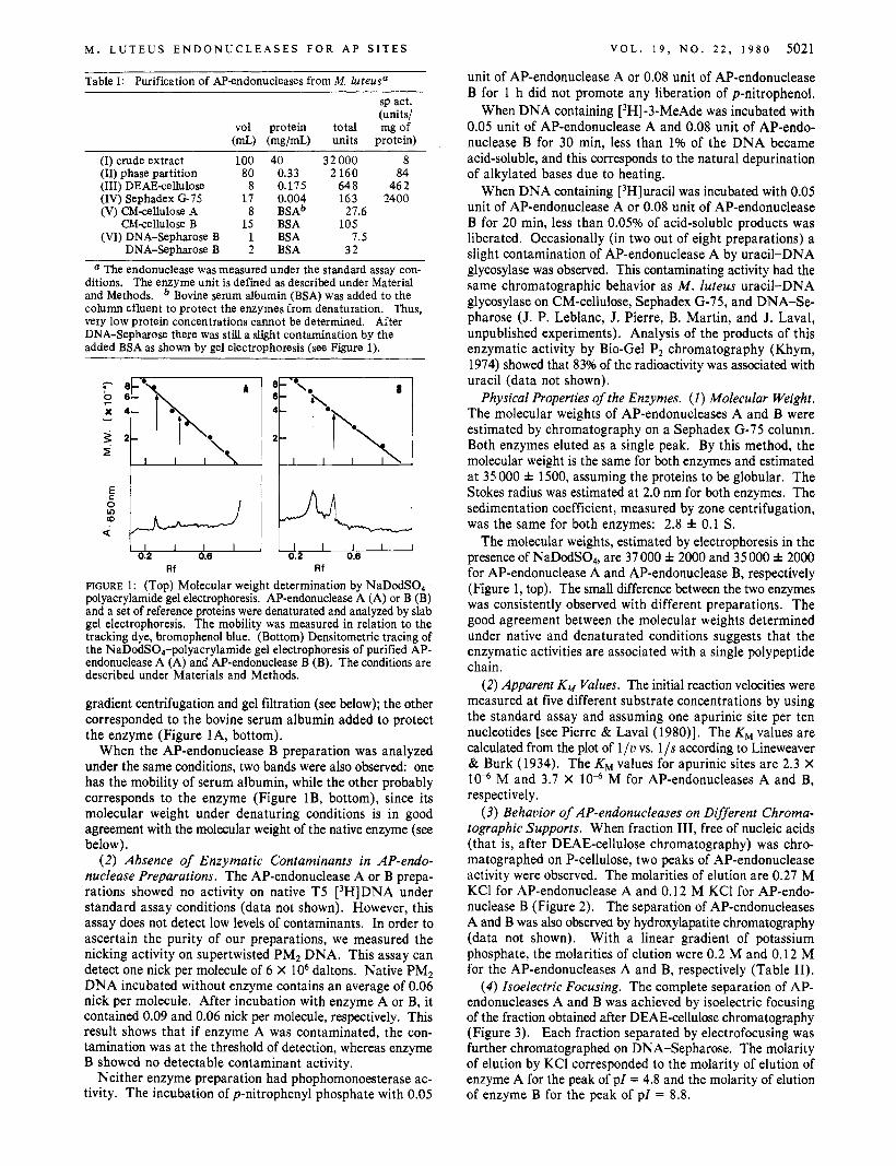
Physical Properties of the Enzymes. (1) Molecular Weight. The molecular weights of AP-endonucleases A and B were estimated by chromatography on a Sephadex G-75 column. Both enzymes eluted as a single peak. By this method, the molecular weight is the same for both enzymes and estimated at 35 000 * 1500, assuming the proteins to be globular. The Stokes radius was estimated at 2.0 nm for both enzymes. The sedimentation coefficient, measured by zone centrifugation, was the same for both enzymes: 2.8 f 0.1 S .
The molecular weights, estimated by electrophoresis in the presence of NaDodSO,, are 37 000 f 2000 and 35 000 * 2000 for AP-endonuclease A and AP-endonuclease B, respectively (Figure 1, top). The small difference between the two enzymes was consistently observed with different preparations. The good agreement between the molecular weights determined under native and denaturated conditions suggests that the enzymatic activities are associated with a single polypeptide chain.
( 2 ) Apparent K,,, Values. The initial reaction velocities were measured at five different substrate concentrations by using the standard assay and assuming one apurinic site per ten nucleotides [see Pierre & Laval (1980)l. The KM values are calculated from the plot of 1 / u vs. 1 /s according to Lineweaver & Burk (1934). The KM values for apurinic sites are 2.3 X 10” M and 3.7 X 10” M for AP-endonucleases A and B, respectively.
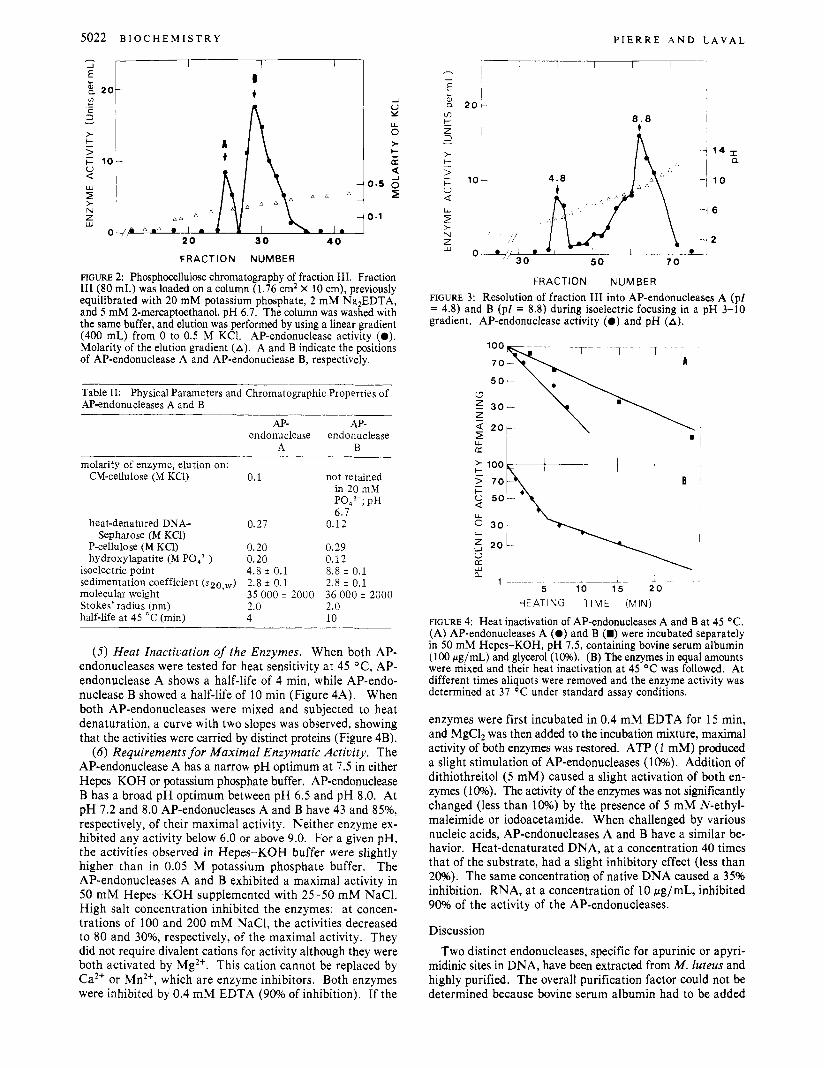
(3) Behavior of AP-endonucleases on Different Chroma- tographic Supports. When fraction 111, free of nucleic acids (that is, after DEAE-cellulose chromatography) was chro- matographed on P-cellulose, two peaks of AP-endonuclease activity were observed. The molarities of elution are 0.27 M KC1 for AP-endonuclease A and 0.12 M KC1 for AP-endo- nuclease B (Figure 2). The separation of AP-endonucleases A and B was also observed by hydroxylapatite chromatography (data not shown). With a linear gradient of potassium phosphate, the molarities of elution were 0.2 M and 0.12 M for the AP-endonucleases A and B, respectively (Table 11).
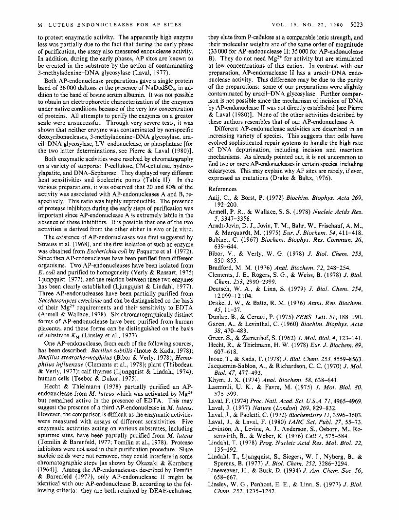
( 4 ) Isoelectric Focusing. The complete separation of AP- endonucleases A and B was achieved by isoelectric focusing of the fraction obtained after DEAE-cellulose chromatography (Figure 3). Each fraction separated by electrofocusing was further chromatographed on DNA-Sepharose. The molarity of elution by KCl corresponded to the molarity of elution of enzyme A for the peak of pZ = 4.8 and the molarity of elution of enzyme B for the peak of p l = 8.8.
Table I: Purification of Mendonucleases from M. ZuteuP
sp act. (units/
vol protein total mg of (mL) (mg/mL) units protein)
(I) crude extract 100 (11) phase partition 80 (111) DEAEcellulose 8 (IV) Sephadex G 7 5 17 (V) CMcellulose A 8
CMcellulose B 15 (VI) DNA-Sepharose B 1
DNA-Sepharose B 2
40 0.33 0.175 0.004
BSA BSA BSA
B S A ~
32000 8 2 160 84
648 462 163 2400
105
32
27.6
7.5
a The endonuclease was measured under the standard assay con- ditions. The enzyme unit is defined as described under Material and Methods. Bovine serum albumin (BSA) was added to the column efluent to protect the enzymes from denaturation. Thus, very low protein concentrations cannot be determined. After DNA-Sepharose there was still a slight contamination by the added BSA as shown by gel electrophoresis (see Figure 1).
0.2 0.0 0.2 0.6 Rf Rf
FIGURE 1 : (Top) Molecular weight determination by NaDodS04- polyacrylamide gel electrophoresis. AP-endonuclease A (A) or B (B) and a set of reference proteins were denaturated and analyzed by slab gel electrophoresis. The mobility was measured in relation to the tracking dye, bromophenol blue. (Bottom) Densitometric tracing of the NaDodS04-polyacrylamide gel electrophoresis of purified AP- endonuclease A (A) and AP-endonuclease B (B). The conditions are described under Materials and Methods.
gradient centrifugation and gel filtration (see below); the other corresponded to the bovine serum albumin added to protect the enzyme (Figure 1 A, bottom).
When the AP-endonuclease B preparation was analyzed under the same conditions, two bands were also observed: one has the mobility of serum albumin, while the other probably corresponds to the enzyme (Figure lB, bottom), since its molecular weight under denaturing conditions is in good agreement with the molecular weight of the native enzyme (see below).
( 2 ) Absence of Enzymatic Contaminants in AP-endo- nuclease Preparations. The AP-endonuclease A or B prepa- rations showed no activity on native T5 [3H]DNA under standard assay conditions (data not shown). However, this assay does not detect low levels of contaminants. In order to ascertain the purity of our preparations, we measured the nicking activity on supertwisted PM2 DNA. This assay can detect one nick per molecule of 6 X lo6 daltons. Native PM2 DNA incubated without enzyme contains an average of 0.06 nick per molecule. After incubation with enzyme A or B, it contained 0.09 and 0.06 nick per molecule, respectively. This result shows that if enzyme A was contaminated, the con- tamination was at the threshold of detection, whereas enzyme B showed no detectable contaminant activity.
Neither enzyme preparation had phophomonoesterase ac- tivity. The incubation of p-nitrophenyl phosphate with 0.05
5022 B I o c H E M I S T R Y
-I u Y
0
3 U
P I E R R E A N D L A V A L
, , * 1 0 . 1
..
FRACTION NUMBER
FIGURE 2: Phosphocellulose chromatography of fraction 111. Fraction I11 (80 mL) was loaded on a column (1.76 cm2 X 10 cm), previously equilibrated with 20 mM potassium phosphate, 2 mM Na2EDTA, and 5 mM 2-mercaptoethanol, pH 6 .7 . The column was washed with the same buffer, and elution was performed by using a linear gradient (400 mL) from 0 to 0.5 M KCI. AP-endonuclease activity (0). Molarity of the elution gradient (A). A and B indicate the positions of AP-endonuclease A and AP-endonuclease B, respectively.
Table 11: Physical Parameters and Chromatographic Properties of AP-endonucleases A and B
AP- AP- endonuclease endonuclease
A B molarity of enzyme, elution on:
CM-cellulose (M KC1)
heat-denatured DNA- Sepharose (M KCI)
P-cellulose (M KCl) hydroxylapatite (M P043-)
isoelectric point sedimentation coefficient ( ~ 2 0 , ~ ) molecular weight Stokes' radius (nm) half-iife at 45 "C (min)
0.1
0.27
0.20 0.20 4.8 i 0.1 2.8 * 0.1 35 000 * 2000 2.0 4
not retained in 20 mhl ~ 0 ~ 3 . ; p~ 6.1
0.12
0.29 0.12 8.8 i 0.1 2.8 i 0.1 36 000 i 2000 2.0 10
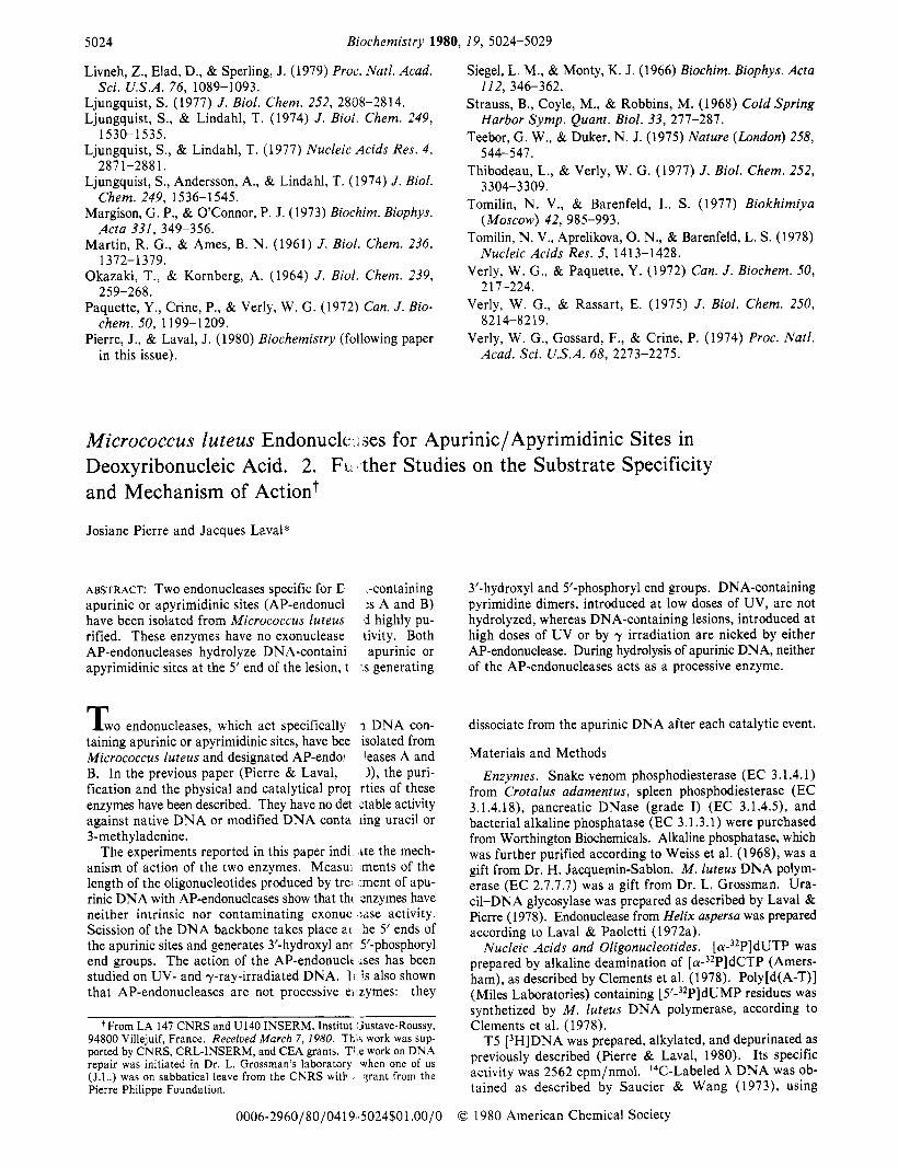
( 5 ) Heat Inactivation of the Enzymes. When both AP- endonucleases were tested for heat sensitivity at 45 OC, AP- endonuclease A shows a half-life of 4 min, while AP-endo- nuclease B showed a half-life of 10 min (Figure 4A). When both AP-endonucleases were mixed and subjected to heat denaturation, a curve with two slopes was observed, showing that the activities were carried by distinct proteins (Figure 4B).
( 6 ) Requirements for Maximal Enzymatic Activity. The AP-endonuclease A has a narrow pH optimum at 7.5 in either Hepe-KOH or potassium phosphate buffer. AP-endonuclease B has a broad pH optimum between pH 6.5 and pH 8.0. At pH 7.2 and 8.0 AP-endonucleases A and B have 43 and 85%, respectively, of their maximal activity. Neither enzyme ex- hibited any activity below 6.0 or above 9.0. For a given pH, the activities observed in Hepes-KOH buffer were slightly higher than in 0.05 M potassium phosphate buffer. The AP-endonucleases A and B exhibited a maximal activity in 50 mM Hepes-KOH supplemented with 25-50 mM NaC1. High salt concentration inhibited the enzymes: at concen- trations of 100 and 200 mM NaC1, the activities decreased to 80 and 3096, respectively, of the maximal activity. They did not require divalent cations for activity although they were both activated by Mg2+. This cation cannot be replaced by Ca2+ or Mn2+, which are enzyme inhibitors. Both enzymes were inhibited by 0.4 mM EDTA (90% of inhibition). If the
m k z
c 0 Q W
2 > N z iL1
8 . 8 I t
0 1
5 0 7 0
i O */30°
\ - 1 2
FRACTION NUMBER FIGURE 3: Resolution of fraction 111 into AP-endonucleases A ( p l = 4.8) and B (p l = 8.8) during isoelectric focusing in a pH 3-10 gradient. AP-endonuclease activity (0 ) and pH (A).
50-
I
m , e--+-+- -+--- B - ; 100
5 7 0
1 L __ -----i-- I- a
5 10 15 20 HEATING TIME (MIN)
FIGURE 4: Heat inactivation of AP-endonucleases A and B at 45 O C .
(A) AP-endonucleases A (0 ) and B (m) were incubated separately in 50 mM Hepes-KOH, pH 7 . 5 , containing bovine serum albumin (100 pg/mL) and glycerol (10%). (B) The enzymes in equal amounts were mixed and their heat inactivation at 45 O C was followed. At different times aliquots were removed and the enzyme activity was determined at 37 O C under standard assay conditions.
enzymes were first incubated in 0.4 mM EDTA for 15 min, and MgC12 was then added to the incubation mixture, maximal activity of both enzymes was restored. ATP (1 mM) produced a slight stimulation of AP-endonucleases (10%). Addition of dithiothreitol (5 mM) caused a slight activation of both en- zymes (10%). The activity of the enzymes was not significantly changed (less than 10%) by the presence of 5 mM N-ethyl- maleimide or iodoacetamide. When challenged by various nucleic acids, AP-endonucleases A and B have a similar be- havior. Heat-denaturated DNA, at a concentration 40 times that of the substrate, had a slight inhibitory effect (less than 20%). The same concentration of native DNA caused a 35% inhibition. RNA, at a concentration of 10 Ng/mL, inhibited 90% of the activity of the AP-endonucleases.
Discussion Two distinct endonucleases, specific for apurinic or apyri-
midinic sites in DNA, have been extracted from M. luteus and highly purified. The overall purification factor could not be determined because bovine serum albumin had to be added
M . L U T E U S E N D O N U C L E A S E S F O R A P S I T E S V O L . 1 9 , N O . 2 2 , 1980 5023
they elute from P-cellulose at a comparable ionic strength, and their molecular weights are of the same order of magnitude (33 000 for AP-endonuclease 11; 35 000 for AP-endonuclease B). They do not need Mg2+ for activity but are stimulated at low concentrations of this cation. In contrast with our preparation, AP-endonuclease I1 has a uracil-DNA endo- nuclease activity. This difference may be due to the purity of the preparations: some of our preparations were slightly contaminated by uracil-DNA glycosylase. Further compar- ison is not possible since the mechanism of incision of DNA by AP-endonuclease I1 was not directly established [see Pierre & Laval (1980)l. None of the other activities described by these authors resembles that of our AP-endonuclease A.
Different AP-endonuclease activities are described in an increasing variety of species. This suggests that cells have evolved sophisticated repair systems to handle the high rate of DNA depurination, including incision and insertion mechanisms. As already pointed out, it is not uncommon to find two or more AP-endonucleases in certain species, including eukaryotes. This may explain why AP sites are rarely, if ever, expressed as mutations (Drake & Baltz, 1976).
References Aaij, C., & Borst, P. (1972) Biochim. Biophys. Acta 269,
Armell, P. R., & Wallace, S . S . (1978) Nucleic Acids Res.
Arndt-Jovin, D. J., Jovin, T. M., Bahr, W., Frischauf, A. M., & Marquardt, M. (1975) Eur. J . Biochem. 54, 411-418.
Babinet, C. (1967) Biochem. Biophys. Res. Commun. 26,
Bibor, V., & Verly, W. G. (1978) J . Biol. Chem. 253,
Bradford, M. M. (1976) Anal. Biochem. 72, 248-254. Clements, J. E., Rogers, S. G., & Weiss, B. (1978) J . Biol.
Deutsch, W. A., & Linn, S. (1979) J . Biol. Chem. 254,
Drake, J . W., & Baltz, R. M. (1976) Annu. Rev. Biochem.
Dunlap, B., & Cerutti, P. (1975) FEBS Lett. 51, 188-190. Garen, A., & Levinthal, C. (1960) Biochim. Biophys. Acta
Greer, S . , & Zamenhof, S . (1962) J. Mol. Biol. 4, 123-141. Hecht, R., & Thielmann, H. W. (1978) Eur. J . Biochem. 89,
Inoue, T., & Kada, T. (1978) J. Biol. Chem. 253, 8559-8563. Jacquemin-Sablon, A., & Richardson, C. C. (1 970) J . Mol.
Khym, J. X. (1974) Anal. Biochem. 58, 638-641. Laemmli, U. K., & Favre, M. (1975) J . Mol. Biol. 80,
Laval, F. (1974) Proc. Natl. Acad. Sci. U.S.A. 71,49654969, Laval, J. (1977) Nature (London) 269, 829-832. Laval, J., & Paoletti, C. (1972) Biochemistry 11, 3596-3603. Laval, J., & Laval, F. (1980) IARC Sci. Publ. 27, 55-73. Levinson, A., Levine, A. J., Anderson, S., Osborn, M., Ro-
Lindahl, T. (1978) Prog. Nucleic Acid Res. Mol. Biol. 22,
Lindahl, T., Ljungquist, S., Siegert, W. I., Nyberg, B., &
Lineweaver, H., & Burk, D. (1934) J . Am. Chem. SOC. 56,
Linsley, W. G., Penhoet, E. E., & Linn, S . (1977) J . Biol.
192-200.
5, 3347-3356.
639-644.
850-8 55.
Chem. 253, 2990-2999.
12099-12 104.
45, 11-37.
38, 470-483.
607-61 8.
Biol. 47, 471-493,
575-599.
senwirth, B., & Weber, K. (1976) Cell 7, 575-584.
135-192.
Sperens, B. (1977) J . Biol. Chem. 252, 3286-3294.
658-667.
Chem. 252, 1235-1242.
to protect enzymatic activity. The apparently high enzyme loss was partially due to the fact that during the early phase of purification, the assay also measured exonuclease activity. In addition, during the early phases, AP sites are known to be created in the substrate by the action of contaminating 3-methyladenine-DNA glycosylase (Laval, 1977).
Both AP-endonuclease preparations gave a single protein band of 36 000 daltons in the presence of NaDodSO,, in ad- dition to the band of bovine serum albumin. It was not possible to obtain an electrophoretic characterization of the enzymes under native conditions because of the very low concentration of proteins. All attempts to purify the enzymes on a greater scale were unsuccessful. Through very severe tests, it was shown that neither enzyme was contaminated by nonspecific deoxyribonucleases, 3-methyladenine-DNA glycosylase, ura- cil-DNA glycosylase, UV-endonuclease, or phosphatase [for the two latter determinations, see Pierre & Laval (1980)l.
Both enzymatic activities were resolved by chromatography on a variety of supports: P-cellulose, CM-cellulose, hydrox- ylapatite, and DNA-Sepharose. They displayed very different heat sensitivities and isoelectric points (Table 11). In the various preparations, it was observed that 20 and 80% of the activity was associated with AP-endonucleases A and B, re- spectively. This ratio was highly reproducible. The presence of protease inhibitors during the early steps of purification was important since AP-endonuclease A is extremely labile in the absence of these inhibitors. It is possible that one of the two activities is derived from the other either in vivo or in vitro.
The existence of AP-endonucleases was first suggested by Strauss et al. (1968), and the first isolation of such an enzyme was obtained from Escherichia coli by Paquette et al. (1972). Since then AP-endonucleases have been purified from different organisms. Two AP-endonucleases have been isolated from E. coli and purified to homogeneity (Verly & Rassart, 1975; Ljungquist, 1977), and the relation between these two enzymes has been clearly established (Ljungquist & Lindahl, 1977). Three AP-endonucleases have been partially purified from Saccharomyces cereuisiae and can be distinguished on the basis of their Mg2+ requirements and their sensitivity to EDTA (Armell & Wallace, 1978). Six chromatographically distinct forms of AP-endonuclease have been purified from human placenta, and these forms can be distinguished on the basis of substrate K M (Linsley et al., 1977).
One AP-endonuclease, from each of the following sources, has been described: Bacillus subtilis (Inoue & Kada, 1978); Bacillus stearothermophilus (Bibor & Verly, 1978); Hemo- philus influenzae (Clements et al., 1978); plant (Thibodeau & Verly, 1977); calf thymus (Ljungquist & Lindahl, 1974); human cells (Teebor & Duker, 1975).
Hecht & Thielmann (1978) partially purified an AP- endonuclease from M . luteus which was activated by Mg2+ but remained active in the presence of EDTA. This may suggest the presence of a third AP-endonuclease in M . luteus. However, the comparison is difficult as the enzymatic activities were measured with assays of different sensitivities. Five enzymatic activities acting on various substrates, including apurinic sites, have been partially purified from M . luteus (Tomilin & Barenfeld, 1977; Tomilin et al., 1978). Protease inhibitors were not used in their purification procedure. Since nucleic acids were not removed, they could interfere in some chromatographic steps [as shown by Okazaki & Kornberg (1 964)l. Among the AP-endonucleases described by Tomilin & Barenfeld (1977), only AP-endonuclease I1 might be identical with our AP-endonuclease B, according to the fol- lowing criteria: they are both retained by DEAE-cellulose,
5024 Biochemistry 1980, 19, 5024-5029
Livneh, Z., Elad, D., & Sperling, J. (1979) Proc. Natl. Acad.
Ljungquist, S. (1977) J. Biol. Chem. 252, 2808-2814. Ljungquist, S., & Lindahl, T. (1974) J. Bioi. Chem. 249,
Ljungquist, S., & Lindahl, T. (1977) Nucleic Acids Res. 4 ,
Ljungquist, S. , Anderson, A., & Lindahl, T. (1974) J . Biol.
Margison, G. P., & O’Connor, P. J. (1973) Biochim. Biophys.
Martin, R. G., & Ames, B. N. (1961) J. Biol. Chem. 236,
Okazaki, T., & Kornberg, A. (1964) J . Biol. Chem. 239,
Paquette, Y. , Crine, P., & Verly, W. G. (1972) Can. J . Bio-
Pierre, J., & Laval, J. (1980) Biochemistry (following paper
Sci. U.S.A. 76, 1089-1093.
1530-1535.
2871-2881.
Chem. 249, 1536-1545.
Acta 331, 349-356.
1372-1379.
259-268.
chem. 50, 1199-1209.
in this issue).
Siegel, L. M., & Monty, K. J. (1966) Biochim. Biophys. Acta
Strauss, B., Coyle, M., & Robbins, M. (1968) Cold Spring
Teebor, G. W., & Duker, N. J. (1975) Nature (London) 258,
Thibodeau, L., & Verly, W. G. (1977) J. Biol. Chem. 252,
Tomilin, N. V., & Barenfeld, L. S. (1977) Biokhimiya
Tomilin, N. V., Aprelikova, 0. N., & Barenfeld, L. S. (1978)
Verly, W. G., & Paquette, Y . (1972) Can. J . Biochem. 50,
Verly, W. G., & Rassart, E. (1975) J. Biol. Chem. 250,
Verly, W. G., Gossard, F., & Crine, P. (1974) Proc. Natl.
112, 346-362.
Harbor Symp. Quant. Biol. 33, 277-287.
544-547.
3 304-3 309.
(MOSCOW) 42, 985-993.
Nucleic Acids Res. 5 , 1413-1428.
217-224.
8 2 14-82 19.
Acad. Sci. U.S.A. 68, 2273-2275.
Micrococcus luteus Endonucle ses for Apurinic/ Apyrimidinic Sites in Deoxyribonucleic Acid. 2. FL~ and Mechanism of Actiont
Josiane Pierre and Jacques Laval*
ABSTRACT: Two endonucleases specific for C apurinic or apyrimidinic sites (AP-endonucl have been isolated from Micrococcus luteus rified. These enzymes have no exonuclease AP-endonucleases hydrolyze DNA-containi apyrimidinic sites at the 5’ end of the lesion, t
T w o endonucleases, which act specifically taining apurinic or apyrimidinic sites, have bee Micrococcus luteus and designated AP-endol B. In the previous paper (Pierre & Laval, fication and the physical and catalytical pror enzymes have been described. They have no del against native DNA or modified DNA conta 3-methyladenine.
The experiments reported in this paper indi
ther Studies on the Substrate Specificity
,-containing :s A and B) j highly pu- tivity. Both apurinic or
.s generating
I DNA con- isolated from leases A and I), the puri-
rties of these ztable activity ling uracil or
ite the mech- - - anism of action of the two enzymes. Measui ments of the length of the oligonucleotides produced by tree .ment of apu- rinic DNA with AP-endonucleases show that thi snzymes have neither intrinsic nor contaminating exonuc :;se activity. Scission of the DNA backbone takes place 21 he 5’ ends of the apurinic sites and generates 3’-hydroxyl anr 5’-phosphoryl end groups. The action of the AP-endonuclr s e s has been studied on UV- and y-ray-irradiated DNA. 1 is also shown that AP-endonucleases are not processive el zymes: they
From LA 147 CNRS and U140 INSERM, Institut ‘hstave-Roussy, 94800 Villejuif, France. Receiued March 7, 1980. Thir work was sup- ported by CNRS, CRL-INSERM, and CEA grants. TI e work on DNA repair was initiated in Dr. L. Grossman’s laboratory when one of us (J.L.) was on sabbatical leave from the CNRS witb qrant from the Pierre Philippe Foundation.
3’-hydroxyl and 5’-phosphoryl end groups. DNA-containing pyrimidine dimers, introduced at low doses of UV, are not hydrolyzed, whereas DNA-containing lesions, introduced at high doses of UV or by y irradiation are nicked by either AP-endonuclease. During hydrolysis of apurinic DNA, neither of the AP-endonucleases acts as a processive enzyme.
dissociate from the apurinic DNA after each catalytic event.
Materials and Methods
Enzymes. Snake venom phosphodiesterase (EC 3.1.4.1) from Crotalus adamentus, spleen phosphodiesterase (EC 3.1.4.18), pancreatic DNase (grade I) (EC 3.1.4.5), and bacterial alkaline phosphatase (EC 3.1.3.1) were purchased from Worthington Biochemicals. Alkaline phosphatase, which was further purified according to Weiss et al. (1968), was a gift from Dr. H. Jacquemin-Sablon. M . luteus DNA polym- erase (EC 2.7.7.7) was a gift from Dr. L. Grossman. Ura- cil-DNA glycosylase was prepared as described by Laval & Pierre (1978). Endonuclease from Helix aspersa was prepared according to Laval & Paoletti (1972a).
Nucleic Acids and Oligonucleotides. [ C ~ - ~ ~ P ] ~ U T P was prepared by alkaline deamination of [ ( u - ~ ~ P ] ~ C T P (Amers- ham), as described by Clements et al. (1978). Poly[d(A-T)] (Miles Laboratories) containing [ 5’-32P]dUMP residues was synthetized by M . luteus DNA polymerase, according to Clements et al. (1978).
T5 [3H]DNA was prepared, alkylated, and depurinated as previously described (Pierre & Laval, 1980). Its specific activity was 2562 cpm/nmol. I4C-Labeled X DNA was ob- tained as described by Saucier & Wang (1973), using
0006-2960/80/0419-5024$01 .OO/O 0 1980 American Chemical Society