microenvironmental control of malignancy exerted by ... · microenvironmental control of malignancy...
TRANSCRIPT

Microenvironmental control of malignancy exerted byRNASET2, a widely conserved extracellular RNaseFrancesco Acquatia, Sabrina Bertilacciob, Annalisa Grimaldia, Laura Montia, Raffaella Cinquettia, Paolo Bonettia,Marta Lualdia, Laura Vidalinoc, Marco Fabbrid, Maria Grazia Saccod, Nico van Rooijene, Paola Campomenosia,Davide Vigettif, Alberto Passif, Cristina Rivag, Carlo Capellag, Francesca Sanvitoh, Claudio Doglionih, Laura Gribaldod,Paolo Macchic, Antonio Sicai,j, Douglas M. Noonank, Paolo Ghiab, and Roberto Taramellia,1
aDipartimento di Biotecnologie e Scienze Molecolari, fDipartimento di Scienze Biomediche Sperimentali e Cliniche, and kDepartment of ExperimentalMedicine, Faculty of Medicine, Università degli Studi dell’Insubria, 21100 Varese, Italy; bLymphoma Unit, Department of Clinical Oncology, and hDepartmentof Pathology, San Raffaele Scientific Institute, 20132 Milan, Italy; cCentre for Integrative Biology, Laboratory of Molecular and Cellular Neurobiology,University of Trento, 38060 Mattarello, Trent, Italy; dEuropean Centre for the Validation of Alternative Methods, Institute for Health and ConsumerProtection, Joint Research Centre, 21027 Ispra, Italy; eDepartment of Molecular Cell Biology, Faculty of Medicine, Vrije Universiteit, N-1081, Amsterdam,The Netherlands; gDepartment of Pathology, Ospedale di Circolo, 21100 Varese, Italy; iIstituto Clinico Humanitas, Istituto Di Ricovero e Cura a CarattereScientifico, 20089 Rozzano, Italy; and jDipartimento di Scienze Chimiche, Alimentari, Farmaceutiche e Farmacologiche, University of Piemonte OrientaleA. Avogadro, 28100 Novara, Italy
Edited* by George Klein, Karolinska Institute, Stockholm, Sweden, and approved November 30, 2010 (received for review September 24, 2010)
A recent body of evidence indicates an active role for stromal(mis)-regulation in the progression of neoplasias. Within thisconceptual framework, genes belonging to the growing but stillpoorly characterized class of tumor antagonizing/malignancysuppressor genes (TAG/MSG) seem to play a crucial role in theregulation of the cross-talk between stromal and epithelial cells bycontrolling malignant growth in vivo without affecting anycancer-related phenotype in vitro. Here, we have functionallycharacterized the human RNASET2 gene, which encodes the firsthuman member of the widespread Rh/T2/S family of extracellularRNases and was recently found to be down-regulated at the tran-script level in several primary ovarian tumors or cell lines and inmelanoma cell lines. Although we could not detect any activityfor RNASET2 in several functional in vitro assays, a remarkable con-trol of ovarian tumorigenesis could be detected in vivo. Moreover,the control of ovarian tumorigenesis mediated by this unique tumorsuppressor gene occurs through modification of the cellular micro-environment and the induction of immunocompetent cells of themonocyte/macrophage lineage. Taken together, the data presentedin this work strongly indicate RNASET2 as a previously unexploredmember of the growing family of tumor-antagonizing genes.
ovarian cancer | xenograft cancer model
Ovarian carcinoma is the leading cause of death from gyne-cological cancer in the Western world, and the majority
(≈70%) of ovarian cancer patients present with an advanceddisease at diagnosis, with a dismal 5-y survival rate of 30% (1). Atpresent, cytoreductive surgery combined with platinum/taxaneschemotherapy is the standard treatment (2). Although suchmultimodality treatment has resulted in increased response ratesand overall survival, the cure rate of the disease has not changedsubstantially. Indeed, whereas more than 75% of patients re-spond to first-line chemotherapy, ≈70% of them eventually re-lapse and die. Among human cancers located in the ovary, thosederived from the epithelium are the most frequent (3), althoughvery heterogeneous at both morphological and biological levels.Although such heterogeneity has contributed to difficulties indefining the molecular alterations associated with ovarian cancerdevelopment (4), all epithelial ovarian cancer subtypes originatefrom the single layer of epithelial cells covering the surface of theovaries (OSE cells) (5) and a solid body of data has provided firmevidence that epithelial ovarian cancers arises from OSE cells(6). These cells undergo repeated cycles of proliferation becauseof the recurrent growth and rupture of ovarian follicles duringthe ovulatory cycle, and this phenomenon is supported by a well-characterized interaction between ovarian mesenchyme and ep-ithelium (7). Alterations of such microenviromental interactions
might therefore contribute to ovarian cancerogenesis and agrowing interest has indeed been placed toward the microenvi-ronment, based on a recent body of evidence suggesting an activerole for stromal misregulation in the progression of neoplasias(7, 8). Within this conceptual framework, a special class of genesseems to play a crucial role in the regulation of the cross-talkbetween stromal and epithelial cells. These genes belong to thegrowing but still poorly characterized class of tumor antagoniz-ing/malignancy suppressor genes (TAG/MSG) (9–12), whoseprincipal feature is their ability to control malignant growth invivo but not in vitro. Such “asymmetric tumor suppression” is inkeeping with earlier studies showing that somatic hybridizationof normal and malignant cells can suppress tumorigenicity in vivobut not cell growth in vitro (13). Moreover, recent theoreticaland experimental evidences indicate that a rather large pro-portion of cancer genes endowed with these peculiar features hasescaped detection to date (9–12). TAG/MSG genes are postu-lated to encode for products required to respond to differenti-ation-inducing signals in vivo, to mediate cellular interactionswith the tumor microenvironment, or to negatively regulate an-giogenesis (9–12). Significantly, although biologically different,all three mechanisms point to the occurrence of a functionalcross-talk between the cancer cell and the local microenviron-ment as a critical factor for cancer suppression. This observationconfers a particular relevance to the identification of novel TAG/MSGs, as the role played by microenvironmental cues in cancerprogression has been increasingly appreciated (7, 8). A few yearsago, we reported the isolation of the RNASET2 gene from hu-man chromosomal region 6q27. This gene encodes the onlyhuman member of the widespread Rh/T2/S family of extracel-lular RNAses (14) and was found to be down-regulated at thetranscript level in several ovarian primary tumors or cell lines,and in melanoma cell lines (15–17). Moreover, RNASET2 down-regulation was reported in other human malignancies, such aslymphomas and gliomas (18, 19), in keeping with the observationthat deletions of chromosome 6q27, where the RNASET2 gene islocated, are associated with a wide range of human neoplasias(20–23). Neither inactivating mutations nor abnormal CpG
Author contributions: F.A., A.P., C.C., C.D., P.M., A.S., D.M.N., P.G., and R.T. designedresearch; F.A., S.B., A.G., L.M., R.C., P.B., M.L., L.V., M.F., M.G.S., P.C., D.V., C.R., and F.S.performed research; N.V.R. contributed new reagents/analytic tools; F.A., S.B., L.G., andR.T. analyzed data; and F.A. and R.T. wrote the paper.
The authors declare no conflict of interest.
*This Direct Submission article had a prearranged editor.1To whom correspondence should be addressed. E-mail: [email protected].
This article contains supporting information online at www.pnas.org/lookup/suppl/doi:10.1073/pnas.1013746108/-/DCSupplemental.
1104–1109 | PNAS | January 18, 2011 | vol. 108 | no. 3 www.pnas.org/cgi/doi/10.1073/pnas.1013746108
Dow
nloa
ded
by g
uest
on
Aug
ust 1
2, 2
020

methylation patterns in the promoter region were detected forthis gene in the ovarian tumor samples under investigation (15).However, functional studies of the human Hey3Met2 ovariancancer and SK-MEL 28 melanoma cell lines showed that trans-fection of the RNASET2 cDNA in these cell lines resulted ina strong inhibition of tumor growth in vivo (15–17). Strikingly,disruption of RNASET2’s catalytic activity did not impair theinhibition of tumorigenicity with respect to the wild-type allele,ruling out ribonuclease activity as an essential requirement forthe control of malignant growth (16).Here, we report a thorough in vivo analysis of RNASET2-
expressing ovarian cancer cells and compare them to a lack ofevident effects in vitro. We also show that control of ovariantumorigenesis by RNASET2 occurs through modification of thecellular microenvironment and involvement of immunocompe-tent cells, thus providing evidence for specific modulations ofcellular responses induced by RNASET2 that might underlayovarian tumorigenesis. Because RNASET2 appears to modulatethe tumor microenvironment to repress cancer growth, we pro-pose this gene as a previously unexplored member of the TAG/MSG gene family.
ResultsRNASET2 Suppresses Tumorigenesis in Vivo but Not in Vitro Pro-liferation. The Hey3Met2 cell line, derived from a highly meta-static subclone of the HEY4 ovarian cancer-cell line (16), showshighly decreased endogenous RNASET2 expression levels whencompared with normal human ovarian epithelial cells. Moreover,because this cell line displayed the lowest levels among severalother ovarian cancer cell lines (Fig. S1), it appeared to representan ideal recipient for RNASET2 re-expression experiments.Thus, Hey3Met2 cells were stably transfected with plasmidsencoding either wild-type RNASET2 or a catalytically dead form(whose cDNA has been previously mutagenized in the two CAScatalytic sites) (16) or with the empty vector as a control. Severalclones transfected with RNASET2 expression vectors showedsignificantly increased levels of the RNASET2 protein and werethus selected for subsequent analyses. The effects of RNASET2expression on several cancer-related parameters were subse-quently investigated in vitro. In all these assays, the effect of bothwild-type and catalytically dead RNASET2 expression in Hey3-Met2 cells was unremarkable (Fig. S2 and Table S1). To evaluatethe role of RNASET2 in vivo, we then turned to a xenograftassay. Nude mice were injected with the same Hey3Met2 clonespreviously investigated in vitro and followed for up to 24 d. Asexpected, large tumors developed in mice inoculated with vector-only transfected cells, reaching a volume 800 mm3 after 24 d.Interestingly, clones expressing either wild-type or the catalyti-cally dead form of RNASET2 were clearly suppressed in theirtumorigenic potential (Fig. 1). These notable in vivo findingsclearly contrast with what was observed in the in vitro context,where RNASET2 was shown to have no effect whatsoever. Toverify that expression of RNASET2 was maintained in vivo,immunohistochemical (IHC) assays with anti-RNASET2 anti-bodies were carried out on sections from excised tumor samples.As shown in Fig. 2 A–C, RNASET2 was clearly detected insections from tumors inoculated with cells previously transfectedwith the RNASET2 expression vectors, whereas expression ofthe protein in the tumors bearing empty vector-transfectedcontrol cells was barely detectable. The results from both in vitroand in vivo functional assays thus showed that RNASET2behaves similarly to those tumor-suppressor genes whose bi-ological function is carried out in vivo but not in vitro (9–12).Under the assumption that these tumor suppressors exert theiraction within the tumor microenvironment, we next investigatedseveral components that define the architectural structure of thetumor tissue.
Morphological Characterization of RNASET2-Expressing Tumors Grownin Vivo. To compare the morphological features of RNASET2-expressing and control tumors, they were excised 23 d after im-plantation, sectioned, and stained with H&E. As shown in Fig. 2D–F, an irregular architectural pattern was observed in bothwild-type and mutant RNASET2-expressing tumors comparedwith control tumors. The latter were mostly filled by vital andrapidly dividing cells, whereas in RNASET2-expressing tumorsscattered cells showing cytological features of apoptosis withcondensation of nuclear chromatin and cytoplasm were alsoobserved (see section below). Moreover, in wild-type and par-ticularly in mutant RNASET2-expressing tumors, neoplasticcells were entrapped by bridges of accumulated connective tissuecontaining an increased population of host-derived cells (Fig. 2D–F). Morphological analyses indicated that the latter weremostly granulocytes/mononuclear cells segregating within con-nective strands in the neoplastic mass. In contrast, a few gran-ulocytes in a diffuse pattern could be seen in control tumors.IHC assays for F4/80 and CD11b markers showed that the cel-lular population infiltrating the neoplastic mass within RNA-SET2-expressing tumors was mostly represented by cells fromthe monocyte/macrophage lineage (Fig. 2 G–L). Accordingly,a Masson’s trichrome staining indicated the prevalence of con-nective structures in the RNASET2-expressing tumors, likelyderived from nonmalignant infiltrating mononuclear cells (Fig.2 M–O).To confirm the murine origin of the cell population infiltrating
the tumors, a chromogenic in situ hybridization (CISH) assaywas performed by taking advantage of the XX chromosomalcomplement of the inoculated Hey2Met3 ovarian cells in thecontext of a XY complement of the recipient male nude mice.This assay clearly showed that the infiltrating cellular componentin RNASET2-expressing tumors was indeed of murine origin(Fig. 2 P–R).To further characterize the tumors in vivo, we then evaluated
their proliferation and apoptosis rates by IHC with Ki67 andcleaved caspase-3 (CCL-3) antibodies, respectively. As shown inFig. 3, in RNASET2-expressing tumors, and particularly in thoseexpressing the mutant form, Ki67 staining showed a decreasedproliferation rate compared with control tumors, whereas CCL-3immunoreactive neoplastic cells showed an opposite pattern,being increased when compared with control tumor (Fig. 3 A–Fand Fig. S3). In clear contrast, no apparent changes in blood-
Fig. 1. Expression of RNASET2 suppresses tumor growth in vivo. Followingsubcutaneous inoculation of nude mice with Hey3Met2 clones stably trans-fected with pcDNA3 control plasmid vector or RNASET2-expressing vectors,tumors were collected at days 22 to 24 for histological and IHC analyses. Atleastfivemicewere inoculated for each tested clone. Bars represent SD values.
Acquati et al. PNAS | January 18, 2011 | vol. 108 | no. 3 | 1105
GEN
ETICS
Dow
nloa
ded
by g
uest
on
Aug
ust 1
2, 2
020

vessel density and morphology could be observed betweenRNASET2-transfected tumors and controls (Fig. 3 G–I).
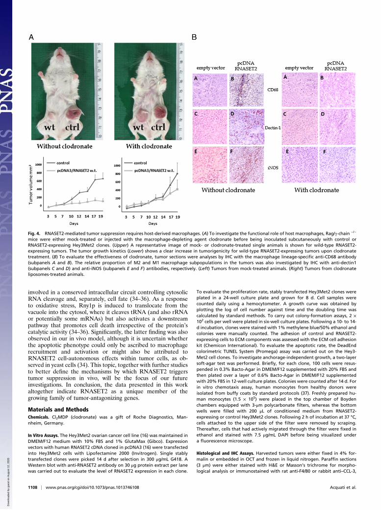
Comprehensive Characterization of the Macrophage InfiltratingPopulation. Because cells from the monocyte/macrophage line-age were shown to represent the main host cellular populationinfiltrating RNASET2-expressing tumors, we aimed at evaluatingwhether these cells could be functionally responsible for RNA-SET2-mediated suppression of tumorigenicity, as already de-scribed in other cancer models (24). To this aim, we exploiteda different xenograft model based on Rag2−/−γc−/− mice, whichlack both lymphocytes and NK cells. As shown in Fig. S4, wild-type and catalytically dead mutant RNASET2-expressing cellswere again clearly suppressed in tumorigenicity. Indeed, whereaslarge tumors developed in mice inoculated with control cells,reaching a 1,400 mm3 volume in 19 d, clones expressing eitherwild-type or mutant RNASET2 produced just a slight increase inthe tumor mass over time. Thus, we used this experimental modelto evaluate the role played by macrophages in the control ofcancer growth in vivo. To this end, Rag2−/−γc−/− mice were pre-treated with the macrophage-depleting agent clodronate beforeinoculation of the RNASET2-expressing Hey3Met2 clones.Noteworthy, the tumor suppressing activity of wild-type RNA-
SET2 turned out to be largely impaired in clodronate-treatedmice, as shown in Fig. 4A. Indeed, in contrast to the remarkabledifference in tumor size between RNASET2-overexpressing vs.control clones that was observed in untreated mice, clodronate-treated animals developed RNASET2-expressing tumors whosesize was very similar to that observed in control clones (Fig.4A). To confirm the efficient depletion of host macrophages inclodronate-treated animals, IHC assay with anti-CD68 antibodieswas carried out on xenograft tumor sections. As shown in Fig. 4B,the treatment was highly effective because no CD68+ cells couldbe detected in RNASET2-expressing tumors from clodronate-injected animals. Unfortunately, this assay was not informativewith mutant RNASET2-expressing tumors, because macrophageswere still detected in clodronate-treated mice following injectionof mutant RNASET2-expressing cells (Fig. S5), probably as a re-sult of the much prominent macrophages infiltration triggered bymutant versus wild-type RNASET2 from neighboring immuno-competent regions (compare Fig. 2 H and I). Taken together,these data strongly suggest that recruitment of murine hostcells from the monocyte/macrophage lineage is a critical step inRNASET2-mediated tumor suppression.Tissue macrophages are known to be endowed with a wide
functional plasticity, which allows them to carry out both pro-tumoral and antitumoral activities depending on microenviron-mental cues (24). Therefore, we investigated the pattern ofmacrophage polarization within RNASET2-expressing tumorxenografts by carrying out IHC assays with markers known todiscriminate the differentiation pattern of macrophages into ei-ther M1 and M2 subtypes. Interestingly, as shown in Fig. 4B(subpanels C–F), most of the infiltrating macrophages were ofthe M1, inducible nitric oxide synthase-positive type, in keepingwith the suggested role of this macrophage subpopulation intumor suppression (24). These data further support the crucialrole for macrophage recruitment in RNASET2-mediated tumorsuppression. As a last point, a preliminary study aimed at de-fining the mechanism by which RNASET2 affects the behavior ofthe monocyte/macrophage cell lineage was carried out. To thisend, in vitro chemotaxis assays were performed with peripheralblood lymphocyte-derived human monocytes, which were cul-tured in the top insert of Boyden chambers in the presence ofconditioned media from RNASET2-expressing or controlHey3Met2 clones added to the bottom wells. Significantly, asshown in Fig. S6, conditioned media of wild-type RNASET2-expressing Hey3Met2 clones turned out to provide a strongchemotactic signal to human monocytes when compared with
Fig. 2. IHC, histological, and CISH analysis of the cell infiltrate in tumorsamples. (A–C) Hey3Met2-derived tumors grown in nude mice were paraffin-embeddedand tissue sectionswereprocessed for IHC assaywith anti-RNASET2antibody. Photomicrographs show the distribution of RNASET2 in tumor xe-nograft derived from pcDNA3/RNASET2-transfected cells in comparison withcontrol cells. (D–F) Tissue sections were also stained with H&E. The arrow-heads mark the massive cellular infiltrate within RNASET2-expressing tumors.(G–L) Sections were also processed for IHCwith anti-F4/80 and anti-CD11b (G–Iand J–L, respectively). As shown in the photomicrographs, both wild type andmutant RNASET2-expressing tumors showed increased infiltration of F4/80and CD11b+ cells compared with control tumors. (M–O) Tumor sections pro-cessed for Masson’s trichrome staining showed an abundant collagen de-position within RNASET2-expressing tumors. (P–R) A CISH assay was carriedout to confirm that cells infiltrating the tumormass were host-derived. A DNAprobe frommouse chromosome Ywas used to discriminate male-host murinecells from the injected human female tumor cells. All photomicrographsshown are representative images of multiple fields examined in at least foursections derived from tumor excised in two to three independent animals foreach experimental group. (Magnifications: A–C and P–R, 100×; D–O, 20×.)
1106 | www.pnas.org/cgi/doi/10.1073/pnas.1013746108 Acquati et al.
Dow
nloa
ded
by g
uest
on
Aug
ust 1
2, 2
020

media conditioned by empty vector-transfected Hey3Met2clones. These results add further evidence for a mechanisticcrosstalk between extracellular RNASET2 and immunocompe-tent inflammatory cells.
DiscussionIn this work, we report an extensive analysis of the biologicalproperties of RNASET2 using ovarian cancer as an experimentalmodel, and show that the remarkable tumor-suppressive activityof this gene is carried out in vivo, but not in vitro. RNASET2-mediated in vivo tumor suppression was observed in two in-dependent xenograft models and was not dependent on thecatalytic activity of RNASET2, in keeping with other reportsfocusing on the biological activities of T2 RNase family members(25). Noteworthy, the control of tumor growth apparently relieson the establishment of a cross-talk between RNASET2-expressing cancer cells and the tumor microenvironment. Morespecifically, cells of the monocyte/macrophage lineage turnedout to represent the target of RNASET2 in vivo.The failure of RNASET2 to suppress tumor growth in vitro is
not unprecedented. Indeed, the lack of any RNASET2 in vitroactivity was also reported by Liu et al. in a panel of severalovarian cancer cell lines, although the authors did not considerany in vivo role for the RNASET2 gene (26). On the other hand,in vivo control of tumorigenicity by RNASET2 has already beenreported in human colon cancer (27) and malignant melanomacell lines (17). However, our group is unique in reportinga comparative study of the in vivo and in vitro properties ofRNASET2, and such comprehensive analyses allowed us to de-fine this gene as a previously unrecorded member of the TAG/MSG gene family. The conceptual bases for the identification of
the TAG family stem from the pioneeristic work of H. Harris andG. Klein (9, 12, 13) with somatic hybrids generated by fusion ofnormal and malignant cells. These hybrids lost their tumorige-nicity as long as they maintained a nearly full set of chromosomesfrom both parental cells, but the subsequent loss of chromo-somes from the normal parent cell was associated with reap-pearance of tumorigenicity. Indeed, later studies showed thatsingle chromosomes from noncancer cells could suppress tu-morigenicity by themselves (12, 13). Strikingly, the normal par-ent-derived chromosomes that were lost in the subclones thatregained a tumorigenic phenotype were not those harboring P53,RB, or any other tumor-suppressor gene known at that time (9,11, 13). Indeed, because these studies entailed the selection of invitro permanently growing somatic hybrid lines that were thentested in vivo for tumorigenicity, the experimental design itselfcould not select tumor suppressor genes that would inducegrowth arrest or apoptosis in vitro (9–12). The genes belongingto the TAG/MSG class are postulated to encode for productsrequired to respond to differentiation-inducing signals in vivo, tomediate normal cellular responses to microenvironment con-trols, or to negatively regulate the process of angiogenesis (11).Initial examples of this emerging category of genes came fromthe work on human chromosomal region 3p12-26 (9, 11). Amongthe TAGs most recently discovered are the hyaluronidaseHYAL1 and HYAL2 genes (28) and the lactoferrin and LIMD1genes, also mapping to chromosome 3p (9, 29). The hypoxia-regulated transcription factor DEC1 mapping in 9q32 and morerecently the WWOX gene mapping in 16q23 have also been re-cently added to the TAG/MSG gene list (30, 31).In this article, the biological properties of RNASET2 allowed
us to ascribe it to the TAG/MSG gene class. Indeed, besides theobserved asymmetric tumor-suppressive activity (which func-tionally define the TAG/MSG family), the fact that RNASET2overexpression triggers a marked response of cells from themonocyte/macrophage lineage suggests a biological mechanismbased on a microenvironmental control of tumorigenesis, whichrepresents one of the hallmarks of the TAG family members. Thecrucial role of macrophage infiltration in the control of tumori-genicity in our model was demonstrated by treating Rag/γ-chaindouble knock-out mice with the macrophage-depleting agentclodronate. These experiments confirmed that macrophage re-cruitment was crucially important for the control of tumorige-nicity mediated by RNASET2, because their effective ablationwas associated with the reappearance of tumors indistinguishablefrom those of the control clones. We have also investigated thepolarization pattern of themacrophage cell population infiltratingRNASET2-expressing tumors, as the M1 or M2 subtypes are as-sociated with rather different biological responses in the contextof tumor progression. As expected, most of the infiltrating mac-rophages were shown to express M1 markers, in keeping witha solid body of evidence pointing to this macrophage subtype as animportant player in the control of cancer growth (24).Noteworthy, a role for T2 RNases in modulating the host im-
mune response is not unprecedented, as epitomized by theRNASET2 ortholog gene from Schistosoma mansoni, whose geneproduct is responsible for priming dendritic cells to trigger a T2polarization pattern in CD4+ cells during human infection by thisparasite (32). Moreover, although the mechanistic details areunknown, an involvement of the innate immune response withpotential downstream consequences on neurodevelopment hasalso been postulated in an autosomal-recessive form of humancystic leukoencephalopathy characterized by mutations in theRNASET2 gene (33).Finally, although our in vitro studies have not shown any
particular effect mediated by RNASET2, we cannot rule out thatthis gene could also play a cell-autonomous role in specific invivo conditions. In this context, it is worth mentioning a recentwork on the yeast orthologue of RNASET2, Rny1p, which is
Fig. 3. IHC detection of tumor apoptotic/proliferating and endothelial cellsin tumor samples. Sections from tumor grown in nude mice were analyzedby IHC with antiactivated caspase 3 (A–C), anti-Ki-67 (D–F), and anti-CD31(G–I) antibodies, to detect apoptotic and proliferating cells and intratumorvessels, respectively. Photomicrographs shown are representative images ofmultiple fields examined in at least four sections derived from tumor excisedin two to three independent animals for each experimental group. (Mag-nifications: A–C, 40×; D–F, 100×; G–I, 10×.)
Acquati et al. PNAS | January 18, 2011 | vol. 108 | no. 3 | 1107
GEN
ETICS
Dow
nloa
ded
by g
uest
on
Aug
ust 1
2, 2
020
involved in a conserved intracellular circuit controlling cytosolicRNA cleavage and, separately, cell fate (34–36). As a responseto oxidative stress, Rny1p is induced to translocate from thevacuole into the cytosol, where it cleaves tRNA (and also rRNAor potentially some mRNAs) but also activates a downstreampathway that promotes cell death irrespective of the protein’scatalytic activity (34–36). Significantly, the latter finding was alsoobserved in our in vivo model, although it is uncertain whetherthe apoptotic phenotype could only be ascribed to macrophagerecruitment and activation or might also be attributed toRNASET2 cell-autonomous effects within tumor cells, as ob-served in yeast cells (34). This topic, together with further studiesto better define the mechanisms by which RNASET2 triggerstumor suppression in vivo, will be the focus of our futureinvestigations. In conclusion, the data presented in this workaltogether indicate RNASET2 as a unique member of thegrowing family of tumor-antagonizing genes.
Materials and MethodsChemicals. Cl2MDP (clodronate) was a gift of Roche Diagnostics, Man-nheim, Germany.
In Vitro Assays. The Hey3Met2 ovarian cancer cell line (16) was maintained inDMEM/F12 medium with 10% FBS and 1% GlutaMax (Gibco). Expressionvectors with human RNASET2 cDNA cloned in pcDNA3 (16) were transfectedinto Hey3Met2 cells with Lipofectamine 2000 (Invitrogen). Single stablytransfected clones were picked 14 d after selection in 300 μg/mL G418. AWestern blot with anti-RNASET2 antibody on 30 μg protein extract per lanewas carried out to evaluate the level of RNASET2 expression in each clone.
To evaluate the proliferation rate, stably transfected Hey3Met2 clones wereplated in a 24-well culture plate and grown for 8 d. Cell samples werecounted daily using a hemocytometer. A growth curve was obtained byplotting the log of cell number against time and the doubling time wascalculated by standard methods. To carry out colony-formation assays, 2 ×10² cells per well were plated in six-well culture plates. Following a 10- to 14-d incubation, clones were stained with 1% methylene blue/50% ethanol andcolonies were manually counted. The adhesion of control and RNASET2-expressing cells to ECM components was assessed with the ECM cell adhesionkit (Chemicon International). To evaluate the apoptotic rate, the DeadEndcolorimetric TUNEL System (Promega) assay was carried out on the Hey3-Met2 cell clones. To investigate anchorage-independent growth, a two-layersoft-agar test was performed. Briefly, for each clone, 100 cells were resus-pended in 0.3% Bacto-Agar in DMEM/F12 supplemented with 20% FBS andthen plated over a layer of 0.6% Bacto-Agar in DMEM/F12 supplementedwith 20% FBS in 12-well culture plates. Colonies were counted after 14 d. Forin vitro chemotaxis assay, human monocytes from healthy donors wereisolated from buffy coats by standard protocols (37). Freshly prepared hu-man monocytes (1.5 × 10⁵) were placed in the top chamber of Boydenchambers equipped with 3-μm polycarbonate filters, whereas the bottomwells were filled with 200 μL of conditioned medium from RNASET2-expressing or control Hey3Met2 clones. Following 2 h of incubation at 37 °C,cells attached to the upper side of the filter were removed by scraping.Thereafter, cells that had actively migrated through the filter were fixed inethanol and stained with 7.5 μg/mL DAPI before being visualized undera fluorescence microscope.
Histological and IHC Assays. Harvested tumors were either fixed in 4% for-malin or embedded in OCT and frozen in liquid nitrogen. Paraffin sections(3 μm) were either stained with H&E or Masson’s trichrome for morpho-logical analysis or immunostained with rat anti-F4/80 or rabbit anti–CCL-3,
Fig. 4. RNASET2-mediated tumor suppression requires host-derived macrophages. (A) To investigate the functional role of host macrophages, Rag/γ-chain −/−
mice were either mock-treated or injected with the macrophage-depleting agent clodronate before being inoculated subcutaneously with control orRNASET2-expressing Hey3Met2 clones. (Upper) A representative image of mock- or clodronate-treated single animals is shown for wild-type RNASET2-expressing tumors. The tumor growth kinetics (Lower) shows a clear increase in tumorigenicity for wild-type RNASET2-expressing tumors upon clodronatetreatment. (B) To evaluate the effectiveness of clodronate, tumor sections were analyses by IHC with the macrophage lineage-specific anti-CD68 antibody(subpanels A and B). The relative proportion of M2 and M1 macrophage subpopulations in the tumors was also investigated by IHC with anti-dectin1(subpanels C and D) and anti-iNOS (subpanels E and F) antibodies, respectively. (Left) Tumors from mock-treated animals. (Right) Tumors from clodronateliposomes-treated animals.
1108 | www.pnas.org/cgi/doi/10.1073/pnas.1013746108 Acquati et al.
Dow
nloa
ded
by g
uest
on
Aug
ust 1
2, 2
020

anti–Ki-67, and anti-RNASET2 (16) antibodies after antigen retrieval. Cry-osections were fixed with 4% PFA and incubated with the rat anti-CD11band rat anti-mouse CD68 (Pharmingen). The immunoreactions were revealedeither by biotinylated-conjugated anti-rat antibody (Vector), horseradishperoxidase-conjugated streptavidin either by rabbit or rat on rodent HRP-polymer (Biocare Medical) and using DAB as chromogen (Biogenex). Slideswere counterstained with Hematoxylin. Photomicrographs were taken usingthe AxioCam HRc (Zeiss) with the AxioVision System 6.4.
To evaluate blood-vessel density in tumor samples, cryosections weretreatedwith 50mM Levamisole to block endogenous alkaline phosphatase. Amouse anti-human PECAM-1 (CD31) antibody (1: 20; DAKO) was then in-cubated for 1 h at 37 °C, followed by a 45-min treatment with a secondarygoat anti-mouse AP-conjugated antibody (1:200). The signal was detectedwith BCIP/NBT Substrate Solution (Sigma). Control reactions were carried outby omitting the primary antibody. For CISH analysis, the ZytoDot probe kit(Histo-Line) was used to hybridize a biotinylated murine chromosome Y-specific probe to serial sections of Hey3Met2-derived tumors.
In Vivo Xenograft Studies. Male nude (five animals per group) or femaleRag2−/−γc−/− (three animals per group) mice were challenged subcutaneouslyin the left flank with 5 × 10⁶ Hey3Met2 cells (parental or Hey3Met2 cellsexpressing wild-type RNASET2 or RNASET2 catalytically dead cells) in 0.15 mLof medium and 0.15 mL matrigel (Sigma). Animals were monitored twicea week for weight and tumor growth (measuring three perpendiculardiameters), and killed when the mean tumor volume of control micereached a dimension of ≥1,000 mm3. Tumors were formalin-fixed, paraffin-embedded, cut at 5-μm thick sections and stained with H&E. Histologicalsections were evaluated in a double-blinded fashion. Macrophages de-pletion in Rag2−/−γc−/− mice was carried out using clodronate liposomes (38).Four animals per group were injected subcutaneously with control Hey3-Met2 cells in the left flank and with Hey3Met2 cells expressing either wild-
type or catalitically impaired mutant RNASET2 in the right flank. Each groupof transplanted animals was either macrophage-depleted or not by sub-cutaneous injection in both flanks of clodronate liposomes (50 μL per flank)every 6 d, starting at day −2 of the tumor challenge. Animals were moni-tored twice a week for weight and tumor growth, and killed when the meantumor volume reached a dimension of ≥800 mm3. Animals were maintainedin a pathogen-free facility and treated in accordance with the EuropeanUnion guidelines under the approval of the Ethical Committee of the IstitutoSan Raffaele (IACUC #418).
Macrophages populations in clodronate-treated and control animals weredetected on cryosections obtained as described above, and treatedwith 0.3%H2O2 in PBS to remove endogenous peroxidase. Washed sections were in-cubated for 1 h at 37 °C with the following biotinylated primary antibodies:rat anti-mouse CD68 (1:100; HyCult Biotechnology b.v.), rat anti-mouseDectin-1 (1:50; Cell Sciences), and rabbit anti-mouse Nos-2 (1: 10; Santa CruzBiotechnology). The washed specimens were incubated for 30 min at roomtemperature with streptavidin-peroxidase (Dako), washed, and signal wasdetected with 0.05% DAB and 0.03% H2O in PBS. Sections were counter-stained with Ematoxylin. Coverslips were mounted and slides were exam-ined with a microscope Olympus. Images were combined with AdobePhotoshop.
Statistical Analysis. The results from in vitro assays were evaluated by one-wayANOVA, whereas the Student t test was used to analyze in vivo data fromthe xenograft assays for statistical significance.
ACKNOWLEDGMENTS. This work was partly supported by Federico Ghido-ni’s memorial funds. F.A. was supported by a grant from the FondazioneComunitaria del Varesotto Organizzazione Non Lucrativa di Utilita’ Sociale(ONLUS).
1. Runnebaum IB, Stickeler E (2001) Epidemiological and molecular aspects of ovariancancer risk. J Cancer Res Clin Oncol 127(2):73–79.
2. McGuire WP, 3rd (2003) Current status of taxane and platinum-based chemotherapyin ovarian cancer. J Clin Oncol 21(10, Suppl):133s–135s.
3. Feeley KM, Wells M (2001) Precursor lesions of ovarian epithelial malignancy.Histopathology 38(2):87–95.
4. Shih IeM, Kurman RJ (2004) Ovarian tumorigenesis: A proposed model based onmorphological and molecular genetic analysis. Am J Pathol 164:1511–1518.
5. Auersperg N, Maines-Bandiera SL, Dyck HG, Kruk PA (1994) Characterization ofcultured human ovarian surface epithelial cells: Phenotypic plasticity andpremalignant changes. Lab Invest 71:510–518.
6. Murdoch WJ, McDonnel AC (2002) Roles of the ovarian surface epithelium inovulation and carcinogenesis. Reproduction 123:743–750.
7. Mueller MM, Fusenig NE (2004) Friends or foes—Bipolar effects of the tumour stromain cancer. Nat Rev Cancer 4:839–849.
8. Liotta LA, Kohn EC (2001) The microenvironment of the tumour-host interface.Nature 411:375–379.
9. Klein G, Imreh S, Zabarovsky ER (2007) Why do we not all die of cancer at an earlyage? Adv Cancer Res 98:1–16.
10. Klein G (2001) Are there large uncharted regions of tumor suppressor genes? IUBMBLife 51(2):83–85.
11. Imreh S, Klein G, Zabarovsky ER (2003) Search for unknown tumor-antagonizinggenes. Genes Chromosomes Cancer 38:307–321.
12. Klein G (2009) Toward a genetics of cancer resistance. Proc Natl Acad Sci USA 106:859–863.
13. Harris H, Miller OJ, Klein G, Worst P, Tachibana T (1969) Suppression of malignancy bycell fusion. Nature 223:363–368.
14. Trubia M, Sessa L, Taramelli R (1997) Mammalian Rh/T2/S-glycoprotein ribonucleasefamily genes: Cloning of a human member located in a region of chromosome 6(6q27) frequently deleted in human malignancies. Genomics 42:342–344.
15. Acquati F, et al. (2001) Cloning and characterization of a senescence inducing andclass II tumor suppressor gene in ovarian carcinoma at chromosome region 6q27.Oncogene 20:980–988.
16. Acquati F, et al. (2005) Tumor and metastasis suppression by the human RNASET2gene. Int J Oncol 26:1159–1168.
17. Monti L, et al. (2008) RNASET2 as a tumor antagonizing gene in a melanoma cancermodel. Oncol Res 17(2):69–74.
18. Steinemann D, et al. (2003) Identification of candidate tumor-suppressor genes in6q27 by combined deletion mapping and electronic expression profiling in lymphoidneoplasms. Genes Chromosomes Cancer 37:421–426.
19. Kim TY, Zhong S, Fields CR, Kim JH, Robertson KD (2006) Epigenomic profiling revealsnovel and frequent targets of aberrant DNA methylation-mediated silencing inmalignant glioma. Cancer Res 66:7490–7501.
20. Morita R, et al. (1991) Common regions of deletion on chromosomes 5q, 6q, and 10qin renal cell carcinoma. Cancer Res 51:5817–5820.
21. Tibiletti MG, et al. (1996) Early involvement of 6q in surface epithelial ovarian tumors.Cancer Res 56:4493–4498.
22. Theile M, et al. (1996) A defined chromosome 6q fragment (at D6S310) harborsa putative tumor suppressor gene for breast cancer. Oncogene 13:677–685.
23. Hauptschein RS, et al. (1998) Cloning and mapping of human chromosome 6q26-q27deleted in B-cell non-Hodgkin lymphoma and multiple tumor types. Genomics 50(2):170–186.
24. Sica A, et al. (2008) Macrophage polarization in tumour progression. Semin CancerBiol 18:349–355.
25. Luhtala N, Parker R (2010) T2 Family ribonucleases: Ancient enzymes with diverseroles. Trends Biochem Sci 35:253–259.
26. Liu Y, et al. (2002) Physical and transcript map of the region between D6S264 andD6S149 on chromosome 6q27, the minimal region of allele loss in sporadic epithelialovarian cancer. Oncogene 21:387–399.
27. Smirnoff P, Roiz L, Angelkovitch B, Schwartz B, Shoseyov O (2006) A recombinanthuman RNASET2 glycoprotein with antitumorigenic and angiogenic characteristics.Cancer 107:2760–2769.
28. Wang F, et al. (2008) HYAL1 and HYAL2 inhibit tumour growth in vivo but not invitro. PLoS ONE 3:e3031.
29. Sharp TV, et al. (2004) LIM domains-containing protein 1 (LIMD1), a tumor suppressorencoded at chromosome 3p21.3, binds pRB and represses E2F-driven transcription.Proc Natl Acad Sci USA 101:16531–16536.
30. Yang L, et al. (2005) Tumor suppressive role of a 2.4 Mb 9q33-q34 critical region andDEC1 in esophageal squamous cell carcinoma. Oncogene 24:697–705.
31. Gourley C, et al. (2009) WWOX gene expression abolishes ovarian cancertumorigenicity in vivo and decreases attachment to fibronectin via integrin alpha3.Cancer Res 69:4835–4842.
32. Steinfelder S, et al. (2009) The major component in schistosome eggs responsible forconditioning dendritic cells for Th2 polarization is a T2 ribonuclease (omega-1). J ExpMed 206:1681–1690.
33. Henneke M, et al. (2009) RNASET2-deficient cystic leukoencephalopathy resemblescongenital cytomegalovirus brain infection. Nat Genet 41:773–775.
34. Thompson DM, Parker R (2009) The RNase Rny1p cleaves tRNAs and promotes celldeath during oxidative stress in Saccharomyces cerevisiae. J Cell Biol 185(1):43–50.
35. Thompson DM, Lu C, Green PJ, Parker R (2008) tRNA cleavage is a conserved responseto oxidative stress in eukaryotes. RNA 14:2095–2103.
36. Thompson DM, Parker R (2009) Stressing out over tRNA cleavage. Cell 138:215–219.37. McNally AK, Chisolm GM, Morel DW, Cathcart MK (1990) Activated human monocytes
oxidize LDL by a lipoxygenase-dependent pathway. J Immunol 145:254–259.38. Sunderkötter C, et al. (2004) Subpopulations of mouse blood monocytes differ in
maturation stage and inflammatory response. J Immunol 172:4410–4417.
Acquati et al. PNAS | January 18, 2011 | vol. 108 | no. 3 | 1109
GEN
ETICS
Dow
nloa
ded
by g
uest
on
Aug
ust 1
2, 2
020