microfabricated tissues for investigating traction forces ...traction forces in terms of traction...
TRANSCRIPT
REVIEW
Microfabricated tissues for investigating traction forces involvedin cell migration and tissue morphogenesis
Bryan A. Nerger1 • Michael J. Siedlik1 • Celeste M. Nelson1,2
Received: 21 September 2016 / Revised: 2 December 2016 / Accepted: 8 December 2016 / Published online: 22 December 2016
� Springer International Publishing 2016
Abstract Cell-generated forces drive an array of biologi-
cal processes ranging from wound healing to tumor
metastasis. Whereas experimental techniques such as
traction force microscopy are capable of quantifying trac-
tion forces in multidimensional systems, the physical
mechanisms by which these forces induce changes in tissue
form remain to be elucidated. Understanding these mech-
anisms will ultimately require techniques that are capable
of quantifying traction forces with high precision and
accuracy in vivo or in systems that recapitulate in vivo
conditions, such as microfabricated tissues and engineered
substrata. To that end, here we review the fundamentals of
traction forces, their quantification, and the use of micro-
fabricated tissues designed to study these forces during cell
migration and tissue morphogenesis. We emphasize the
differences between traction forces in two- and three-di-
mensional systems, and highlight recently developed
techniques for quantifying traction forces.
Keywords Traction force microscopy �Extracellular matrix � Mechanotransduction �Morphodynamics
Abbreviations
2D Two-dimensional
3D Three-dimensional
AFM Atomic force microscopy
ECM Extracellular matrix
FRET Fluorescence resonance energy transfer
MDCK Madin–Darby canine kidney
PA Polyacrylamide
PEG Polyethylene glycol
TFM Traction force microscopy
Introduction
Mechanical forces play an integral role in cellular behav-
iors as diverse as proliferation [1, 2], differentiation [3, 4],
gene expression [5, 6], wound healing [7], tumor metastasis
and invasion [8–10], collective migration [11, 12], and
tissue morphogenesis [13]. In particular, traction stress,
which refers to the force per unit area acting on the
deformed cell–matrix interface, is a key mechanical cue in
cellular systems. Traction forces originate from the con-
tractile actomyosin network and can alter cellular behavior
through a number of mechanisms collectively referred to as
mechanotransduction [14]. However, the biophysical and
biochemical mechanisms by which these forces induce
changes in tissue form are not fully understood. The ability
to accurately and precisely calculate traction forces is,
therefore, important and will strengthen our quantitative
understanding of their role in cell- and tissue-level
processes.
The idea that the laws of mechanics can be applied to
cells was first postulated in the late 1800s by Wilhelm
His [15, 16]. Using the developing chicken embryo as a
model, he proposed that differential growth rates in the
germ layers gave rise to mechanical forces that are
responsible for producing the final tissue form [15, 17].
& Celeste M. Nelson
1 Department of Chemical and Biological Engineering,
Princeton University, 303 Hoyt Laboratory, William Street,
Princeton, NJ 08544, USA
2 Department of Molecular Biology, Princeton University, 303
Hoyt Laboratory, William Street, Princeton, NJ 08544, USA
Cell. Mol. Life Sci. (2017) 74:1819–1834
DOI 10.1007/s00018-016-2439-z Cellular and Molecular Life Sciences
123
Yet, despite this early insight, quantifying cell-generated
mechanical forces such as traction forces—either in vivo
or in culture—presents a tremendous challenge. The
inherently small size of cells (*10 lm in diameter)
means that traction forces act over a small area. When
coupled with the fact that the magnitude of the corre-
sponding forces is *10 nN [18], it becomes clear that
traction forces are inaccessible to macroscopic force
measurements. Furthermore, mechanical forces act over
both cell- and tissue-level length scales, which further
complicates any attempts at measurement, since the
scale at which forces should be measured is a non-trivial
consideration [19]. Despite these challenges, a diverse
group of experimental techniques enables the quantifi-
cation of traction forces in two (2D) and three
dimensions (3D) [20].
Several excellent reviews [20–23] have summarized
the role of traction forces in various biological processes
and the corresponding tools available to quantify them.
Here, we focus specifically on the advancement of
traction force microscopy (TFM) and microfabricated
tissues for quantifying traction forces in the context of
cell migration and tissue morphogenesis. Throughout the
review, we discuss the differences between traction
forces in 2D and 3D systems and highlight recent
developments for traction force quantification. It should
be noted that we use the term tractions in cases where
traction stress and traction force can be used inter-
changeably or for consistency when referring to
published work that used the term tractions. TFM has
emerged as the most widely accepted approach for
quantifying traction forces owing to a number of
advantages [21]. Above all, TFM can be performed
without specialized equipment, and tractions can be
calculated using MATLAB code available online [21],
which makes the technique readily accessible to most
research labs. TFM is also remarkably versatile owing to
the fact that force calculations are not inherently limited
to any length scale [21].
We begin with a description of experimental progress in
traction force quantification. Next, we discuss computa-
tional approaches to calculate traction forces, and provide
specific examples for the application of TFM to cell
migration and morphogenesis. Finally, we discuss the
design and use of TFM in microfabricated tissues as well as
strategies for overcoming limitations of conventional TFM.
While both TFM [20–22] and micropatterning [24–26]
have been previously reviewed separately, here we focus
specifically on traction force quantification in the context
of multidimensional cell migration and tissue
morphogenesis.
Traction forces: multiple decades of experimentalprogress
The quantification of cell-generated traction forces was
pioneered by Harris and co-workers in 1980 [27]. In this
seminal study, traction forces were determined by mea-
suring substratum deformations in the form of cell-induced
wrinkles at the surface of a thin flexible silicone membrane
with known elastic properties. This technique built off two
similar approaches that had previously been used to study
distortions and birefringence in gelatin [28] and thin
plasma clots [29], but which were limited by substratum
stability [27]. Quantifying traction forces from wrinkles in
the substratum is inherently challenging, because the
wrinkles are often larger than the cells and form gradually
over time in a non-linear and chaotic manner [30]. Harris’s
technique was subsequently improved by introducing a
stretchable non-wrinkling silicone substratum that incor-
porated beads as fiducial markers [31, 32]. The 2D
displacement of these beads was measured and used to
calculate traction forces with higher accuracy than could be
obtained by measuring wrinkles in the substratum [31, 32].
Modifications to the deformable substratum were also
investigated to obtain additional improvements in traction
force quantification. Burton and Taylor developed a new
silicone polymer substratum with UV-tunable stiffness to
control the size of wrinkles, such that the length could be
measured and the movement of the substratum could be
minimized [33]. An alternative approach quantified traction
forces from the deformation of collagen gels, which were
assumed to approximate an elastic material and were more
representative of the ECM encountered by cells in an
in vivo microenvironment [34]. After polyacrylamide (PA)
gels were introduced as a cell culture platform [35, 36],
these substrata were used to investigate the mechanical
forces exerted by cells [37]. Compared to silicone substrata,
PA gels exhibit a number of advantages: the stiffness can be
tuned by changing the relative concentrations of acrylamide
and bisacrylamide; substratum deformation is proportional
to force applied over a range of forces; the optical properties
of the material enable high-magnification imaging; sub-
stratum porosity allows media penetration, such that cells
can be embedded within the gel; and PA is relatively inert
[37, 38]. Thick PA gels have been used to produce spatial
maps of traction forces exerted by fibroblasts during steady
locomotion [30]. Recently, work using PA gels has
improved computational and spatial resolution using fidu-
cial markers that can be imaged in separate fluorescence
channels [39]. Finally, tunable viscoelastic substrata have
been used to study cell tractions in a micro environment that
more closely represents human tissues [40].
1820 B. A. Nerger et al.
123
The spatial resolution of traction force quantification was
also improved with the creation of micropatterned substrata,
such as arrays of microposts. Similar to PA gels,
micropatterned substrata allow mechanical properties and
surface chemistry to be tuned independently. These systems
typically consist of elastic pillars that deform from cell-
generated forces [41]. Alternative approaches have also
used discrete arrays of cantilever sensors [42] to quantify
traction forces at different positions under the cell. Provided
that the density of pillars or cantilevers is high enough, the
traction forces can be quantified at different focal adhesions
within the same cell [43]. Arrays of microposts have also
been used to measure the velocity and power of migrating
cells [44]. In using these approaches, the density of pillars
needs to be sufficiently high such that the surface can be
approximated as a continuum [45]. In addition, any calcu-
lations performed must account for substratum warping that
can occur if the pillars are made from the same elastic
material as their underlying support [46].
Initially, 2D traction force calculations relied on an in-
plane assumption that there was no stress normal to the
substratum beneath the cells [47]. However, this assumption
was found to be invalid under certain experimental condi-
tions, because cellular contractility can induce traction
forces normal to the underlying substratum [47, 48]. As a
result, 2.5D TFMmethods were developed to calculate both
in-plane and normal traction forces [18, 47, 49]. Here, 2.5D
refers to the ability of the TFM technique to quantify 3D
traction forces—both in-plane and normal—for cells on a
2D planar surface. Both in-plane and normal traction forces
were found to be of the same order of magnitude during the
migration of many types of individual cells [47, 50]. The
first quantification of traction forces in a purely 3D setting
used the displacements of beads generated by cells
embedded in gels of polyethylene glycol (PEG), from which
the corresponding tractions were determined using linear
elastic theory [51]. Additional studies have since quantified
traction forces for individual cells embedded in highly non-
linear biopolymer networks consisting of collagen, fibrin,
and Matrigel [52]. Traction force calculations have also
been performed for single cells [53] and tissues [54–56]
embedded in 3D matrices of collagen and Matrigel. Taken
together, these techniques are well suited to quantify cell-
induced tractions in many different contexts and provide the
user with the flexibility to choose the setup that best mat-
ches experimental requirements.
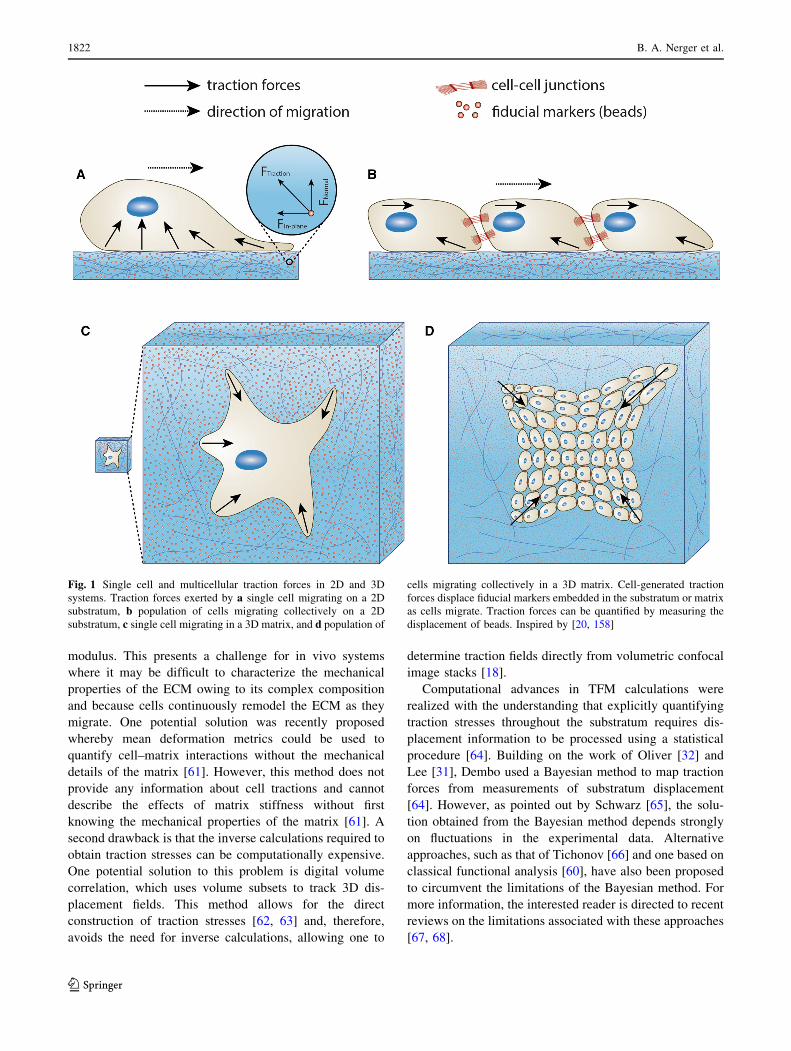
Quantifying traction forces
As described previously, traction forces are not measured
explicitly. Rather, TFM relies on measuring the physical
displacement of fiducial markers or the deformation of a
substratum. For single isolated cells, local displacement
data are combined with the appropriate constitutive equa-
tions to calculate local traction forces (Fig. 1a, c). For
multicellular systems, global forces can be calculated from
these local tractions using Newton’s laws of motion
(Fig. 1b, d). In this section, we outline a previously
described computational approach [21] for calculating
local traction forces directly from experimentally measured
displacement data. We further discuss the limitations
associated with traction force calculations as well as some
recent developments that aim to improve computational
efficiency.
For sufficiently small deformations of elastic substrata,
discrete traction forces (F) can be calculated using Hooke’s
law (Eq. 1):
F ¼ kx; ð1Þ
where k is the spring constant and x is the displacement
[57]. The accuracy of the calculation is a function of the
material properties of the substratum, the resolution of the
displacement measurements, and the validity of the
assumption that the deformations are sufficiently small
[16]. Although Hooke’s law provides a simple approach to
calculate traction forces at discrete locations, traction
forces are actually distributed continuously throughout the
substratum [21]. As a result, it is convenient to represent
traction forces in terms of traction stress, which represents
the traction force per unit contact area between the cell and
the underlying substratum or the surrounding matrix [21].
Furthermore, if the substratum is assumed to be a linearly
elastic solid, then the traction stress, rðxÞ, is related to the
displacement, uðxÞ, by Hooke’s law in tensor form [21, 58]
(Eq. 2):
r ¼ E
1� m1
2ruþruT� �
þ mr � u1� 2m
I
� �; ð2Þ
where m is Poisson’s ratio, E is the elastic modulus, and I is
the identity tensor. It is important to note that boundary
conditions at the top and bottom surfaces of the substratum
and the assumption of mechanical equilibrium (r � r ¼ 0)
are both required to solve Eq. 2 [58]. These assumptions
give the following equation [21]:
ð1� 2mÞr2uþrðr � uÞ ¼ 0; ð3Þ
which can be solved using a variety of mathematical
manipulations, including Fourier transforms [21, 59] and
finite-element analysis [49, 60]. Using similar approaches,
traction forces have been mapped for single cells and
populations of cells cultured on a planar substratum
(Fig. 2a, b) as well as embedded in a matrix (Fig. 2c, d).
One drawback of the aforementioned approach is that
calculating the traction stresses requires knowledge about
the properties of the underlying matrix, such as the elastic
Microfabricated tissues for investigating traction forces involved in cell migration and… 1821
123
modulus. This presents a challenge for in vivo systems
where it may be difficult to characterize the mechanical
properties of the ECM owing to its complex composition
and because cells continuously remodel the ECM as they
migrate. One potential solution was recently proposed
whereby mean deformation metrics could be used to
quantify cell–matrix interactions without the mechanical
details of the matrix [61]. However, this method does not
provide any information about cell tractions and cannot
describe the effects of matrix stiffness without first
knowing the mechanical properties of the matrix [61]. A
second drawback is that the inverse calculations required to
obtain traction stresses can be computationally expensive.
One potential solution to this problem is digital volume
correlation, which uses volume subsets to track 3D dis-
placement fields. This method allows for the direct
construction of traction stresses [62, 63] and, therefore,
avoids the need for inverse calculations, allowing one to
determine traction fields directly from volumetric confocal
image stacks [18].
Computational advances in TFM calculations were
realized with the understanding that explicitly quantifying
traction stresses throughout the substratum requires dis-
placement information to be processed using a statistical
procedure [64]. Building on the work of Oliver [32] and
Lee [31], Dembo used a Bayesian method to map traction
forces from measurements of substratum displacement
[64]. However, as pointed out by Schwarz [65], the solu-
tion obtained from the Bayesian method depends strongly
on fluctuations in the experimental data. Alternative
approaches, such as that of Tichonov [66] and one based on
classical functional analysis [60], have also been proposed
to circumvent the limitations of the Bayesian method. For
more information, the interested reader is directed to recent
reviews on the limitations associated with these approaches
[67, 68].
Fig. 1 Single cell and multicellular traction forces in 2D and 3D
systems. Traction forces exerted by a single cell migrating on a 2D
substratum, b population of cells migrating collectively on a 2D
substratum, c single cell migrating in a 3D matrix, and d population of
cells migrating collectively in a 3D matrix. Cell-generated traction
forces displace fiducial markers embedded in the substratum or matrix
as cells migrate. Traction forces can be quantified by measuring the
displacement of beads. Inspired by [20, 158]
1822 B. A. Nerger et al.
123
Application 1: forces exerted during cell migration
The study of mechanical forces in cell migration was first
performed qualitatively by examining changes in cell shape
during the migration of isolated epithelial cells and cell
sheets in 2D cell cultures [69]. While qualitative observa-
tions provide relative information about the magnitude of
mechanical forces, these approaches are inherently limited
by their inability to explicitly quantify forces in a repro-
ducible manner. To that end, a variety of experimental
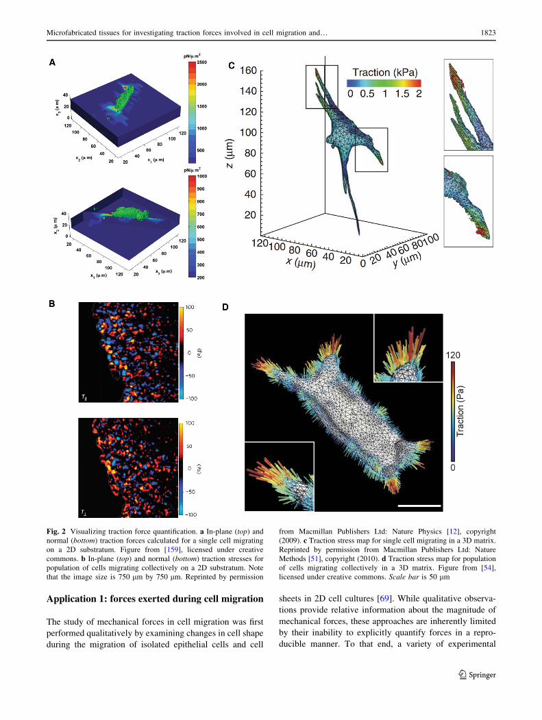
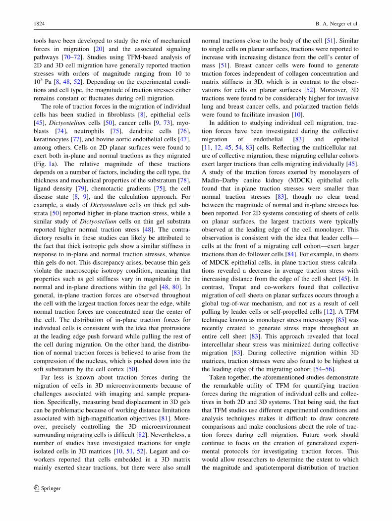
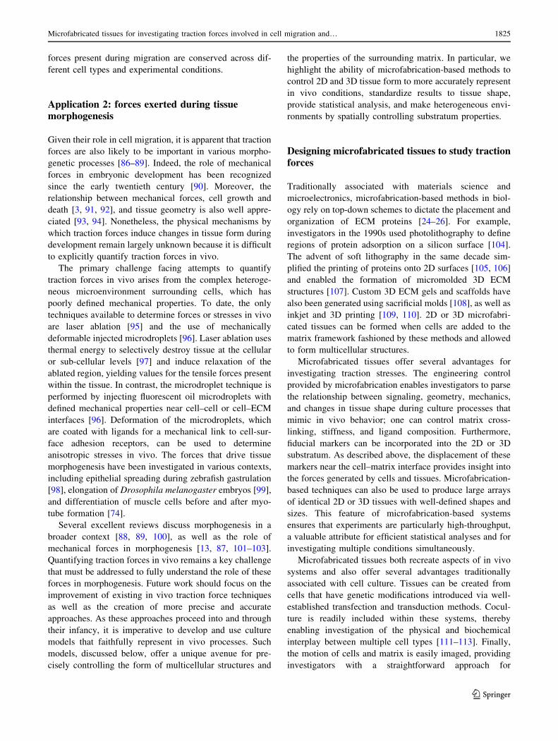
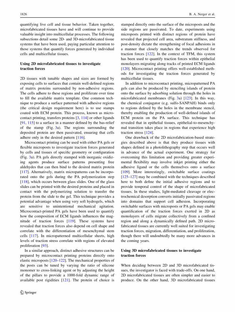
Fig. 2 Visualizing traction force quantification. a In-plane (top) and
normal (bottom) traction forces calculated for a single cell migrating
on a 2D substratum. Figure from [159], licensed under creative
commons. b In-plane (top) and normal (bottom) traction stresses for
population of cells migrating collectively on a 2D substratum. Note
that the image size is 750 lm by 750 lm. Reprinted by permission
from Macmillan Publishers Ltd: Nature Physics [12], copyright
(2009). c Traction stress map for single cell migrating in a 3D matrix.
Reprinted by permission from Macmillan Publishers Ltd: Nature
Methods [51], copyright (2010). d Traction stress map for population
of cells migrating collectively in a 3D matrix. Figure from [54],
licensed under creative commons. Scale bar is 50 lm
Microfabricated tissues for investigating traction forces involved in cell migration and… 1823
123
tools have been developed to study the role of mechanical
forces in migration [20] and the associated signaling
pathways [70–72]. Studies using TFM-based analysis of
2D and 3D cell migration have generally reported traction
stresses with orders of magnitude ranging from 10 to
103 Pa [8, 48, 52]. Depending on the experimental condi-
tions and cell type, the magnitude of traction stresses either
remains constant or fluctuates during cell migration.
The role of traction forces in the migration of individual
cells has been studied in fibroblasts [8], epithelial cells
[45], Dictyostelium cells [50], cancer cells [9, 73], myo-
blasts [74], neutrophils [75], dendritic cells [76],
keratinocytes [77], and bovine aortic endothelial cells [47],
among others. Cells on 2D planar surfaces were found to
exert both in-plane and normal tractions as they migrated
(Fig. 1a). The relative magnitude of these tractions
depends on a number of factors, including the cell type, the
thickness and mechanical properties of the substratum [78],
ligand density [79], chemotactic gradients [75], the cell
disease state [8, 9], and the calculation approach. For
example, a study of Dictyostelium cells on thick gel sub-
strata [50] reported higher in-plane traction stress, while a
similar study of Dictyostelium cells on thin gel substrata
reported higher normal traction stress [48]. The contra-
dictory results in these studies can likely be attributed to
the fact that thick isotropic gels show a similar stiffness in
response to in-plane and normal traction stresses, whereas
thin gels do not. This discrepancy arises, because thin gels
violate the macroscopic isotropy condition, meaning that
properties such as gel stiffness vary in magnitude in the
normal and in-plane directions within the gel [48, 80]. In
general, in-plane traction forces are observed throughout
the cell with the largest traction forces near the edge, while
normal traction forces are concentrated near the center of
the cell. The distribution of in-plane traction forces for
individual cells is consistent with the idea that protrusions
at the leading edge push forward while pulling the rest of
the cell during migration. On the other hand, the distribu-
tion of normal traction forces is believed to arise from the
compression of the nucleus, which is pushed down into the
soft substratum by the cell cortex [50].
Far less is known about traction forces during the
migration of cells in 3D microenvironments because of
challenges associated with imaging and sample prepara-
tion. Specifically, measuring bead displacement in 3D gels
can be problematic because of working distance limitations
associated with high-magnification objectives [81]. More-
over, precisely controlling the 3D microenvironment
surrounding migrating cells is difficult [82]. Nevertheless, a
number of studies have investigated tractions for single
isolated cells in 3D matrices [10, 51, 52]. Legant and co-
workers reported that cells embedded in a 3D matrix
mainly exerted shear tractions, but there were also small
normal tractions close to the body of the cell [51]. Similar
to single cells on planar surfaces, tractions were reported to
increase with increasing distance from the cell’s center of
mass [51]. Breast cancer cells were found to generate
traction forces independent of collagen concentration and
matrix stiffness in 3D, which is in contrast to the obser-
vations for cells on planar surfaces [52]. Moreover, 3D
tractions were found to be considerably higher for invasive
lung and breast cancer cells, and polarized traction fields
were found to facilitate invasion [10].
In addition to studying individual cell migration, trac-
tion forces have been investigated during the collective
migration of endothelial [83] and epithelial
[11, 12, 45, 54, 83] cells. Reflecting the multicellular nat-
ure of collective migration, these migrating cellular cohorts
exert larger tractions than cells migrating individually [45].
A study of the traction forces exerted by monolayers of
Madin–Darby canine kidney (MDCK) epithelial cells
found that in-plane traction stresses were smaller than
normal traction stresses [83], though no clear trend
between the magnitude of normal and in-plane stresses has
been reported. For 2D systems consisting of sheets of cells
on planar surfaces, the largest tractions were typically
observed at the leading edge of the cell monolayer. This
observation is consistent with the idea that leader cells—
cells at the front of a migrating cell cohort—exert larger
tractions than do follower cells [84]. For example, in sheets
of MDCK epithelial cells, in-plane traction stress calcula-
tions revealed a decrease in average traction stress with
increasing distance from the edge of the cell sheet [45]. In
contrast, Trepat and co-workers found that collective
migration of cell sheets on planar surfaces occurs through a
global tug-of-war mechanism, and not as a result of cell
pulling by leader cells or self-propelled cells [12]. A TFM
technique known as monolayer stress microscopy [85] was
recently created to generate stress maps throughout an
entire cell sheet [83]. This approach revealed that local
intercellular shear stress was minimized during collective
migration [83]. During collective migration within 3D
matrices, traction stresses were also found to be highest at
the leading edge of the migrating cohort [54–56].
Taken together, the aforementioned studies demonstrate
the remarkable utility of TFM for quantifying traction
forces during the migration of individual cells and collec-
tives in both 2D and 3D systems. That being said, the fact
that TFM studies use different experimental conditions and
analysis techniques makes it difficult to draw concrete
comparisons and make conclusions about the role of trac-
tion forces during cell migration. Future work should
continue to focus on the creation of generalized experi-
mental protocols for investigating traction forces. This
would allow researchers to determine the extent to which
the magnitude and spatiotemporal distribution of traction
1824 B. A. Nerger et al.
123
forces present during migration are conserved across dif-
ferent cell types and experimental conditions.
Application 2: forces exerted during tissuemorphogenesis
Given their role in cell migration, it is apparent that traction
forces are also likely to be important in various morpho-
genetic processes [86–89]. Indeed, the role of mechanical
forces in embryonic development has been recognized
since the early twentieth century [90]. Moreover, the
relationship between mechanical forces, cell growth and
death [3, 91, 92], and tissue geometry is also well appre-
ciated [93, 94]. Nonetheless, the physical mechanisms by
which traction forces induce changes in tissue form during
development remain largely unknown because it is difficult
to explicitly quantify traction forces in vivo.
The primary challenge facing attempts to quantify
traction forces in vivo arises from the complex heteroge-
neous microenvironment surrounding cells, which has
poorly defined mechanical properties. To date, the only
techniques available to determine forces or stresses in vivo
are laser ablation [95] and the use of mechanically
deformable injected microdroplets [96]. Laser ablation uses
thermal energy to selectively destroy tissue at the cellular
or sub-cellular levels [97] and induce relaxation of the
ablated region, yielding values for the tensile forces present
within the tissue. In contrast, the microdroplet technique is
performed by injecting fluorescent oil microdroplets with
defined mechanical properties near cell–cell or cell–ECM
interfaces [96]. Deformation of the microdroplets, which
are coated with ligands for a mechanical link to cell-sur-
face adhesion receptors, can be used to determine
anisotropic stresses in vivo. The forces that drive tissue
morphogenesis have been investigated in various contexts,
including epithelial spreading during zebrafish gastrulation
[98], elongation of Drosophila melanogaster embryos [99],
and differentiation of muscle cells before and after myo-
tube formation [74].
Several excellent reviews discuss morphogenesis in a
broader context [88, 89, 100], as well as the role of
mechanical forces in morphogenesis [13, 87, 101–103].
Quantifying traction forces in vivo remains a key challenge
that must be addressed to fully understand the role of these
forces in morphogenesis. Future work should focus on the
improvement of existing in vivo traction force techniques
as well as the creation of more precise and accurate
approaches. As these approaches proceed into and through
their infancy, it is imperative to develop and use culture
models that faithfully represent in vivo processes. Such
models, discussed below, offer a unique avenue for pre-
cisely controlling the form of multicellular structures and
the properties of the surrounding matrix. In particular, we
highlight the ability of microfabrication-based methods to
control 2D and 3D tissue form to more accurately represent
in vivo conditions, standardize results to tissue shape,
provide statistical analysis, and make heterogeneous envi-
ronments by spatially controlling substratum properties.
Designing microfabricated tissues to study tractionforces
Traditionally associated with materials science and
microelectronics, microfabrication-based methods in biol-
ogy rely on top-down schemes to dictate the placement and
organization of ECM proteins [24–26]. For example,
investigators in the 1990s used photolithography to define
regions of protein adsorption on a silicon surface [104].
The advent of soft lithography in the same decade sim-
plified the printing of proteins onto 2D surfaces [105, 106]
and enabled the formation of micromolded 3D ECM
structures [107]. Custom 3D ECM gels and scaffolds have
also been generated using sacrificial molds [108], as well as
inkjet and 3D printing [109, 110]. 2D or 3D microfabri-
cated tissues can be formed when cells are added to the
matrix framework fashioned by these methods and allowed
to form multicellular structures.
Microfabricated tissues offer several advantages for
investigating traction stresses. The engineering control
provided by microfabrication enables investigators to parse
the relationship between signaling, geometry, mechanics,
and changes in tissue shape during culture processes that
mimic in vivo behavior; one can control matrix cross-
linking, stiffness, and ligand composition. Furthermore,
fiducial markers can be incorporated into the 2D or 3D
substratum. As described above, the displacement of these
markers near the cell–matrix interface provides insight into
the forces generated by cells and tissues. Microfabrication-
based techniques can also be used to produce large arrays
of identical 2D or 3D tissues with well-defined shapes and
sizes. This feature of microfabrication-based systems
ensures that experiments are particularly high-throughput,
a valuable attribute for efficient statistical analyses and for
investigating multiple conditions simultaneously.
Microfabricated tissues both recreate aspects of in vivo
systems and also offer several advantages traditionally
associated with cell culture. Tissues can be created from
cells that have genetic modifications introduced via well-
established transfection and transduction methods. Cocul-
ture is readily included within these systems, thereby
enabling investigation of the physical and biochemical
interplay between multiple cell types [111–113]. Finally,
the motion of cells and matrix is easily imaged, providing
investigators with a straightforward approach for
Microfabricated tissues for investigating traction forces involved in cell migration and… 1825
123
quantifying live cell and tissue behavior. Taken together,
microfabricated tissues have and will continue to provide
valuable insight into multicellular processes. The following
subsections detail some 2D- and 3D-microfabricated tissue
systems that have been used, paying particular attention to
those systems that quantify forces generated by individual
cells and multicellular tissues.
Using 2D microfabricated tissues to investigate
traction forces
2D tissues with tunable shapes and sizes are formed by
exposing cells to surfaces that contain well-defined regions
of matrix proteins surrounded by non-adhesive regions.
The cells adhere to these regions and proliferate over time
to fill the available matrix template. One common tech-
nique to produce a surface patterned with adhesive regions
(the critical design requirement here) is to use stamps
coated with ECM protein. This process, known as micro-
contact printing, transfers proteins [3, 114] or other ligands
[91, 115] to a surface in a manner defined by the bas-relief
of the stamp (Fig. 3a). The regions surrounding the
deposited protein are then passivated, ensuring that cells
adhere only in the desired pattern [116].
Microcontact printing can be used with either PA gels or
flexible microposts to investigate traction forces generated
by cells and tissues of specific geometry or configuration
(Fig. 3a). PA gels directly stamped with inorganic oxidiz-
ing agents produce surface patterns presenting free
aldehydes that can then bind to the desired matrix protein
[117]. Alternatively, matrix micropatterns can be incorpo-
rated onto the gels during the PA polymerization step
[118], which occurs between glass slides. One of the glass
slides can be printed with the desired proteins and placed in
contact with the polymerizing solution to transfer the
protein from the slide to the gel. This technique provides a
potential advantage when using very soft hydrogels, which
are sensitive to unintentional mechanical agitation.
Microcontact-printed PA gels have been used to quantify
how the composition of ECM ligands influences the mag-
nitude of traction forces [119]. These systems have
revealed that traction forces also depend on cell shape and
correlate with the differentiation of mesenchymal stem
cells [117]. In micropatterned multicellular sheets, high
levels of traction stress correlate with regions of elevated
proliferation [93].
In a similar approach, distinct adhesive structures can be
prepared by microcontact printing proteins directly onto
elastic microposts [120–122]. The mechanical properties of
the posts can be tuned by varying the ratio of silicone
monomer to cross-linking agent or by adjusting the height
of the pillars to provide a 1000-fold dynamic range of
available post rigidities [121]. The protein of choice is
stamped directly onto the surface of the microposts and the
side regions are passivated. To date, experiments using
microposts printed with distinct regions of protein have
revealed that projected cell area, substratum stiffness, and
post-density dictate the strengthening of focal adhesions in
a manner that closely matches the trends observed for
traction forces [122]. In the context of TFM, this system
has been used to quantify traction forces within epithelial
monolayers migrating along tracks of printed ECM ligands
[120]. Microcontact printing offers well-established meth-
ods for investigating the traction forces generated by
multicellular tissues.
In addition to microcontact printing, micropatterned PA
gels can also be produced by stenciling islands of protein
onto the surface by adsorbing solution through the holes in
microfabricated membranes (Fig. 3a) [123]. In this way,
the chemical conjugator (e.g. sulfo-SANPAH) binds only
to regions defined by the holes in the membrane stencil,
thereby enabling the production of well-defined islands of
ECM protein on the PA surface. This technique has
revealed that in epithelial tissues, epithelial-to-mesenchy-
mal transition takes place in regions that experience high
traction stress [124].
One drawback of the 2D microfabrication-based strate-
gies described above is that they produce tissues with
shapes defined in a photolithography step that occurs well
in advance of the actual experiment. One strategy for
overcoming this limitation and providing greater experi-
mental flexibility may involve inkjet printing either the
adhesive ligand or the cells themselves onto a surface
[109]. More interestingly, switchable surface coatings
[125–127] may be combined with the techniques described
here to both define the initial adhesive regions and to
provide temporal control of the shape of microfabricated
tissues. In these studies, light-mediated cleavage or elec-
trochemical desorption converts initially passivated regions
into domains that support cell adhesion. Incorporating
switchable surfaces with microposts or PA gels may enable
quantification of the traction forces exerted in 2D as
monolayers of cells migrate collectively from a confined
region and along a dynamically defined path. 2D micro-
fabricated tissues are currently well suited for investigating
traction forces, migration, differentiation, and proliferation,
though there will undoubtedly be many more advances in
the coming years.
Using 3D microfabricated tissues to investigate
traction forces
When deciding between 2D and 3D microfabricated tis-
sues, the investigator is faced with trade-offs. On one hand,
2D microfabricated tissues are often simpler and easier to
produce. On the other hand, 3D microfabricated tissues
1826 B. A. Nerger et al.
123
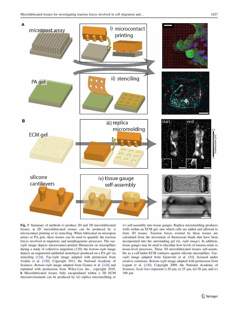
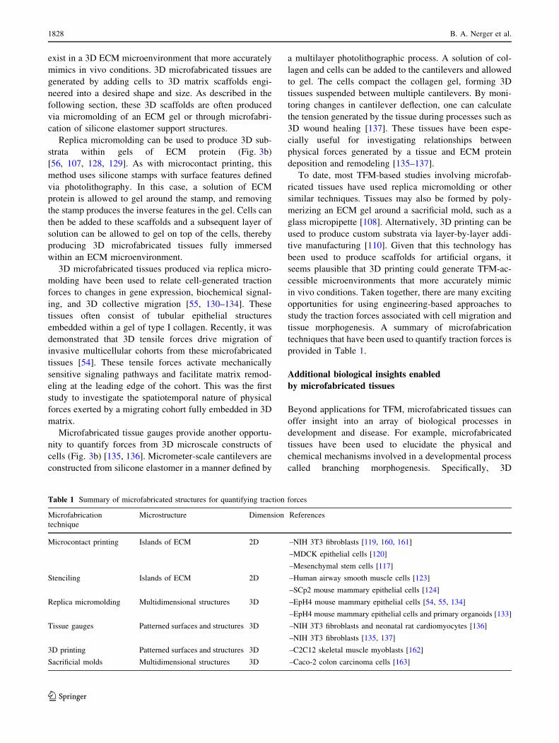
Fig. 3 Summary of methods to produce 2D and 3D microfabricated
tissues. a 2D microfabricated tissues can be produced by i)
microcontact printing or ii) stenciling. When fabricated on micropost
arrays or PA gels, these tissues can be used to quantify the traction
forces involved in migratory and morphogenetic processes. The top-
right image depicts microcontact-printed fibronectin on micropillars
during a study of collective migration [120]; the bottom-right image
depicts an engineered epithelial monolayer produced on a PA gel via
stenciling [124]. Top-right image adapted with permission from
Vedula et al. [120]. Copyright 2012, the National Academy of
Sciences. Bottom-right image adapted from Gomez et al. [124] and
reprinted with permission from Wiley-Liss Inc., copyright 2010.
b Microfabricated tissues fully encapsulated within a 3D ECM
microenvironment can be produced by iii) replica micromolding or
iv) self-assembly into tissue gauges. Replica micromolding produces
wells within an ECM gel, into which cells are added and allowed to
form 3D tissues. Traction forces exerted by these tissues are
calculated from the movement of fluorescent beads that have been
incorporated into the surrounding gel (iii, right image). In addition,
tissue gauges may be used to elucidate how levels of tension relate to
tissue-level processes. These 3D microfabricated tissues self-assem-
ble as a cell-laden ECM contracts against silicone micropillars. Top-
right image adapted from Gjorevski et al. [54], licensed under
creative commons. Bottom-right image adapted with permission from
Legant et al. [136]. Copyright 2009, the National Academy of
Sciences. Scale bars represent i) 20 lm, ii) 25 lm, iii) 50 lm, and iv)
100 lm
Microfabricated tissues for investigating traction forces involved in cell migration and… 1827
123
exist in a 3D ECM microenvironment that more accurately
mimics in vivo conditions. 3D microfabricated tissues are
generated by adding cells to 3D matrix scaffolds engi-
neered into a desired shape and size. As described in the
following section, these 3D scaffolds are often produced
via micromolding of an ECM gel or through microfabri-
cation of silicone elastomer support structures.
Replica micromolding can be used to produce 3D sub-
strata within gels of ECM protein (Fig. 3b)
[56, 107, 128, 129]. As with microcontact printing, this
method uses silicone stamps with surface features defined
via photolithography. In this case, a solution of ECM
protein is allowed to gel around the stamp, and removing
the stamp produces the inverse features in the gel. Cells can
then be added to these scaffolds and a subsequent layer of
solution can be allowed to gel on top of the cells, thereby
producing 3D microfabricated tissues fully immersed
within an ECM microenvironment.
3D microfabricated tissues produced via replica micro-
molding have been used to relate cell-generated traction
forces to changes in gene expression, biochemical signal-
ing, and 3D collective migration [55, 130–134]. These
tissues often consist of tubular epithelial structures
embedded within a gel of type I collagen. Recently, it was
demonstrated that 3D tensile forces drive migration of
invasive multicellular cohorts from these microfabricated
tissues [54]. These tensile forces activate mechanically
sensitive signaling pathways and facilitate matrix remod-
eling at the leading edge of the cohort. This was the first
study to investigate the spatiotemporal nature of physical
forces exerted by a migrating cohort fully embedded in 3D
matrix.
Microfabricated tissue gauges provide another opportu-
nity to quantify forces from 3D microscale constructs of
cells (Fig. 3b) [135, 136]. Micrometer-scale cantilevers are
constructed from silicone elastomer in a manner defined by
a multilayer photolithographic process. A solution of col-
lagen and cells can be added to the cantilevers and allowed
to gel. The cells compact the collagen gel, forming 3D
tissues suspended between multiple cantilevers. By moni-
toring changes in cantilever deflection, one can calculate
the tension generated by the tissue during processes such as
3D wound healing [137]. These tissues have been espe-
cially useful for investigating relationships between
physical forces generated by a tissue and ECM protein
deposition and remodeling [135–137].
To date, most TFM-based studies involving microfab-
ricated tissues have used replica micromolding or other
similar techniques. Tissues may also be formed by poly-
merizing an ECM gel around a sacrificial mold, such as a
glass micropipette [108]. Alternatively, 3D printing can be
used to produce custom substrata via layer-by-layer addi-
tive manufacturing [110]. Given that this technology has
been used to produce scaffolds for artificial organs, it
seems plausible that 3D printing could generate TFM-ac-
cessible microenvironments that more accurately mimic
in vivo conditions. Taken together, there are many exciting
opportunities for using engineering-based approaches to
study the traction forces associated with cell migration and
tissue morphogenesis. A summary of microfabrication
techniques that have been used to quantify traction forces is
provided in Table 1.
Additional biological insights enabled
by microfabricated tissues
Beyond applications for TFM, microfabricated tissues can
offer insight into an array of biological processes in
development and disease. For example, microfabricated
tissues have been used to elucidate the physical and
chemical mechanisms involved in a developmental process
called branching morphogenesis. Specifically, 3D
Table 1 Summary of microfabricated structures for quantifying traction forces
Microfabrication
technique
Microstructure Dimension References
Microcontact printing Islands of ECM 2D –NIH 3T3 fibroblasts [119, 160, 161]
–MDCK epithelial cells [120]
–Mesenchymal stem cells [117]
Stenciling Islands of ECM 2D –Human airway smooth muscle cells [123]
–SCp2 mouse mammary epithelial cells [124]
Replica micromolding Multidimensional structures 3D –EpH4 mouse mammary epithelial cells [54, 55, 134]
–EpH4 mouse mammary epithelial cells and primary organoids [133]
Tissue gauges Patterned surfaces and structures 3D –NIH 3T3 fibroblasts and neonatal rat cardiomyocytes [136]
–NIH 3T3 fibroblasts [135, 137]
3D printing Patterned surfaces and structures 3D –C2C12 skeletal muscle myoblasts [162]
Sacrificial molds Multidimensional structures 3D –Caco-2 colon carcinoma cells [163]
1828 B. A. Nerger et al.
123
microfabricated mammary epithelial tissues were used to
reveal the interplay between tissue geometry and factors
such as mechanical stress [55], autocrine inhibitory mor-
phogen concentration [133], and epithelial-mesenchymal
transition [124]. Microfabricated tissues have also been
used as models to study diseases including cancer, where
they revealed the relationship between mechanical stress
and breast cancer cell invasiveness [113] as well as the
interplay between interstitial fluid pressure, gene expres-
sion, and collective invasion of engineered breast tumors
[108]. Additional biological processes, such as collective
cell migration [54], wound healing [137], and cell sorting
[138], have also been studied using microfabricated tissues.
Interested readers can find a more comprehensive discus-
sion of tissue micropatterning in literature [139].
Strategies for overcoming the limitationsof conventional TFM
The spatiotemporal accuracy of quantified traction forces
has been significantly improved since the original work of
Harris and colleagues. Nevertheless, the accuracy of TFM
is inherently limited because force is calculated implicitly
from measured displacement data. Moreover, this calcula-
tion procedure can be computationally expensive, which
limits the complexity of the biological systems that can be
studied. Additional factors, including the resolution of the
fiducial markers, substratum properties, and the validity of
the underlying constitutive model used in force calcula-
tions, limit the accuracy of any traction forces that may be
reported [16]. Limitations also arise from the difficulty
associated with creating systems that recapitulate the
in vivo microenvironment and that have well-defined
mechanical properties. For example, a combination of
spatial heterogeneities and large pore sizes in collagen gels
can result in a breakdown of the continuous medium
assumption [140]. Thus, the relevant mechanical properties
of collagen are difficult to determine because of discrep-
ancies between bulk rheology and micromechanics of the
collagen network [140]. Furthermore, discrete fiducial
markers cannot fully capture the deformation of a contin-
uous substratum, and any deformations of the matrix in
between markers remain uncertain [64, 65]. Finally,
obtaining high-resolution images can be expensive and
may limit the accessibility of the technology.
A recent alternative for measuring substratum defor-
mation involves imaging deformations of fibrous matrix
materials as opposed to embedded beads [141, 142]. These
studies offer a new approach for calculating 3D displace-
ment fields that may be advantageous in systems where
bead displacement techniques are not feasible or have
limited accuracy. For example, during remodeling of a
fibrous matrix, reflection microscopy can be used to cal-
culate matrix displacements and obtain qualitative
information on cell tractions [141, 142]. Quantitative cell
traction calculations rely on the assumption that the fibrous
network is isotropic and can be approximated as linearly
elastic [141]. In addition, molecular force sensors offer one
strategy for improving the spatial resolution of quantified
traction forces [143–147]. In contrast to TFM-based
approaches that can only quantify traction forces exerted
by the cell on its external microenvironment, molecular
force sensors can be used to probe internal cellular forces
[100]. One example is molecular tension-based fluores-
cence microscopy, which uses immobilized DNA hairpins
that have a tunable force threshold to measure tension
across integrins [148]. Another example uses a combina-
tion of super-resolution light microscopy and polypeptide
fluorescence resonance energy transfer (FRET)-based ten-
sion sensors to visualize forces across individual molecules
that are present within focal adhesions [149]. Myosin I has
been used as a sensor for molecular force [150], and DNA
hairpins have been used to probe traction forces with high
spatial resolution [151]. A final example consists of a
molecular tension sensor that uses a PEG polymer to create
spatiotemporal maps of mechanical forces exerted by cell-
surface receptors [152]. This tension sensor has well-
characterized mechanical properties, is biocompatible, and
shows minimal non-specific interactions [152]. That being
said, a number of challenges must be addressed, including
in vivo calibration [145], improved understanding of the
relationship between measurements in culture and in vivo,
and improved understanding of the appropriate simplifying
assumptions [100].
Additional information about cell-generated mechanical
forces can also be obtained by combining TFM with other
experimental techniques. For example, AFM, which can be
used to directly measure the mechanical properties of cells,
has been combined with TFM to study how cells respond to
applied forces [153, 154]. In a similar approach, incorpo-
rating magnetic particles within silicone micropillars
enables one to study the response of cells to precisely
applied forces [155]. Cellular traction forces have also been
studied by combining TFM with optical tweezers. In par-
ticular, optical tweezers have been used to dictate the
placement of beads, so that traction forces can be quanti-
fied locally [156, 157]. It has also been suggested that
integrating TFM with molecular force sensors may provide
a way to understand the interplay between extrinsic and
intrinsic cellular forces [100]. While the aforementioned
techniques have made improvements to conventional TFM,
developing a complete understanding of traction forces in
biological systems will require versatile techniques that can
produce spatial maps of forces in vivo with high accuracy
and precision.
Microfabricated tissues for investigating traction forces involved in cell migration and… 1829
123
Concluding remarks
Insight into the biophysical and biochemical mechanisms
through which traction forces act is required for under-
standing a vast array of biological processes. Here, we
describe experimental techniques and computational
methods to quantify traction forces. Despite considerable
progress and improvement in experimental and computa-
tional approaches, current TFM techniques cannot be used
to quantify traction forces exerted within in vivo
microenvironments, nor can they account for the complex
heterogeneity present in native ECMs. As described here,
microfabricated tissues provide an experimentally
tractable means to quantify the traction forces exerted by
and within multicellular cohorts. Coupled with improved
computational approaches, microfabricated tissues will
shed light on the tractions exerted by cells in heterogeneous
non-linear microenvironments and provide valuable insight
into processes that resemble in vivo migrational and mor-
phogenetic movements.
Acknowledgements Work from the authors’ group was supported in
part by grants from the NIH (GM083997, HL110335, HL118532,
HL120142, and CA 187692), the NSF (CMMI-1435853), the David
and Lucile Packard Foundation, the Alfred P. Sloan Foundation, the
Camille and Henry Dreyfus Foundation, and the Burroughs Wellcome
Fund. M.J.S. was supported in part by the NSF Graduate Research
Fellowship Program. C.M.N. was supported in part by a Faculty
Scholars Award from the Howard Hughes Medical Institute.
References
1. Maniotis AJ, Chen CS, Ingber DE (1997) Demonstration of
mechanical connections between integrins, cytoskeletal fila-
ments, and nucleoplasm that stabilize nuclear structure. Proc
Natl Acad Sci USA 94(3):849–854. doi:10.1073/pnas.94.3.849
2. Thery M, Jimenez-Dalmaroni A, Racine V, Bornens M, Julicher
F (2007) Experimental and theoretical study of mitotic spindle
orientation. Nature 447(7143):493–496. doi:10.1038/nature05786
3. Chen CS, Mrksich M, Huang S, Whitesides GM, Ingber DE
(1997) Geometric control of cell life and death. Science
276(5317):1425–1428. doi:10.1126/science.276.5317.1425
4. Engler AJ, Sen S, Sweeney HL, Discher DE (2006) Matrix
elasticity directs stem cell lineage specification. Cell
126(4):677–689. doi:10.1016/j.cell.2006.06.044
5. Desprat N, Supatto W, Pouille P-A, Beaurepaire E, Farge E
(2008) Tissue deformation modulates twist expression to
determine anterior midgut differentiation in Drosophila
embryos. Dev Cell 15(3):470–477. doi:10.1016/j.devcel.2008.
07.009
6. Farge E (2003) Mechanical induction of twist in the Drosophila
foregut/stomodeal primordium. Curr Biol 13(16):1365–1377.
doi:10.1016/S0960-9822(03)00576-1
7. Hinz B, Mastrangelo D, Iselin CE, Chaponnier C, Gabbiani G
(2001) Mechanical tension controls granulation tissue contrac-
tile activity and myofibroblast differentiation. Am J Pathol
159(3):1009–1020. doi:10.1016/S0002-9440(10)61776-2
8. Munevar S, Wang Y, Dembo M (2001) Traction force micro-
scopy of migrating normal and H-ras transformed 3T3
fibroblasts. Biophys J 80(4):1744–1757. doi:10.1016/S0006-
3495(01)76145-0
9. Peschetola V, Laurent VM, Duperray A, Michel R, Ambrosi D,
Preziosi L, Verdier C (2013) Time-dependent traction force
microscopy for cancer cells as a measure of invasiveness.
Cytoskeleton. doi:10.1002/cm.21100 (1949-3592 (Electronic))
10. Koch TM, Munster S, Bonakdar N, Butler JP, Fabry B (2012)
3D Traction forces in cancer cell invasion. PLoS One
7(3):e33476. doi:10.1371/journal.pone.0033476
11. Reffay M, Parrini MC, Cochet-Escartin O, Ladoux B, Buguin A,
Coscoy S, Amblard F, Camonis J, Silberzan P (2014) Interplay
of RhoA and mechanical forces in collective cell migration
driven by leader cells. Nat Cell Biol 16(3):217–223. doi:10.
1038/ncb2917
12. Trepat X, Wasserman MR, Angelini TE, Millet E, Weitz DA,
Butler JP, Fredberg JJ (2009) Physical forces during collective
cell migration. Nat Phys 5(6):426–430. doi:10.1038/nphys1269
13. Heisenberg C-P, Bellaıche Y (2013) Forces in tissue morpho-
genesis and patterning. Cell 153(5):948–962. doi:10.1016/j.cell.
2013.05.008
14. Gov NS (2009) Traction forces during collective cell motion.
HFSP Journal 3(4):223–227. doi:10.2976/1.3185785
15. His W (1874) Unsere Korperform und das physiologische
Problem ihrer Entstehung Briefe an einen befreundeten Natur-
forscher/von Wilhelm His. F.C.W. Vogel, Leipzig. doi:10.5962/
bhl.title.28860
16. Paluch EK, Nelson CM, Biais N, Fabry B, Moeller J, Pruitt BL,
Wollnik C, Kudryasheva G, Rehfeldt F, Federle W (2015)
Mechanotransduction: use the force(s). BMC Biol 13(1):1–14.
doi:10.1186/s12915-015-0150-4
17. Richards RJ (2008) The tragic sense of life: ernst haeckel and
the struggle over evolutionary thought. University of Chicago
Press, Chicago
18. Maskarinec SA, Franck C, Tirrell DA, Ravichandran G (2009)
Quantifying cellular traction forces in three dimensions. Proc
Natl Acad Sci USA 106(52):22108–22113. doi:10.1073/pnas.
0904565106
19. Mok S, Moraes C (2016) Thinking big by thinking small:
advances in mechanobiology across the length scales. Integr
Biol 8(3):262–266. doi:10.1039/C6IB90008A
20. Polacheck WJ, Chen CS (2016) Measuring cell-generated for-
ces: a guide to the available tools. Nat Meth 13(5):415–423.
doi:10.1038/nmeth.3834
21. Style RW, Boltyanskiy R, German GK, Hyland C, MacMinn
CW, Mertz AF, Wilen LA, Xu Y, Dufresne ER (2014) Traction
force microscopy in physics and biology. Soft Matter 10(23):
4047–4055. doi:10.1039/C4SM00264D
22. Ribeiro AJS, Denisin AK, Wilson RE, Pruitt BL (2016) For
whom the cells pull: hydrogel and micropost devices for mea-
suring traction forces. Methods 94:51–64. doi:10.1016/j.ymeth.
2015.08.005
23. Gupta M, Kocgozlu L, Sarangi BR, Margadant F, Ashraf M,
Ladoux B (2015) Chapter 16—micropillar substrates: a tool for
studying cell mechanobiology. In: Ewa KP (ed) Methods in cell
biology, vol 125. Academic Press, London, pp 289–308. doi:10.
1016/bs.mcb.2014.10.009
24. Nelson CM, Tien J (2006) Microstructured extracellular matri-
ces in tissue engineering and development. Curr Opin
Biotechnol 17(5):518–523. doi:10.1016/j.copbio.2006.08.011
25. Tien J, Nelson CM (2013) Microstructured extracellular matri-
ces in tissue engineering and development: an update. Ann
Biomed Eng 42(7):1413–1423. doi:10.1007/s10439-013-0912-5
26. Zorlutuna P, Annabi N, Camci-Unal G, Nikkhah M, Cha JM,
Nichol JW, Manbachi A, Bae H, Chen S, Khademhosseini A
(2012) Microfabricated biomaterials for engineering 3D tissues.
Adv Mater 24(14):1782–1804. doi:10.1002/adma.201104631
1830 B. A. Nerger et al.
123
27. Harris AK, Wild P, Stopak D (1980) Silicone rubber substrata: a
new wrinkle in the study of cell locomotion. Science
208(4440):177–179. doi:10.1126/science.6987736
28. Harris JK (1978) A photoelastic substrate technique for dynamic
measurements of forces exerted by moving organisms. J Mi-
crosc 114(2):219–228. doi:10.1111/j.1365-2818.1978.tb00132.x
29. Harris AK (1973) Cell surface movements related to cell loco-
motion. In: Locomotion of tissue cells, Ciba Foundation
Symposium, Amsterdam, Elsevier, pp 3–26. doi:10.1002/
9780470719978.ch2
30. Dembo M, Wang Y-L (1999) Stresses at the cell-to-substrate
interface during locomotion of fibroblasts. Biophys J
76(4):2307–2316. doi:10.1016/S0006-3495(99)77386-8
31. Lee J, Leonard M, Oliver T, Ishihara A, Jacobson K (1994)
Traction forces generated by locomoting keratocytes. J Cell Biol
127(6):1957–1964. doi:10.1083/jcb.127.6.1957
32. Oliver T, Jacobson K, Dembo M (1995) Traction forces in
locomoting cells. Cell Motil Cytoskeleton 31(3):225–240.
doi:10.1002/cm.970310306
33. Burton K, Taylor DL (1997) Traction forces of cytokinesis
measured with optically modified elastic substrata. Nature
385(6615):450–454. doi:10.1038/385450a0
34. Roy P, Petroll WM, Cavanagh HD, Chuong CJ, Jester JV (1997)
An in vitro force measurement assay to study the early
mechanical interaction between corneal fibroblasts and collagen
matrix. Exp Cell Res 232(1):106–117. doi:10.1006/excr.1997.
3511
35. Brandley BK, Weisz OA, Schnaar RL (1987) Cell attachment
and long-term growth on derivatizable polyacrylamide surfaces.
J Biol Chem 262(13):6431–6437
36. Pelham RJ, Wang Y-L (1997) Cell locomotion and focal
adhesions are regulated by substrate flexibility. Proc Natl Acad
Sci USA 94(25):13661–13665. doi:10.1073/pnas.94.25.13661
37. Wang Y-L, Pelham RJ Jr (1998) Preparation of a flexible, por-
ous polyacrylamide substrate for mechanical studies of cultured
cells. Methods in Enzymology, vol 298. Academic Press, Lon-
don, pp 489–496. doi:10.1016/S0076-6879(98)98041-7
38. Beningo KA, Wang Y-L (2002) Flexible substrata for the
detection of cellular traction forces. Trends Cell Biol
12(2):79–84. doi:10.1016/S0962-8924(01)02205-X
39. Sabass B, Gardel ML, Waterman CM, Schwarz US (2008) High
resolution traction force microscopy based on experimental and
computational advances. Biophys J 94(1):207–220. doi:10.1529/
biophysj.107.113670
40. Lautscham LA, Lin CY, Auernheimer V, Naumann CA, Gold-
mann WH, Fabry B (2014) Biomembrane-mimicking lipid
bilayer system as a mechanically tunable cell substrate. Bio-
materials 35(10):3198–3207. doi:10.1016/j.biomaterials.2013.
12.091
41. Tan JL, Tien J, Pirone DM, Gray DS, Bhadriraju K, Chen CS
(2003) Cells lying on a bed of microneedles: an approach to
isolate mechanical force. Proc Natl Acad Sci USA
100(4):1484–1489. doi:10.1073/pnas.0235407100
42. Galbraith CG, Sheetz MP (1997) A micromachined device
provides a new bend on fibroblast traction forces. Proc Natl
Acad Sci USA 94(17):9114–9118. doi:10.1073/pnas.94.17.9114
43. Balaban NQ, Schwarz US, Riveline D, Goichberg P, Tzur G,
Sabanay I, Mahalu D, Safran S, Bershadsky A, Addadi L, Geiger
B (2001) Force and focal adhesion assembly: a close relation-
ship studied using elastic micropatterned substrates. Nat Cell
Biol 3(5):466–472. doi:10.1038/35074532
44. Rodriguez ML, Graham BT, Pabon LM, Han SJ, Murry CE,
Sniadecki NJ (2014) Measuring the contractile forces of human
induced pluripotent stem cell-derived cardiomyocytes with
arrays of microposts. J Biomech Eng 136(5):051005. doi:10.
1115/1.4027145
45. du Roure O, Saez A, Buguin A, Austin RH, Chavrier P, Siberzan
P, Ladoux B (2005) Force mapping in epithelial cell migration.
Proc Natl Acad Sci USA 102(7):2390–2395. doi:10.1073/pnas.
0408482102
46. Schoen I, Hu W, Klotzsch E, Vogel V (2010) Probing cellular
traction forces by micropillar arrays: contribution of substrate
warping to pillar deflection. Nano Lett 10(5):1823–1830. doi:10.
1021/nl100533c
47. Hur SS, Zhao Y, Li Y-S, Botvinick E, Chien S (2009) Live cells
exert 3-dimensional traction forces on their substrata. Cell Mol
Bioeng 2(3):425–436. doi:10.1007/s12195-009-0082-6
48. del Alamo JC, Meili R, Alvarez-Gonzalez B, Alonso-Latorre B,
Bastounis E, Firtel R, Lasheras JC (2013) Three-dimensional
quantification of cellular traction forces and mechanosensing of
thin substrata by Fourier traction force microscopy. PLoS One
8(9):e69850. doi:10.1371/journal.pone.0069850
49. Legant WR, Choi CK, Miller JS, Shao L, Gao L, Betzig E, Chen
CS (2013) Multidimensional traction force microscopy reveals
out-of-plane rotational moments about focal adhesions. Proc
Natl Acad Sci USA 110(3):881–886. doi:10.1073/pnas.
1207997110
50. Delanoe-Ayari H, Rieu JP, Sano M (2010) 4D traction force
microscopy reveals asymmetric cortical forces in migrating
dictyostelium cells. Phys Rev Lett 105(24):248103. doi:10.1103/
PhysRevLett.105.248103
51. Legant WR, Miller JS, Blakely BL, Cohen DM, Genin GM,
Chen CS (2010) Measurement of mechanical tractions exerted
by cells in three-dimensional matrices. Nat Meth 7(12):969–971.
doi:10.1038/nmeth.1531
52. Steinwachs J, Metzner C, Skodzek K, Lang N, Thievessen I,
Mark C, Munster S, Aifantis KE, Fabry B (2016) Three-di-
mensional force microscopy of cells in biopolymer networks.
Nat Meth 13(2):171–176. doi:10.1038/nmeth.3685
53. Hall MS, Long R, Feng X, Huang Y, Hui C-Y, Wu M (2013)
Toward single cell traction microscopy within 3D collagen
matrices. Exp Cell Res 319(16):2396–2408. doi:10.1016/j.yexcr.
2013.06.009
54. Gjorevski N, Piotrowski AS, Varner VD, Nelson CM (2015)
Dynamic tensile forces drive collective cell migration through
three-dimensional extracellular matrices. Sci Rep 5:11458.
doi:10.1038/srep11458
55. Gjorevski N, Nelson CM (2012) Mapping of mechanical strains
and stresses around quiescent engineered three-dimensional
epithelial tissues. Biophys J 103(1):152–162. doi:10.1016/j.bpj.
2012.05.048
56. Piotrowski AS, Varner VD, Gjorevski N, Nelson CM (2015)
Three-dimensional traction force microscopy of engineered
epithelial tissues. In: Nelson CM (ed) Tissue morphogenesis:
methods and protocols. Springer, New York, pp 191–206.
doi:10.1007/978-1-4939-1164-6_13
57. Landau LD, Lifshitz EM (1986) Theory of elasticity, vol 7.
Course of theoretical physics, 3 edn. Elsevier Butterworth-
Heinemann, Institute of Physical Problems, USSR Academy of
Sciences, Moscow USSR
58. Landau LD, Lifshitz EM (1976) Mechanics, vol 1. Course of
Theoretical Physics, 3 edn. Elsevier Butterworth-Heinemann,
Institute of Physical Problems, USSR Academy of Sciences,
Moscow USSR
59. Butler JP, Tolic-Nørrelykke IM, Fabry B, Fredberg JJ (2002)
Traction fields, moments, and strain energy that cells exert on
their surroundings. Am J Physiol Cell Physiol 282(3):C595–
C605. doi:10.1152/ajpcell.00270.2001
60. Ambrosi D (2006) Cellular Traction as an Inverse Problem.
SIAM J Numer Anal 66(6):2049–2060. doi:10.1137/060657121
61. Stout DA, Bar-Kochba E, Estrada JB, Toyjanova J, Kesari H,
Reichner JS, Franck C (2016) Mean deformation metrics for
Microfabricated tissues for investigating traction forces involved in cell migration and… 1831
123
quantifying 3D cell–matrix interactions without requiring
information about matrix material properties. Proc Natl Acad
Sci USA 113(11):2898–2903. doi:10.1073/pnas.1510935113
62. Franck C, Hong S, Maskarinec SA, Tirrell DA, Ravichandran G
(2007) Three-dimensional full-field measurements of large
deformations in soft materials using confocal microscopy and
digital volume correlation. Exp Mech 47(3):427–438. doi:10.
1007/s11340-007-9037-9
63. Bar-Kochba E, Toyjanova J, Andrews E, Kim KS, Franck C
(2015) A fast iterative digital volume correlation algorithm for
large deformations. Exp Mech 55(1):261–274. doi:10.1007/
s11340-014-9874-2
64. Dembo M, Oliver T, Ishihara A, Jacobson K (1996) Imaging the
traction stresses exerted by locomoting cells with the elastic
substratum method. Biophys J 70(4):2008–2022. doi:10.1016/
S0006-3495(96)79767-9
65. Schwarz US, Balaban NQ, Riveline D, Bershadsky A, Geiger B,
Safran SA (2002) Calculation of forces at focal adhesions from
elastic substrate data: the effect of localized force and the need
for regularization. Biophys J 83(3):1380–1394. doi:10.1016/
S0006-3495(02)73909-X
66. Hansen P (1998) Rank-deficient and discrete ill-posed problems
Mathematical modeling and computation. Soc Ind Appl Math.
doi:10.1137/1.9780898719697
67. Schwarz US (1853) Soine JRD (2015) Traction force micro-
scopy on soft elastic substrates: a guide to recent computational
advances. Biochim Biophys Acta 11, Part B:3095–3104. doi:10.
1016/j.bbamcr.2015.05.028
68. Plotnikov SV, Sabass B, Schwarz US, Waterman CM (2014)
Chapter 20—high-resolution traction force microscopy. In:
Jennifer CW, Torsten W (eds) Methods in cell biology, vol 123.
Academic Press, London, pp 367–394. doi:10.1016/B978-0-12-
420138-5.00020-3
69. Vaughan RB, Trinkaus JP (1966) Movements of epithelial cell
sheets in vitro. J Cell Sci 1(4):407–413
70. Siedlik MJ, Varner VD, Nelson CM (2016) Pushing, pulling,
and squeezing our way to understanding mechanotransduction.
Methods. doi:10.1016/j.ymeth.2015.08.019
71. Borghi N, Farge E, Lavelle C (2016) Experimental approaches
in mechanotransduction: from molecules to pathology. Methods
94:1–3. doi:10.1016/j.ymeth.2016.01.007
72. Miller CJ, Davidson LA (2013) The interplay between cell
signalling and mechanics in developmental processes. Nat Rev
Genet 14(10):733–744. doi:10.1038/nrg3513
73. Molino D, Quignard S, Gruget C, Pincet F, Chen Y, Piel M,
Fattaccioli J (2016) On-chip quantitative measurement of
mechanical stresses during cell migration with emulsion dro-
plets. Sci Rep 6:29113. doi:10.1038/srep29113
74. Mann C, Leckband D (2010) Measuring traction forces in long-
term cell cultures. Cell Mol Bioeng 3(1):40–49. doi:10.1007/
s12195-010-0108-0
75. Jannat RA, Dembo M, Hammer DA (2011) Traction forces of
neutrophils migrating on compliant substrates. Biophys J
101(3):575–584. doi:10.1016/j.bpj.2011.05.040
76. Ricart BG, Yang MT, Hunter CA, Chen CS, Hammer DA
(2011) Measuring traction forces of motile dendritic cells on
micropost arrays. Biophys J 101(11):2620–2628. doi:10.1016/j.
bpj.2011.09.022
77. Soon FC, Tee SK, Youseffi M, Denyer CM (2015) Tracking
traction force changes of single cells on the liquid crystal sur-
face. Biosensors 5(1):13–24. doi:10.3390/bios5010013
78. Engler A, Bacakova L, Newman C, Hategan A, Griffin M,
Discher DE (2004) Substrate compliance versus ligand density
in cell on gel responses. Biophys J 86(1):617–628. doi:10.1016/
S0006-3495(04)74140-5
79. Reinhart-King CA, Dembo M, Hammer DA (2005) The
dynamics and mechanics of endothelial cell spreading. Biophys
J 89(1):676–689. doi:10.1529/biophysj.104.054320
80. Maloney JM, Walton EB, Bruce CM, Van Vliet KJ (2008)
Influence of finite thickness and stiffness on cellular adhesion-
induced deformation of compliant substrata. Phys Rev E
78(4):041923. doi:10.1103/PhysRevE.78.041923
81. Zhou DW, Garcıa AJ (2015) Measurement systems for cell
adhesive forces. J Biomech Eng 137(2):020908. doi:10.1115/1.
4029210
82. Ruder WC, LeDuc PR (2012) Cells gain traction in 3D. Proc
Natl Acad Sci USA 109(28):11060–11061. doi:10.1073/pnas.
1208617109
83. Tambe DT, Corey Hardin C, Angelini TE, Rajendran K, Park
CY, Serra-Picamal X, Zhou EH, Zaman MH, Butler JP, Weitz
DA, Fredberg JJ, Trepat X (2011) Collective cell guidance by
cooperative intercellular forces. Nat Mater 10(6):469–475.
doi:10.1038/nmat3025
84. Friedl P, Wolf K, Zegers MM (2014) Rho-directed forces in
collective migration. Nat Cell Biol 16(3):208–210. doi:10.1038/
ncb2923
85. Tambe DT, Croutelle U, Trepat X, Park CY, Kim JH, Millet E,
Butler JP, Fredberg JJ (2013) Monolayer stress microscopy:
limitations, artifacts, and accuracy of recovered intercellular
stresses. PLoS One 8(2):e55172. doi:10.1371/journal.pone.
0055172
86. Murray JD, Oster GF (1984) Cell traction models for generating
pattern and form in morphogenesis. J Math Biol 19(3):265–279.
doi:10.1007/BF00277099
87. Szabo A, Mayor R (2015) Cell traction in collective cell migra-
tion and morphogenesis: The chase and run mechanism. Cell Adh
Migr 9(5):380–383. doi:10.1080/19336918.2015.1019997
88. Varner VD, Nelson CM (2014) Cellular and physical mecha-
nisms of branching morphogenesis. Development 141(14):
2750–2759. doi:10.1242/dev.104794
89. Siedlik MJ, Nelson CM (2015) Regulation of tissue morpho-
dynamics: an important role for actomyosin contractility. Curr
Opin Genet Dev 32:80–85. doi:10.1016/j.gde.2015.01.002
90. Thompson DAW (1917) On growth and form. Cambridge
University Press, Cambridge
91. Singhvi R, Kumar A, Lopez GP, Stephanopoulos GN, Wang DI,
Whitesides GM, Ingber DE (1994) Engineering cell shape and
function. Science 264(5159):696–698. doi:10.1126/science.
8171320
92. Huang S, Ingber DE (1999) The structural and mechanical
complexity of cell-growth control. Nat Cell Biol 1(5):E131–
E138. doi:10.1038/13043
93. Nelson CM, Jean RP, Tan JL, Liu WF, Sniadecki NJ, Spector
AA, Chen CS (2005) Emergent patterns of growth controlled by
multicellular form and mechanics. Proc Natl Acad Sci USA
102(33):11594–11599. doi:10.1073/pnas.0502575102
94. Chanet S, Martin AC (2014) Chapter thirteen—mechanical
force sensing in tissues. In: Adam JE, Sanjay K (eds) Progress in
molecular biology and translational science, vol 126. Academic
Press, London, pp 317–352. doi:10.1016/B978-0-12-394624-9.
00013-0
95. Hutson MS, Tokutake Y, Chang M-S, Bloor JW, Venakides S,
Kiehart DP, Edwards GS (2003) Forces for morphogenesis
investigated with laser microsurgery and quantitative modeling.
Science 300(5616):145–149. doi:10.1126/science.1079552
96. Campas O, Mammoto T, Hasso S, Sperling RA, O’Connell D,
Bischof AG, Maas R, Weitz DA, Mahadevan L, Ingber DE
(2014) Quantifying cell-generated mechanical forces within
living embryonic tissues. Nat Meth 11(2):183–189. doi:10.1038/
nmeth.2761
1832 B. A. Nerger et al.
123

97. Rauzi M, Lenne PF (2015) Probing cell mechanics with sub-
cellular laser dissection of actomyosin networks in the early
developing Drosophila embryo. In: Nelson CM (ed) Tissue
morphogenesis: methods and protocols. Springer, New York,
pp 209–218
98. Behrndt M, Salbreux G, Campinho P, Hauschild R, Oswald F,
Roensch J, Grill SW, Heisenberg C-P (2012) Forces driving
epithelial spreading in zebrafish gastrulation. Science
338(6104):257–260. doi:10.1126/science.1224143
99. Rauzi M, Verant P, Lecuit T, Lenne P-F (2008) Nature and
anisotropy of cortical forces orienting Drosophila tissue mor-
phogenesis. Nat Cell Biol 10(12):1401–1410. doi:10.1038/
ncb1798
100. Varner VD, Nelson CM (2013) Let’s push things forward: dis-
ruptive technologies and the mechanics of tissue assembly.
Integr Biol 5(9):1162–1173. doi:10.1039/C3IB40080H
101. Farge E (2011) Chapter eight—mechanotransduction in devel-
opment. In: Michel L (ed) Current topics in developmental
biology, vol 95. Academic Press, London, pp 243–265. doi:10.
1016/B978-0-12-385065-2.00008-6
102. Mammoto T, Ingber DE (2010) Mechanical control of tissue and
organ development. Development (Cambridge, England)
137(9):1407–1420. doi:10.1242/dev.024166
103. Rauzi M, Lenne P-F (2011) Chapter four—cortical forces in cell
shape changes and tissue morphogenesis. In: Michel L (ed)
Current topics in developmental biology, vol 95. Academic
Press, London, pp 93–144. doi:10.1016/B978-0-12-385065-2.
00004-9
104. Mooney JF, Hunt AJ, McIntosh JR, Liberko CA, Walba DM,
Rogers CT (1996) Patterning of functional antibodies and other
proteins by photolithography of silane monolayers. Proc Natl
Acad Sci USA 93(22):12287–12291. doi:10.1073/pnas.93.22.
12287
105. Alom Ruiz S, Chen CS (2007) Microcontact printing: A tool to
pattern. Soft Matter 3(2):168–177. doi:10.1039/B613349E
106. Shen K, Qi J, Kam LC (2008) Microcontact printing of proteins
for cell biology. JoVE 22:e1065. doi:10.3791/1065
107. Nelson CM, Inman JL, Bissell MJ (2008) Three-dimensional
lithographically defined organotypic tissue arrays for quantita-
tive analysis of morphogenesis and neoplastic progression. Nat
Protocols 3(4):674–678. doi:10.1038/nprot.2008.35
108. Piotrowski-Daspit AS, Tien J, Nelson CM (2016) Interstitial
fluid pressure regulates collective invasion in engineered human
breast tumors via Snail, vimentin, and E-cadherin. Integr Biol
8(3):319–331. doi:10.1039/C5IB00282F
109. Xu T, Jin J, Gregory C, Hickman JJ, Boland T (2005) Inkjet
printing of viable mammalian cells. Biomaterials 26(1):93–99.
doi:10.1016/j.biomaterials.2004.04.011
110. Murphy SV, Atala A (2014) 3D bioprinting of tissues and
organs. Nat Biotech 32(8):773–785. doi:10.1038/nbt.2958
111. Tien J, Nelson CM, Chen CS (2002) Fabrication of aligned
microstructures with a single elastomeric stamp. Proc Natl Acad
Sci USA 99(4):1758–1762. doi:10.1073/pnas.042493399
112. Pavlovich AL, Manivannan S, Nelson CM (2010) Adipose
stroma induces branching morphogenesis of engineered epithe-
lial tubules. Tissue Eng Part A 16(12):3719–3726. doi:10.1089/
ten.tea.2009.0836
113. Boghaert E, Gleghorn JP, Lee K, Gjorevski N, Radisky DC,
Nelson CM (2012) Host epithelial geometry regulates breast
cancer cell invasiveness. Proc Natl Acad Sci USA
109(48):19632–19637. doi:10.1073/pnas.1118872109
114. Bernard A, Renault JP, Michel B, Bosshard HR, Delamarche E
(2000) Microcontact printing of proteins. Adv Mater
12(14):1067–1070. doi:10.1002/1521-4095(200007)12:14\1067:
AID-ADMA1067[3.0.CO;2-M
115. Wilbur JL, Kumar A, Kim E, Whitesides GM (1994) Micro-
fabrication by microcontact printing of self-assembled
monolayers. Adv Mater 6(7–8):600–604. doi:10.1002/adma.
19940060719
116. Nelson CM, Raghavan S, Tan JL, Chen CS (2003) Degradation
of micropatterned surfaces by cell-dependent and -independent
processes. Langmuir 19(5):1493–1499. doi:10.1021/la026178b
117. Lee J, Abdeen AA, Tang X, Saif TA, Kilian KA (2015) Geo-
metric guidance of integrin mediated traction stress during stem
cell differentiation. Biomaterials 69:174–183. doi:10.1016/j.
biomaterials.2015.08.005
118. Polio SR, Smith ML (2014) Chapter 2—patterned hydrogels for
simplified measurement of cell traction forces. In: Matthieu P,
Manuel T (eds) Methods in cell biology, vol 121. Academic Press,
London, pp 17–31. doi:10.1016/B978-0-12-800281-0.00002-6
119. Polio SR, Parameswaran H, Canovic EP, Gaut CM, Aksyonova
D, Stamenovic D, Smith ML (2014) Topographical control of
multiple cell adhesion molecules for traction force microscopy.
Integr Biol 6(3):357–365. doi:10.1039/C3IB40127H
120. Vedula SRK, Leong MC, Lai TL, Hersen P, Kabla AJ, Lim CT,
Ladoux B (2012) Emerging modes of collective cell migration
induced by geometrical constraints. Proc Natl Acad Sci USA
109(32):12974–12979. doi:10.1073/pnas.1119313109121. Yang MT, Fu J, Wang Y-K, Desai RA, Chen CS (2011)
Assaying stem cell mechanobiology on microfabricated elas-
tomeric substrates with geometrically modulated rigidity. Nat
Protocols 6(2):187–213. doi:10.1038/nprot.2010.189
122. Han SJ, Bielawski KS, Ting LH, Rodriguez ML, Sniadecki NJ
(2012) Decoupling substrate stiffness, spread area, and micro-
post density: a close spatial relationship between traction forces
and focal adhesions. Biophys J 103(4):640–648. doi:10.1016/j.
bpj.2012.07.023
123. Wang N, Ostuni E, Whitesides GM, Ingber DE (2002)
Micropatterning tractional forces in living cells. Cell Motil
Cytoskeleton 52(2):97–106. doi:10.1002/cm.10037
124. Gomez EW, Chen QK, Gjorevski N, Nelson CM (2010) Tissue
geometry patterns epithelial–mesenchymal transition via inter-
cellular mechanotransduction. J Cell Biochem 110(1):44–51.
doi:10.1002/jcb.22545
125. Rolli CG, Nakayama H, Yamaguchi K, Spatz JP, Kemkemer R,
Nakanishi J (2012) Switchable adhesive substrates: revealing
geometry dependence in collective cell behavior. Biomaterials
33(8):2409–2418. doi:10.1016/j.biomaterials.2011.12.012
126. Raghavan S, Desai RA, Kwon Y, Mrksich M, Chen CS (2010)
Micropatterned dynamically adhesive substrates for cell migra-
tion. Langmuir 26(22):17733–17738. doi:10.1021/la102955m
127. Jiang X, Ferrigno R, Mrksich M, Whitesides GM (2003) Elec-
trochemical desorption of self-assembled monolayers
noninvasively releases patterned cells from geometrical con-
finements. J Am Chem Soc 125(9):2366–2367. doi:10.1021/
ja029485c
128. Gomez EW, Nelson CM (2011) Lithographically defined two-
and three-dimensional tissue microarrays. In: Khademhosseini
A, Suh K-Y, Zourob M (eds) Biological microarrays: methods
and protocols. Humana Press, Totowa, pp 107–116. doi:10.
1007/978-1-59745-551-0_5
129. Manivannan S, Gleghorn JP, Nelson CM (2012) Engineered
tissues to quantify collective cell migration during morphogen-
esis. In: Michos O (ed) Kidney development: methods and
protocols. Humana Press, Totowa, pp 173–182. doi:10.1007/
978-1-61779-851-1_16
130. Zhu W, Nelson CM (2013) PI3K regulates branch initiation and
extension of cultured mammary epithelia via Akt and Rac1
respectively. Dev Biol 379(2):235–245. doi:10.1016/j.ydbio.
2013.04.029
Microfabricated tissues for investigating traction forces involved in cell migration and… 1833
123
131. Lee K, Gjorevski N, Boghaert E, Radisky DC, Nelson CM
(2011) Snail1, Snail2, and E47 promote mammary epithelial
branching morphogenesis. EMBO J 30(13):2662–2674. doi:10.
1038/emboj.2011.159
132. Pavlovich AL, Boghaert E, Nelson CM (2011) Mammary
branch initiation and extension are inhibited by separate path-
ways downstream of TGFb in culture. Exp Cell Res
317(13):1872–1884. doi:10.1016/j.yexcr.2011.03.017
133. Nelson CM, VanDuijn MM, Inman JL, Fletcher DA, Bissell MJ
(2006) Tissue geometry determines sites of mammary branching
morphogenesis in organotypic cultures. Science 314(5797):298–
300. doi:10.1126/science.1131000
134. Gjorevski N, Nelson CM (2010) Endogenous patterns of
mechanical stress are required for branching morphogenesis.
Integr Biol 2(9):424–434. doi:10.1039/C0IB00040J
135. Legant WR, Chen CS, Vogel V (2012) Force-induced fibro-
nectin assembly and matrix remodeling in a 3D microtissue
model of tissue morphogenesis. Integr Biol 4(10):1164–1174.
doi:10.1039/C2IB20059G
136. Legant WR, Pathak A, Yang MT, Deshpande VS, McMeeking
RM, Chen CS (2009) Microfabricated tissue gauges to measure
and manipulate forces from 3D microtissues. Proc Natl Acad Sci
USA 106(25):10097–10102. doi:10.1073/pnas.0900174106
137. Sakar MS, Eyckmans J, Pieters R, Eberli D, Nelson BJ, Chen CS
(2016) Cellular forces and matrix assembly coordinate fibrous
tissue repair. Nat Commun 7:11036. doi:10.1038/ncomms11036
138. Mori H, Gjorevski N, Inman JL, Bissell MJ, Nelson CM (2009)
Self-organization of engineered epithelial tubules by differential
cellular motility. Proc Natl Acad Sci USA 106(35):14890–
14895. doi:10.1073/pnas.0901269106
139. Thery M (2010) Micropatterning as a tool to decipher cell
morphogenesis and functions. J Cell Sci 123(24):4201. doi:10.
1242/jcs.075150
140. Jones CAR, Cibula M, Feng J, Krnacik EA, McIntyre DH,
Levine H, Sun B (2015) Micromechanics of cellularized
biopolymer networks. Proc Natl Acad Sci USA 112(37):E5117–
E5122. doi:10.1073/pnas.1509663112
141. Kim J, Jones CAR, Groves NS, Sun B (2016) Three-dimensional
reflectance traction microscopy. PLoS One 11(6):e0156797.
doi:10.1371/journal.pone.0156797
142. Notbohm J, Lesman A, Tirrell DA, Ravichandran G (2015)
Quantifying cell-induced matrix deformation in three dimen-
sions based on imaging matrix fibers. Integr Biol 7(10):1186–
1195. doi:10.1039/C5IB00013K
143. Cost AL, Ringer P, Chrostek-Grashoff A, Grashoff C (2015)
How to measure molecular forces in cells: a guide to evaluating
genetically-encoded FRET-based tension sensors. Cell Mol
Bioeng 8(1):96–105. doi:10.1007/s12195-014-0368-1
144. Gayrard C, Borghi N (2016) FRET-based molecular tension
microscopy. Methods 94:33–42. doi:10.1016/j.ymeth.2015.07.
010
145. de Souza N (2012) Pulling on single molecules. Nat Meth
9(9):873–877. doi:10.1038/nmeth.2149
146. Liu B, Kim T-J, Wang Y (2010) Live cell imaging of
mechanotransduction. J R Soc. Interface 7(Suppl 3):S365–S375.
doi:10.1098/rsif.2010.0042.focus
147. Leidel C, Longoria RA, Gutierrez FM, Shubeita GT (2012)
Measuring molecular motor forces in vivo: implications for tug-
of-war models of bidirectional transport. Biophys J 103(3):492–
500. doi:10.1016/j.bpj.2012.06.038
148. Zhang Y, Ge C, Zhu C, Salaita K (2014) DNA-based digital
tension probes reveal integrin forces during early cell adhesion.
Nat Commun. doi:10.1038/ncomms6167
149. Morimatsu M, Mekhdjian AH, Chang AC, Tan SJ, Dunn AR
(2015) Visualizing the interior architecture of focal adhesions
with high-resolution traction maps. Nano Lett 15(4):2220–2228.
doi:10.1021/nl5047335
150. Laakso JM, Lewis JH, Shuman H, Ostap EM (2008) Myosin I
can act as a molecular force sensor. Science 321(5885):133–136.
doi:10.1126/science.1159419
151. Blakely BL, Dumelin CE, TrappmannB,McGregor LM, Choi CK,
Anthony PC, Duesterberg VK, Baker BM, Block SM, Liu DR,
Chen CS (2014) A DNA-based molecular probe for optically
reporting cellular traction forces. Nat Meth 11(12):1229–1232.
doi:10.1038/nmeth.3145
152. Stabley DR, Jurchenko C, Marshall SS, Salaita KS (2012)
Visualizing mechanical tension across membrane receptors with a
fluorescent sensor. Nat Meth 9(1):64–67. doi:10.1038/nmeth.1747
153. Zeinab A-R, Andrew EP (2013) Cross talk between matrix
elasticity and mechanical force regulates myoblast traction
dynamics. Phys Biol 10(6):066003. doi:10.1088/1478-3975/10/
6/066003
154. Haase K, Pelling AE (2015) Investigating cell mechanics with
atomic force microscopy. J R Soc Interface 12(104):20140970.
doi:10.1098/rsif.2014.0970
155. Sniadecki NJ, Anguelouch A, Yang MT, Lamb CM, Liu Z,
Kirschner SB, Liu Y, Reich DH, Chen CS (2007) Magnetic
microposts as an approach to apply forces to living cells. Proc
Natl Acad Sci USA 104(37):14553–14558. doi:10.1073/pnas.
0611613104
156. Mejean CO, Schaefer AW, Millman EA, Forscher P, Dufresne
ER (2009) Multiplexed force measurements on live cells with
holographic optical tweezers. Opt Express 17(8):6209–6217.
doi:10.1364/OE.17.006209
157. van der Horst A, Forde NR (2008) Calibration of dynamic
holographic optical tweezers for force measurements on bio-
materials. Opt Express 16(25):20987–21003. doi:10.1364/OE.
16.020987
158. Ladoux B (2009) Biophysics: Cells guided on their journey. Nat
Phys 5(6):377–378. doi:10.1038/nphys1281
159. Franck C, Maskarinec SA, Tirrell DA, Ravichandran G (2011)
Three-dimensional traction force microscopy: a new tool for
quantifying cell–matrix interactions. PLoS One 6(3):e17833.
doi:10.1371/journal.pone.0017833
160. Rape AD, Guo W-H, Wang Y-L (2011) The regulation of
traction force in relation to cell shape and focal adhesions.
Biomaterials 32(8):2043–2051. doi:10.1016/j.biomaterials.2010.
11.044
161. Polio SR, Rothenberg KE, Stamenovic D, Smith ML (2012) A
micropatterning and image processing approach to simplify
measurement of cellular traction forces. Acta Biomater
8(1):82–88. doi:10.1016/j.actbio.2011.08.013
162. Cvetkovic C, Raman R, Chan V, Williams BJ, Tolish M, Bajaj
P, Sakar MS, Asada HH, Saif MTA, Bashir R (2014) Three-
dimensionally printed biological machines powered by skeletal
muscle. Proc Natl Acad Sci USA 111(28):10125–10130. doi:10.
1073/pnas.1401577111
163. Sung JH, Yu J, Luo D, Shuler ML, March JC (2011) Microscale
3-D hydrogel scaffold for biomimetic gastrointestinal (GI) tract
model. Lab Chip 11(3):389–392. doi:10.1039/C0LC00273A
1834 B. A. Nerger et al.
123