microfluidic compartmentalized directed evolution
TRANSCRIPT

Chemistry & Biology
Article
Microfluidic Compartmentalized Directed EvolutionBrian M. Paegel1,* and Gerald F. Joyce2,*1Department of Chemistry, The Scripps Research Institute, 130 Scripps Way, Jupiter, FL 33458, USA2Departments of Chemistry and Molecular Biology and The Skaggs Institute for Chemical Biology, The Scripps Research Institute,
10550 N. Torrey Pines Road, La Jolla, CA 92037, USA*Correspondence: [email protected] (B.M.P.), [email protected] (G.F.J.)
DOI 10.1016/j.chembiol.2010.05.021
SUMMARY
Directed evolution studies often make use of water-in-oil compartments, which conventionally are pre-pared by bulk emulsification, a crude process thatgenerates nonuniform droplets and can damagebiochemical reagents. A microfluidic emulsificationcircuit was devised that generates uniform water-in-oil droplets (21.9 ± 0.8 mm radius) with highthroughput (107–108 droplets per hour). The circuitcontains a radial array of aqueous flow nozzles thatintersect a surrounding oil flow channel. This devicewas used to evolve RNA enzymes with RNA ligaseactivity, selecting enzymes that could resist inhibi-tion by neomycin. Each molecule in the populationhad the opportunity to undergo 108-fold selectiveamplification within its respective compartment.Then the progeny RNAs were harvested and usedto seed new compartments. During five rounds ofthis procedure, the enzymes acquired mutationsthat conferred resistance to neomycin and causedsome enzymes to become dependent on neomycinfor optimal activity.
INTRODUCTION
Darwinian evolution involves the amplification and mutation of
genetic information, coupled with the selection of corresponding
phenotypic traits. In biological organisms, this coupling is
achieved through processes of transcription and translation,
and by the colocalization of genes and gene products within
a common cellular environment. In multicellular organisms,
phenotypic traits may be expressed at higher levels of organiza-
tion but still must be coupled to their corresponding genes
through physical colocalization. The in vitro Darwinian evolution
of molecules does not employ cells or organisms but nonethe-
lessmust provide away to couple genes and their corresponding
traits. For certain functional nucleic acids, the genotype and
phenotype are embodied within the same molecule and
compartmentalization is unnecessary. For other nucleic acids,
however, the phenotype is expressed through the action of
separate molecules, and it is necessary to devise a means for
colocalizing the gene and its functional consequences.
The two most prominent approaches in directed evolution
for coupling gene and trait are phage display (Scott and
Chemistry & Biology 17,
Smith, 1990; Smith and Petrenko, 1997) and in vitro compart-
mentalization (IVC) (Tawfik and Griffiths, 1998; Miller et al.,
2006). In phage display, phage particles are assembled within
a host cell, where a modified phage gene is packaged together
with a corresponding modified phage coat protein. Phage
particles are harvested based on the function of the modified
coat protein, and the genes responsible for that function are
recovered and amplified. This method is limited by the require-
ment to transform host cells but nonetheless has proved to be
very powerful in evolving peptides (Cwirla et al., 1990; Devlin
et al., 1990), antibodies (McCafferty et al., 1990; Barbas et al.,
1991), and protein enzymes (McCafferty et al., 1991; Soumillion
et al., 1994) that can be displayed on the surface of phage.
Related display methods have been devised for bacterial and
eukaryotic cells (Francisco et al., 1993; Boder and Wittrup,
1997), ribosome particles (Hanes and Pluckthun, 1997), and
mRNAs that are directly linked to their translation product
(Roberts and Szostak, 1997).
IVC involves the production of artificial cell-like compartments,
most commonly by forming a water-in-oil emulsion, where each
water droplet contains one or a few genetic molecules together
with their corresponding gene products. The droplets can be
subjected to a high-throughput screen (e.g., by fluorescence-
activated cell sorting) (Bernath et al., 2004; Aharoni et al.,
2005; Mastrobattista et al., 2005) or selection based on modifi-
cation of a coencapsulated genetic element (Ghadessy et al.,
2001; Lee et al., 2002). IVC has been applied primarily to the
directed evolution of proteins, in which case the droplets must
contain a gene and the entire transcription-translation
machinery. IVC also has been used to evolve RNA enzymes,
for example, that catalyze a Diels-Alder cycloaddition reaction
with multiple turnover (Agresti et al., 2005), that ligate two
separate RNA substrates (Levy et al., 2005), or that catalyze
RNA polymerization on a separate template (Zaher and Unrau,
2007). In all these studies, the water-in-oil emulsions were
prepared in mayonnaise-like fashion, employing a chilled oil-
surfactant mixture to which the aqueous components were
slowly added while stirring continuously. This procedure has
considerable variability but can be mastered by a skilled
practitioner. Even with best practices, however, the biochemical
constituents of the mixture may become damaged due to the
vigorous mixing that occurs during homogenization.
The yield of droplets from IVC is typically 107 mL-1, with a mean
droplet diameter of 2 mm (30 fL volume). However, there is
typically 5- to 10-fold variation in droplet diameter, with the
degree of polydispersity depending on the oil-surfactant
formulation and the stirring procedure (Miller et al., 2006). For
certain applications, such as multiple-turnover reactions or
717–724, July 30, 2010 ª2010 Elsevier Ltd All rights reserved 717

RaqRin Rout
1 mm
A
B
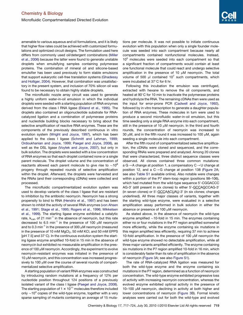
Figure 1. Microfluidic Device for Generating Uniform Water-in-Oil
Emulsions
(A) Schematic of the device, showing central aqueous input reservoir (Raq) with
110 radial nozzles that empty into an annular oil flow channel. An oil-surfactant
mixture is supplied at the oil input reservoir (Rin), and the emulsion is collected
at the output reservoir (Rout). The reservoirs are contacted by microbore
tubing, which delivers material to and from the device.
(B) Photograph of the device during operation, with magnified view on the left.
For visualization, the aqueous phase contained bromophenol blue in a solution
of 15 mM MgCl2, 50 mM KCl, and 50 mM EPPS (pH 7.5).
Chemistry & Biology
Microfluidic Compartmentalized Directed Evolution
compartmentalized replication, functional molecules that
happen to reside in a larger droplet will enjoy a selective advan-
tage because of the greater carrying capacity of their local envi-
ronment.
Efforts have been made to employ microfluidic technology to
produce water-in-oil emulsions that are highly uniform
(Umbanhowar et al., 2000; Thorsen et al., 2001). The general
approach is to cause a narrow aqueous stream (10–100 mm
diameter) to intersect with an oil stream, relying on interfacial
surface tension to break up the laminar flow and form individual
droplets. However, even at flow rates of 0.1 ml min-1 (requiring
10–20 psi), the rate of production of 20 mm droplets is
only �105 droplets per hour (Umbanhowar et al., 2000). This
would not be sufficient to maintain a sizeable population of
evolving molecules that are contained within separate fluidic
compartments. Recently, a variety of more sophisticated droplet
generators have been described (for review, seeChristopher and
Anna, 2007), including devices that can operate with substan-
tially higher throughput (Mazutis et al., 2009; Agresti et al.,
2010; Zeng et al., 2010). However, the complicated procedures
required to fabricate these devices may limit their widespread
use in chemical biology applications. In addition, back pressure
issues and topological constraints on the oil inputs make these
circuits technically challenging to implement in arrayed formats.
An alternative method for fabricating circuits that generate mi-
crofluidic droplets employs soft lithography and does not require
access to a cleanroom facility (Duffy et al., 1998). Devices of this
kind have been used to conduct protein crystallization screens
(Zheng et al., 2003), to synthesize semiconductor nanocrystals
(Chan et al., 2005), and to culture cells and even multicellular
organisms within microfluidic droplets (Clausell-Tormos et al.,
2008). However, low throughput continues to be a challenge in
applying this technology to IVC for the purposes of in vitro
evolution experiments.
Here amicrofluidic device is described that is easily fabricated
using soft lithography and generates uniform water-in-oil drop-
lets with high throughput and scalability. This device was used
to carry out the compartmentalized directed evolution of an
RNA enzyme, with multiple rounds of selective amplification
occurring within individual droplets. As a selective pressure,
neomycin was introduced at a concentration that inhibited the
catalytic activity of the parental RNA enzymes. The evolving pop-
ulation developed resistance to the inhibitor through the
acquisition of phenotypic traits that could be understood in
terms of the structural properties of the RNA.
RESULTS
Amultichannel droplet generator was fabricated in polydimethyl-
siloxane (PDMS), which can be used to produce highly uniform
water-in-oil emulsions for compartmentalized directed evolu-
tion. As shown in the circuit schematic (Figure 1A), the device
consists of a circular nozzle array with 110 fluidic channels
(20 mm wide, 20 mm step height) that fan out from a central
aqueous input reservoir. The tips of the nozzles empty into an
oil flow channel (500 mm wide, 150 mm step height) that is
supplied by an oil-surfactant input reservoir. Droplets form at
the nozzle tips and are swept into the oil phase for collection at
the output reservoir. The oil phase, which is driven by a syringe
718 Chemistry & Biology 17, 717–724, July 30, 2010 ª2010 Elsevier L
pump, bifurcates at the entrance to the oil flow channel. No
nozzles are present at the poles of the channel because there
is lower shear at these positions, which would yield nonuniform
droplets.
Droplet generation in the device was assessed by dispersing
a buffered aqueous solution that contained bromophenol blue
dye to aid in visualizing individual droplets. The oil phase
contained 70% Ar20 silicone oil (polyphenyl-methylsiloxane),
26% mineral oil, and 4% Abil EM90 (a nonionic silicone-based
emulsifier). The flow rates of the aqueous and oil phases were
5 and 70 ml min-1, respectively. Micrographs depicting the forma-
tion of the dye-containing droplets (Figure 1B) illustrate the
uniformity of droplets produced by this method. The images
were processed to assess the droplet size in situ. The droplets
have a radius of 21.9 ± 0.8 mm (4%SD), each containing a volume
of 44 ± 2 pl.
Using the silicone oil/mineral oil/silicone-based emulsifier
combination, the device can generate droplets continuously at
a rate of 1.9 kHz (�107 droplets per hour). It can emulsify aqueous
solutions at even higher flow rates, exceeding 100 ml min-1, which
generates droplets at a frequency of nearly 40 kHz (108 droplets
per hour). At these very high flow rates, however, droplet shearing
in the output channel starts to become a problem. The device is
td All rights reserved
Chemistry & Biology
Microfluidic Compartmentalized Directed Evolution
amenable to various aqueous and oil formulations, and it is likely
that higher flow rates could be achieved with customized formu-
lations and optimized circuit designs. The formulation used here
differs from commonly used oil/detergent combinations (Miller
et al., 2006) because the latter were found to generate unstable
droplets when emulsifying samples containing polymerase
proteins. The combination of mineral oil and silicone-based
emulsifier has been used previously to form stable emulsions
that support eukaryotic cell-free translation systems (Ghadessy
and Holliger, 2004). However, that combination was unsatisfac-
tory in the present system, and inclusion of 70% silicon oil was
found to be necessary to obtain highly stable droplets.
The microfluidic nozzle array circuit was used to generate
a highly uniform water-in-oil emulsion in which the individual
droplets were seededwith a starting population of RNA enzymes
derived from the class I RNA ligase (Ekland et al., 1995). The
droplets also contained an oligonucleotide substrate for RNA-
catalyzed ligation and a combination of polymerase proteins
and nucleotide building blocks necessary to bring about the
selective amplification of reactive RNA enzymes. These are the
components of the previously described continuous in vitro
evolution system (Wright and Joyce, 1997), which has been
applied to the class I ligase (Schmitt and Lehman, 1999;
Ordoukhanian and Joyce, 1999; Paegel and Joyce, 2008), as
well as the DSL ligase (Voytek and Joyce, 2007), but only in
bulk solution. The droplets were seededwith a low concentration
of RNA enzymes so that each droplet contained none or a single
parent molecule. The droplet volume and the concentration of
reactants allowed each parent molecule to give rise to �108
progeny through repeated rounds of selective amplification
within the droplet. Afterward, the droplets were harvested and
the RNAs (and their corresponding cDNAs) were collected and
processed.
The microfluidic compartmentalized evolution system was
used to develop variants of the class I ligase that are resistant
to inhibition by the antibiotic neomycin. Neomycin has a strong
propensity to bind to RNA (Hendrix et al., 1997) and has been
shown to inhibit the activity of several RNA enzymes (von Ahsen
et al., 1991; Stage et al., 1995; Rogers et al., 1996; Mikkelsen
et al., 1999). The starting ligase enzyme exhibited a catalytic
rate, kcat, of 21 min-1 in the absence of neomycin, but this rate
decreased to 6.5 min-1 in the presence of 100 mM neomycin
and to 0.3 min-1 in the presence of 300 mM neomycin (measured
in the presence of 10 mM MgCl2, 50 mM KCl, and 50 mM EPPS
[pH 7.5] and 37�C). In the continuous evolution system the start-
ing ligase enzyme amplified 10-fold in 15 min in the absence of
neomycin but exhibited no measurable amplification in the pres-
ence of 100 mMneomycin. Accordingly, the experiment to evolve
neomycin-resistant enzymes was initiated in the presence of
10 mMneomycin, and this concentration was increased progres-
sively to 100 mM over the course of several rounds of compart-
mentalized selective amplification.
A starting population of variant RNA enzymeswas constructed
by introducing random mutations at a frequency of 12% per
nucleotide position throughout 124 positions of a previously
isolated variant of the class I ligase (Paegel and Joyce, 2008).
The starting population of 13 1011 molecules therefore included
only �104 copies of the wild-type enzyme, together with a very
sparse sampling of mutants containing an average of 15 muta-
Chemistry & Biology 17,
tions per molecule. It was not possible to initiate continuous
evolution with this population when only a single founder mole-
cule was seeded into each compartment because nearly all
compartments contained nonfunctional molecules. Instead,
105 molecules were seeded into each compartment so that
a significant fraction of compartments would contain at least
one functional molecule that could react and undergo selective
amplification in the presence of 10 mM neomycin. The total
volume of 500 ml contained 107 such compartments, which
were incubated at 37�C for 6 hr.
Following this incubation the emulsion was centrifuged,
extracted with hexane to remove the oil components, and
heated at 90�C for 10 min to inactivate the polymerase proteins
and hydrolyze the RNA. The remaining cDNAs then were used as
the input for error-prone PCR (Cadwell and Joyce, 1992),
followed by in vitro transcription to generate a daughter popula-
tion of RNA enzymes. These molecules in turn were used to
produce a second microfluidic water-in-oil emulsion, but this
time seeding only a single RNA enzyme into each compartment,
still in the presence of 10 mM neomycin. In the third and fourth
rounds, the concentration of neomycin was increased to
30 mM, and in the fifth round it was increased to 100 mM, again
seeding a single molecule into each compartment.
After the fifth round of compartmentalized selective amplifica-
tion, the cDNAs were cloned and sequenced, and the corre-
sponding RNAs were prepared and analyzed. Among 20 clones
that were characterized, three distinct sequence classes were
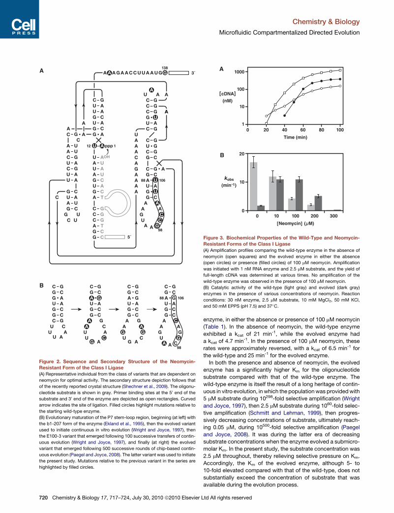
observed. All clones contained three common mutations:
a G/A change at position 1, a compensatory C/U change at
position 12, and a C/G change at position 138 (Figure 2A;
see also Table S1 available online). Also notable were changes
within a portion of the P7 stem-loop region (positions 98–106),
which had mutated from the wild-type sequence 50-CGGACCC
AG-30 (still present in six clones) to either 50-GCCAGCCAG-30
(in seven clones) or 50-GCCAACUAU-30 (in six clones; changes
underlined). All three major classes of variants, together with
the starting wild-type enzyme, were evaluated in a selective
amplification assay performed in bulk solution in either the
absence or presence of 100 mM neomycin.
As stated above, in the absence of neomycin the wild-type
enzyme amplified �10-fold in 15 min. The enzymes containing
either no or four mutations in the P7 region amplified somewhat
more efficiently, while the enzyme containing six mutations in
this region amplified less efficiently, requiring 37 min to achieve
10-fold amplification. In the presence of 100 mM neomycin the
wild-type enzyme showed no detectable amplification, while all
three major variants amplified efficiently. The enzyme containing
six mutations in the P7 region amplified 10-fold in 16 min, which
is considerably faster than its rate of amplification in the absence
of neomycin (Figure 3A; see also Figure S1).
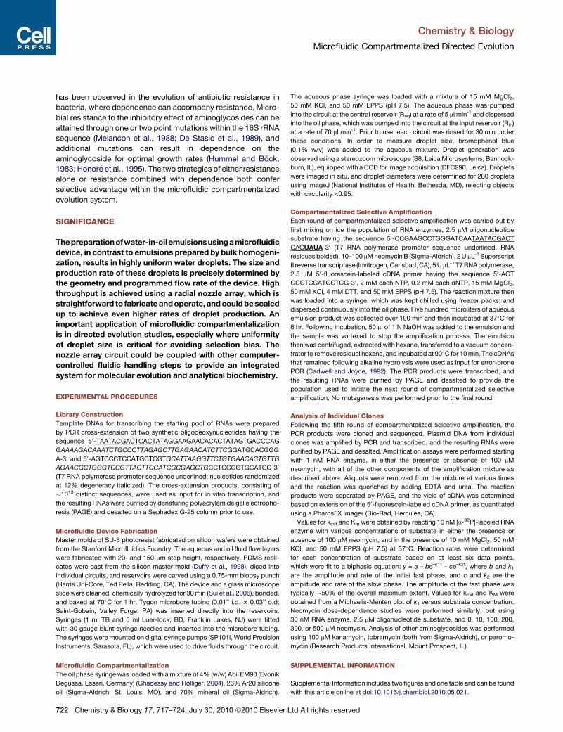
The rate of RNA-catalyzed RNA ligation was measured for
both the wild-type enzyme and the enzyme containing six
mutations in the P7 region, determined as a function of neomycin
concentration. The wild-type enzyme exhibited progressive loss
of activity with increasing neomycin concentration, whereas the
evolved enzyme exhibited optimal activity in the presence of
10–100 mM neomycin, declining in activity at both higher and
lower concentrations of neomycin (Figure 3B). Formal kinetic
analyses were carried out for both the wild-type and evolved
717–724, July 30, 2010 ª2010 Elsevier Ltd All rights reserved 719
A
B
ppp
A
A
A
U
U
U
U
5´
G
U
GG
CC
UAU
AUA
A
OHA
GC
AUCG
CG
CGCG
3´
T
T
A
CGA
GC
GCG
AA
U
AA
CA G
GUC
GC
CG
AGU
GCGCGCAU
GC
U
A
U
AUA
CG
UA
GUC
CG
AUAUG
C
UC
GC
UA
C
A
AG
A
A
U
U
A
CG
CG
AUG
1
98
12
138
C
CG
C
AA A
UA A
G
A
A
A
U
AG
AA
A
A
A
C
AA UA G GA A C C U U A A G
A
AA
A
GC
98
10688
C
G
C
CGA
10688
GCG
AU
AA A
UA
G GG
G
G
G
A G
C
C
GC
G
C
G
C
CGA
GCG
AU
AA
UG A
GC
G
C
CG
GCG
AU
C
UA
U A
GC
G
C
ACG
G
GCG
AU
CU C
U AU A
Figure 2. Sequence and Secondary Structure of the Neomycin-Resistant Form of the Class I Ligase
(A) Representative individual from the class of variants that are dependent on
neomycin for optimal activity. The secondary structure depiction follows that
of the recently reported crystal structure (Shechner et al., 2009). The oligonu-
cleotide substrate is shown in gray. Primer binding sites at the 50 end of the
substrate and 30 end of the enzyme are depicted as open rectangles. Curved
arrow indicates the site of ligation. Filled circles highlight mutations relative to
the starting wild-type enzyme.
(B) Evolutionary maturation of the P7 stem-loop region, beginning (at left) with
the b1-207 form of the enzyme (Ekland et al., 1995), then the evolved variant
used to initiate continuous in vitro evolution (Wright and Joyce, 1997), then
the E100-3 variant that emerged following 100 successive transfers of contin-
uous evolution (Wright and Joyce, 1997), and finally (at right) the evolved
variant that emerged following 500 successive rounds of chip-based contin-
uous evolution (Paegel and Joyce, 2008). The latter variant was used to initiate
the present study. Mutations relative to the previous variant in the series are
highlighted by filled circles.
1
10
1000
100
200 40
Time (min)
60 80 100
0
10
20
100
[Neomycin] (µ M)
[cDNA](nM)
kobs
(min-1)
200 300100
A
B
Figure 3. Biochemical Properties of the Wild-Type and Neomycin-
Resistant Forms of the Class I Ligase
(A) Amplification profiles comparing the wild-type enzyme in the absence of
neomycin (open squares) and the evolved enzyme in either the absence
(open circles) or presence (filled circles) of 100 mM neomycin. Amplification
was initiated with 1 nM RNA enzyme and 2.5 mM substrate, and the yield of
full-length cDNA was determined at various times. No amplification of the
wild-type enzyme was observed in the presence of 100 mM neomycin.
(B) Catalytic activity of the wild-type (light gray) and evolved (dark gray)
enzymes in the presence of various concentrations of neomycin. Reaction
conditions: 30 nM enzyme, 2.5 mM substrate, 10 mM MgCl2, 50 mM KCl,
and 50 mM EPPS (pH 7.5) and 37�C.
Chemistry & Biology
Microfluidic Compartmentalized Directed Evolution
720 Chemistry & Biology 17, 717–724, July 30, 2010 ª2010 Elsevier L
enzyme, in either the absence or presence of 100 mM neomycin
(Table 1). In the absence of neomycin, the wild-type enzyme
exhibited a kcat of 21 min-1, while the evolved enzyme had
a kcat of 4.7 min-1. In the presence of 100 mM neomycin, these
rates were approximately reversed, with a kcat of 6.5 min-1 for
the wild-type and 25 min-1 for the evolved enzyme.
In both the presence and absence of neomycin, the evolved
enzyme has a significantly higher Km for the oligonucleotide
substrate compared with that of the wild-type enzyme. The
wild-type enzyme is itself the result of a long heritage of contin-
uous in vitro evolution, in which the population was providedwith
5 mM substrate during 10298-fold selective amplification (Wright
and Joyce, 1997), then 2.5 mM substrate during 1060-fold selec-
tive amplification (Schmitt and Lehman, 1999), then progres-
sively decreasing concentrations of substrate, ultimately reach-
ing 0.05 mM, during 10500-fold selective amplification (Paegel
and Joyce, 2008). It was during the latter era of decreasing
substrate concentrations when the enzyme evolved a submicro-
molar Km. In the present study, the substrate concentration was
2.5 mM throughout, thereby relieving selective pressure on Km.
Accordingly, the Km of the evolved enzyme, although 5- to
10-fold elevated compared with that of the wild-type, does not
substantially exceed the concentration of substrate that was
available during the evolution process.
td All rights reserved

Table 1. Kinetic Properties of the Wild-Type and Evolved
Enzymes
Wild-Type Evolved
Neomycin 0 100 mM 0 100 mM
kcat (min-1) 21 ± 0.8 6.5 ± 0.3 4.7 ± 0.1 25 ± 1
KM (mM) 0.4 ± 0.05 0.7 ± 0.1 4.2 ± 0.4 3.1 ± 0.7
Reaction conditions: 10 mM MgCl2, 50 mM KCl, 50 mM EPPS
(pH 7.5), 37�C.
Chemistry & Biology
Microfluidic Compartmentalized Directed Evolution
The wild-type and evolved enzymes were tested for catalytic
activity in the presence of 100 mM concentration of three other
aminoglycosides: kanamycin, paromomycin, and tobramycin.
The wild-type is inhibited by all three of these compounds, but
to a lesser extent than it is inhibited by neomycin (Figure 3B;
see also Figure S2). The evolved enzyme, which is stimulated
by neomycin, is also stimulated by the other three aminoglyco-
sides, but to a lesser extent. Thus the neomycin dependence
of the evolved enzyme is somewhat specific for that ligand.
The wild-type and evolved enzymes also were tested in the pres-
ence of various concentrations of Mg2+, measured in either the
absence or presence of 100 mM neomycin (Figure 3B; see also
Figure S2). In the absence of neomycin, the wild-type enzyme
showed no dependence on Mg2+ concentration, whereas the
neomycin-activated enzyme showed a slight increase in activity
with increasing Mg2+ over the range of 10–50 mM. In the pres-
ence of neomycin, both enzymes showed no significant depen-
dence on Mg2+ concentration. Thus it is unlikely that neomycin
and Mg2+ occupy the same binding site in the evolved enzyme.
Rather, the enzyme appears to have evolved a specific binding
site for neomycin that enhances catalytic activity.
DISCUSSION
In vitro compartmentalization provides a way to link genotype
and phenotype during the course of a directed evolution exper-
iment (Tawfik and Griffiths, 1998). Compartmentalization is
achieved by emulsifying the aqueous phase, which contains
the biochemical components, in an oil phase that includes emul-
sion stabilizers. This procedure results in large numbers of
water-in-oil droplets, each harboring one or a few individuals
from the population. In past practice, however, the droplets
were highly variable in size, which can bias the effect of selection
because individuals that happen to occupy a larger droplet will
benefit from the greater carrying capacity of their local environ-
ment. This bias is especially a concern when carrying out
multiple-turnover reactions or iterative selective amplification
within the compartments. In some screening applications it is
possible to apply a correction factor to the observed phenotype
to compensate for differences in compartment volume, or to
consider only those droplets that fall within a particular size
range (Aharoni et al., 2005). It would be far preferable, however,
to generate droplets that are highly uniform in size, provided that
large numbers of such droplets can be obtained.
Microfluidic emulsification has proved to be a viable approach
for preparing highly uniform droplet trains, but the yield has been
much lower than what can be obtained by bulk emulsification
(Thorsen et al., 2001). The nozzle array circuit described here
achieves substantial throughput, generating 107–108 droplets
Chemistry & Biology 17,
per hour with the initial design. It does so by significantly
reducing backpressure, which is the primary cause of device
failure and would otherwise limit the flow rate and therefore the
population size. Vigorous mixing during homogenization is elim-
inated, preventing damage to the biochemical reagents.
The nozzle array is topologically equivalent to single-channel
circuits for generating slugs of fluid (Song et al., 2003), but the
depth and width of the oil flow channel are greater, which mini-
mizes fluidic resistance at the sites of droplet formation, thereby
preventing oil from intruding into the nozzles. The circuit is easily
fabricated in PDMS using a simple dual-height mold. Multiple
such circuits could be made to operate in parallel to provide
even greater throughput.
The directed evolution of RNA enzymes was carried out within
microfluidic water-in-oil droplets to demonstrate the capabilities
of the new system. The system allows large numbers of
functional molecules to operate within separate compartments,
although the demonstration case reported here did not critically
depend on compartmentalization and could have been carried
out in bulk solution. As a selection pressure, the evolving RNA
enzymes were challenged to escape inhibition by neomycin.
This compound is a member of the family of aminoglycoside
antibiotics, many of which exhibit potent antimicrobial activity
through their ability to bind 16S ribosomal RNA and thereby
inhibit protein synthesis (Moazed and Noller, 1987). Aminoglyco-
sides have a strong propensity to bind to structured RNAs and
have been shown to inhibit the function of various biological
RNAs and ribonucleoproteins (Walter et al., 1999; Schroeder
et al., 2000). The catalytic activity of the starting class I RNA
ligase is substantially inhibited in the presence of 10–100 mM
neomycin, which prevents it from undergoing selective amplifi-
cation in the continuous in vitro evolution system. Following
five rounds of compartmentalized evolution, however, novel vari-
ants arose that are resistant to neomycin. Compared with the
wild-type, the evolved enzymes have a 4-fold faster catalytic
rate in the presence of 100 mM neomycin, which enables them
to undergo efficient selective amplification.
Genetic changes within the evolved molecules were clustered
mainly in the P7 stem-loop region of the enzyme (Figure 2A).
Over the course of several previous in vitro evolution experi-
ments involving the class I ligase (Wright and Joyce, 1997;
Schmitt and Lehman, 1999; Paegel and Joyce, 2008), this region
underwent significant changes (Figure 2B). The changes were
confined largely to the loop portion of the region, the only excep-
tion being inversion of a noncanonical G88,A106 pair in the stem
(to an A,G pair), which occurred several hundred rounds of
evolution ago. In the present study, this same base pair became
mutated to a Watson-Crick pair for the first time, accompanied
bymutation of the G90,C104 pair to a G,Uwobble pair. Elimina-
tion of the A,Gpair within the stem is especially notable in light of
a recent study demonstrating that such motifs have a special
propensity to bind neomycin (Disney et al., 2008).
It is intriguing that the evolved RNA enzyme depicted in
Figure 2A has a 5-fold faster catalytic rate (kcat) and 8-fold
greater catalytic efficiency (kcat Km-1) in the presence compared
with the absence of 100 mM neomycin (Table 1). Other evolved
individuals that contain fewer mutations within the P7 stem-
loop also are resistant to neomycin, but are not dependent on
neomycin for optimal activity. This result echoes a theme that
717–724, July 30, 2010 ª2010 Elsevier Ltd All rights reserved 721
Chemistry & Biology
Microfluidic Compartmentalized Directed Evolution
has been observed in the evolution of antibiotic resistance in
bacteria, where dependence can accompany resistance. Micro-
bial resistance to the inhibitory effect of aminoglycosides can be
attained through one or two point mutations within the 16S rRNA
sequence (Melancon et al., 1988; De Stasio et al., 1989), and
additional mutations can result in dependence on the
aminoglycoside for optimal growth rates (Hummel and Bock,
1983; Honore et al., 1995). The two strategies of either resistance
alone or resistance combined with dependence both confer
selective advantage within the microfluidic compartmentalized
evolution system.
SIGNIFICANCE
Thepreparationofwater-in-oilemulsionsusingamicrofluidic
device, in contrast to emulsions prepared by bulk homogeni-
zation, results in highly uniform water droplets. The size and
production rate of these droplets is precisely determined by
the geometry and programmed flow rate of the device. High
throughput is achieved using a radial nozzle array, which is
straightforward to fabricate andoperate, andcouldbescaled
up to achieve even higher rates of droplet production. An
important application of microfluidic compartmentalization
is in directed evolution studies, especially where uniformity
of droplet size is critical for avoiding selection bias. The
nozzle array circuit could be coupled with other computer-
controlled fluidic handling steps to provide an integrated
system for molecular evolution and analytical biochemistry.
EXPERIMENTAL PROCEDURES
Library Construction
Template DNAs for transcribing the starting pool of RNAs were prepared
by PCR cross-extension of two synthetic oligodeoxynucleotides having the
sequence 50-TAATACGACTCACTATAGGAAGAACACACTATAGTGACCCAG
GAAAAGACAAATCTGCCCTTAGAGCTTGAGAACATCTTCGGATGCACGGG
A-30 and 50-AGTCCCTCCATGCTCGTGCATTAAGGTTCTGTGAACACTGTTG
AGAACGCTGGGTCCGTTACTTCCATCGCGAGCTGCCTCCCGTGCATCC-30
(T7 RNA polymerase promoter sequence underlined; nucleotides randomized
at 12% degeneracy italicized). The cross-extension products, consisting of
�1013 distinct sequences, were used as input for in vitro transcription, and
the resulting RNAs were purified by denaturing polyacrylamide gel electropho-
resis (PAGE) and desalted on a Sephadex G-25 column prior to use.
Microfluidic Device Fabrication
Master molds of SU-8 photoresist fabricated on silicon wafers were obtained
from the Stanford Microfluidics Foundry. The aqueous and oil fluid flow layers
were fabricated with 20- and 150-mm step height, respectively. PDMS repli-
cates were cast from the silicon master mold (Duffy et al., 1998), diced into
individual circuits, and reservoirs were carved using a 0.75-mm biopsy punch
(Harris Uni-Core, Ted Pella, Redding, CA). The device and a glass microscope
slide were cleaned, chemically hydrolyzed for 30min (Sui et al., 2006), bonded,
and baked at 70�C for 1 hr. Tygon microbore tubing (0.01’’ i.d. 3 0.03’’ o.d;
Saint-Gobain, Valley Forge, PA) was inserted directly into the reservoirs.
Syringes (1 ml TB and 5 ml Luer-lock; BD, Franklin Lakes, NJ) were fitted
with 30 gauge blunt syringe needles and inserted into the microbore tubing.
The syringes were mounted on digital syringe pumps (SP101i, World Precision
Instruments, Sarasota, FL), which were used to drive fluids through the circuit.
Microfluidic Compartmentalization
The oil phase syringe was loaded with amixture of 4% (w/w) Abil EM90 (Evonik
Degussa, Essen, Germany) (Ghadessy and Holliger, 2004), 26% Ar20 silicone
oil (Sigma-Aldrich, St. Louis, MO), and 70% mineral oil (Sigma-Aldrich).
722 Chemistry & Biology 17, 717–724, July 30, 2010 ª2010 Elsevier L
The aqueous phase syringe was loaded with a mixture of 15 mM MgCl2,
50 mM KCl, and 50 mM EPPS (pH 7.5). The aqueous phase was pumped
into the circuit at the central reservoir (Raq) at a rate of 5 ml min-1 and dispersed
into the oil phase, which was pumped into the circuit at the input reservoir (Rin)
at a rate of 70 ml min-1. Prior to use, each circuit was rinsed for 30 min under
these conditions. In order to measure droplet size, bromophenol blue
(0.1% w/v) was added to the aqueous mixture. Droplet generation was
observed using a stereozoommicroscope (S8, LeicaMicrosystems, Bannock-
burn, IL), equippedwith a CCD for image acquisition (DFC290, Leica). Droplets
were imaged in situ, and droplet diameters were determined for 200 droplets
using ImageJ (National Institutes of Health, Bethesda, MD), rejecting objects
with circularity <0.95.
Compartmentalized Selective Amplification
Each round of compartmentalized selective amplification was carried out by
first mixing on ice the population of RNA enzymes, 2.5 mM oligonucleotide
substrate having the sequence 50-CCGAAGCCTGGGATCAATAATACGACT
CACUAUA-30 (T7 RNA polymerase promoter sequence underlined, RNA
residues bolded), 10–100 mMneomycinB (Sigma-Aldrich), 2UmL-1 Superscript
II reverse transcriptase (Invitrogen,Carlsbad,CA), 5UmL-1 T7RNApolymerase,
2.5 mM 50-fluorescein-labeled cDNA primer having the sequence 50-AGT
CCCTCCATGCTCG-30, 2 mM each NTP, 0.2 mM each dNTP, 15 mM MgCl2,
50 mM KCl, 4 mM DTT, and 50 mM EPPS (pH 7.5). The reaction mixture then
was loaded into a syringe, which was kept chilled using freezer packs, and
dispersed continuously into the oil phase. Five hundred microliters of aqueous
emulsion product was collected over 100 min and then incubated at 37�C for
6 hr. Following incubation, 50 ml of 1 N NaOH was added to the emulsion and
the sample was vortexed to stop the amplification process. The emulsion
then was centrifuged, extracted with hexane, transferred to a vacuum concen-
trator to remove residual hexane, and incubated at 90�C for 10min. The cDNAs
that remained following alkaline hydrolysis were used as input for error-prone
PCR (Cadwell and Joyce, 1992). The PCR products were transcribed, and
the resulting RNAs were purified by PAGE and desalted to provide the
population used to initiate the next round of compartmentalized selective
amplification. No mutagenesis was performed prior to the final round.
Analysis of Individual Clones
Following the fifth round of compartmentalized selective amplification, the
PCR products were cloned and sequenced. Plasmid DNA from individual
clones was amplified by PCR and transcribed, and the resulting RNAs were
purified by PAGE and desalted. Amplification assays were performed starting
with 1 nM RNA enzyme, in either the presence or absence of 100 mM
neomycin, with all of the other components of the amplification mixture as
described above. Aliquots were removed from the mixture at various times
and the reaction was quenched by adding EDTA and urea. The reaction
products were separated by PAGE, and the yield of cDNA was determined
based on extension of the 50-fluorescein-labeled cDNA primer, as quantitated
using a PharosFX imager (Bio-Rad, Hercules, CA).
Values for kcat and Kmwere obtained by reacting 10 nM [a-32P]-labeled RNA
enzyme with various concentrations of substrate in either the presence or
absence of 100 mM neomycin, and in the presence of 10 mM MgCl2, 50 mM
KCl, and 50 mM EPPS (pH 7.5) at 37�C. Reaction rates were determined
for each concentration of substrate based on at least six data points,
which were fit to a biphasic equation: y = a – be–k1t – ce–k2t, where b and k1are the amplitude and rate of the initial fast phase, and c and k2 are the
amplitude and rate of the slow phase. The amplitude of the fast phase was
typically �50% of the overall maximum extent. Values for kcat and KM were
obtained from a Michaelis-Menten plot of k1 versus substrate concentration.
Neomycin dose-dependence studies were performed similarly, but using
30 nM RNA enzyme, 2.5 mM oligonucleotide substrate, and 0, 10, 100, 200,
300, or 500 mM neomycin. Analysis of other aminoglycosides was performed
using 100 mM kanamycin, tobramycin (both from Sigma-Aldrich), or paromo-
mycin (Research Products International, Mount Prospect, IL).
SUPPLEMENTAL INFORMATION
Supplemental Information includes two figures and one table and can be found
with this article online at doi:10.1016/j.chembiol.2010.05.021.
td All rights reserved

Chemistry & Biology
Microfluidic Compartmentalized Directed Evolution
ACKNOWLEDGMENTS
This work was supported by National Science Foundation grant no. MCB-
0614614 (G.F.J.), and NIH Pathway to Independence Career Development
Award no. GM083155 (B.M.P.). The authors declare that there is no conflict
of interest.
Received: January 6, 2010
Revised: March 29, 2010
Accepted: May 19, 2010
Published: July 29, 2010
REFERENCES
Agresti, J.J., Kelly, B.T., JIschke, A., and Griffiths, A.D. (2005). Selection of
ribozymes that catalyse multiple-turnover Diels-Alder cycloadditions by using
in vitro compartmentalization. Proc. Natl. Acad. Sci. USA 102, 16170–16175.
Agresti, J.J., Antipov, E., Abate, A.R., Ahn, K., Rowat, A.C., Baret, J.C.,
Marquez, M., Klibanov, A.M., Griffiths, A.D., and Weitz, D.A. (2010). Ultra-
high-throughput screening in drop-based microfluidics for directed evolution.
Proc. Natl. Acad. Sci. USA 107, 4004–4009.
Aharoni, A., Amitai, G., Bernath, K., Magdassi, S., and Tawfik, D.S. (2005).
High-throughput screening of enzyme libraries: thiolactonases evolved by
fluorescence-activated sorting of single cells in emulsion compartments.
Chem. Biol. 12, 1281–1289.
Barbas, C.F., Kang, A.S., Lerner, R.A., and Benkovic, S.J. (1991). Assembly of
combinatorial antibody libraries on phage surfaces: the gene III site. Proc. Natl.
Acad. Sci. USA 88, 7978–7982.
Bernath, K., Hai, M.T., Mastrobattista, E., Griffiths, A.D., Magdassi, S., and
Tawfik, D.S. (2004). In vitro compartmentalization by double emulsions: sorting
and gene enrichment by fluorescence activated cell sorting. Anal. Biochem.
325, 151–157.
Boder, E.T., and Wittrup, K.D. (1997). Yeast surface display for screening
combinatorial polypeptide libraries. Nat. Biotechnol. 15, 553–557.
Cadwell, R.C., and Joyce, G.F. (1992). Randomization of genes by PCRmuta-
genesis. PCR Methods Appl. 2, 28–33.
Chan, E.M., Alivisatos, A.P., and Mathies, R.A. (2005). High-temperature mi-
crofluidic synthesis of CdSe nanocrystals in nanoliter droplets. J. Am. Chem.
Soc. 127, 13854–13861.
Christopher, G.F., and Anna, S.L. (2007). Microfluidic methods for generating
continuous droplet streams. J. Phys. D Appl. Phys. 40, R319–R336.
Clausell-Tormos, J., Lieber, D., Baret, J.C., El-Harrak, A., Miller, O.J., Frenz, L.,
Blouwolff, J., Humphry, K.J., Koster, S., Duan, H., et al. (2008). Droplet-based
microfluidic platforms for the encapsulation and screening of mammalian cells
and multicellular organisms. Chem. Biol. 15, 427–437.
Cwirla, S.E., Peters, E.A., Barrett, R.W., and Dower, W.J. (1990). Peptides on
phage: a vast library of peptides for identifying ligands. Proc. Natl. Acad. Sci.
USA 87, 6378–6382.
De Stasio, E.A., Moazed, D., Noller, H.F., and Dahlberg, A.E. (1989). Mutations
in 16S ribosomal RNA disrupt antibiotic-RNA interactions. EMBO J. 8,
1213–1216.
Devlin, J.J., Panganiban, L.C., and Devlin, P.E. (1990). Random peptide
libraries: a source of specific protein binding molecules. Science 249,
404–406.
Disney, M.D., Labuda, L.P., Paul, D.J., Poplawski, S.G., Pushechnikov, A.,
Tran, T., Velagapudi, S.P., Wu, M., and Childs-Disney, J.L. (2008). Two-dimen-
sional combinatorial screening identifies specific aminoglycoside-RNA
internal loop partners. J. Am. Chem. Soc. 130, 11185–11194.
Duffy, D.C., McDonald, J.C., Schueller, O.J.A., and Whitesides, G.M. (1998).
Rapid prototyping of microfluidic systems in poly(dimethylsiloxane). Anal.
Chem. 70, 4974–4984.
Ekland, E.H., Szostak, J.W., and Bartel, D.P. (1995). Structurally complex and
highly active RNA ligases derived from random RNA sequences. Science 269,
364–370.
Chemistry & Biology 17,
Francisco, J.A., Campbell, R., Iverson, B.L., and Georgiou, G. (1993). Produc-
tion and fluorescence-activated cell sorting of Escherichia coli expressing
a functional antibody fragment on the external surface. Proc. Natl. Acad.
Sci. USA 90, 10444–10448.
Ghadessy, F.J., and Holliger, P. (2004). A novel emulsion mixture for in vitro
compartmentalization of transcription and translation in the rabbit reticulocyte
system. Protein Eng. Des. Sel. 17, 201–204.
Ghadessy, F.J., Ong, J.L., and Holliger, P. (2001). Directed evolution of poly-
merase function by compartmentalized self-replication. Proc. Natl. Acad.
Sci. USA 98, 4552–4557.
Hanes, J., and Pluckthun, A. (1997). In vitro selection and evolution of func-
tional proteins by using ribosome display. Proc. Natl. Acad. Sci. USA 94,
4937–4942.
Hendrix, M., Alper, P.B., Priestley, E.S., and Wong, C.-H. (1997). Hydroxy-
amines as a newmotif for the molecular recognition of phosphodiesters: impli-
cations for aminoglycoside-RNA interactions. Angew. Chem. Int. Ed. Engl. 36,
95–98.
Honore, N., Marchal, G., and Cole, S.T. (1995). Novel mutation in 16S rRNA
associated with streptomycin dependence in Mycobacterium tuberculosis.
Antimicrob. Agents Chemother. 39, 769–770.
Hummel, H., and Bock, A. (1983). On the basis of aminoglycoside-dependent
growth of mutants from E. coli: physiological studies. Mol. Gen. Genet. 191,
167–175.
Lee, Y.-F., Tawfik, D.S., and Griffiths, A.D. (2002). Investigating the target
recognition of DNA cytosine-5 methyltransferase HhaI by library selection
using in vitro compartmentalisation. Nucleic Acids Res. 30, 4937–4944.
Levy, M., Griswold, K.E., and Ellington, A.D. (2005). Direct selection of trans-
acting ligase ribozymes by in vitro compartmentalization. RNA 11, 1555–1562.
Mastrobattista, E., Taly, V., Chanudet, E., Treacy, P., Kelly, B.T., and Griffiths,
A.D. (2005). High-throughput screening of enzyme libraries: in vitro evolution of
a b-galactosidase by fluorescence-activated sorting of double emulsions.
Chem. Biol. 12, 1291–1300.
Mazutis, L., Araghi, A.F., Miller, O.J., Baret, J.C., Frenz, L., Janoshazi, A., Taly,
V., Miller, B.J., Hutchison, J.B., Link, D., et al. (2009). Droplet-based microflui-
dic systems for high-throughput single DNAmolecule isothermal amplification
and analysis. Anal. Chem. 81, 4813–4821.
McCafferty, J., Griffiths, A.D., Winter, G., and Chiswell, D.J. (1990). Phage anti-
bodies: filamentous phage displaying antibody variable domains. Nature 348,
552–554.
McCafferty, J., Jackson, R.H., and Chiswell, D.J. (1991). Phage-enzymes:
expression and affinity chromatography of functional alkaline phosphatase
on the surface of bacteriophage. Protein Eng. 4, 955–961.
Melancon, P., Lemieux, C., and Brakier-Gingras, L. (1988). A mutation in the
530 loop of Escherichia coli 16S ribosomal RNA causes resistance to strepto-
mycin. Nucleic Acids Res. 16, 9631–9639.
Mikkelsen, N.E., BrInnvall, M., Virtanen, A., and Kirsebom, L.A. (1999). Inhibi-
tion of RNase P RNA cleavage by aminoglycosides. Proc. Natl. Acad. Sci. USA
96, 6155–6160.
Miller, O.J., Bernath, K., Agresti, J.J., Amitai, G., Kelly, B.T., Mastrobattista, E.,
Taly, V., Magdassi, S., Tawfik, D.S., and Griffiths, A.D. (2006). Directed evolu-
tion by in vitro compartmentalization. Nat. Methods 3, 561–570.
Moazed, D., and Noller, H.F. (1987). Interaction of antibiotics with functional
sites in 16S ribosomal RNA. Nature 327, 389–394.
Ordoukhanian, P., and Joyce, G.F. (1999). A molecular description of the
evolution of resistance. Chem. Biol. 6, 881–889.
Paegel, B.M., and Joyce, G.F. (2008). Darwinian evolution on a chip. PLoS Biol.
6, e85.
Roberts, R.W., and Szostak, J.W. (1997). RNA-peptide fusions for the in vitro
selection of peptides and proteins. Proc. Natl. Acad. Sci. USA 94,
12297–12302.
Rogers, J., Chang, A.H., von Ahsen, U., Schroeder, R., and Davies, J. (1996).
Inhibition of the self-cleavage reaction of the human hepatitis delta virus ribo-
zyme by antibiotics. J. Mol. Biol. 259, 916–925.
717–724, July 30, 2010 ª2010 Elsevier Ltd All rights reserved 723

Chemistry & Biology
Microfluidic Compartmentalized Directed Evolution
Schmitt, T., and Lehman, N. (1999). Non-unity molecular heritability demon-
strated by continuous evolution in vitro. Chem. Biol. 6, 857–869.
Schroeder, R., Waldisch, C., andWank, H. (2000). Modulation of RNA function
by aminoglycoside antibiotics. EMBO J. 19, 1–9.
Scott, J.K., and Smith, G.P. (1990). Searching for peptide ligands with an
epitope library. Science 249, 386–390.
Shechner, D.M., Grant, R.A., Bagby, S.C., Koldobskaya, Y., Piccirilli, J.A., and
Bartel, D.P. (2009). Crystal structure of the catalytic core of an RNA-poly-
merase ribozyme. Science 326, 1271–1275.
Smith, G.P., and Petrenko, V.A. (1997). Phage display. Chem. Rev. 97,
391–410.
Song, H., Tice, J.D., and Ismagilov, R.F. (2003). A microfluidic system for
controlling reaction networks in time. Angew. Chem. Int. Ed. Engl. 42,
768–772.
Soumillion, P., Jespers, L., Bouchet, M., Marchand-Brynaert, J., Winter, G.,
and Fastrez, J. (1994). Selection of b-lactamase on filamentous bacteriophage
by catalytic activity. J. Mol. Biol. 237, 415–422.
Stage, T.K., Hertel, K.J., and Uhlenbeck, O.C. (1995). Inhibition of the hammer-
head ribozyme by neomycin. RNA 1, 95–101.
Sui, G.D., Wang, J., Lee, C.C., Lu, W., Lee, S.P., Leyton, J.V., Wu, A.M., and
Tseng, H.R. (2006). Solution-phase surface modification in intact poly(dime-
thylsiloxane) microfluidic channels. Anal. Chem. 78, 5543–5551.
Tawfik, D.S., and Griffiths, A.D. (1998). Man-made cell-like compartments for
molecular evolution. Nat. Biotechnol. 16, 652–656.
724 Chemistry & Biology 17, 717–724, July 30, 2010 ª2010 Elsevier L
Thorsen, T., Roberts, R.W., Arnold, F.H., and Quake, S.R. (2001). Dynamic
pattern formation in a vesicle-generating microfluidic device. Phys. Rev.
Lett. 86, 4163–4166.
Umbanhowar, P.B., Prasad, V., and Weitz, D.A. (2000). Monodisperse emul-
sion generation via drop break off in a coflowing stream. Langmuir 16,
347–351.
von Ahsen, U., Davies, J., and Schroeder, R. (1991). Antibiotic inhibition of
group I ribozyme function. Nature 353, 368–370.
Voytek, S.B., and Joyce, G.F. (2007). Emergence of a fast-reacting ribozyme
that is capable of undergoing continuous evolution. Proc. Natl. Acad. Sci.
USA 104, 15288–15293.
Walter, F., Vicens, Q., and Westhof, E. (1999). Aminoglycoside-RNA interac-
tions. Curr. Opin. Chem. Biol. 3, 694–704.
Wright, M.C., and Joyce, G.F. (1997). Continuous in vitro evolution of catalytic
function. Science 276, 614–617.
Zaher, H.S., and Unrau, P.J. (2007). Selection of an improved RNA polymerase
ribozyme with superior extension and fidelity. RNA 13, 1017–1026.
Zeng, Y., Novak, R., Shuga, J., Smith, M.T., and Mathies, R.A. (2010). High-
performance single cell genetic analysis using microfluidic emulsion generator
arrays. Anal. Chem. 82, 3183–3190.
Zheng, B., Roach, L.S., and Ismagilov, R.F. (2003). Screening of protein crys-
tallization conditions on a microfluidic chip using nanoliter-size droplets.
J. Am. Chem. Soc. 125, 11170–11171.
td All rights reserved