microscopic agglutination and polyacrylamide gel electrophoresis
TRANSCRIPT

JOURNAL OF CLINICAL MICROBIOLOGY, Aug. 1986, p. 282-2870095-1137/86/080282-06$02.00/0Copyright C 1986, American Society for Microbiology
Microscopic Agglutination and Polyacrylamide Gel ElectrophoresisAnalyses of Oral Anaerobic Spirochetes
BEN D. TALL AND ROBERT K. NAUMAN*
Department of Microbiology, Dental School, University of Maryland, Baltimore, Maryland 21201
Received 13 January 1986/Accepted 30 April 1986
Microscopic agglutination (MA) analysis and sodium dodecyl sulfate-polyacrylamide gel electrophoresis(SDS-PAGE) were used to determine strain and species similarities and dissimilarities among three species oforal anaerobic spirochetes, Treponema denticola, Treponema pectinovorum, and Treponema vincentii. The MAanalysis revealed a diversity of serologic reactivity or sharing of common antigens within each species.However, there was no cross-reactivity or sharing of common antigens among the three species. DistinctSDS-PAGE whole-cell electrophoretograms for each species were obtained. The banding patterns for 16 T.denticola strains revealed 30 distinct proteins, while the banding patterns for 5 strains of T. pectinovorum and2 strains of T. vincentii revealed 26 and 35 distinct proteins, respectively. Analysis of the electrophoretogramsshowed that their respective banding patterns could be used to distinguish the three species from one another.In addition, strain differences within each species could be detected. There was a correlation between MAanalysis and SDS-PAGE analysis. It is thus suggested that both MA and SDS-PAGE analyses be included inclassification schemes for the identification of oral spirochetes.
The oral anaerobic spirochetes represent a diversifiedgroup of nutritionally fastidious microorganisms which are
present as indigenous components of the oral flora of hu-mans and other dentulous animals. There is strong evidencethat an association exists between the presence, progres-sion, and severity of periodontal diseases and the presenceof oral anaerobic spirochetes (1, 7, 11, 14, 15, 18-20, 22, 23,32, 34). Although these studies do not provide direct evi-dence implicating oral spirochetes as etiologic agents inperiodontal diseases, they do, based on microscopic as wellas immunological and cultural observations, provide evi-dence that spirochetes contribute to the periodontal diseasestate and that the periodontal pocket is a suitable ecologicalniche for their proliferation (14, 42).At present, the oral anaerobic spirochetes are classified
under the genus Treponema (35). This classification scheme,based on the ultrastructural studies of Listgarten andSocransky (21), divides the oral spirochetes into three maingroups based on the diameter of the protoplasmic cylinder,the morphology of the outer membrane, and the number ofperiplasmic flagella originating from each cell pole. Aspointed out by Breznak (3), the taxonomic status of the oralanaerobic spirochetes is confusing. Presently five species oforal anaerobic spirochetes are recognized: T. denticola, T.scoliodontum, T. vincentii, T. pectinovorum, and newlydescribed T. socranskii (23, 36, 37, 42).By using distinctive phenotype evidence of the similarity
and dissimilarity of strains within a species and betweenspecies, a number of investigators have shown that poly-acrylamide gel electrophoresis (PAGE) (4, 6, 9, 12, 16, 26,33, 39), sodium dodecyl sulfate (SDS)-PAGE (5, 8, 25, 38),and urea-PAGE (13, 27, 28) of soluble proteins can be usedto differentiate species of bacteria. Identifications of isolatesbased on these kinds of data closely agree with separationsbased on DNA-DNA homology studies (2, 26). Therefore,
* Corresponding author.
SDS-PAGE appears to be a useful and highly sensitive toolfor screening multiple isolates for comparative identification.
In the present study electrophoretograms of 16 strains ofT. denticola, 5 strains of T. pectinovorum, and 2 strains of T.vincentii were examined. In addition, the results of SDS-PAGE analysis were compared with those of microscopicagglutination (MA) testing. The protein patterns and agglu-tination results obtained were of value in determining thesimilarity among well-documented members of each speciesand in demonstrating differences among these three speciesof oral anaerobic spirochetes.
MATERIALS AND METHODSOrganisms and growth conditions. The Treponema strains
used in this study and their origins are listed in Table 1.Stock cultures of the T. denticola strains were maintained on
a semisolid medium consisting of PPLO broth, withoutcrystal violet (BBL Microbiology Systems, Cockeysville,Md.) plus 15 Fig of cocarboxylase (Sigma Chemical Co., St.Louis, Mo.) per ml, 10% sterile rabbit serum (GIBCOLaboratories, Grand Island, N.Y.), 0.05% sodium thiogly-colate (Sigma Chemical Co.), and 0.1% agar. Stock culturesof the T. pectinovorum strains were maintained on the NOSmedium described by Leschine and Canale-Parola (17),supplemented with 0.3% sodium glucuronate (Sigma Chem-ical Co.) and 0.1% agar (43). Stock cultures of T. vincentiiN-9 and T. vincentii LA1 were both maintained on themedium described by Mangan et al. (24) supplemented with15 ptg of cocarboxylase per ml and 10% sterile rabbit serum.
The same media lacking agar were used for liquid cultiva-tion. Electron microscopic analysis of negatively stainedspecimens of T. denticola, T. pectinovorum, and T. vincentiiindicated that they possessed the 2-4-2, 1-2-1, and 5-10-5periplasmic flagellum arrangements, respectively. Stock cul-tures, as well as cultures used in the experiments, wereincubated at 37°C in a Coy anaerobic chamber (Coy Labo-ratory Products, Ann Arbor, Mich.), as previously described(14).
282
Vol. 24, No. 2
on March 19, 2018 by guest
http://jcm.asm
.org/D
ownloaded from
MA AND ELECTROPHORETIC ANALYSIS OF ORAL SPIROCHETES
TABLE 1. Spirochete strains used in the studySpecies and strain Source"
T. denticolaTVd....... UMDS, APWd ... .... UMDS, APlld ....... UMDS,APMSd....... UMDS, ANUGlW ... .... UMDS, ANUGMI ... .... UMDS, ANUGJP428d....... UMDS, GJP23....... UMDS,ANUG22d ....... UMDS, ANUG1022....... UMDS, ANUGJD1....... R. Mink, WLC; tongue isolatedJD2....... R. Mink, WLC; tongue isolatedJD3....... R. Mink, WLC; tongue isolated8A ... .... R. Mink, WLC; gingivitis2513.......M. Listgarten, U. PAe2516 ....... M. Listgarten, U. PAe2519....... M. Listgarten, U. PAeMRBd.... E. Hunter, CDC; healthyUSAd..... UMDS, monkey (Macaca mulatta)
T. pectinovorump2d...... E. Canale-Parola, U. MASSep3d...... E. Canale-Parola, U. MASSeP4...... E. Canale-Parola, U. MASSeP5...... E. Canale-Parola, U. MASSEp8d...... E. Canale-Parola, U. MASSe
T. vincentiiN-9"......P. Hardy, Johns Hopkins UniversityeLAid...... M. Listgarten, U. PAe
a Organisms labeled T. denticola were identified as such based on periplas-mic flagellum arrangement, dark-field morphology, and gas-liquid chromatog-raphy of metabolic end products. The organisms labeled T. pectinovorum andT. vincentii were identified as such based on periplasmic flagellum arrange-ment, dark-field morphology, and nutritional requirements.
b T. denticola W and il are now ATCC 33520 and 33521, respectively, andT. vincentii LA1 is now ATCC 35580. The arrangements of periplasmicflagella by species were as follows: T. denticola, 2-4-2; T. pectinovorum, 1-2-1; T. vincentùi, 5-10-5.
C T. denticola USA was isolated from a rhesus monkey (Macaca mulatta)with periodontal disease. Abbreviations: U. MASS, University of Massachu-setts, Amherst; UMDS, University of Maryland Dental School, Baltimore; U.PA, University of Pennsylvania, Philadelphia; AP, advanced periodontitis;ANUG, acute necrotizing ulcerative gingivitis; GJP, generalized juvenileperiodontitis; WLC, Warner-Lambert Company, Morris Plains, N.J.
d Treponema strains used in antiserum preparation.eOral health of the individual from whom the organism was isolated was
not defined.
Antiserum preparation. Specific rabbit antiserum was pre-pared against eight strains of T. denticola, three strains of T.pectinovorum, and two strains of T. vincentii in NewZealand White rabbits by inoculating 2.5 ml of a 5- to7-day-old broth culture (2.5 x 10⁸ cells per ml) into themarginal ear vein. The immunization schedule consisted of asingle inoculation per week for a total of 4 weeks. Blood wascollected 3 to 5 days after the last inoculation by cardiacpuncture, and the serum was separated and stored at -20'Cuntil needed. Before the first inoculation, nonimmune serumwas obtained from each rabbit in the same fashion as for thehyperimmune rabbit antisera.MA analysis. MA analysis was performed by a method
used for Leptospira analysis (10). This consisted of prepar-ing serial 10-fold dilutions (0.5-ml volume) ranging from 1:5to 1:500,000 of each rabbit antiserum diluted in 0.85% NaCI.This was followed by the addition of 0.5 ml of an activelygrowing 5- to 7-day broth culture, adjusted to 1 x 10⁸ cells
per ml as determined by direct count in a Petroff-Hausserchamber, of either the homologous or heterologous spiro-chetal strains. The tubes were incubated at 25°C for 2 h withintermittent agitation every 15 min. Controls includednonimmnune rabbit serum reacted with each isolate as well assaline, instead of antiserum, to test for autoagglutination.After the 2-h incubation period, 1 drop of each reactionmixture was placed on a microscope slide, and agglutinationwas recorded at 125 diameters with a Dialux 20 microscope(Leitz/Opto-Metric Div. of E. Leitz Inc., Rockleigh, N.J.),equipped with a low-power dark-field condenser. The recip-rocal of the highest dilution of each serum which resulted inagglutination of at least 50% of the spirochetal cells wasconsidered as the endpoint.SDS-PAGE. The gels and solutions used were modifica-
tions of those previously described by Payne (31) andSwindlehurst et al. (39). The spirochete isolates were grownin 175-ml broth cultures for 5 to 7 days and harvested bycentrifugation at 16,000 x g for 30 min. The cell pellets werewashed three times with 0.1 M Tris buffer (pH 6.8) and weresuspended in the same buffer to an optical density of 1.5 at650 nm. The cells were then frozen and thawed four times,divided into 50-puI fractions, and either stored at -20°C orfrozen and thawed a fifth time and immediately used. Sam-ples were then digested by boiling 25 pI of each cellsuspension for 2 min in a mixture containing 0.6 M Tris, 2%SDS, 5% mercaptoethanol, 10% sucrose, and 0.001% bro-mophenol blue. After cooling to room temperature, 30 pi ofeach suspension was added to wells of a 1.5-mm-thickvertical discontinuous polyacrylamide gel slab composed ofa 5% acrylamide stacking gel (0.125 M Tris hydrochloride,pH 6.8) and a 12.5% acrylamide separating gel (0.375 M Trishydrochloride, pH 8.8). Electrophoresis was performed byusing a vertical gel electrophoresis system (model V16,Bethesda Research Laboratories, Inc., Gaithersburg, Md.)at 150 V of constant voltage, in a Tris-glycine buffer (0.025 MTris, 0.192 M glyçine [pH 8.3]) until the bromophenol bluetracking dye approached the bottom of the gel.The gel was removed from its glass plate sandwich and
fixed for 2 h in an aqueous solution containing 10% (wt/vol)trichloroacetic acid and 5% (wt/vol) sulfosalicylic acid(Sigma Chemical Co.). The fixative was then removed, andthe gel was equilibrated for 1 h in an aqueous solution of 25%(vol/vol) methanol and 5% (vol/vol) acetic acid. After equil-ibration, the gel was then stained for 6 h in 0.1% Coomassiebrilliant R-250 blue in an aqueous solution of 25% (vol/vol)methanol and 5% (vol/vol) acetic acid and then destained bysoaking the gel in the equilibration solution until the back-ground was clear. Once the gels were destained, they werephotographed with Polaroid 55 P/N film. Approximate mo-lecular sizes were determined by the method of Weber andOsborn (44). The protein molecular size standards used toestablish the molecular sizes of spirochetal proteins areshown in Fig. 1.
RESULTS
Serologic analysis of spirochetes. The MA test was used todetermine whether the spirochetes T. denticola, T.pectinovorum, and T. vincentii shared common antigens.MA titers ranging from 103 to 106 were obtained when eachstrain was reacted against its homologous rabbit antiserum(Table 2). When each isolate was reacted against the heter-ologous antisera, several consistent patterns were found: (i)there was a variety of cross-reactivity displayed within theT. denticola strains, ranging from no reactivity to a titer as
VOL. 24, 1986 283
on March 19, 2018 by guest
http://jcm.asm
.org/D
ownloaded from
284 TALL AND NAUMAN
TABLE 2. MA analysis of rabbit antisera reacted with homologous and heterologous strains of oral spirochetesMA titera of antisera prepared against:
Antigen T. denticola T. pectinovorum T. vincentii
MS JP428 USA TT il W 22 MRB P2 P3 P8 N-9 LA1
MS 16 102 0 12 O 102 101 0 0 0 0 O 0JP428 o 1i4o 101 0 101 102 0 0 0 0USA 102 102 102 10l 105 103 102 O O O O OTT 102 10l O 103 10' 10' O 102 o o o o oil 102 101 O 101 104 10' O 102 o o o o ow 1 1 0 3 103 10' 10' 106 102 10' O O O O O22 102 102 102 102 O 103 103 0 0 0 0 0 0MRB 103 0 0 0 O O 102 103 O 0 0 0 0DW 103 101 O O 1î4 O 10l 103 0 0 0 0 0JD, 0 0 102 0 0 0 0 0 - _ O8A 0 0 102 0 10 0 - O0P2 O O O O O O O O 104 103 103 0P3 0 0 0 0 0 0 0 0 102 103 103 0P4 O O O O O O O O 103 103 103 0P8 O O O O O O O O 103 103 103 0N-9 O O O O O O O O O O O 103 0LAi O O O O O O O O O ° ° 102 106
a Reciprocal of highest dilution resulting in agglutination of 50% of cells. -, Not tested.
high as 105; (ii) there was a much closer range of MA titersassociated with the T. pectinovorum strains, indicating thatthese strains possess a high degree of cross-reactivity; (iii)rabbit anti-T. vincentii N-9 serum cross-reacted to a minimalextent with T. vincentii LA1 although the converse, rabbitanti-T. vincentii LA1 serum, showed no cross-reactivity orsharing of common antigens with strain N-9; and (iv) therewas ho detectable serologic cross-reactivity or sharing ofcommon antigens among T. denticola, T. pectinovorum, andT. vincentil.SD9-PAGE. Electrophoretic protein banding patterns for
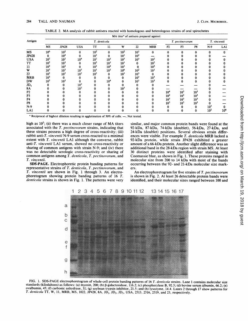
representative strains of T. denticola, T. pectinovorum, andT. vincentii are shown in Fig. 1 through 3. An electro-phoretogram showing protein banding patterns of 16 T.denticola strains is shown in Fig. 1. The patterns were very
similar, and major common protein bands were found at the92-kDa, 87-kDa, 74-kDa doublett), 56-kDa, 27-kDa, and24-kDa (doublet) positions. Several obvious strain differ-ences were visible. For example T. denticola MRB lacked a92-kDa protein, while strain JP428 exhibited a greateramount of a 66-kDa protein. Another slight difference was anadditional band in the 28-kDa region with strain MS. At least30 distinct proteins were identified after staining withCoomassie blue, as shown in Fig. 1. These proteins ranged in'molecular size from 200 to 14 kDa with môst of the bandsoccurring between the 92- and 21-kDa molecular size mark-ers.An electrophoretogram for five strains of T. pectinovorum
is shown in Fig. 2. At least 26 detectable protein bands wereidentified, and their molecular sizes ranged between 100 and
1 2 3 4 5 6 7 8 9 10 1112 131415 16 17
aG_- _- .dnosa. LII_ba_ ( _ b I 1 I p9 b I à 6c d_ s I III ld_ 2I a 2 i
e-f_..
g~_FI.1DSPG letohreormofwoecelpoei adngptenso_6Tdn ic strinsLae1cotis3oeursz
stnad(ioatos sfolw:() _sn 20 b glcosds,16.;()phshrlaeB 2.;()bviesrm lui, 62 eovîui,45 t aboi nyrse 1 g syentypiniior_15 nd()lsye 44ans2trug_7sopten oT.dnioaT ,1, RM,102_P2,-ADJDJ3 SA25, 256 259 an 23 epctvl.
J. CLIN. MICROBIOL.
on March 19, 2018 by guest
http://jcm.asm
.org/D
ownloaded from

MA AND ELECTROPHORETIC ANALYSIS OF ORAL SPIROCHETES
1 2 3 4 5
e - .. '.#S ..
g _ ............
FIG. 2. SDS-PAGE electrophoretogram of whole-cell proteinbanding patterns of five T. pectinovorum strains. The positions ofmolecular size standards are the same as those listed in the legend toFig. 1. Lanes 1 through 5 show patterns for T. pectinovorum PR,, P3,P4, P5, and P8, respectively.
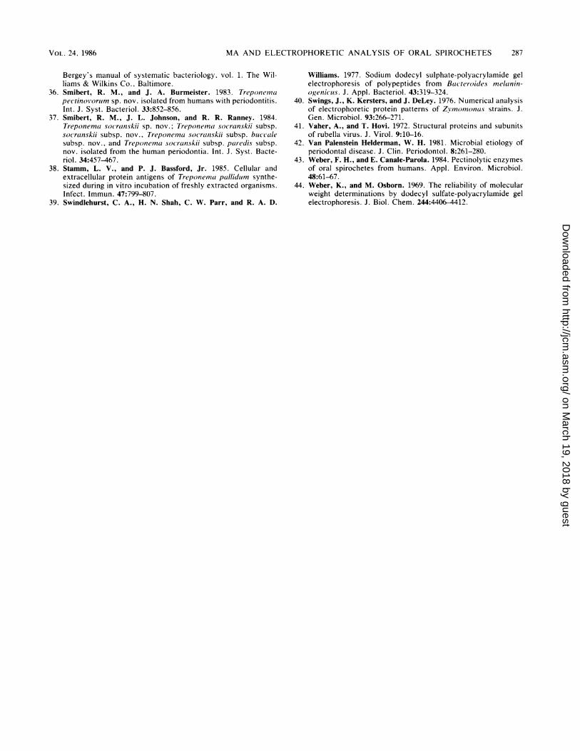
21 kDa. The protein banding patterns observed for thesestrains were essentially identical. The only apparent differ-ence observed was a slight variation in the banding positionof a 29- to 31-kDa protein.An electrophoretogram showing the electrophoretic pro-
tein banding patterns for two T. vincentii strains is shown inFig. 3. At least 35 distinct proteins were identified, withmolecular sizes ranging from 100 to 14 kDa and with most ofthe proteins falling between the 92- and 21-kDa molecularsize markers. The two banding patterns had regions ofhomogeneity, but for the most part presented many differ-ences. The protein bands present that were unique to eachof the strains of T. vincentii are indicated by arrows inFig. 3. The remaining bands were shared between the twostrains.The possibility existed that the protein banding patterns
observed were influenced by the medium used to grow eachspecies. To rule out this possibility, a strain of T. vincentiiwas grown in the medium used to grow T. denticola, and astrain of T. denticola was grown in the medium used for T.vincentii. Although the growth obtained was not ideal foreach species, we were able to prepare SDS-polyacrylamidegel protein banding patterns for each species and to comparethe patterns with those observed for cells grown in theoriginal growth medium. The results obtained (data notshown) indicated that the growth medium did not change theobserved protein banding patterns.By comparing several regions of the electrophoretic pro-
tein banding patterns for the three species of spirochetes, wecould distinguish each species from the others. Strains of T.denticola shared distinctive species protein band markerswith approximate molecular sizes of 74 to 92, 46 to 56, and 24
to 27 kDa. T. pectinovorum strains shared distinctive speciesbands with approximate molecular sizes of 120, 92, 66, 56,and 25 to 31 kDa, whereas the T. vincentii strains sharedbands at the 116-, 92-, 66-, 56-, 47-, and 24-kDa positions.
DISCUSSIONOver the past decade, SDS-PAGE has gained increasing
popularity for determining taxonomic similarities and differ-ences in a wide variety of microorganisms (5, 8, 25, 39, 40)and for revealing single- or multiple-protein components ofmicrobial structures and antigens (29, 38, 41). The presentinvestigation describes the use of SDS-PAGE to determinewhole-cell protein, banding patterns for 23 strains of oralanaerobic spirochetes, representing three species ofTreponema. A total of sixteen T. denticola strains, 5 T.pectinovorum strains, and 2 T. vincentii strains were evalu-ated. Distinct protein banding patterns were obtained foreach of the three species of oral anaerobic spirochetes, andthese patterns were found to be independent of the mediumwhich was used to grow each species. To compare MA withSDS-PAGE, a total of 11 T. denticola strains, 4 T.pectinovorum strains, and 2 T. vincentii strains were evalu-ated by MA. MA analysis revealed a diversity of serologicreactivity or sharing of common antigens within each spe-cies. However, there was no cross-reactivity or sharing ofcommon antigehs among the three species. The dissimilarityin banding patterns seen among the three species agreed withthe lack of cross-reactivity determined by MA. In addition,the similarity in banding patterns observed among strains ofthe three spirochetal species agreed with the strain variationseen among strains by MA. The MA and electrophoreticbanding patterns for T. vincentii N-9 and LA1 suggest thatthese two spirochetes may be two distinct species.
12
-a
:.1A
A_-C
d-iÀ .4-h
FIG. 3. SDS-PAGE electrophoretogram of whole-cell proteinbanding patterns of T. vincentii N-9 and LA1. The positions ofmolecular size standards are the same as those listed in the legend toFig. 1. Arrows indicate protein bands unique to each strain of T.vincenlii.
VOL. 24, 1986 285
on March 19, 2018 by guest
http://jcm.asm
.org/D
ownloaded from
286 TALL AND NAUMAN
Our electrophoresis data were consistent with those re-ported by Stamm and Bassford (38) in that [35S]methionine-labeled T. denticola W and 11 possessed some 30 to 34identifiable labeled proteins, whereas the Coomassie blue-stained electrophoretograms of T. denticola presented hereidentified at least 30 stained proteins. The difference in totalidentifiable proteins observed in these two studies may beattributed to the greater sensitivity of radiolabeling tech-niques versus protein staining. In addition, the protein bandsidentified by Stamm and Bassford (38) and those reportedhere possessed visually similar banding patterns, with themajority of the proteins that were identified in both studiesbeing positioned within the 21- and 92-kDa molecular sizerange. Lastly, our data support the rRNA oligonucleotidecatalog technique of Paster et al. (30), who have shown thatT. denticola and T. pectinovorum (P5) possess a singlespirochetal ancestor, but that these organisms diverge sep-arately into two distinct species.
In summary, SDS-PAGE of oral anaerobic spirochetes is auseful technique for determining species-to-species and spe-cies-to-strain relatedness, and a high degree of correlationwas found between the results of MA and SDS-PAGEanalyses. We recommended that SDS-PAGE and MA anal-yses be included in classification schemes for the identifica-tion of oral anaerobic spirochetes.
LITERATURE CITED1. Armitage, G. C., W. R. Dickinson, R. J. Jenderseck, S. M.
Levine, and D. W. Chambers. 1982. Relationship between thepercentage of subgingival spirochetes and the severity ofperiodontal disease. J. Periodontol. 53:550-556.
2. Biavati, B., V. Scardovi, and W. E. C. Moore. 1982. Electro-phoretic patterns of proteins in the genus Bifidobacterium andproposal of four new species. Int. J. Syst. Bacteriol. 32:358-373.
3. Breznak, J. A. 1973. Biology of non-pathogenic host associatedspirochetes. Crit. Rev. Microbiol. 21:457-489.
4. Cato, E. P., D. E. Hash, L. V. Holdeman, and W. E. C. Moore.1982. Electrophoretic study of Clostridium species. J. Clin.Microbiol. 15:688-702.
5. Daniels, M. J., and B. M. Meddins. 1973. Polyacrylamide gelelectrophoresis of Mycoplasma proteins in sodium dodecylsulphate. J. Gen. Microbiol. 76:239-242.
6. El-Sharkawy, T. A., and D. Huisingh. 1971. Differentiationamong Xanthomonas species by polyacrylamide gel electropho-resis of soluble proteins. J. Gen. Microbiol. 68:155-165.
7. Fiehn, N. E., and J. Westergaard. 1984. Cultivation on solidmedia of spirochetes in subgingival plaque from advancedmarginal periodontitis in humans. Scand. J. Periodontal Res.92:426-435.
8. Fotos, P. G., M. A. Gerencser, and D. B. Yelton. 1984. Straindifferentiation of Rothia dentocariosa and related isolates bysodium dodecyl sulfate-polyacrylamide gel electrophoresis. Int.J. Syst. Bacteriol. 34:102-106.
9. Fox, R. H., and D. E. McClain. 1974. Evaluation of thetaxonomic relationship of Micrococcus cryophilus, Bran-hamella catarrhalis, and neisseriae by comparative polyacryl-amide gel electrophoresis of soluble proteins. Int. J. Syst.Bacteriol. 24:172-176.
10. Gochenour, W. S., R. H. Yager, P. W. Wetmore, and J. A.Hightower. 1953. Laboratory diagnosis of leptospirosis. Am. J.Public Health 43:405-410.
11. Goldhaber, P., and D. B. Giddon. 1964. Present conceptsconcerning the etiology and treatment of acute necrotizingulcerative gingivitis. Int. Dent. J. 14:468-496.
12. Gross, C. S., D. A. Ferguson, Jr., and C. S. Cummins. 1978.Electrophoretic protein patterns and enzyme mobilities inanaerobic coryneforms. Apple. Environ. Microbiol. 35:1102-1108.
13. Hanna, J., S. D. Neill, J. J. O'Brien, and W. A. Ellis. 1983.
Comparison of aerotolerant and reference strains of Campylo-bacter species by polyacrylamide gel electrophoresis. Int. J.Syst. Bacteriol. 33:143-146.
14. Jacob, E., A. L. Allen, and R. K. Nauman. 1979. Detection oforal anaerobic spirochetes in dental plaque by the indirectfluorescent antibody technique. J. Clin. Microbiol. 10:934-936.
15. Jacob, E., T. F. Meiller, and R. K. Nauman. 1982. Detection ofelevated serum antibodies to T. denticola in patients withadvanced periodontitis by an enzyme-linked immunosorbentassay. J. Periodontal Res. 17:145-153.
16. Kersters, K., and J. DeLey. 1975. Identification and grouping ofbacteria by numerical analysis of their electrophoretic proteinpatterns. J. Gen. Microbiol. 87:333-342.
17. Leschine, S. B., and E. Canale-Parola. 1980. Rifampin as aselective agent for isolation of oral spirochetes. J. Clin. Micro-biol. 12:792-795.
18. Listgarten, M. A. 1965. Electron microscopic observations onthe bacterial flora of acute necrotizing ulcerative gingivitis. J.Periodontol. 36:328-339.
19. Listgarten, M. A., and L. Heliden. 1978. Relative distribution ofbacteria at clinically healthy and periodontally diseased sites inhumans. J. Clin. Periodontol. 5:115-132.
20. Listgarten, M. A., H. E. Mayo, and R. Tremblay. 1975. Devel-opment of dental plaque on epoxy resin crowns in man: a lightand electron microscopic study. J. Periodontol. 46:10-26.
21. Listgarten, M. A., and S. S. Socransky. 1965. Electron micros-copy as an aid in the taxonomic differentiation of oral spiro-chetes. Arch. Oral Biol. 10:127-138.
22. Loe, H., E. Theilade, and S. B. Jensen. 1965. Experimentalgingivitis in man. J. Periodontol. 36:177-187.
23. Loesche, W. J., and B. E. Laughon. 1982. Role of spirochetes inperiodontal disease, p. 62-75. In R. J. Genco and S. E.Mergenhagen (ed.), Host-parasite interactions in periodontaldiseases. American Society for Microbiology, Washington,D.C.
24. Mangan, D. F., B. E. Laughon, B. Bower, and D. E. Lopatin.1982. In vitro lymphocyte blastogenic responses and titers ofhumoral antibodies from periodontitis patients to oral spiro-chete isolates. Infect. Immun. 37:445-451.
25. Mocca, L. F., and C. E. Frasch. 1982. Sodium dodecyl sulfate-polyacrylamide gel typing system for characterization of Neis-seria meningitidis isolates. J. Clin. Microbiol. 16:240-244.
26. Moore, W. E. C., D. E. Hash, L. V. Holdeman, and E. P. Cato.1980. Polyacrylamide slab gel electrophoresis of soluble pro-teins for studies of bacterial floras. Appl. Environ. Microbiol.39:900-907.
27. Morris, J. A. 1973. The use of polyacrylamide gel electropho-resis in taxonomy of Brucella. J. Gen. Microbiol. 76:231-237.
28. Morris, J. A., and R. W. A. Park. 1973. A comparison using gelelectrophoresis of cell proteins of campylobacters (Vibrios)associated with infertility, abortion and swine dysentery. J.Gen. Microbiol. 78:165-178.
29. Palva, E. T., and P. H. Makela. 1980. Lipopolysaccharideheterogeneity in Salmonella typhimurium analyzed by sodiumdodecyl sulfate-polyacrylamide gel electrophoresis. Eur. J.Biochem. 107:137-143.
30. Paster, B. J., E. Stackebrandt, R. B. Héspell, G. M. Hahn, andC. R. Woese. 1984. The phylogeny of the spirochetes. Syst.Appl. Microbiol. 5:337-351.
31. Payne, J. W. 1976. Electrophoresis of proteins in sodiumdodecyl sulphate-polyacrylamide gels, p. 321-326. In Ivor Smith(ed.), Chromatographic and electrophoretic techniques, vol. 2.Zonal electrophoresis, 4th ed. Yearbook Medical Publishers,Chicago; 111.
32. Schultz-Haudt, S., M. A. Bruce, and B. G. Bibby. 1954. Bacterialfactors in nonspecific gingivitis. J. Dent. Res. 33:454-458.
33. Seiter, J. A., and J. M. Jay. 1980. Application of polyacrylamidegel electrophoresis to the characterization and identification ofArthrobacter species. Int. J. Syst. Bacteriol. 30:460-465.
34. Slots, J., P. Mashimo, M. J. Levine, and R. J. Genco. 1979.Periodontal therapy in humans. J. Periodontol. 49:495-509.
35. Smibert, R. M. 1984. Genus III. Treponema Schaudinn 1905,1728, p. 49-57. In R. E. Buchanan and N. E. Gibbons (ed.),
J. CLIN. MICROBIOL.
on March 19, 2018 by guest
http://jcm.asm
.org/D
ownloaded from
MA AND ELECTROPHORETIC ANALYSIS OF ORAL SPIROCHETES
Bergey's manual of systematic bacteriology. vol. 1. The Wil-liams & Wilkins Co., Baltimore.
36. Smibert, R. M., and J. A. Burmeister. 1983. Tr-epoteeinapectinoiworirn sp. nov. isolated from humans with periodontitis.Int. J. Syst. Bacteriol. 33:852-856.
37. Smibert, R. M., J. L. Johnson, and R. R. Ranney. 1984.Treponemna socranskii sp. nov.; Treponemta socranskii subsp.soc ranskii subsp. nov., Treponeina socranskii subsp. buccalesubsp. nov., and Treponema socranskii subsp. paredis subsp.nov. isolated from the human periodontia. tnt. J. Syst. Bacte-riol. 34:457-467.
38. Stamm, L. V., and P. J. Bassford, Jr. 1985. Cellular andextracellular protein antigens of Treponeina pallidumn synthe-sized during in vitro incubation of freshly extracted organisms.Infect. Immun. 47:799-807.
39. Swindlehurst, C. A., H. N. Shah, C. W. Parr, and R. A. D.
Williams. 1977. Sodium dodecyl sulphate-polyacrylamide gelelectrophoresis of polypeptides from Bacteroides melanin-ogenicus. J. Appl. Bacteriol. 43:319-324.
40. Swings, J., K. Kersters, and J. DeLey. 1976. Numerical analysisof electrophoretic protein patterns of Zyinotnonas strains. J.Gen. Microbiol. 93:266-271.
41. Vaher, A., and T. Hovi. 1972. Structural proteins and subunitsof rubella virus. J. Virol. 9:10-16.
42. Van Palenstein Helderman, W. H. 1981. Microbial etiology ofperiodontal disease. J. Clin. Periodontol. 8:261-280.
43. Weber, F. H., and E. Canale-Parola. 1984. Pectinolytic enzymesof oral spirochetes from humans. Appl. Environ. Microbiol.48:61-67.
44. Weber, K., and M. Osborn. 1969. The reliability of molecularweight determinations by dodecyl sulfate-polyacrylamide gelelectrophoresis. J. Biol. Chem. 244:4406-4412.
VOL. 24, 1986 287
on March 19, 2018 by guest
http://jcm.asm
.org/D
ownloaded from