microscopic anatomy of selected male and female reproductive organs
DESCRIPTION
Microscopic Anatomy of Selected Male and Female Reproductive Organs. Testes. Each lobule contains one to four seminiferous tubules Tightly coiled structures Function as sperm-forming factories Empty sperm into the rete testis . Figure 16.1. Testes. - PowerPoint PPT PresentationTRANSCRIPT
Microscopic Anatomy of Selected Male and Female
Reproductive Organs
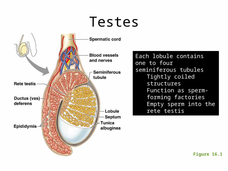
Testes
Figure 16.1
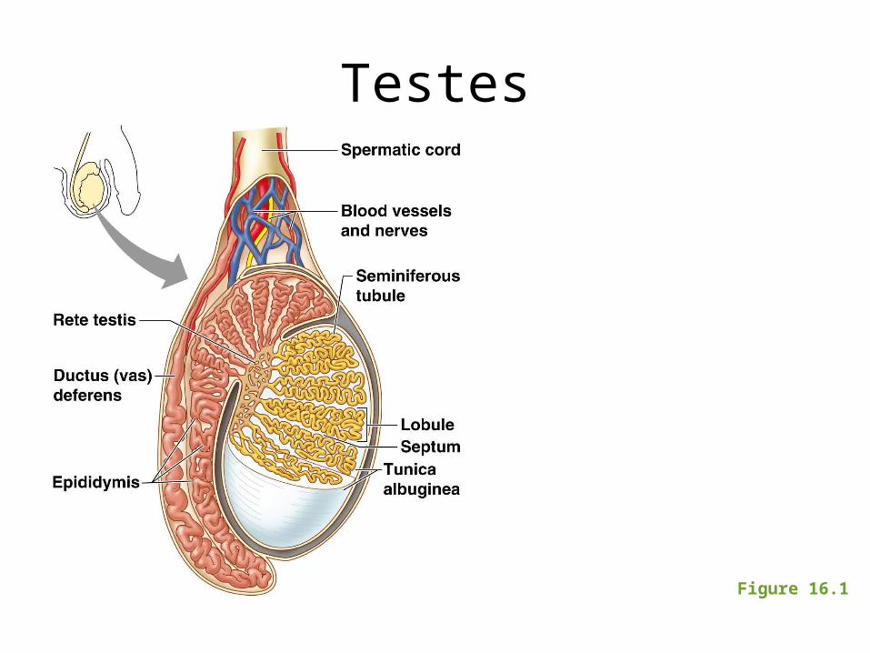
Each lobule contains one to four seminiferous tubules
Tightly coiled structuresFunction as sperm-forming factoriesEmpty sperm into the rete testis
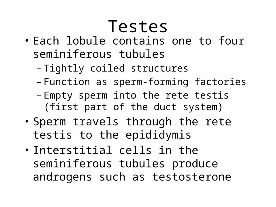
Testes• Each lobule contains one to four
seminiferous tubules– Tightly coiled structures– Function as sperm-forming factories– Empty sperm into the rete testis (first part of
the duct system)• Sperm travels through the rete testis to the
epididymis• Interstitial cells in the seminiferous tubules
produce androgens such as testosterone
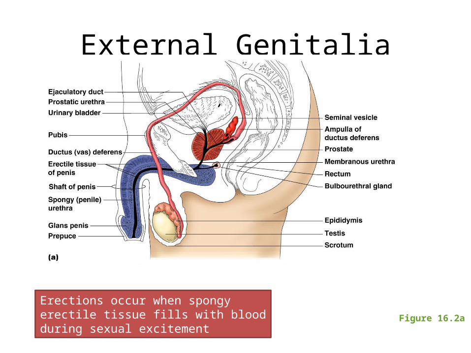
External Genitalia
Figure 16.2a
Erections occur when spongy erectile tissue fills with blood during sexual excitement
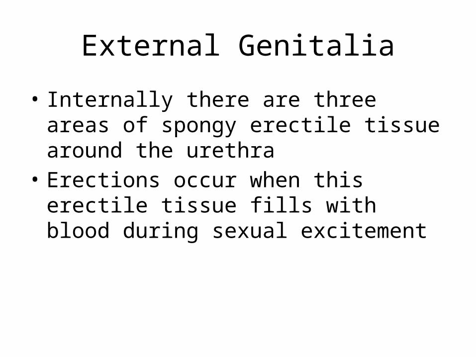
External Genitalia
• Internally there are three areas of spongy erectile tissue around the urethra
• Erections occur when this erectile tissue fills with blood during sexual excitement
Testes
Figure 16.1
Ovaries
Figure 16.7
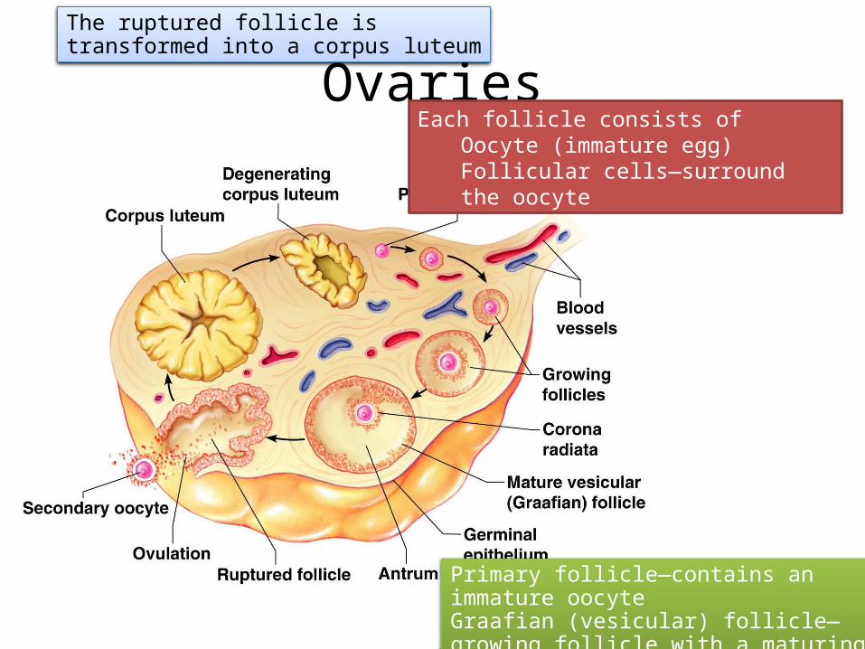
Each follicle consists of Oocyte (immature egg)Follicular cells—surround the oocyte
Primary follicle—contains an immature oocyteGraafian (vesicular) follicle—growing follicle with a maturing oocyte
The ruptured follicle is transformed into a corpus luteum
Ovaries
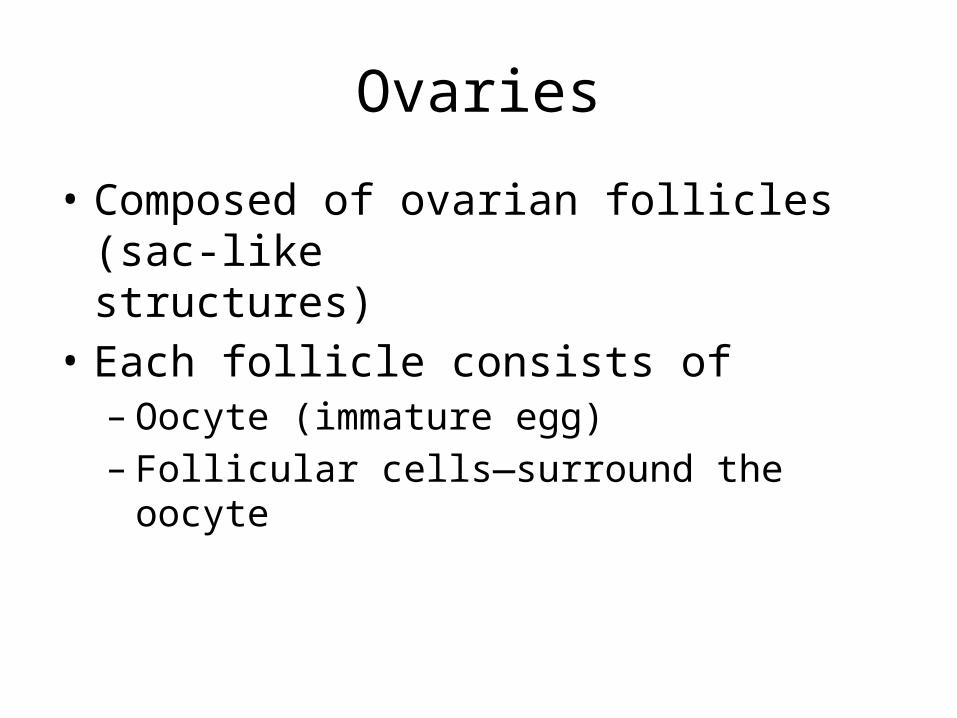
• Composed of ovarian follicles (sac-like structures)
• Each follicle consists of – Oocyte (immature egg)– Follicular cells—surround the oocyte
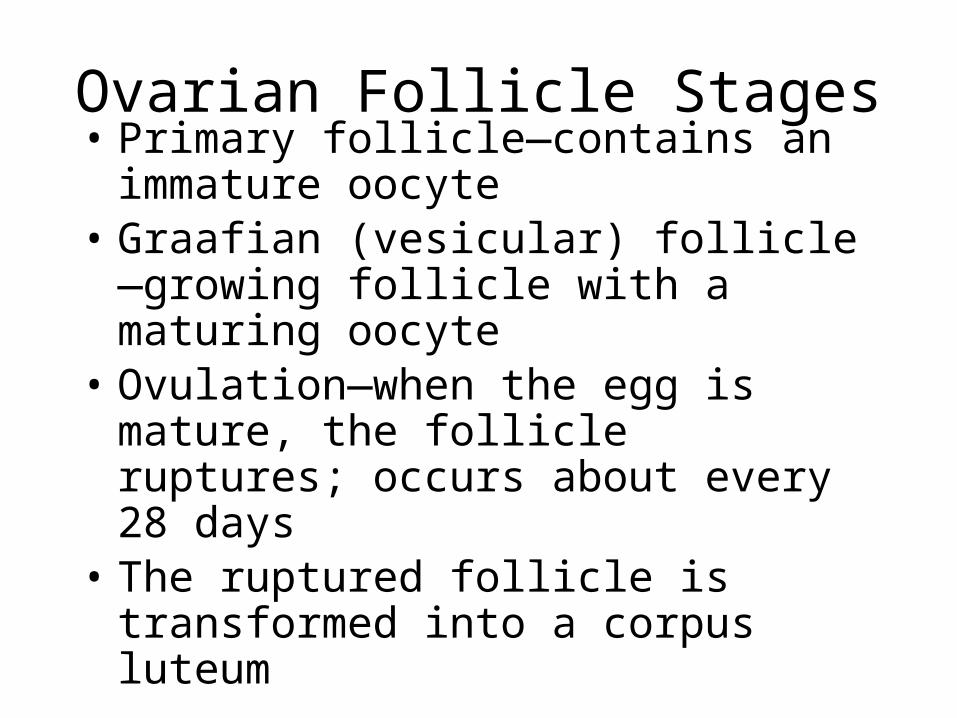
Ovarian Follicle Stages• Primary follicle—contains an immature
oocyte• Graafian (vesicular) follicle—growing follicle
with a maturing oocyte• Ovulation—when the egg is mature, the
follicle ruptures; occurs about every 28 days
• The ruptured follicle is transformed into a corpus luteum
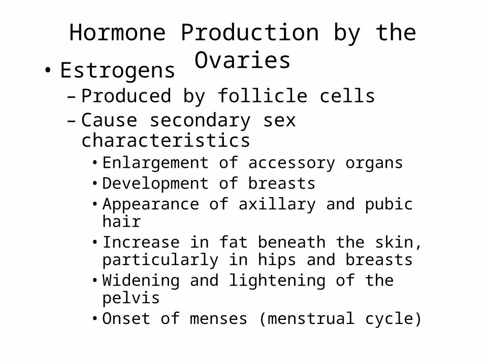
Hormone Production by the Ovaries• Estrogens– Produced by follicle cells– Cause secondary sex characteristics• Enlargement of accessory organs• Development of breasts• Appearance of axillary and pubic hair• Increase in fat beneath the skin, particularly in hips
and breasts• Widening and lightening of the pelvis• Onset of menses (menstrual cycle)
Hormone Production by the Ovaries
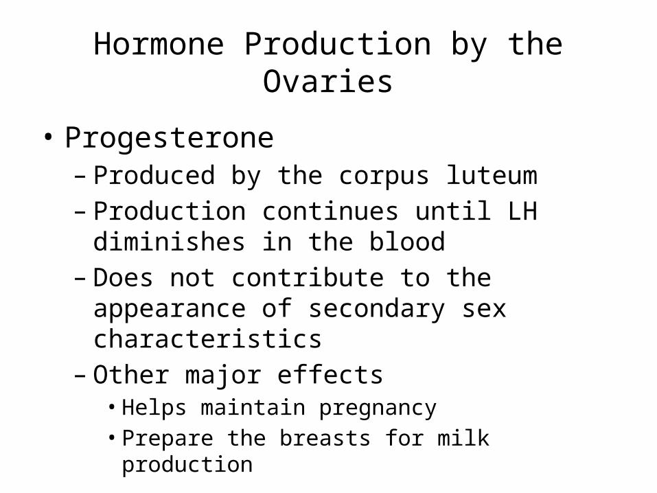
• Progesterone– Produced by the corpus luteum– Production continues until LH diminishes in the
blood– Does not contribute to the appearance of
secondary sex characteristics– Other major effects• Helps maintain pregnancy• Prepare the breasts for milk production
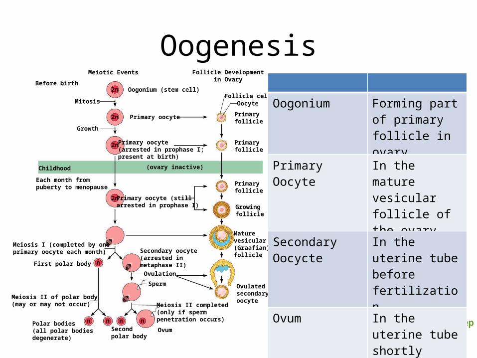
Oogenesis
Figure 16.10, step 9
Meiotic Events Follicle Developmentin OvaryBefore birth
Childhood
Primary oocyte
Primary oocyte (stillarrested in prophase I)
Maturevesicular(Graafian)follicle
Primaryfollicle
Primaryfollicle
Primaryfollicle
Oocyte
Ovulatedsecondaryoocyte
Growingfollicle
Primary oocyte(arrested in prophase I;present at birth)
Oogonium (stem cell)
Each month frompuberty to menopause
Meiosis I (completed by oneprimary oocyte each month)
First polar body
Mitosis
Growth
Meiosis II of polar body(may or may not occur)
Polar bodies(all polar bodiesdegenerate)
OvumSecondpolar body
Meiosis II completed(only if spermpenetration occurs)
SpermOvulation
Secondary oocyte(arrested inmetaphase II)
Follicle cells
(ovary inactive)
2n
2n
2n
2n
n
nnn n
Oogonium Forming part of primary follicle in ovary
Primary Oocyte In the mature vesicular follicle of the ovary
Secondary Oocycte
In the uterine tube before fertilization
Ovum In the uterine tube shortly after sperm penetration
Oogenesis and the Ovarian Cycle
• Oogonia—female stem cells found in a developing fetus
• Oogonia undergo mitosis to produce primary oocytes
• Primary oocytes are surrounded by cells that form primary follicles in the ovary
• Oogonia no longer exist by the time of birth
Oogenesis and the Ovarian Cycle
• Primary oocytes are inactive until puberty • Follicle stimulating hormone (FSH) causes
some primary follicles to mature each month• Cyclic monthly changes constitute the ovarian
cycle
Oogenesis and the Ovarian Cycle
• Meiosis starts inside maturing follicle• Produces a secondary oocyte and the first polar
body• Follicle development to the stage of a vesicular
follicle takes about 14 days• Ovulation of a secondary oocyte occurs with the
release of luteinizing hormone (LH)• Secondary oocyte is released and surrounded by
a corona radiata
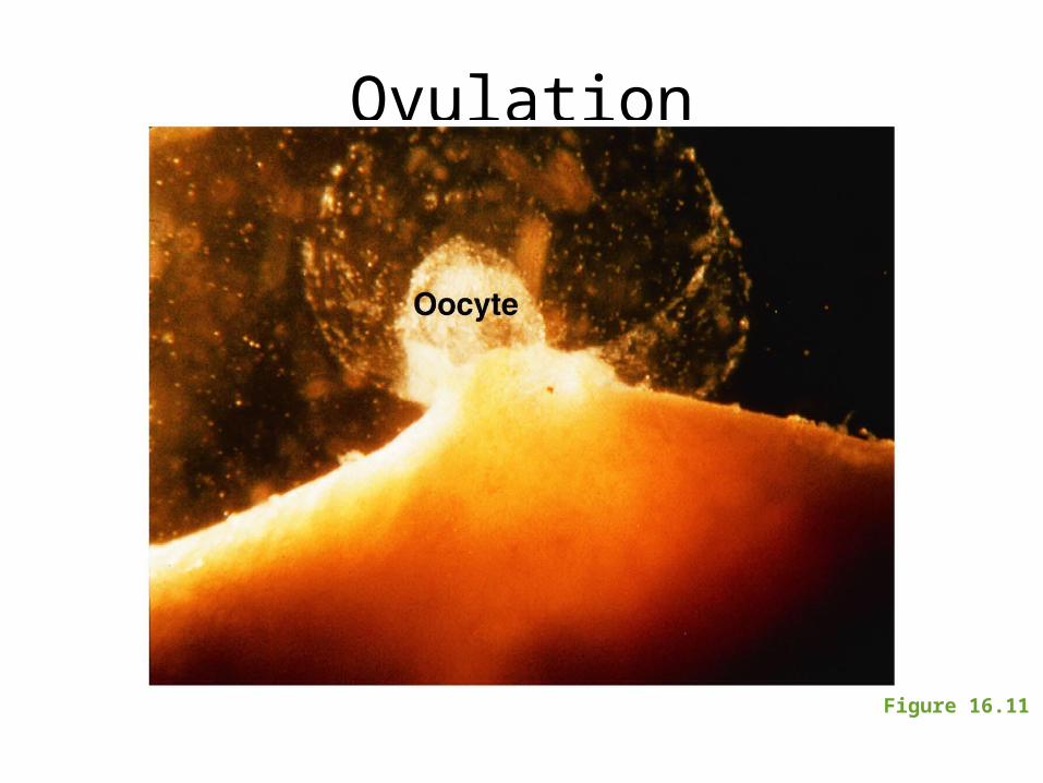
Ovulation
Figure 16.11
Oogenesis and the Ovarian Cycle• Meiosis is completed after ovulation only if
sperm penetrates– Ovum is produced – Two additional polar bodies are produced
• Once ovum is formed, the 23 chromosomes can be combined with those of the sperm to form the fertilized egg (zygote)
• If the secondary oocyte is not penetrated by a sperm, it dies and does not complete meiosis to form an ovum
