microsoft word - wound_assessment_guideline w… · web viewthe equality act also requires regard...
TRANSCRIPT

Wound Assessment Clinical GuidelineSkin Care Service January 2016
Page 1 of 36
Policy Number
LCH-121
This document has been reviewed in line with the Policy Alignment Process for Liverpool Community Health NHS Trust Services. It is a valid Mersey Care document, however due to organisational change this FRONT COVER has been added so the reader is aware of any changes to their role or to terminology which has now been superseded. When reading this document please take account of the changes highlighted in Part B and C of this form.Part A – Information about this DocumentPolicy Name Wound Assessment Guideline
Policy Type Board Approved (Trust-wide) ☐ Trust-wide ☐ Divisional / Team / Locality ☒
Action No Change ☐ Minor
Change ☐ MajorChange ☐ New
Policy ☒ No LongerNeeded ☐
Approval
As Mersey Care’s Executive Director / Lead for this document, I confirm that this document:a) complies with the latest statutory / regulatory requirements,b) complies with the latest national guidance,c) has been updated to reflect the requirements of clinicians and officers, andd) has been updated to reflect any local contractual requirements
Signature: Date:Part B – Changes in Terminology (used with ‘Minor Change’, ‘Major Changes’ & ‘New Policy’ only)
Terminology used in this Document New terminology when reading this Document
Part C – Additional Information Added (to be used with ‘Major Changes’ only)Section /
Paragraph NoOutline of the information that has been added to this document – especially where it may
change what staff need to do
Part D – Rationale (to be used with ‘New Policy’ & ‘Policy No Longer Required’ only)Please explain why this new document needs to be adopted or why this document is no longer required
Part E – Oversight Arrangements (to be used with ‘New Policy’ only)Accountable Director
Recommending Committee
Approving Committee
Next Review Date
LCH Policy Alignment Process – Form 1

Wound Assessment Clinical GuidelineSkin Care Service January 2016
Page 2 of 36
SUPPORTING STATEMENTS This document should be read in conjunction with the following statements:
SAFEGUARDING IS EVERYBODY’S BUSINESS
All Mersey Care NHS Foundation Trust employees have a statutory duty to safeguard and promote the welfare of children and adults, including: being alert to the possibility of child / adult abuse and neglect through their observation of
abuse, or by professional judgement made as a result of information gathered about the child / adult;
knowing how to deal with a disclosure or allegation of child / adult abuse; undertaking training as appropriate for their role and keeping themselves updated; being aware of and following the local policies and procedures they need to follow if they
have a child / adult concern; ensuring appropriate advice and support is accessed either from managers, Safeguarding
Ambassadors or the trust’s safeguarding team; participating in multi-agency working to safeguard the child or adult (if appropriate to your
role); ensuring contemporaneous records are kept at all times and record keeping is in strict
adherence to Mersey Care NHS Foundation Trust policy and procedures and professional guidelines. Roles, responsibilities and accountabilities, will differ depending on the post you hold within the organisation;
ensuring that all staff and their managers discuss and record any safeguarding issues that arise at each supervision session
EQUALITY AND HUMAN RIGHTS
Mersey Care NHS Foundation Trust recognises that some sections of society experience prejudice and discrimination. The Equality Act 2010 specifically recognises the protected characteristics of age, disability, gender, race, religion or belief, sexual orientation and transgender. The Equality Act also requires regard to socio-economic factors including pregnancy /maternity and marriage/civil partnership.
The trust is committed to equality of opportunity and anti-discriminatory practice both in the provision of services and in our role as a major employer. The trust believes that all people have the right to be treated with dignity and respect and is committed to the elimination of unfair and unlawful discriminatory practices.
Mersey Care NHS Foundation Trust also is aware of its legal duties under the Human Rights Act 1998. Section 6 of the Human Rights Act requires all public authorities to uphold and promote Human Rights in everything they do. It is unlawful for a public authority to perform any act which contravenes the Human Rights Act.
Mersey Care NHS Foundation Trust is committed to carrying out its functions and service delivery in line the with a Human Rights based approach and the FREDA principles of Fairness, Respect, Equality Dignity, and Autonomy

Wound Assessment Clinical GuidelineSkin Care Service January 2016
Page 3 of 36
Title Wound Assessment Clinical Guideline
Guideline referencenumber
121
Aim and purpose of clinical document
To provide evidence-based guidance on the wound assessment process
Author Skin Care Service
TypeNew document
Reviewed document √
Review Date February 2018 – 1 month extension agreed at Clinical Standards Group meeting 24.01.18
Person/groupaccountable for review
Skin Care Service
Type of Evidencebase used
C: Evidence which includes published and/orunpublished studies and expert opinion (limited scientific evidence)
Issue date January 2016
Authorised byClinical Standards Group
January 2016 – amendment noted at meeting on 25th
July 17
Impact AssessmentUndertaken Yes √ date when
undertaken 03.02.2015
No
Evidencecollated √

Wound Assessment Clinical GuidelineSkin Care Service January 2016
Page 4 of 36
Version SevenRatified by: Clinical Standards GroupDate of Approval: January 2016Name of originator / author Skin Care ServiceApproving body / committee: Clinical Standards Group
Date issued: January 2016Review date: February 2018Target audience: Organisation WideName of lead Director /Managing Director
Director of Nursing
Changes / Alterations made to previous version
• Additional information regarding Sepsis
• Counting in and out of dressings amended to include all wounds, not just cavity wounds
• Chronic Wound Pathway added

Wound Assessment Clinical GuidelineSkin Care Service January 2016
Page 5 of 36
Contents Page
Purpose of the Guideline 4
Scope of the Guideline 4
Definitions 4
Wound Assessment 4
Training requirements 4
Documentation and Record Keeping 5
Monitoring Tool 7
Development of the Guideline, Contributions and Peer Review 7
Equality Analysis 7
Dissemination / Distribution method 7
References 8
Linked areas / Information 9
Key objectives of wound assessment
Factors that could delay wound healing 9
Specific assessment of the wound 12
Summary of the wound assessment process 16
Appendix 1
Audit Tool 18
Appendix 2
General Practice Screening and Action Tool 21
Appendix 3
UKST Toolkit: General Practice management of Sepsis 22
Appendix 4
Chronic Wound Pathway 36

Wound Assessment Clinical GuidelineSkin Care Service January 2016
Page 6 of 36
Purpose of the Guideline
This guideline has been developed to provide evidence-based guidance on the wound assessment process. It aims to improve clinical practice and reduce variations in standards of care within the primary and intermediate care setting.
Scope of the Guideline
This guideline applies to all registered health professionals employed by Liverpool Community Health NHS Trust (LCH) who are involved in the management of patients with wounds.
Definitions
Taken from The Free Dictionary Online (Medical) 2013 unless otherwise stated
A b sc e ss: A collection of pus associated with damaged and inflamed
tissues.
AI D S: Acquired Immune Deficiency Syndrome
C o ll a g en : A protein that is the principle constituent of white fibrous connective tissue. Found in tendons, skin, bone, cartilage and ligaments.
C omp l e m e n t: A substance in the blood that aids the body’s defence mechanisms when antibodies combine with invading antigens.
C onne c t i v e t i ss ue : Supportive or packing material consisting of a fibrous gel made up of collagen and elastin fibres in a mucopolysaccharide ground substance, bathed in extracellular fluid with scattered cells, blood vessels, lymphatics and nerve fibres transversing it (The Faber Pocket Medical Dictionary, Faber and Faber Limited, London, 1966).
C on t e m p o r an e ou s: Existing or occurring at the same time.
E r y t hema : Abnormal flushing of the skin caused by dilatation of the blood capillaries.
Fi st u l a : An abnormal communication between two hollow organs or between a hollow organ and the exterior. Baranoski and Ayello (2004) add that fistulae are named using the point of origin and point of exit e.g. a rectovaginal fistula would originate in the rectum and terminate in the vagina. Fistulae that connect an organ with the skin is termed an ‘enterocutanoeus fistula’.
H ae m o s e r ou s : Serous fluid that contains blood or is blood-stained.
N eo v a sc u l a ri s a t i on : new blood vessel formation in abnormal tissue or in abnormal

Wound Assessment Clinical GuidelineSkin Care Service January 2016
Page 7 of 36
positions

Wound Assessment Clinical GuidelineSkin Care Service January 2016
Page 8 of 36
Mi t o s i s: A type of cell division in which a single cell produces two genetically identical daughter cells. It is the way new body cells are produced for both growth and repair.
Ost e o m y e li t i s: Inflammation of the bone marrow due to infection.
P e ri : A prefix denoting near, around or enclosing.
P u r u l e n t: Forming, consisting of or containing pus.
P u s: A thick yellow-green liquid formed at the site of infection. Pus contains dead white cells, both living and dead bacteria and fragments of dead tissue.
S a c r u m : A curved, triangular element of the backbone consisting of five fused vertebrae. It articulates with the last lumbar vertebra above, the coccyx below and the hip bones laterally.
S ep s i s : A life threatening condition that arises when the body’s response to an infection injures its own tissues and organs (Slade 2003).
S e r ou s: Relates to, contains or resembles serum. Serum is the fluid that separates from clotted blood or blood plasma that is allowed to stand. It is similar in composition to plasma but lacks fibrinogen and other substances that are used in the coagulation process.
S i nu s: In wound healing terms as sinus is an infected tract leading from the focus of infection to the surface of the skin or a hollow organ.
T e n s il e St r e n g t h : Resistance to breaking under tension (The Oxford Compact English Dictionary, Oxford University Press, 1996).
T i ss u e R e g ene r a t i on : Natural re-growth of tissue lost through injury. Calvin (1998) states that it implies complete re-establishment of the original tissue structure.
T i ss u e R e p a ir : Original tissue lost through injury is replaced with non-specific connective tissue, forming a functionally inferior scar (Calvin 1998).
V a s od il a t i on : An increase in the diameter of the blood vessels.
Wound Assessment
A wound is defined as “a disruption of the integrity and function of the tissues in the body” (Baharestani 2004). Any injury to the skin interrupts continuity and protective and functional capacity, rendering the individual physically and emotionally vulnerable, therefore the priority is to heal the void as quickly as possible. In order to plan and implement appropriate management, a full, holistic patient assessment must be undertaken.
Assessment is defined as “information obtained via observation, questioning, physical examination and clinical investigation in order to establish a baseline”

Wound Assessment Clinical GuidelineSkin Care Service January 2016
Page 9 of 36
(Collins et al 2002). In any clinical situation, a thorough assessment is fundamental in ensuring correct diagnosis of the presenting problem. Once a differential diagnosis

Wound Assessment Clinical GuidelineSkin Care Service January 2016
Page 10 of 36
is made an appropriate management plan can be developed. This same principle must be adopted in the treatment of patients with wounds in order to promote wound healing (Miller 1999a).
It must be remembered that the process of wound healing is a natural, normal response by the body to injury, which results in tissue repair or regeneration. In normal wound healing, the application of a dressing to a wounded area merely provides protection to a vulnerable area and allows normal wound healing to take place in an optimal environment (see guidelines on dressing selection). The key to effective wound management lies in the identification and optimisation of factors that could potentially delay the normal wound healing process and is therefore not just the act of applying a dressing.
There is no dressing that can compensate for an undiagnosed and untreated pathologic condition (Bolton 1990).
There are many factors that can potentially delay the wound healing process. It is vital that these factors are identified through assessment and optimised where possible therefore minimising their effect on wound healing.
Training Requirements
LCH employees will be expected to act all times in such a manner as to safeguard and promote the interests of patients and clients. Registered health professionals must have the knowledge and skills for safe and effective practice; recognise and work within the limits of personal competence (Nursing and Midwifery Council [NMC] 2015)
All employees must be made aware of the organisations guidelines before commencement in post, as part of their local induction process.
Elements of this guideline have been incorporated in the development of the wound assessment competency programme. This is available to all registered health professionals involved in the management of patients with wounds and can be accessed through the Learning and Development Bureau at: l ea r n i n g . de v e l o p m e n t @ li v e r poo l . nh s . u k
Documentation and Record Keeping
Effective documentation is of paramount importance for the following reasons:
• To comply with NMC guidance on record keeping• Contemporaneous written records provide the evidence and rationale on
which care delivery is based and are a legal requirement. They could be scrutinised at any time in a court of law and must therefore accurately reflect and justify the actions and decisions made by health care professionals
• Accurate records improve communication between health care professionals regarding individual patient care

Wound Assessment Clinical GuidelineSkin Care Service January 2016
Page 11 of 36
• Accurate, comprehensive records enhance continuity of care
Wound assessment documentation is again another contentious issue (Russell 2002b) and there is still no nationally agreed wound assessment tool. Each organisation should therefore develop a wound assessment tool that is based on best practice and encompasses all relevant information.
Monitoring Tool
The monitoring of the use of this guideline should be undertaken by individual services and localities where wound care is performed as part of their local audit plan.
Development of the Guideline, Contributions and Peer Review
The guideline was developed and peer reviewed by members of the Skin Care Service and ratified by LCH Clinical Policies Group.
Equality Analysis
This has been undertaken and the evidence has been retained by the authors and the Equality and Diversity Lead of LCH
Distribution/Dissemination Method
On ratification by the Clinical Policies Group, this guideline will be added to the Clinical Policies intranet site and communicated via the weekly Communication Bulletin. Knowledge of the guideline will also be communicated to staff via the Learning and Development Bureau:l ea r n i n g . de v e l o p m e n t @ li v e r poo l p ct. n h s. u k and also by the Skin Care Service who support community nurses in the management of patients with wounds.

Wound Assessment Clinical GuidelineSkin Care Service January 2016
Page 12 of 36
References
Baharestani M M (2004) Quality of life and ethical issues. In: Baranoski S and Ayello E A (Eds) W ou n d C a r e Ess e n t i a l s: P r a ct i ce P ri n c i p l e s Lippincott, Williams and Wilkins, Philadelphia
Baranoski S, Ayello E A (2004) (Eds) W o u n d C a r e Ess e n t i a l s: P r a ct i ce P ri n c i p l e s Lippincott, Williams and Wilkins, Philadelphia
Bolton L (1990) Dressing’s effects on wound healing. W ou nd s 2, 126
Calvin M (1998) Cutaneous wound repair. W oun d s: A C o mp e nd i u m o f Cli n i c a l R e s ea r ch an d P r a ct i c e . 10 (1) 12-32
Collier M (2002) A ten-point assessment plan for wounds J o u r n a l o f C o m mun i ty N u r s i n g 16 (6) 22-26
Collins F, Hampton S, White R (2002) A - Z Di ct i ona r y o f W ou n d C a r e . Quay Books, Mark Allen Publishing, Surrey
Collins F, Hampton S, (2004) Tissue Viability Whurr Publishers Ltd, London.
Dale J and Gibson B (1986) The epidemiology of leg ulcers P r o f e s s i ona l N u r se 1 (8) 215-216
Davidson M (2002) Sharpen your wound assessment skills N u r s i n g 32 (10) 1-4
Dealey C (1994) T h e C a r e o f W ou nd s . First Edition. Blackwell Scientific, Oxford
Dealey C (1999) T h e C a r e o f W o u n d s . Second Edition. Blackwell Scientific Publications, Oxford
Desai H (1997) Ageing and wounds: Part 2 J o u r na l o f w oun d C a r e 6 (5) 237-239
Flanagan M (1996) A practical framework for wound assessment 2: methods B ri t i sh J ou r na l o f N u r s i n g 5 (22) 1391-7
Flanagan M (1997) Acc e ss to Cli n i c a l E du c a t i on : W ou n d M an ag emen t Churchill Livingstone
Gilchrist B (1999) Wound infection. In: Miller M, Glover D (Eds) W oun d M ana g e m en t: T h e o r y an d P r a ct i ce Emap Healthcare Ltd, London
Miller M (1999a) Wound assessment. In: Miller M, Glover D (Eds) W o u n d M ana g e m en t: T h e o r y an d P r a ct i ce Emap Healthcare Ltd, London
Miller M (1999b) Nursing assessment of patients with non-acute wounds B ri t i sh J ou r na l o f N u r s i n g 8 (1) 10-16
Morison M J (1992) A C o l ou r G u i d e to t h e N u r s i n g M ana g em e n t o f W o u n d s . First Edition. Wolfe, London

Wound Assessment Clinical GuidelineSkin Care Service January 2016
Page 13 of 36
Nobbs S (1998) Medication and wound healing P r a ct i ce N u r s i n g 9 (14) 28-30
Nursing and Midwifery Council (2015) The Code: Professional Standards of Practice and Behaviour for Nurses and Midwives. NMC, London
Russell L (2002a) The importance of patients’ nutritional status in wound healing. In: White R (Ed) T r e n d s i n W ou n d C a r e . Quay Books Division, Mark Allen Publishing Ltd, Bath
Russell L (2002b) The importance of wound documentation and classification. In: White R (Ed) T r e n d s i n W ou n d C a r e . Quay Books Division, Mark Allen Publishing Ltd, Bath
The UK Sepsis Trust 2014 Executive Summary: General Practice Management of SepsisAvailable at: ht t p:// s e p s i st r ust.o r g / w p - conten t / f il e s _ m f/ 1 40 9 3 2 24 98 GPtoo l k i t 20 1 4. pd f Accessed on 02.02.15
Silhi N (1998) Diabetes and wound healing J ou r na l o f W ou n d C a r e 7 (1) 47-51
Slade E (2003) The Surviving Sepsis Campaign: raising awareness to reduce mortality Cri t i c a l C a r e ; 7, 1-2.
Young T (2004) Wound assessment and dressing selection T i ss u e V i ab ili ty S o c i e ty R e g i ona l St u d y D a y Trafford General Hospital, Manchester (1.12.2004)
Linked Areas/Information
Other related Policies, Procedures or Work Instructions of Liverpool Community Health are:
• Accident and Incident Reporting and Management Policy• Consent to Treatment Policy• Risk Assessment Policy and Guidance• Other Skin Care Service clinical guidelines• A Practical Guide to the Management and Treatment of Wounds in Primary Care
Key objectives of wound assessment
• Thorough, holistic and systematic assessment.• Identification of factors that could potentially delay wound healing.• Diagnosis of underlying cause of the wound.• Assessment of current care and local wound management priorities.• Regular, planned evaluation of the effectiveness of treatment and
reassessment of the patients’ situation where necessary.• DOCUMENTATION – of assessment, treatment plan and rationale,
reassessment, review and evaluation of treatment given.

Wound Assessment Clinical GuidelineSkin Care Service January 2016
Page 14 of 36
Factors that could delay wound healing
• Age: The ageing process itself brings about many natural changes in the structure and functional capacity of the skin. The inflammatory and proliferative responses are delayed (Desai 1997). The skin loses much of its tensile strength and natural moisture and its capacity to repair, regenerate and fight bacterial invasion is reduced.
• Nutrition: Nutrition is a key aspect of assessment as wound healing is dependent upon good nutrition. All cellular activity requires oxygen and nutrients therefore patients with wounds must be nutritionally optimised for effective wound healing to take place. Russell (2002a) states that patients require adequate levels of calories, protein, vitamins and minerals to support wound healing.
Holistic assessment of nutrition and early detection of malnutrition are essential. The nutritional assessment tool of choice should be MUST (Malnutrition Universal Screening Tool) (ww w . bapen .c o . u k ) with a local agreed care plan in place. It is vital that nutritional intake is discussed on initial assessment. If the assessing practitioner has any cause for concern or indicated at the appropriate stage of the agreed care plan a referral to a dietician is required to ensure a more expert assessment is undertaken.
• Associated disease processes: There are various identified disease processes that can have a negative effect on wound healing. Diabetes is associated with an increased risk of peripheral vascular disease, both of the large and small vessels, and is known to impair wound healing (Silhi 1998). Uncontrolled hyperglycaemia can delay the wound healing process and increase the risk of wound infection.
Anaemia, chronic heart disease and chronic pulmonary disease can all potentially reduce the amount of oxygen supplied to the tissues. An adequate oxygen supply to any area of tissue damage is necessary in order to promote wound healing.
Peripheral arterial disease results in a compromised arterial blood supply to the periphery. In situations where the blood supply is inadequate, oxygen and nutrients will not be delivered to the tissues and wound healing will be significantly impaired.
Rheumatoid arthritis is an inflammatory condition of connective tissue and is associated with delayed wound healing.
Other disease processes that may delay healing and must be considered include; malabsorption disorders, malignancy and other immune deficiency disorders such as AIDS (Flanagan 1997).
• Mobility: Mobility can be affected by many underlying pathologies.Restricted mobility or total immobility increase the risk of delayed wound healing for various reasons. Directly, the inability to change position will result in prolonged pressure and possible pressure damage (refer to Trust guidelines on pressure ulcer prevention and management). Indirectly, reduced mobility may affect the patients’ ability to self care and attend to fundamental
Wound Assessment Clinical GuidelineSkin Care Service January 2016
Page 15 of 36
activities of daily living including shopping for food and preparing meals, maintaining personal hygiene and elimination and getting in/out of bed. Furthermore, the patients’ inability to mobilise may affect their quality of life and lead to emotional and psychological distress and social isolation.
• Medication: Although most of the research to date regarding the effects of drugs on wound healing has been performed on animals, the relevance of these findings to humans should be considered (Nobbs 1998)
o St e r o i d s – exert an anti-inflammatory effect therefore inhibiting the inflammatory phase of wound healing. This increases the risk of infection and a reduction in degradation of cellular debris and foreign materials. They also inhibit epithelialisation and decrease the mechanical strength of the wound.
o N on - st e r o i da l an t i-i n f l a m m a t o r y d r u g s – also have an anti-inflammatory effect therefore reduce the effectiveness of the inflammatory phase of wound healing. May also impair the tensile strength of new granulation tissue. Aspirin increases the risk of haematoma due to its’ anti-platelet activity.
o I m mu n o s up r e ss a n ts – reduce the body’s natural immune response therefore increasing the risk of infection and reducing clearance of debris and devitalised tissue.
o C y t o t o x i c d r u g s – target all rapidly dividing cells, reducing protein synthesis and cell division.
o R ad i o t h e r ap y – reduces cell mitosis, increases vascular damage and detrimental to fibroblasts thus reducing collagen production.
o Mi sc e ll aneou s d r u g s – including anticoagulants, local anaesthetics, Penicillamine, alcohol and nicotine. These may all delay the wound healing process.
• Infection: Clinical infection delays wound healing by prolonging the inflammatory phase, depleting the components of the complement cascade, disrupting normal clotting mechanisms, preventing neovascularisation and formation of new granulation tissue (Gilchrist 1999).
• Sepsis: A life threatening condition that arises when the body’s response to an infection injures its own tissues and organs. The source of which may stem from wound/surgical site infection. Sepsis can lead to shock, multiple organ failure and death especially if not recognised early and treated promptly. If sepsis is detected early enough and has not yet affected vital organs, it may be possible to treat the condition at home with appropriate antibiotic treatment. Routine clinical observations can play a vital role in detecting sepsis (Slade 2003). The UK Sepsis Trust (UKST) (2014) suggests that as well as the general impression at the time of initial assessment, the presence of abnormal observations should be enough to initiate evaluation for sepsis.UKST (2014) advise that clinical evaluation for sepsis should be undertaken in patients:
o With clinical evidence of systemic infection o In whom antibiotics are being considered o Suspected fluo Suspected gastroenteritiso Who are obviously unwell without clear cause

Wound Assessment Clinical GuidelineSkin Care Service January 2016
Page 16 of 36
o Who are elderly or immunocompromised and present with signs of infection
o Who have deteriorated on antibiotic therapy
• Psychological perspective: The lack of research in the field of health psychology and its’ application to wound healing is lacking however there is a push to address these issues. Stress and anxiety can result from many wound related factors including chronicity and subsequent necessary lifestyle changes, changes in body image, stigma and sexuality. The patient’s ability to understand their condition and available treatment options will impact on their concordance with treatment regimes. A patient’s ‘desire to heal’ will also have a major impact on wound healing.
• Social environment and social support: People from a poorer, more deprived social environment are generally more likely to smoke and eat a less nutritious diet, both of which have the potential to delay wound healing. Social isolation and lack of formal and informal social support networks may be due to the debilitating effects of underlying pathologies or may be a direct result of having a wound and may contribute to delayed healing.
• Mechanical forces: Mechanical forces can be defined as pressure, shear and friction and are extrinsic factors that should be considered in the assessment of patients with pressure ulceration (Dealey 1999) (refer to Trust guidelines on pressure ulcer prevention and management). Mechanical forces directly cause pressure damage and assessment of their impact on the patient is required in order to reduce associated risk, appropriately manage patients with pressure damage and prevent further tissue breakdown.
Specific assessment of the wound
• Number of wounds and location: If the patient has more than one wound, it is important to assess each wound individually. The location of the wound will give an indication of the possible aetiology. For example, a wound on the sacrum or an area with prominent bone and little skin covering could be caused by pressure damage.
• Size: It is important to accurately measure the dimensions of the wound to provide a baseline of wound size on assessment. To increase the accuracy of wound measurement the simplest tool is to take a trace/map of the wound and to note the maximum length and width. It is also useful to photograph the wound on initial assessment. Wound mapping and photography provide accurate visual evidence to enhance the documented assessment and will also provide a baseline on which to evaluate the effectiveness of treatment and wound progress (Flanagan 1996).
• Depth: Measuring wound depth is inherently difficult due to the irregular nature of wounds and fear of practitioners in causing further damage by probing areas of unknown origin (Flanagan 1996). Although subjective, a gloved finger or round ended probe can be useful in discovering the depth and direction of sinus tracts (Hampton, Collins 2004). Baranoski and

Wound Assessment Clinical GuidelineSkin Care Service January 2016
Page 17 of 36
Ayello (2004) advocates measuring depth when:

Wound Assessment Clinical GuidelineSkin Care Service January 2016
Page 18 of 36
o Wounds on the foot or in close proximity to bone: Wounds on the foot or those assessed as being close to bone should be assessed for depth and contact with bone using a sterile probe due to the increased risk of osteomyelitis. The wound should be probed in all directions and variations in depth documented in the assessment documentation. Baranoski and Ayello (2004) suggest that the most reliable method of documenting wound depth is to use a ‘clock face’ analogy, using the patient’s head as the 12 o’clock position. For example, the wound could be described as a “maximum of 4cm deep at 2 o’clock”.
o Undermining: Again, this is often a characteristic of pressure ulceration that has been subject to shearing forces. The wound edges pull away from the wound bed resulting in areas of subcutaneous and underlying tissue destruction at the wound perimeter covered by intact skin. These areas are at risk of debris build up whilst the top layers may close prematurely creating dead space and increasing the risk of abscess formation.
o Tracking: Sinus or track formation can occur in any part of the wound bed. A small channel or pathway forms that may pass through subcutaneous tissue and/or muscle. Again, these should be assessed as current research suggests sinuses or tracks within wounds should be li g h t l y an d g en t l y filled to encourage granulation. Packing the wound too tightly may cause the patient unnecessary pain and could cause local ischaemia through capillary occlusion. This will delay the formation of new granulation tissue. Premature wound closure results in increased risk of abscess formation. If on assessment it is not possible to find an end to a sinus or track within a wound bed it is advisable to request further investigation to ensure that the track is not in contact with another body organ.
o Wound dressings: When using multiple dressings to dress a wound, the number of dressings applied / inserted m u s t be documented as well of the number of dressings removed at each dressing change, to reduce the risk of any dressings being left within the wound. When planning new treatment for the wound/s it is necessary to recheck allergies as with any other change in medication
• Grade: Grade is only applicable when assessing pressure damage. A visual tool is used to assist the practitioner in assessing the extent of tissue destruction caused by pressure damage. A grade is applied to the ulcer dependant upon both the depth and type of tissue damage (Refer to Trust guidelines on pressure ulcer prevention and management).
• Duration: The length of time the patient has had the wound will provide an idea of what type of wound the practitioner is aiming to treat i.e. is it a new/acute wound or is it a wound that has failed to heal within an expected time frame and can thus be termed as ‘chronic’ (Baranoski and Ayello 2004).

Wound Assessment Clinical GuidelineSkin Care Service January 2016
Page 19 of 36
This information will assist the practitioner in decisions regarding treatment options and setting of realistic treatment outcomes.
• Cause: Collier (2002) suggests that wounds can be broadly split into four categories; mechanical injuries (surgical/traumatic wounds), burns (chemical or thermal injuries), malignant wounds (primary lesions e.g. melanomas) and chronic wounds (pressure ulcers, leg ulcers). The initial cause of a lower leg wound is often a mechanical injury such as a laceration, accidental knock/scratch or an insect bite. Foot wounds are commonly caused by inappropriate pressure, particularly in diabetic patients with neuropathy and other patient groups who have decreased sensation. Some wounds will heal without delay, however chronic wounds, defined as those that do not heal within the expected time frame (currently 6 weeks for wounds on the lower limb – Dale and Gibson 1986), would necessitate further investigations to ascertain any underlying aetiology. Further guidance on the management of chronic wounds can be found on the Ch r on i c W oun d P a t h w a y (Appendix 4).
• Aetiology: Aetiology is defined as the cause of a specific disease. It is important to determine the aetiology of the wound in order to formulate an appropriate treatment regime. Different aetiologies will necessitate specific diagnostic investigations in addition to routine clinical investigations e.g. leg ulceration is a symptom of an underlying aetiology. Different investigations will be required to establish whether this aetiology is venous, arterial, a combination of both or some other underlying disease process.
• Nature of wound fluid: Exudate is produced at any site of tissue injury as a result of the body’s normal immune response to injury. During vasodilation there is an increased blood supply to the wounded area to ensure that important cells such as platelets and white blood cells are delivered to effect haemostasis and fight off infection. Capillary permeability is increased which allows fluid to escape into the tissue spaces causing the classic signs of inflammation; oedema, swelling, redness, heat and pain. Wound exudate contains bacteria and cell debris along with important cells, nutrients and oxygen for wound healing. Although exudate is important in promoting natural healing, excessive exudate must be controlled to prevent maceration of the surrounding skin. The important considerations when assessing exudate is the amount and type as this may provide information regarding the bacterial burden in the wound bed and the stage of wound healing. It is recommended that exudate should be described as; serous, haemoserous or purulent (see glossary for definition of terms). It is difficult to quantify amounts of exudate as one person’s idea of moderate will be very different to the next. The measurement is subjective and therefore lacks validity and reliability. Therefore exudate volume should not be viewed in isolation but in conjunction with other methods of assessing exudate amount. Young (2004) suggested measuring exudate in terms of the ability of the dressing to contain wound fluid (e.g. noting the number of dressing changes required) and prevent strike- through. Therefore exudate can be described as increasing/decreasing.
• Pain: It is important to assess the patients’ wound-associated pain yet it is an area that tends to be overlooked by health professionals (Baharestani 2004). Particular reference should be made to cause, type/nature and severity of pain experienced by the patient as this will determine the type, dose and

Wound Assessment Clinical GuidelineSkin Care Service January 2016
Page 20 of 36
frequency of analgesia required. Any coping mechanisms the patient may
Wound Assessment Clinical GuidelineSkin Care Service January 2016
Page 21 of 36
have developed in order to manage their pain should also be taken into account. The use of a visual pain scale is recommended in order to accurately assess pain as pain is a perception and can only be described by the sufferer.
• Assessment of the surrounding skin: The condition of the skin surrounding the wound must also be assessed, as it may be indicative of problems associated directly with the wound or with the current management plan. It is important to determine whether the peri-wound skin demonstrates any of the following characteristics:
o Erythema – which may be due to pressure, presence of micro- organisms that are not multiplying (colonisation) or the presence of micro-organisms that are multiplying and causing a host response such as changes in exudates (critical colonisation/infection). A rapidly spreading erythema is often associated with spreading cellulitis and developing systemic infection.
o Excoriation – stripping of the upper layers of the epidermis due to prolonged exposure to toxins on the skin surface.
o Induration – a hardened texture of the skin.o Maceration – softening/sogginess of the skin due to excessive
moisture retention.(Collier 2002)
o Rash and irritation – this may be indicative of an eczematous type reaction. This could be exogenous in nature; a contact irritant or allergic dermatitis caused by exposure to skin irritants or an allergic reaction to a particular dressing component. It may be as a result of endogenous factors such as varicose eczema associated with venous hypertension
• Tissue type: It is important to classify the particular tissue type found within the wound bed, as this can be indicative of the stage of healing and the progress of the wound. It will also assist in determining initial treatment priorities.
Classification of wounds is a contentious issue but consensus opinion seems to suggest that a ‘colour-coded’ method of assessment is most appropriate (Flanagan 1996):
• Black necrotic wounds: in many cases hard, necrotic tissue requires removal in order to ensure that the wound progresses through the healing process (refer to wound debridement guidelines)
• Yellow sloughy wounds: again, slough should be removed from the wound bed to encourage granulation and epithelialisation.
• Red granulating wounds: granulation tissue is a healthy red-pink colour and takes on a ‘granular’ appearance due to new growth of capillary buds. It should be moist but not wet; granulating tissue is fragile in nature and has to be protected from trauma by dressings that will not adhere to the wound. Bleeding granulation tissue is no t a sign of a healthy wound but more indicative of a traumatised wound (Young 2004).
• Pink epithelialising wounds: epithelial tissue is classically pink-white

Wound Assessment Clinical GuidelineSkin Care Service January 2016
Page 22 of 36
in nature and is observed migrating across the newly formed

Wound Assessment Clinical GuidelineSkin Care Service January 2016
Page 23 of 36
granulation tissue to cover the wound. It migrates from the edges of the wound and can occasionally be observed developing around hair follicles within the dermis.
Although again contentious, it is important as part of the wound assessment to denote the % of different tissue types within the wound bed. Not all wounds will demonstrate one particular tissue type only and more commonly two or more tissue types within a wound bed may be observed. In documenting this information on initial assessment it will provide a baseline for evaluation of wound progress. For example, it has no meaning to state that; “following four weeks of treatment the wound bed now displays 20% necrotic tissue and 80% slough” if tissue types were not classified on initial assessment. From this statement it is not possible to conclude that the wound is improving, deteriorating or remains static as there is no documented evidence of the tissue type on initial assessment. It could be that four weeks prior to commencing treatment the wound was 100% necrotic therefore improvement is noted and treatment effective however, it could be that four weeks ago the wound was 20% slough and 80% granulation, indicating deterioration and perhaps ineffective treatment.
Summary of the wound assessment process
Action Rationale Supportingevidence and level
A full, systematic andholistic patient assessment should be undertaken using the Trust wound assessment tool
To provide a ‘prompt’ that ensuresall important aspects of wound assessment are considered.
To ensure that all factors that could possibly delay wound healing are identified and optimised.
To ensure accurate identification of the stage of wound healing, the development of realistic care outcomes and an appropriate management plan
C
All information gatheredon assessment should be documented in a clear and concise manner using the Trust wound assessment tool.
To ensure that accurate baselineinformation is obtained and recorded.This provides a baseline for subsequent evaluation of treatment and wound progress.
C
The patient and thewound should be reviewed on a regular basis.
A review can take place
Evaluation of wound managementshould take place at regular intervals, which will be determined by the assessing practitioner and will depend on the individual patient situation, the wound type and
C

Wound Assessment Clinical GuidelineSkin Care Service January 2016
Page 24 of 36
on set dates or sooner ifrequired
Evaluation dates should be documented within the records.
Review should include progress of the wound and evaluation of current management plan
agreed care outcomes.
Wound healing is not always predictable and problems may arise at any time in an episode of care. If the practitioner is concerned regarding the progress of the wound, a review should be undertaken. The outcome and action taken should be documented.If patients with wounds are not reviewed at regular intervals it increases the risk of prolonged inappropriate treatment regimes to which the patient and the wound are not responding.

Wound Assessment Clinical GuidelineSkin Care Service January 2016
Page 25 of 36
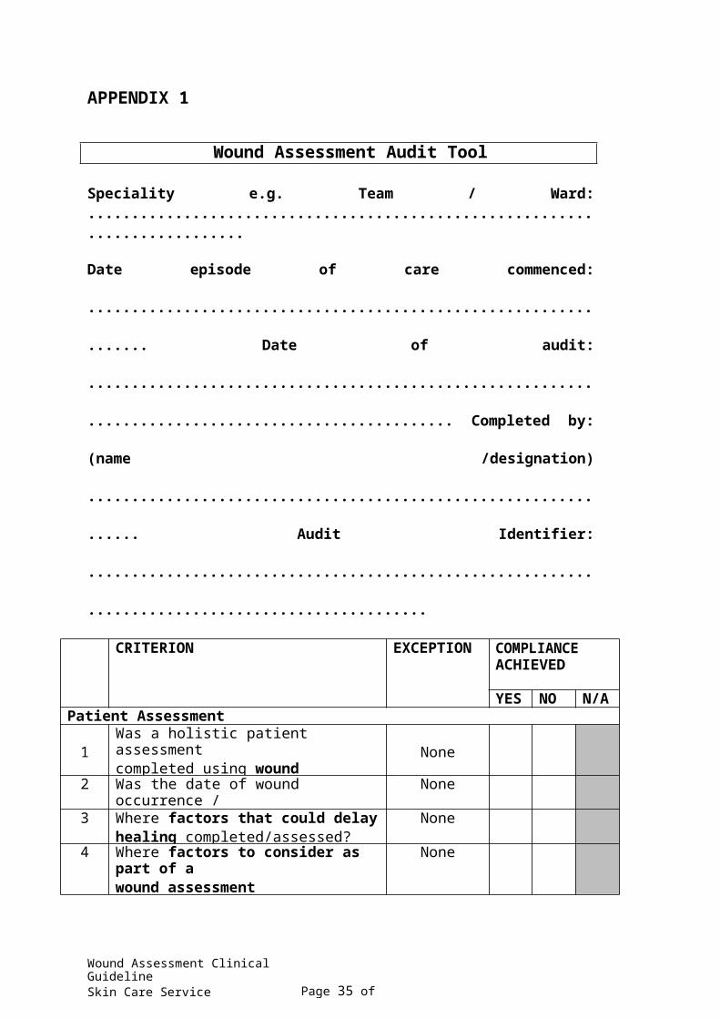
APPENDIX 1
Wound Assessment Audit Tool
Speciality e.g. Team / Ward: ............................................................................
Date episode of care commenced: .................................................................
Date of audit: ....................................................................................................
Completed by: (name /designation) ................................................................
Audit Identifier: .................................................................................................
CRITERION EXCEPTION COMPLIANCE ACHIEVED
YES NO N/APatient Assessment
1Was a holistic patient assessmentcompleted using wound assessment / care form or other systems?
None
2 Was the date of wound occurrence /duration has been recorded?
None
3 Where factors that could delayhealing completed/assessed?
None
4 Where factors to consider as part of awound assessment completed / assessed?*
None
5 *If an issue was identified eg wound pain or nutritional problems was there evidence that this was sufficientlyaddressed within the patient records?
No issues identified
Specific assessment of the wound6 Was the type / cause of wound been
identified (e.g. trauma / surgical)?None
7 Was the site of the wound beenidentified?
None
8 Is there documented evidence that thewound has been mapped
A valid reasonis documented for not mapping the wound
9 Has the dimensions of the wound beenrecorded (length / width / depth)?
None
10 Is there documented evidence that thewound was assessed for exudate levels?
None
11 Is there documented evidence that the wound has been assessed for any
None

Wound Assessment Clinical GuidelineSkin Care Service January 2016
Page 26 of 36
12 Is there documented evidence that thecondition of the surrounding skin has been recorded?
None
CRITERION EXCEPTION COMPLIANCEACHIEVED
YES NO N/A13 Is there documented evidence that the
% tissue type in the wound bed has been
None
14 Is there documented evidence that the patient and the wound have been assessed for clinical signs of infection?
None
If there is documented evidence that clinical signs of infection are presentplease also complete infected wound audit tool.Communication15 Is there documented evidence that the
risks, benefits and alternative treatments have been discussed with the patient?
None
16 Is there documented evidence thatconsent to treatment has been obtained?
None
Care Planning and Evaluation17 Is there documented evidence that the
wound management plan (e.g. wound dressing selection) has been recorded
None
18 Was the wound dressing used in line withLCH formulary? *
Clinical rationale
provided fornon formulary prescribing*
*Identify which non formulary productswhere used.(if applicable)
19 Did the wound dressing selection appearappropriate for the clinical characteristics of the wound? if NO provide details below*
None
*20 Is there documented evidence that a
wound care plan has been recorded with review date identified?
Where onlyone visit was
required21 Did the frequency of dressing change
appear sufficient for clinical need?Where only
one visit was required
22 Is there documented evidence that the expected result of wound care plan (egwound debridement / granulation etc) has been identified?
Where only one visit
wasrequired
23 Is there documented evidence that the wound care plan has been reviewed atidentified review dates?
Where only one visit
wasrequired

Wound Assessment Clinical Guideline Skin Care ServiceJanuary 2016 Page 20 of 36
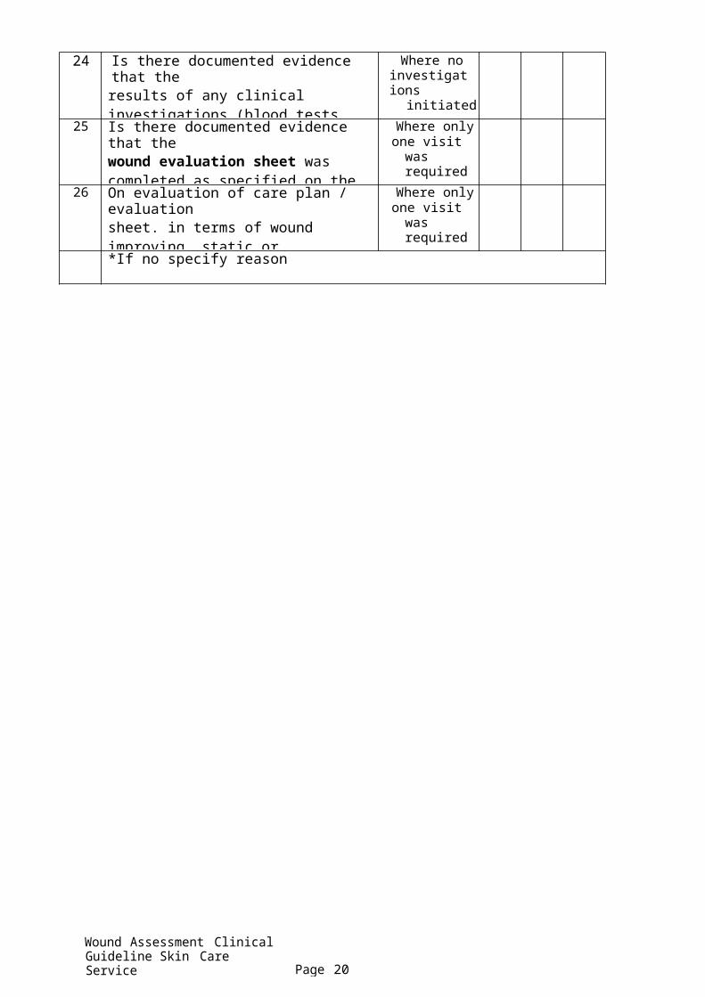
24 Is there documented evidence that theresults of any clinical investigations (blood tests, wound swabs) have been followed up?
Where no investigations
initiated
25 Is there documented evidence that thewound evaluation sheet was completed as specified on the wound care plan or when a change in the wound was noted?
Where onlyone visit was
required
26 On evaluation of care plan / evaluationsheet. in terms of wound improving, static or deterioration was appropriate decision making / actions documented*
Where onlyone visit was
required
*If no specify reason

Appendix 2
Wound Assessment Clinical Guideline Skin Care ServiceJanuary 2016 Page 21 of 36
General Practice Sepsis Screening and

Appendix3
Wound Assessment Clinical Guideline Skin Care ServiceJanuary 2016 Page 22 of 36
THE UK SEPSIS TRUST
Toolkit: General Practice management of Sepsis
This dinical toolkit has dQwloped in partn..-ship with thQ Royal Colk>gQ of General Practitione-rs. It is designed to provide operational solutions to the complexitiQS challenging the reliable ide-ntification and management of sepsis in the primary care setting, and complements dinical toolkits d4?Signed for other clinical are?aS•.
Sepsis is a medical emergency. It is responsible for 37,000 deaths annually in the U nited Ki ngdom and vere sepsis has a five fold higher mortality than ITEH I or stroke. The reliable re<ognition of sepsis is the responsibility of all heal th professional!. The campaign in $e<Ondary care has increased awareness and helped to structu re the management of sepsis once the patient reaches hospital. However, it is essential that psis is re<ognized early for the patient to reach hospital soon enough to avoid serious complication or death . This documen t provides primary care clinicians with a toolkit for managing at risk of psis. I t is part of a wider campaign to increase patient awareness of the signs of sepsis and to mist other pr hospital rvices in the task of early re<ognition.
There are significant challenges and barrien to reliable psis identification in a Primary Care tting. Sepsis is a complex condition and its presentation variable. General Practitioners will be experienced in assessing need for hospital assenment in patients with probable lf·limit ing infe<tion: it is not practicable to expect differentiation between uncomplicated viral and bacterial illness in all cases. Patient! who are obviously critically ill are likely to be identified without the need for new effort!. However, there are some patients with severe sepsis with less immediately obvioll! signs of critical illness. lome of this group m ight be identif.ed earlier with greater awarenen and targeted clinical assessment
This toolkit aims to make GPs and other primary care clinicians familiar with the significant morbidity and mortality associated with severe sepsis and to structure their knowledge and skills so that they can recognize the condition earlier. It advises on spe<ific safety netting in patient! pre nting with signs and symptoms of infection and addresses the need to work collaboratively with health profenional! in other clinical areas to ensure that appropriate further assenment is undertaken and time·critical care is delivered rapidly when ne<essary.
This toolkit is compatible with international guidelines on sepsis managemen with the Department of Health's document 'Start Smart·then focus', and with guidance on infection management in primary care issued by the Health Prote<tion Agency.
UKST PRIMARY CARE TOOLKIT2014 j 1

Wound Assessment Clinical GuidelineSkin Care Service January 2016 Page 23 of
36
Background:
SQ.psis is defined as the body's response to an inf ion . Conse-nsus dfinitions1
dQSCI"ib<> th spons as th Syst mic Inflammatory !Wsponse Syndrom {'SIRS',Box 1). SIRS is not spECific to sepsis:it can bEl caused by non-infQCC.iw conditions such as pancreatitits, trauma and bums. WhEn Sl RS is felt to arise in r4?Sp0nse to a new infECtion, this is sepsis. Du to th lack of sprolicity of SIRS,fgw data ar availabl on ttM? prQvalence of sepsis prior to its progression to severe SQ-psis.
Severe psis occurs when sepsis gives rise to organ dysfunction. Criteria for the idMtification of SE-vere se-psis are given be-low in Box 2. howe'Wor these are focused on a hospital tting and d mand full laboratory sorvic with rapid liv"')' of r sults. These criteria are less appropriate to those charged with ide-ntifying acute Illness in th community. In or r to addr ss this gap, th UK psis Trust has vel th novel pt of 'R Flag psis'.Using abnormal physiology rather than waiting for lab r_,lts, R Flag psis is dQSCI"i in d tail b<>low. From an operational peorspectiw- in Primary Care-, this should be- consider synonymous with severeS-Eopsis .
Figure I: Relationship between SIRS, Infection,Sepsis and Red Flag Sepsis
SIRS can be caused bv multiple triuersSIRSdu t to infettion = StpSiS
Sepsl:$ + Of831'1 dydu!'lt:tfon = Red Fl g 5e:pSi$ ($e e sepi'or.c:e tens tonfltmin hospil31)5ept1c sho<kIs a subset of sevete sepsis
The overall mortality rate for patients admitted with seve sepsis is 35%approximat ly 5 ti s highr than for ST lvation myocardial infarction and strokand is r sponsibl for approximately 37,000 d ths and I00,000 hospital admissions in th Unit Kingdom {UK) r -yW. Th majority of pisod aris from community-acquirKI, rathe-r than healthcare-assodate?d, infection . Though evide-nce is
2 IUKST PRIMARY CARE TOOLKIT 2014

Wound Assessment Clinical GuidelineSkin Care Service January 2016 Page 24 of 36
.
.
focused on th hospital SQtting, data from patients presenting to Emergency Departments (EDs) indicates that severe sepsis is prwalent in the community.
In the United States, the number of pati<>nts transported to hospital by Em<>rg<>ncy MEdical Servic.Qs with severe sepsis now oumumbe-rs those with heart attack or stroke'. In 2007 in the UK severe sepsis was found to account for 12% of early inpatient aths after ED admission: this is likely to haVQ an underestimat du to a furthe-r 26% of dQaths coded as of respiratory caus". Hospitalizations for th condition ha'VQ more- than doubled aver th last I 0 yearsS-.6
$Qvere sepsis is a time-critical condition. In th most SQvere caSQS, septic shock, for very hour that appropriate antibiotic administration is delayed, there is an 8% inere-as in mortality7 ThQ Sepsis Six is an initial resuscitation bundl dQsigned to offer basic intervention within th first hour. In a prospective observational study across a district general hospital, it was ind<>pendently associated with survival suggesting that,if it alon wer r4?Sp0nsibl for outcome differi?OCC?S, the numbe-r nto tr"Qat(NNl) to preV<>nt one death is 4.68. This compares to an NNT of 42 for Aspirin in major heart attack and 45-90 for PCI in ST elevation myocardial infarction.
Sepsis is poorly recognized and treated. A 24-month , large scale prospective improveme-nt programm across 30 countrie?S measuring the delivery of the Severe Sepsis RC?Suscitation Bundle showed compliance rising from I0 to just 2 1% in selfselected centres'. More reoently in 20 13 in the UK, the College of Em<>rg<>ncy Medicine (CEM) audited performanoe against self-imposed standards for the management of severe sepsis and septic shock and identified similarly concerningresults, with antibiotics administerl?d in only 33% of patie-nts within the first hour from time of arrival in the ED". Developing a whole-system solution akin to that for chest pain and strok is likely to rais th profil of thQ patient with sepsis, encourage hospitals to respond robustly, and signifocantly reduce variation and time to therapy.
Fixing our healt system's response to sepsis will not be easy. To do so will demand that all Malth professionals invotv4?d in the patient's journey are working to thQ same end. This clinical toolkit will complement those for othe-r community-basedhealthcare settings, including for residential care facilities, NHS Pathways and II I ,and ambulance services.
Professional responsibility & accountability
NHS England has established s<>psis as a future indicator in both Domains I and 5 of th National Outcome?S Fram work, and issued a 'stagQ 2 alert' on sepsis in September 20 14. This communication to all NHS Chief Executives and Regional Medical Directors established sepsis as a clinical priority for the NHS. It signposted todinical toolkits such as this on . to l?ducation programme?S, exampiQs of good practice , and other available resouroes. NHS England is working with the UK Sepsis Trust and profC?SSional body stakeholders to idQntify and accrMit xemplar acute CE-ntres, ambulance services and primary facilities from which others can IC?am.
UKST PRIMARY CARE TOOLKIT2014 j 3

Wound Assessment Clinical GuidelineSkin Care Service January 2016 Page 25 of 36
In r report of Soptom r 2013 entided 'A Time to Act', the Parliamentary and Health Service Ombudsman called upon the NHS and the Department of Health to act rapidly to rt"duce unnEC.QSS3ry dQaths from se-psis. As a direct result of this work. NICE will produce a clinical guideline and Quality Standard against sepsis.
We will learn valuable l ons from the re-port arising from the recent SUI'VQY on sepsis oonducted by the National Confidential Enquiry into Patient Outcome and Death (NCEPOD). Though retrospective and narrative, this report will analyse how many patie-nts arriving in hospital with sepsis had previously consultro ir General Practitioner. and the outcome of consultation. Until that time, it is the r ponsibility of those commissioning servicQS from, dQSigning clinical systems for. and working within primary care that their efforts focus on early r ition induding through the use of safety OQtting, and urge-nt inte-rvention using existing consensus guideline?S from chQ UK Sepsis Trust and Surviving Sepsis Campaign in arQaS whe- transit times may prolonged.
Delivering Excellent Sepsis Care:
International consensus definitions require adaptation for use outside acute hospitals
The following section dC?SCribQS internationalconsensus definitions in the recognition of sepsis. They inclu for completQOQss' sake. but are in the main not relevant in the Primary Care. SC?tting: the definitions have a hospital focus.
Sepsis arises when the body's rQSponse to infection causQS systQ01ic effQCCS that manifest as two or more of t Systemic Inflammatory Response Syndrome (SIRS) criteria (Box I) triggE!f'ed by a new infection'. Some patients will develop end-organ dysfunction, dMoting severe sepsis. Criteria for the identification of severe- sepsis (ot r than septic shock) are not relevant in t absence of full laboratory services, soare omitted here but can bQ reviQwed in the consensus definitions paper' or inhospital-focused toolkits. For Primary Care, the novel concept of Red Flag Sepsis (see below) will be used as an ope-rational solution- it should be considered synonymous with severe- SC?psis.
Septic shock is a subset of se re sepsis, which strictly speaking (according to international consensus definition) is identified by SC?psis with hypopeorfusion r4?Sistant to fluid therapy (Box 2). The General PractitionE!f' should assume septic shock is pre?S4?nt in any patient with a clinical suspicion of infection, the presMce of SIRS, and eithe-r hypotension or (if available) an elevated serum lactate abow 4 mmoVI.
ltQ01S in black font are- available in all. or most, Primary Care settings. Criteria in orange font (white blood cell count , lactate) are worthy of evaluation for point-of care testing to increase reliability of recognition of sepsis.
4 IUKST PRIMARY CARE TOOLKIT2014

Wound Assessment Clinical Guideline Skin Care ServiceJanuary 2016 Page 26 of 36
Box I : Systemic Inflammatory Response Syndrome (SI RS):
SIRS is present if there are at least 2 of:
Temperature >38.3 or <36.0°C N ew confusion/drowsiness
Pulse > 90/min IIBC 2 01 4.G JIL
RR >10/min Blood glucose >7.7 mmoi!L (not if diabetic)
Box 2 : Defining the severity of sepsis
Severity Definition Group mortalityU ncomplicare4 sepsis
SIRS + presum ed or confirmed infection 10%
Severesepsis Sepsis + oneor more organ dysfunction criteria(other than shock)
JS%
Septic shock Sepsis + shock"' 50%
*Shock criteria:- lactate >4mmolj1 at any time point- Hypotension persisting after JOml/kg intravenous flu id,defined as Systolic Blood
Pressu re <90mmHg, Hean Blood Pressu re <65mmHg,or a fall of >40mmHg fromthe patient's usual Systolic Blood Pressure
UKST PRIMARY CARE TOOLKIT 2014 j 5

Describing the solutions -how can we be good at managing sepsis?
I) Early Recognition:
In Primary Care, the lad< of laboratory SErvices or point of care tests limit our abihy to distinguish between sepsis, s<!'lere sepsis and septic shock (according to intN!lational defmitK:Ins} in many c:ases. Conscious of the challenges this presents in operationalizilg care pathways outside hospitals, the UK Sepsis Trust has developed the concept of 'Red Rag Sepsis', based upon criteria within the National Early Warning Score (NEWS).
Sepsis is identified through the presence of SIRS (Box I) in the contQXt of a dinical suspicion of infection. Pathogens triggering sepsis are almost exclusM>Iy bacteria, and the list of causative inf0Ctions- in dea-easing order of frequency, respiratOJY. abdominal urinary and skin and soft tissues- reflects this.
In high-risk patients, fungi may trigger sepsis, and there are features of certain viral infections (e.g. HI N I, Ebola) that can mimic sepsis. A full description is outside the scope of this document, but General Practitioners and others working in Primary Care should be wary of these potential confounders particularly during a pandemic.
A high degree of vigilance is required for early identification of the septic pationt. In the primary care setting. where perceived infection is one of the most oommon reasons for presentation, the dinicaJ acumen of the General Practitioner is essential in determining which patients to evaluate for sepsis.
Box 3: Suggested clinical indications for undertaking evaluation for sepsis
UndertakQ clinK:al evaluation for S9psis in patients:
with clinical evidence of systemic infection (such as recont history of fever)in whom you are oonsidNing antibiotic prescription or stewardship discussionyou suspect to have "flu'"you suspect to have gastroenteritiswho are obviousty unwell without oiQaf causewho are eklerly or immunosuppressed and present with signs of infKtionwho have deteriorated on antibiotic therapy
As w l as thQ gQOeral impression at the time of initial assessment, the presence of abnormal observations should be enough to initi:atQ evaluation for sepsis. Some GPs are now evaluating the use of Early Warning Scoros {such as NEWS, see UKST appendix 3- NEWS) and must decide the lowest score that will trigger evaluation for SEpsis. As yet unpubUshed data suggests that 94% of patients who wore later found
6 j UKST PRIMARY CARE TOOLKIT 2014
Wound Assessment Clinical Guideline Skin Care ServiceJanuary 2016 Page 27 of 36

to have severe sepsis or septic shock presented to Emergency Departments with aNEWS soore of 3or higher.
The use of a two-part screening process to detennine severity
Sepsis screening should be done as a two-part process; screening for SIRS followed by, where sepsis is identifK>d, screening for Red Flag Sepsis (Box 4). NHS England and the UK Sepsis Trust recommend that patients with Red Rag Sepsis be transferred immediately for hospital assessment.
Box 4. SlRS screening and evaluation for Red Flag Sepsis
a. Screening for SIRS
SIRS is oonfirmed ifAN( lWO of the following are present:
Immediate)>- New onset of Confusion or Ntered Mental State)>- Tomperature >38.3 or <36 degrees Celsius)- Heart Rate >90 beats pN minute•)>- Respiratory Rate (counted over 60 seconds) >20 breaths per minute'
POCT (oornmorq available))>- Blood Glucose> 7.7mmoi/L in the absence of known diabetes
POCT (no< yet widoly available):;.. WCC> I2or<4xi0'/L
b. Evaluation for Red Flag Sepsis
Aa imrnediatoly ifAN( ONE of the following are present:)>- Systolic BP <90mmHg (or >40mmHg fall from basoline)•)- Heart rate > 130 per minute)>- Oxygen saturations <91% §)>- Respiratory rate >25 per minute§)>- Responds only to voice or pain/ unresponsive
POCT (no< yet widoly available):;.. Lactate >2.OmmoV
•Values are guides.. lnterprel observations in the context of the normal physiology for the patienl. For example. in a young man who n.ns 3 times a week and has a baseline pulse of 56 a heart rate of 90 is very sv-r.cantfy raised. whereas it hi be relativety normal for an older patient with lritralregurgitation. SWnilarty for an older person,a BP of I06160 is likety to be kit: lower than their baselineBP, whereas for an athlete a systolic of <90 may be perfectly normal.
t Some patients with ctronic pu1monary tisease may display low oxygen saturations and elevated respiratory rates 'norrnaly'. Consider whether values are abnormal for the individual patient
UKST PRIMARY CARE TOOLKIT 2014 j 7

Wound Assessment Clinical Guideline Skin Care ServiceJanuary 2016 Page 2B of 36
Wound Assessment Clinical GuidelineSkin Care Service January 2016 Page 2 of 36
Sepsis screening should th£-re.fo comme-nce with basic observations to indude measureme-nt of he-art rate. re-spiratory rate, blood pre-ssure, te-mperature and conscious lewl.
Whe: resources pennit, the measureme-nt of oxyge-n saturations using a pu lse oxi TK"te-r should be performt"d in addition to basic physiological as:sQSsment. General Practitioners should evaluate whether there is utility in providing point of care testing for lactate and white blood cell count. These syste-ms are now robust, and may assist in rec::ognition of se-psis and Rt"d Flag Sis and in transfer decision planning. Nonnal resu lts are likely to reassure and reinforce dec.isions to manage in the community. and abnormal resu lts may identify deterioration before the dinical picture becomes evide-nt. This approach will be of panicular relevanc.Q in re-mote locations where trans.it times to hospital can be prolonged.
The use of Point of Care Testing (POCT)
White blood cell count forms part of conse-nsus definitions for SIRS. and diffe-re-ntial white aoll count can help distinguish betw'*n infECtion of viral or bacterial origin, and non-infeoctive cauSQS of inflammation. POCT devia?s using optical t£"Chnology, wh ich re-quire minimal or no mainte-nance. are now available. Studies are f'K'Eided to help evaluate the clinical utility of these devices in primary care in dec.ision-making regarding hospital as:sQSSment and antimicrobial prescription .
The lactate level in sepsis is highly predictive of death" (s'* Box 5) and poor outcomes and, whe-n initially elevated, the degrEoe of reduction following resuscitation ('lactate clearance') predicts survival". A significant proportion of patients with sepsis who have nonnal blood prC?SSure have e-levated serum lactate and outcomes for these patients with 'cryptic shock• are as poor as for thosQ with overt se-ptic shock13•
Commercial POCT devi«>s, as used by athletes in performanao training, are readily available and have been validated against benchtop assays.
Box 5: The relationship oflactate levelin sepsis to mortality
lactate Mor-t al ity
<2 1 5%
2-4 25%
>4 38%
From: Tr..e<i;)l( S,Ddlirlaer RP,Ctord<:y ME,Atnold' RC. Schorr C,Miordl 8.rtJ!lrm-Mive Uri:M«/2007.33(6):9
8 IUKST PRIMARY CARE TOOLKIT2014

Wound Assessment Clinical GuidelineSkin Care Service January 2016 Page 3 of 36
2. Urgent Action
The key immediate int ntions that increase survival are de?Sai in a bundle termEd t Sepsis Six (Box 6). This bund!Q has bQQn shown to bQ assodatE<I with significant mortality rt"ductions whe-n appiK-d within the first hour".
Box 6: The Sepsis Six (Sou e::hm:lf:e-21i:uua.o?)
I. Administer high-flow oxygen2. Take blood rultures and consider infective source3. Administer intravenous antibiotics4. Give intravenous fluid resuscitation5. Check haemoglobin and serial lactates6. Commence hourly urine output measurement
While fE w Ge-neral Practiti rs will haw the rC?SOurCQS to provide much of this bundle of care, it is included to illustrate the time-critical nature of Red Flag Sepsis and t nmfor collaborativQ care pathways.
Action for pa< ents with REd Flag SQps s
For patiMts identifiEd with REd Flag SQpsis, immEdiate arrangement should bQ made for urge-nt transfer for hospital ass me-nt. This should bQ by 'blue light' ambulance with a Paramedic CrQW. A brief. de?ar handowr should accompany the patiEmt to include observations. any relevant mEdical history and antibiotic history including al!Qrgies.
Whe-rQ n sources pennit, Ge-neral Practitioners should initiate high flow oxygenthe-rapy. Patie-nts with sepsis are exempt from British Thoracic Sodety guidelin for the administration of oxygM to acutely ill adults as me pathophysiology of sepsis is such mat organs bQcome critically hypoxic. Hypoxia will kill bQfore hypercapniaCurr t ry sugg ts that hypercapnia occurring in n spot'lSQ to oxyge-n the-rapy inpatients with pulmonary disf?aSe is due not to the unproven theory of a loss of'hypoxic drivQ' but to changes in ventilation-pe-rfusion matching. Oxygen will not cause sudd«a apnoea in such patients. Therefore whe-re patients are known to have moderate to severe pulmonary disease (and where available). we rQComme-nd that oxyge-n be- administered to maintain targe-t oxygen saturations above 92%.
Particularly in re-mote areas, consideration should be- given to the de-liv of othe-r ek-me-nts of the Sepsis Six.
Once a patient arrives in hospital, me All Party Parliamentary Group (APPG) onse-psis has made a recomme-ndation that organizations should give 'consideration to
14the developme-nt of S is Teams' . Comparisons with hC?a.rt attack and stroke.
UKST PRIMARY CARE TOOLKIT 2014 j 9

Wound Assessment Clinical GuidelineSkin Care Service January 2016 Page 31 of
36
where teams arQ availabiQ to bQ mobilizro whQn prMospitalse-rviCQs or Primary CarQ.pre?-alert a suspearo casQ.would makQ this see-m obvious.
It is vital that patie-nts with SQVNQ SQPSis should bQ rQvtewro at thQ QMiiQSt opportunity by thQ most nior availabiQ doctor. This should includQ in Primary CarQ if a doctor in training identifiQS RQd Flag SQPsis, a senior doctor should reviQw thQ findings immQdiatQiy or as soon as practicabiQ.
Many pati nts with sepsis will have multiple co-morbidities, and may be elderly or frail. !For such patients, dQOsions should bQ taken at se-nior IQWI(in consultation with the pati nt and their family as appropriate) ding the appropriateness of QScalation of carQ to hospital.
Actionfor pat Qnts with psis without s gns of Red Flag $Qpsis
Wh re no 'Red Flag' signs are id ntified, dinical judgment will det rmine appropriateactiom.
Patients with as yet uncomplicated SQPSis can deterioratQ rapidly. Fit adults and young peoplecan compensate physiologically in the early stages of psis with minimal chartgQs in their ob rvations ,right up to thQ point where shock ensue?S, and catQ.fulconsidQI"3tion should bQ taken as to thQ nQC?d for a requQst for hospital assC?SSment . Those over 80, patients on chQ01ot:herapy or immunothQrapy « thosQ unwell despitQ antibiotic trQatmoot arQ all high risk groups. If the patient is not to bQ rQferrQd for hospital 3SSQSS01C?nt, therQ must bQ adequatQ safQty nQtting. OnQ QXampiQ of a safQty netting tool is in the UK Sepsis Trust's 'Symptom Checker Cards' produced in collaboration with a survivor group. Any safety netting advice given should be clearly documentro in d\Q patie-nt's notC?S,together with ob rvations and antimicrobial therapy offerro. Itis good practiCQ to agree- a planned OQXt day rQvi£-w assQSsment in any patient with s psis (SIRS, no Red Flag} managed in the community, togethr withan invitation for open SQif-refQITalshould the patient d«erioratQ « bQ concerned.
1IUKST PRIMARY CARE TOOLKIT2014

Wound Assessment Clinical GuidelineSkin Care Service January 2016 Page 32 of
36
Suggested clinical guidelines for the management of patients attending with sepsis in Primary Care
St>psis (no Rt>d Flag signs):
A docuiTK'ntro eX-cision whe-the-r to manage pati£-nt in the community or re.ferto hospitalDiscussion with a nior doaor (where initial asse?SSmeot has n by trainEoe} within 30 minute?S of diagnosisA full set of observations including heart rate, respiratory rate, blood prwsure.te-mperature , conscious level recorded and documentedIf to bQ treated in the community, safety OQtting advice offErEd and docume-ntedIf to bQ treated in the community, arrangements to bQ madQ for review within24 hoursIf to bQ referrE<I for hospital asSE:\ssment. handowr including rQievant clinicalhistory and antibiotic history induding all<>rgios to b<? provided
'Rt>d Ffag' st>psis p<>nding confirmatory costs:
Immediate discussion with a senior doctor (where initial assQSsme-nt has beenby train<?<?)Immediate requ<?St for 999 Ambulance with Paramedic crewHandove-r including rel t clinical history and antibiotic history indudingallergi<?S to b<? providedWhere- resourCQs available, administ£-r oxygen therapyWhere- transfer tim£-s may bQ prolonged , consider need for intrave-nous antibiotics and fluid the-rapy if available
UKST PRIMARY CARE TOOLKIT2014 j 1

Wound Assessment Clinical GuidelineSkin Care Service January 2016 Page 33 of
36
Exemplar Standards for Primary Care management of Sepsis
The standards below are those which have been identified by the UK S<>psis Trust and the APPG for sepsis as important in the management of s<>psis with specifoc relevancQ to Primary Care. They are the 'Exemplar Standards' which organizations should aspire to deliver. Achieving these standards will place a Primary Care organization IIon the road to the provision of QXCC?Ik-nt psis .
I. Oear written gu dance,policies and dnical pathways to be in place for the
rf?Cognition and management of SQpsis and RQC! Flag Sepsis.
2. Oear written criteria for which patients shou ld be screen for sepsis.
3. According to k>cal criteria for screening, I00% of patients satisfying criteria to
have, as a minimu m, heart rate, respiratory rate.blood pressun.
conscious level, oxygen sa[Urations and blood gluco m ur and r
ded {unless precluded by equ ipment failure)
4. Risk assessment to be u ndertaken and maintain rE-garding the need for
Poim of Care Testing for lactate and wh te blood cQIIcount
5. I 00% of patients identified with Red Flag Sepsis to be transported for hospital
as ssmem unless limitations of trQatmQflt agrQQd
6. Oear written and ve,rbal handov rs to accompany all patients
rferred for hospital assessm nt
7. Oxygen therapy to be available and considered for all patients with Red Flag
Sepsis
8. Where transit times to hospital ar routin ly in excess of 60 minutes,
risk as ssment to bQ unden:aken and maintained regarding th nm
for administration of antibiotics and intrav n oos flu ids
9. Documented decision to treat in the comm unity or transfe:r to hospital in
I 00% of pati nts with s psis withou t RQCI Flag signs
I 0.Where patients with sepsiswithout R Rag signs are to be managed in th
community.docu mntQd sa_f ty n tting advi and reviw plans to be in
placQ for all patie:nts.
1IUKST PRIMARY CARE TOOLKIT 2014

Wound Assessment Clinical GuidelineSkin Care Service January 2016 Page 34 of 36
References
I. Levy MM, Fink MP, Marshall JC et al. 200 I SCCM/ESICM/ACCP/ATS/SIS intNnational psis definitions confe-re-nce. lntensivQ Care MMicine 2003; 29: 53
2. Daniels R. Surviving the first hours in Sepsis:getting the basics right (an lntensMst's perspectiw). Joumal of Antimicrobial Chemotherapy 20 I I; 66(Suppl iQ: 1 1-23
3. Seymour ON, Rea TO, Kahn )M, et al. Severe Sepsis in Pre-Hospital Emergency Care: Analysis of lndd. Care, and Outcome.American Journal of Respiratory and Critical Care Medicine 2012; 186(12): 1264-1271
4. Nafsi T, Russell R, Reid CM, et al. Audit of deaths less than a we<>k after admission through an emergency department: how aocurate was
the ED diagnosis and were any deaths preventable? Emergency Medidne )oumal2007; 24: 691 95
5. Vincent JL. Sakr Y. Sprung CL e.t al. Sepsis in European intensive care units:results of the SOAP study. Critical Care Medicine 2006; 34:344-53
6. Hall M), Williams SN, O.Frances C),et al.:Inpatient care for septicenn ia orse-psis: Achallenge for patients and hospitals. NCHS data brio>f HyattsVille, MD:National C..nter for Health Statistics 20 I I ; 62
7. Kumar A, Roberts D.Wood KE et al. Duration of hypotension prior to initiation of Q.ffectivQ antimicrobial therapy is the critical determinant of survival in human septic shock. Critical Care Medicine 2006; 34: 1589-96
8. Daniels R. Nud>eam I, McNamara G et al. The se-psis six and the SQVNQ se-psis uscitation bundle-: a prospectivQ observational cohort study. EITK'rgency Medidne )oumal 20 II; 28(6): 459-460
9. Levy MM, Dellinger RP, Townsend SR et al. The Surviving Sepsis Campaign: ults of an international guide-line-- performance improvement program targo>ting severe sepsis. Critical Care Medidne 20 I 0; 38(2):3 67-74
I 0.Available via the College of Emergency Medidne website at httpJ/www.collemergencyrned.ac .uk/ShopFioor/Ci i nicai% 20Au dit!Previous%20Aud its last accessed August 2014
I I Tn..ciakS, Chansky ME, Delling >< PR et al. Operationalizing the use of serum lactate me?aSure-ment for identifying high risk of dQath in a dinical practiCQ algorithm for suspearo severe sepsis. AcadEfTl ic Eme-rge-ncy Medicine 2006; 13: 150-1
UKST PRIMARY CARE TOOLKIT2014 j 1

Wound Assessment Clinical GuidelineSkin Care Service January 2016
Page 35 of 36
Acrobat Document

Wound Assessment Clinical GuidelineSkin Care Service January 2016
Page 36 of 36
Appendix 4
Chronic Wound Pathway
Does the patient have a chronic wound (present forlonger than 4 weeks) which is No
displaying signs of delayed healing?
Continue with current care plan and review at planned regular intervals.
Yes
Is the wound a pressure ulcer?
Yes Refer to P r e ss u r e Ul c e r P a t h w a y .
No
Is the wound a leg ulcer?
Yes
Refer toLo w e r L i m b P a t h w a y
No If you suspect there is aninfection, refer to I n f e c t e d Wound Algorithm
Have you reviewed all factorsassociated with delayed Nohealing and formulatedrelevant care plans to manage them?
Review all factors associated with delayed healing and incorporate into existing management/care plans.
If you have reviewed all factors causing delayed healing and you still require support, refer toSkin Care Service.Examples of wounds that may require referral:
• Not responding to antibiotics / anti-microbial dressing• Not responding to management plan• Not healing within expected time frame

Wound Assessment Clinical GuidelineSkin Care Service January 2016
Page 37 of 36
• Non-adherence to recommended treatment plan