microstructural characterisation of fatigue crack growth
TRANSCRIPT

University of Birmingham
Microstructural characterisation of fatigue crackgrowth fracture surfaces of lamellarTi45Al2Mn2Nb1BYang, J.; Li, H.; Hu, D.; Dixon, M.
DOI:10.1016/j.intermet.2013.10.011
License:Creative Commons: Attribution (CC BY)
Document VersionPublisher's PDF, also known as Version of record
Citation for published version (Harvard):Yang, J, Li, H, Hu, D & Dixon, M 2014, 'Microstructural characterisation of fatigue crack growth fracture surfacesof lamellar Ti45Al2Mn2Nb1B', Intermetallics, vol. 45, pp. 89-95. https://doi.org/10.1016/j.intermet.2013.10.011
Link to publication on Research at Birmingham portal
Publisher Rights Statement:Eligibility for repository : checked 03/06/2014
General rightsUnless a licence is specified above, all rights (including copyright and moral rights) in this document are retained by the authors and/or thecopyright holders. The express permission of the copyright holder must be obtained for any use of this material other than for purposespermitted by law.
•Users may freely distribute the URL that is used to identify this publication.•Users may download and/or print one copy of the publication from the University of Birmingham research portal for the purpose of privatestudy or non-commercial research.•User may use extracts from the document in line with the concept of ‘fair dealing’ under the Copyright, Designs and Patents Act 1988 (?)•Users may not further distribute the material nor use it for the purposes of commercial gain.
Where a licence is displayed above, please note the terms and conditions of the licence govern your use of this document.
When citing, please reference the published version.
Take down policyWhile the University of Birmingham exercises care and attention in making items available there are rare occasions when an item has beenuploaded in error or has been deemed to be commercially or otherwise sensitive.
If you believe that this is the case for this document, please contact [email protected] providing details and we will remove access tothe work immediately and investigate.
Download date: 20. Mar. 2022
lable at ScienceDirect
Intermetallics 45 (2014) 89e95
Contents lists avai
Intermetallics
journal homepage: www.elsevier .com/locate/ intermet
Microstructural characterisation of fatigue crack growth fracturesurfaces of lamellar Ti45Al2Mn2Nb1B
J. Yang a,b, H. Li b, D. Hu a,*, M. Dixon c
a Interdisciplinary Research Centre in Materials, University of Birmingham, Edgbaston, Birmingham B15 2TT, UKb School of Metallurgy and Materials Science, University of Birmingham, Edgbaston, Birmingham B15 2TT, UKcRolls-Royce Plc, Derby DE24 8BJ, UK
a r t i c l e i n f o
Article history:Received 16 April 2013Received in revised form10 October 2013Accepted 17 October 2013Available online 2 November 2013
Keywords:A. Titanium aluminides, based on TiAlB. Fatigue resistance and crack growthB. Fracture modeD. Microstructure (as-cast)
* Corresponding author. Tel.: þ44 1214147840.E-mail address: [email protected] (D. Hu).
0966-9795/$ e see front matter � 2013 Elsevier Ltd.http://dx.doi.org/10.1016/j.intermet.2013.10.011
a b s t r a c t
Investment cast Ti45Al2Mn2Nb1B with fine lamellar microstructures was subject to fatigue crackpropagation testing at 650 �C and a stress ratio of R ¼ 0.1. The fracture surfaces were examined underSEM and the observed features are correlated with both stress intensity range (DK) and lamellarorientation. Translamellar fracture is primary fracture mode for most of the lamellar orientations.Interlamellar fracture is influenced by a combination of the DK and lamellar orientation. At low DK onlythe lamellar colonies with their lamellar interfaces almost perpendicular to the stress axis fractured viainterlamellar fracture mode. At high DK interlamellar fracture can occur in lamellar colonies with anyorientations.
� 2013 Elsevier Ltd. All rights reserved.
1. Introduction
Being potential high performance high temperature structurematerials TiAl-based alloys have been subjected to intensityinvestigation since late 1980s [1e3]. Fatigue crack propagationbehaviour in TiAl alloys attracted immense attention in 1990sowing to the alloys’most likely fatigue fracture critical applicationsin turbines [4e15]. Research activities in this area, alongside withthose in other areas in TiAl alloys, ebbed after 2000, but has revivedrecently to some extent with the success in certification of TiAlcomponents in a commercial aero-engine [16e18]. Over the yearsmany data have been accumulated and the basic understanding onthe microstructure-fatigue crack propagation behaviour relation-ship has been established. The main findings are: a) lamellar TiAlalloys offers better crack propagation resistance than their duplexand equiaxed gamma counterparts [5,6,11,13e15], b) lamellar TiAlalloys have two major fracture modes, interlamellar and trans-lamellar fracture [8], and c) crack propagation resistance is lamellarorientation dependent [7,10]. In spite of those important findingsthere are still many aspects remaining unclear. For example, inpolycrystalline lamellar TiAl alloys under what conditions thespecific fracture mode prevail and what is the micro-mechanism of
All rights reserved.
lamellae fracture. In addition, the characterisation of the fatiguecrack propagation fracture surfaces has been patchy and there wasonly one paper dedicated to fracture surface characterisation [8].Therefore, many features could have been left unnoticed and theymight hold important clues to the micro-mechanism of lamellaefracture during fatigue crack propagation. This paper is aimed atpresenting systematic observations on the fracture surfaces of fine-grained lamellar Ti45Al2Mn2Nb1B subject to crack propagationtesting at 650 �C with the attempt to relate the observed fracturesurface features to the lamellar orientation and stress intensityrange (DK).
2. Experimental
Ti45Al2Mn2Nb1B (at%), was cast into test bars using investmentcasting technique and the test bars were subject to a proprietaryHIP and heat treatment cycle. The actual composition is very closeto the nominal composition with oxygen concentration at about600 wtppm.
The material was machined into testpieces containing a gaugelength of square cross section of 5 � 5 mm2. The gauge length is25 mm. A notch was introduced at one corner of each testpiece to adepth of 0.75 mm as measured from the corner position using finewire (f30 mm) EDM cutting. Many fine cracks were generatedduring the re-cast process of EDM at the notch surface and they canbe 10 micron in depth. The fine cracks form an almost continuous

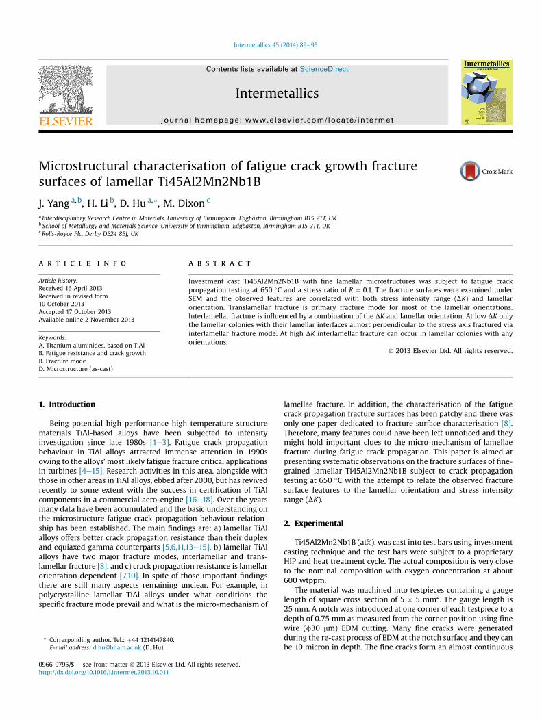
Fig. 1. (a) SEM BSE image showing the lamellar microstructure of investment cast Ti45Al2Mn2Nb1B and (b) the EBSD (0001)a2 pole figure shows the random lamellar orientationdistribution. (c) Fatigue crack propagation curves at 650 �C with R ¼ 0.1.
J. Yang et al. / Intermetallics 45 (2014) 89e9590
network. During testing those with the favourable orientations willpropagate under certain stress conditions. It should be noted herethat using EDM cut instead of precrack is not an established con-ventional method. The feasibility of this method in testing TiAlalloys has been under investigation for a few years and the detailswill be published in a separate paper. Nevertheless, owing to thebrittle nature of the TiAl alloys the measured threshold values arevery close to those using precracking method under the conditionof similar microstructures. Testing to determine the thresholdstress intensity factor and fatigue crack growth rates was con-ducted under constant amplitude load at 650 �C in air. A stress ratio,R, of 0.1 and a frequency of 10 Hz were employed in all tests. Due tothe brittleness and special crack growth characteristics of the ma-terial, the threshold stress intensity factor was measured directlyfrom the EDM sharp notch by step increasing load (stress intensityfactor range, DK) from an initial valuewell below the threshold. Thetotal number of cycles spent at each load level is 5 � 104 cycles atminimum. The load was kept unchanged once consistent crackgrowth was observed and the crack was allowed to grow until apredetermined crack length was reached. A direct current potentialdifference (d.c.p.d.) technique was utilised to automatically moni-toring the extension of the fatigue crack using a calibrated
relationship between crack length and potential drop. After fatiguecrack growth testing, samples were loaded monotonically to failureto allow a rough estimation of the fracture toughness to be made.The fracture surfaces were examined using scanning electron mi-croscopy (SEM).
The microstructure of the alloy is shown in Fig. 1(a) and iscomposed of fine lamellar colonies with an average lamellar col-ony size of about 100 mm determined using linear interceptmethod and a maximum grain size of 240 mm. The lamellar colonyorientation was measured using electron back-scattering diffrac-tion (EBSD) and the (0001) pole figure of the alpha2 phase isshown in Fig. 1(b). Owing to the fact that each lamellar colonydescended from one prior alpha grain, the orientation of each(0001)a2 spot is the orientation of the lamellar interfaces in eachcolony. Thus, it is shown by this pole figure that the lamellarcolonies are randomly oriented. The orientation of the fracturesurface images in this paper is as that the stress axis is perpen-dicular to the images and the overall crack propagation direction isupwards for Figures 3, 5e7.
Fatigue crack growth rate (da/dN) versus stress intensity factorrange (DK) curves of 5 testpieces are shown in Fig. 1(c). At 650 �C inair the threshold stress intensity factor range (DKth) is measured to

Fig. 3. Interlamellar fracture at high DK; (a) 15.3 and (b) 14.1 MPaOm.
J. Yang et al. / Intermetallics 45 (2014) 89e95 91
be between 6 and 7 MPaOm with the R ratio of 0.1. Once thethreshold is exceeded the fatigue crack growth rate increasessharply with the increase of DK demonstrating a cliff edgeappearance of the curves at the near threshold region. This char-acteristic is commonly seen at elevated temperatures in TiAl alloys.
3. Results
3.1. Overview
The overview of the typical fracture surface along the diagonalof a broken testpiece is shown in Fig. 2 with the crack growth di-rection from right to left. Starting from the right edge Fig. 2 shows astraight notch front, a large area of fatigue crack growth and a finalmonotonic tensile fracture area. The boundary between the fatigueand tensile fracture regions can be easily identified under opticalmicroscopy through different shades of discolouration caused byoxidation, which cannot be seen under SEM. However the SEMimages bring out the topological contrast which is very subtle butstill distinguishable for the investigated material. The fatigue re-gion in Fig. 2 spans a DK range from approximately 7 MPaOm rightahead of the EDM notch to 18 MPaOm at the end of fatigue. Twomajor fracture modes, interlamellar fracture and translamellarfracture, have been observed. No striation, which is the signature ofstable fatigue crack growth in ductile materials, can be observed inthe fatigue zone.
3.2. Interlamellar fracture
Interlamellar fracture was found throughout the fracture sur-faces and the facets could have contrast darker or brighter thanother areas in secondary electron SEM images, depending on theirorientations. Those parallel to or having small angles with thefracture plane appear darker whilst those appearing like cliffs in theimages, are often brighter than their surroundings. More facets canbe seen at the left side (high DK region) of the image than at theright side (low DK region) in Fig. 2. The cliff-like facets are difficultto see in lowmagnification SEM images. Fig. 3 shows two examplesof interlamellar fracture surfaces from two lamellar colonies withan angle of about 50e60� off the stress axis.
Pole figure is used to describe the orientation of lamellarcolonies on the fracture surface and the three axes are defined assuch that the centre is the applied stress direction, N, the A axis isat the top of the pole figure for the overall crack propagationdirection and to the right of the pole figure is the B axis, as shownin Fig. 4. The orientation of the colonies is represented by the
Fig. 2. SEM montage showing the fracture surface of a sample tested at 650 �C. The fatigu
poles of their lamellar interface normal in the pole figures. Theorientation of a lamellar colony can be described by two anglesshown in Fig. 4(b) for the pole labelled ‘16.1’. The first is the anglebetween lamellar interface normal and the N axis, here referredto as the ‘Orientation’ for simplicity. The second angle, an acuteangle, is that between the projection of lamellar interface normalonto the fracture plane and the A axis, reflecting the relationbetween the lamellar interfaces and the overall crack propagationdirection. It is referred to as the ‘Sub-Orientation’ in this work.
e crack propagation direction is to the left and the range of the DK is 7e18 MPaOm.

Fig. 4. (a) Definition of pole figure directions with respect to the stress axis and the fracture plane. (b)e(f) Pole figures showing the orientations of interlamellar-cracked coloniesobserved on the fracture surfaces of 5 samples together with their local stress intensity range.
J. Yang et al. / Intermetallics 45 (2014) 89e9592

J. Yang et al. / Intermetallics 45 (2014) 89e95 93
Thus, the two interlamellar cracking facets in Fig. 3 havesimilar Orientations but very different Sub-Orientations. Theformer is nearly facing the overall crack propagation directionand the latter is almost perpendicular to it. The orientation ofinterlamellar-cracked colonies in the fatigue zones was measuredin a SEM using an image rotation method. The sample stage wasrotated to maximise or minimise the shown area of a specificfacet and the orientation of this facet (lamellar interface) can beworked out from the rotations with an accuracy of about 5�. Theresults from 5 samples are shown in pole figures in Fig. 4(bef), inwhich each dot represents the normal of a facet from aninterlamellar-cracked colony. The stress intensity range DK ofeach colony is given next to its pole. It is noted here that thestress intensity range for specific points on the fracture surfaceswas obtained through linear interpolation between two measuredcrack lengths which were determined using d.c.p.d method. Thismethod is unable to guarantee the accuracy to the first decimalpoint of the stress intensity range values. Therefore, the resultsshown here should be treated as qualitative or semi-quantitativeat most.
A few points are highlighted by the pole figures. Firstly thelamellar colonies with Orientations close to 0� can be fractured ininterlamellar fracture mode at both high and low DK levels. Sec-ondly cliff-like interlamellar fracture (the fractured colonies withhigh angle Orientations) only occurred at high DK level and finally,changing the Sub-Orientation while keeping the Orientation con-stant makes almost no difference in interlamellar cracking behav-iour. It can be affirmed that the occurrence and fracture surfacemorphology of interlamellar fracture is related to lamellar orien-tation AND stress intensity range DK. At low DK (up to 9e10 MPaOm, based on the observation of about 10 specimens)interlamellar cracking only occurs in the colonies with theirlamellar interfaces perpendicular to the stress axis with a deviation
Fig. 5. Translamellar fracture at low DK;
up to w30�. Results from all 5 analysed samples show the samecharacteristics although the DK range of each of them is slightlydifferent.
3.3. Translamellar fracture
Translamellar fracture occurred throughout the whole fracturesurfaces and it is the primary fracture mode. It was found in thisstudy that the fracture surface morphology differs with the stressintensity range DK. Fig. 5 shows the translamellar fracture surfacemorphology at low DK (7.5, 6.4 and 6.5 MPaOm for 5a, 5b and 5crespectively). The three colonies have high angle Orientations withdifferent Sub-Orientations (being A, B and AB respectively). Thefracture surfaces are fairly smooth, compared to those observed athigh DK values such as those in Fig. 6, regardless of their Sub-Orientations. There are some fine linear features lying across thelamellae and those within the same lamella are almost parallel toeach other. Those linear features bear no relation with the overallcrack propagation direction and are more likely to be related totheir own crystalline structures [19]. Also it should be noted thatthe lamellar interfaces are intact and free from secondary cracking.
The morphology of the facture surfaces at high DK is verydifferent. Fig. 6 shows two translamellar-cracked colonies at DKvalues of 15.5 and 16.2MPaOm. Again, both of them have high angleOrientation and the one in Fig. 6(a) has w80� Sub-Orientation andthe other is about 30� from the A direction. The fracture surfaces arevery rough. The roughness was caused by the large steps formed bysecondary interlamellar cracking (two deep vertical interfacialcracks in Fig. 6(a)) and small steps within/along each lamella. Thesmall steps were not observed during fracture at lowDK. At high DKinterlamellar fracture occurs simultaneously as the secondarycracking in the lamellar colonies while cracks grow predominantlyin translamellar fracture manner.
(a) 7.5, (b) 6.4, and (c) 6.5 MPaOm.

Fig. 6. Translamellar fracture at high DK; (a)15.5 and (b) 16.2 MPaOm.
J. Yang et al. / Intermetallics 45 (2014) 89e9594
3.4. Other fine features
An interesting observation is the transversal ridges (with respectto the lamellar interface traces) on the translamellar-fracturedcolonies adjacent to interlamellar-fractured colonies. Fig. 7(a)shows an interlamellar-fractured colony right next to a notch and itis surrounded by a few translamellar-fractured colonies. The colonyin the middle has a faceted fracture surface. Some ridges are on thefracture surfaces of those surrounding colonies and they are ar-ranged in a way of radiating out from the facet. Such ridges onfracture surfaces usually are an indication of direction of the actualcrack propagation and the convergent point of the ridges would bethe source of cracking. Thus, it is strongly suggested that in thisimage the colony in the middle (with an Orientation close to 0�)cracked first and then propagated into the surrounding colonies.
Fig. 7(b) shows the ridges on the fracture surface of atranslamellar-cracked colony next to/below an interlamellar-cracked colony. From the direction of the ridges it can be judgedthat the actual crack front moved downward (shown with thewhite arrow) which is opposite to the overall crack propagationdirection. Fig. 7(c) shows a case of crackingwithin a lamellar colonywith an Orientation of about 30�. Cracking within this colonyoccurred on lamellar interfaces at different depths and the cracktorn through the lamellae between them. All the cracked lamellarinterfaces are shown as facets. A high magnification image of thearea just under ‘A’ in Fig. 7(c) is given in Fig. 7(d) where a riverpattern can be seen clearly. This river pattern tells that the local
crack front moved downwards along the white arrow. Therefore inthis colony the crack on lamellar interface A should have beenfractured first and moved onto interfaces at B via tearing throughthe lamellae between A and B. Again, the actual local crack frontmoving direction is different to the overall crack propagationdirection.
4. Discussion
Lamellar TiAl alloys are brittle in nature and a large part of theirshown ductility (up to 2% before brittleeductile transition) isaccompanied by cracks. Unlike in ductile materials where plasticdeformation can be achieved to a large extent before formation ofvoids and cracks, cracking in lamellar TiAl alloys occurs at the verybeginning of plastic deformation. An earlier study revealed that thefirst detected cracking under monotonic tension was at about 2/3e3/4 of the 0.2% proof stress, which is just above the elastic limit [20].This is an intrinsic feature of the lamellar TiAl alloys arising fromboth limited slip systems in the L10 gamma phase and the lowinterface transparency for slip transfer between lamellae [21]. Insuch lamellar structures interfacial cracking is the most effectiveway to accommodate deformation discontinuity from cross-lamellaslips. As a consequence the fracture surface of TiAl alloys commonlycomprises interlamellar facets, typically up to the full size of therelated colonies, under monotonic loading. The fracture surface oflamellar TiAl alloys under fatigue loading are in general analogouswith those under monotonic loading condition, which is similar toother brittle materials such as ceramics. This observation obtainedin this study is in agreement with early findings [22].
Nonetheless the fracture surfaces from fatigue crack propaga-tion test have their own characteristics and of the most importanceis the relationship between the fracture modes (interlamellarfracture and translamellar fracture) and the stress intensity rangeDK and lamellar orientation. The observations that there are moreinterlamellar cracking facets in the regionwith high DK than in theregion with low DK, as shown in Fig. 2, and the fact that at low DKinterlamellar cracking only occurred in the lamellar colonies withvery low angle Orientations, as shown in Fig. 4, led to the notionthat translamellar prevails at low DK. It seems that at low DK theintrinsic coherence stresses of lamellar interfaces are enough toovercome the applied stress component causing interfacedebonding unless the applied stress direction is close to the inter-face normal. The morphology of interlamellar cracking is alsoaffected by lamellar orientation and the stress intensity range.
The local cracking direction opposite to the overall crack prop-agation direction shown in Fig. 7 was resulted from the inter-lamellar fracture in front of the main crack during fatigue testing.The crack propagates sideway and/or backward to sever the liga-ment in between. This is a prominent feature of fracture in lamellarTiAl alloys if not unique. This phenomenonwas already reported insome early work inwhich the observationwas made on the sides oftestpieces and theywere only observed on the sample surfaces [13].What presented in this work shows the evidence of interlamellarcrack in front of the main crack from the actual fracture surfaceswell inside the samples.
The effect of stress intensity range on fatigue fracture surfacemorphology was recognised in early work but not studied in detail[8,23]. James observed that interlamellar cracking in translamellar-fractured colonies only at highDKwhich is the same as in this work.However, the very smooth fracture surfaces of translamellar-fractured colonies, as shown in Fig. 5, at low DK were reportedhere for the first time. Understanding this phenomenon is impor-tant since all those smooth fracture surfaces were found next to thenotch where the DK is low and close to the threshold. In view of thefact that the observed notch areas in this work were primarily

Fig. 7. Linear features on the translamellar fracture surfaces adjacent to interlamellar fracture areas. DK ¼ (a) 6.3, (b) 10.2, and (c) 8.9 MPaOm.
J. Yang et al. / Intermetallics 45 (2014) 89e95 95
fractured via translamellar mode and the easiness of translamellarfracture in such a smooth manner should dictate the threshold.
Interlamellar cracking within colonies is easy and fast, i.e. thecrack growing across the whole lamellar colony almost instanta-neously. Therefore it could not offer as much cracking resistance byitself as does the translamellar cracking. However it may diversifythe local crack propagation direction, changing the Mode I crackingto mixed-mode cracking. Also the formation of interlamellar cracksin front of the main crack may dissipate energy which may helpreducing crack growth rates.
The observed difference in the efficacy of influencing crackingmode by the Orientation and Sub-Orientation of lamellar coloniesmay came at a surprise. The Sub-Orientation of the lamellar colonyshowed little effect on the cracking mode which the Orientationdemonstrated a dominating role together with the local stress in-tensity range. The difference in the two orientations may comefrom their relationship with the activation of slip/twinning sys-tems. It can be worked out easily that the Orientation affects theSchmid Factor of the slip/twinning systems in the lamellae whilstthe Sub-Orientation does not.
5. Summary
The main findings of this work are summarised as following:The fracture surfaces of lamellar Ti45Al2Mn2Nb1B subjected to
fatigue crack propagation testing at 650 �C in air with R ¼ 0.1 aresimilar to those from monotonic tensile test, featured mainly withinterlamellar cracking and translamellar cracking.
The operation of the fracture modes was dictated by bothlamellar orientation and stress intensity range DK. At high DKinterlamellar fracture occurred in colonies with any orientationwhilst at low DK it only occurred in colonies with lamellar interfacenormal close to stress axis. The combination of interlamellar
cracking and translamellar crackingwithin the same colonies formsdifferent fracture surface features.
Acknowledgement
Financial support from the EPSRC through the Strategic Part-nership is gratefully acknowledged. JY would like to thank theEPSRC for the Dorothy Hodgkin Postgraduate Awards (DHPAGAS0206).
References
[1] Lipsitt HA. JOM 1987;39(7):6.[2] Kim Y- W. JOM 1995;47(7):39.[3] Yamaguchi M, Inui H, Ito K. Acta Mater 2000;48:307.[4] Soboyejo WO, Deffeyes JE, Aswath PB. Mater. Sci. Eng. A 1991;A138:95.[5] James AW, Bowen P. Mater. Sci Eng. A 1992;A153:486.[6] Davidson DL, Campbell JB. Met. Trans.. A 1993;24A:1555. n P. Bowen.[7] Bowen P, Chavel RA, James AW. A192/193:443. Mater. Sci. Eng. A 1995.[8] Balsone SJ, Worth BD, Larsen JM, Jones JW. Scripta Metall. Mater 1995;32:
1653.[9] Soboyejo WO, Aswath PB, Mercer C. Scripta Metall. Mater 1995;33:1169.
[10] Henaff G, Bittar B, Marbru C, Petit J, Bowen P. Mater. Sci. Eng. A 1996;219:212.[11] Worth BD, Larsen JM, Balsone SJ, Jones JW. Metall. Mater. Trans.. A 1997;28A:
825.[12] McKelvey AL, Venkateswara Rao KT, Ritchie RO. Scripta Mater 1997;37:1797.[13] Chan KS, Shih DS. Metall. Mater. Trans.. A 1998;29A:73.[14] Gloanec A-L, Henaff G, Bertheau D, Belaygue P, Grange M. Scripta Mater
2003;49:825.[15] Henaff G, Gloanec A- L. Intermetallics 2005;13:543.[16] Appel F, Paul JDH, Ohering M. Gamma Titanium Aluminide Alloys. Weinheim,
Germany: Wiley-VCH; 2011p1.[17] Filippini M, Beretta S, Patriarca L, Pasquero G, Sabbadini S. Procedia Engi-
neering 2011;10:3677.[18] Mine Y, Takashima K, Bowen P. Materials Mater. Sci. Eng. A 2012;A532:13.[19] Huang Z, Bowen P. Scripta Mater 2001;45:931.[20] Hu D, Huang A, Jiang H, Mota-Solis N, Wu X. Intermetallics 2006;14:82.[21] Hu D, Loretto MH. Intermetallics 1999;7:1299.[22] Ritchie RO. International Journal of Fracture 1999;100:55.[23] James AW. PhD thesis 1996. University of Birmingham.