microwave-mediated analysis for sugar, fatty acid and ... · microwave-mediated analysis for sugar,...
TRANSCRIPT
Microwave-Mediated Analysis for Sugar, Fatty Acid and Sphingoid
Compositions of Glycosphingolipids
Saki Itonori1*, Masato Takahashi1, Tomonori Kitamura1, Kazuhiro Aoki2,
John T. Dulaney3 and Mutsumi Sugita1
1 Department of Chemistry, Faculty of Liberal Arts and Education, Shiga University,
Hiratsu, Otsu, Shiga 520-0862, Japan 2 Department of Applied Molecular Biology, Division of Integrated Life Science, Graduate
School of Biostudies, Kyoto University, Kitashirakawa Oiwake-cho, Sakyo-ku, Kyoto
606-8502, Japan 3 Department of Medicine, Division of Nephrology, The University of Tennessee, Court
Avenue, Memphis TN 38163, USA
* To whom correspondence should be addressed: Tel/Fax: +81-77-537-7728
e-mail: [email protected]
Running title: Rapid analytical method of glycosphingolipids
Supplementary key words: microwave, acid hydrolysis, alkaline hydrolysis,
glycosphingolipid
1
by guest, on July 14, 2018w
ww
.jlr.orgD
ownloaded from
Abbreviations: Cer, ceramide; Gal, galactose; Glc, glucose; Man, mannose; Fuc, fucose;
Ara, arabinose; GalNAc, N-acetylgalactosamine; GlcNAc, N-acetylglucosamine; GlcN,
glucosamine; HexNAc, N-acetylhexosamine; NeuAc, N-acetylneuraminic acid; NeuGc,
N-glycolylneuraminic acid; Fuc3Me, 3-O-methylfucose; Xyl3Me, 3-O-methylxylose;
GalNAc3Me, 3-O-methyl-N-acetylgalactosamine; CMS, ceramide monosaccharide; CTS,
ceramide trisaccharide; ganglioside GM1, Galβ1-3GalNAcβ1-4(NeuAcα2-3)Galβ1-
4Glcβ1-1Cer; lyso-CMS, Galβ1-1sphingosine; lyso-CTS, GlcNAcβ1-3Manβ1-4Glcβ1-
1sphingosine; GC, gas-liquid chromatography; GC-MS, gas chromatograph-mass
spectrometry; TMS, trimethylsilyl; MALDI-TOF MS, matrix-assisted laser desorption
ionization time-of-flight mass spectrometry.
2
by guest, on July 14, 2018w
ww
.jlr.orgD
ownloaded from
Abstract
For chemical characterization of glycosphingolipids, it is necessary to determine the
chemical compositions of three constituents, i.e. sugars, fatty acids and sphingolipids. A
new rapid analytical method is described using a one-pot reaction in a household
microwave oven, producing sugars, fatty acids, and especially sphingoids free of
by-products, from a single aliquot of a biological sample. Glycosphingolipids were
hydrolyzed by microwave exposure with 0.1 M NaOH/CH3OH for 2 min followed by 1 M
HCl/CH3OH for 45 sec. The alkaline methanolysis step produced intermediate
lysoglycosphingolipids virtually free of by-products such as the O-methyl ethers usually
seen. The fatty acid methyl esters were extracted with n-hexane, and other reaction
products were dried, taken up in aqueous alkaline methanol, and shaken with chloroform.
Sphingoids partitioned into the organic phase under these conditions, while the sugar
portion that partitioned into the aqueous phase was re-N-acetylated and re-methanolyzed
for 30 sec by microwave exposure. Analysis of the profiles of glycosphingolipid
constituents using the microwave oven method showed that they were quantitatively and
qualitatively comparable to those obtained by time-consuming conventional methods,
which require reaction for several hours. Analysis of the three constituents, including
analysis by gas chromatography, may be obtained within one day using the method
described here. JLR. A new rapid analytical method using a one-pot reaction in a
household microwave oven, producing sugars, fatty acids, and sphingoids in
glycosphingolipids.
3
by guest, on July 14, 2018w
ww
.jlr.orgD
ownloaded from

Introduction
Lipids are basic structural components of cell membranes in the animal kingdom. In
Protostomia, the composition of these lipids is different from that of higher animals. In
particular, membrane glycosphingolipids have different sugar species and aliphatic chain
lengths, unsaturation positions and/or presence of hydroxyl group, as has been elucidated in
our laboratory (1-12). To determine the glycosphingolipid structures, it is necessary to
separate the three major components, i.e. sugars, fatty acids and sphingoids, and derivatize
them before subjecting them to analysis by gas-liquid chromatography (GC) and combined
gas-liquid chromatography-mass spectrometry (GC-MS). These chemical
characterizations are usually achieved by separate procedures for sugars, fatty acids and
sphingoid moieties (13-15). It is important to develop a rapid analytical method that
would consume smaller amounts of precious biological samples.
Methanolysis is a useful method of transesterification for obtaining methylglycosides and
fatty acid methy lesters from glycosphingolipids (13, 14). Aqueous methanolysis is
another useful method to obtain sphingoid moieties from the same class of compounds (15).
The conventional method requires reaction for several hours inside a tube in a boiling water
bath or heating oven, which is a time-consuming step. Recently, reaction time for the
esterification of fatty acids and the methanolysis of sugar components has been reduced
using a microwave oven (10, 16-18). The high yield preparation of
lysoglycosphingolipids also has been developed with an alkaline hydrolysis by using a
household microwave oven (19-21). Lysoglycosphingolipid lacking the fatty acyl group
did not produce any O-methylsphingoids as artifacts even by anhydrous methanolysis (22).
Taking these facts together, a new methodology of systematic structural analysis suggested
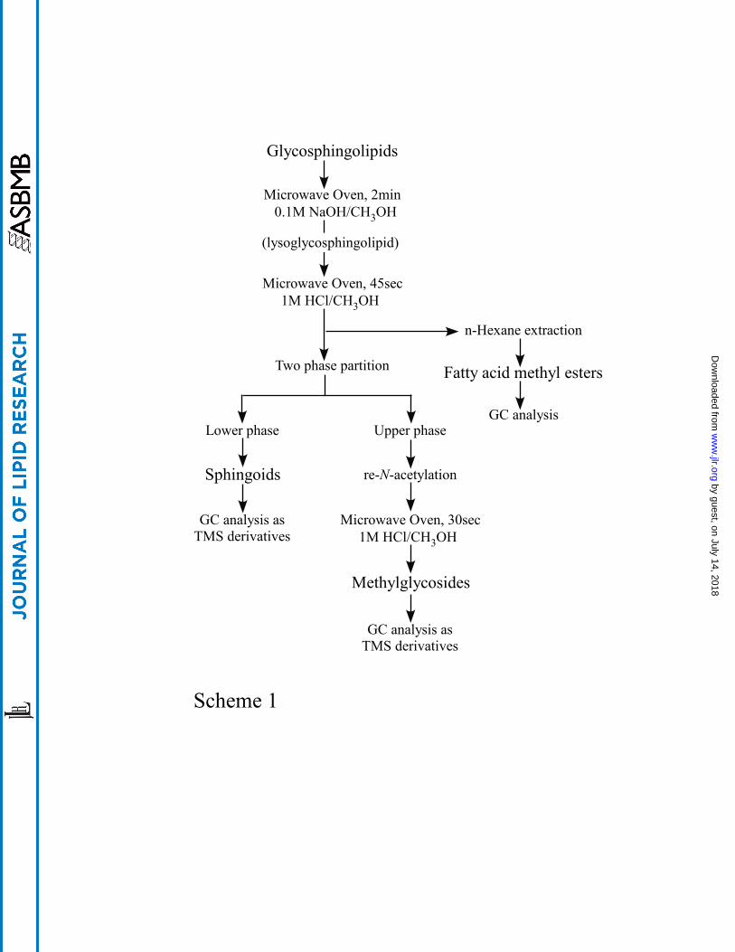
itself and is presented in this paper (Scheme 1).
Though household microwave ovens are easily available, there are several key points to
selection of an instrument, and confirmation of the optimum condition (10, 16-19). Here,
we describe a method for applying microwave-mediated reaction to glycosphingolipid
structural analysis including sphingoid moieties with an alkaline condition by a one-pot
4
by guest, on July 14, 2018w
ww
.jlr.orgD
ownloaded from

reaction, and confirmation of sugar and fatty acid components consequently obtained.
[Scheme 1]
5
by guest, on July 14, 2018w
ww
.jlr.orgD
ownloaded from

MATERIALS AND METHODS
Reagents
All solvents were purchased from Nacalai Tesque Co. (Kyoto, Japan) and were glass
distilled before use. The solution of 0.1 M NaOH/CH3OH was freshly prepared in our
laboratory by dissolving a reagent grade NaOH (Nacalai Tesque Co., purity 96%) in
distilled methanol, and 1 M HCl/CH3OH was prepared by passing dry HCl gas through
distilled methanol (23).
Glycosphingolipids
All glycosphingolipids isolated in our laboratory (1) were available for this experiment.
These included ceramide mono- (CMS, Galβ1-1Cer) and nonasaccharide
[Fuc3Meα1-2Xyl3Meβ1-4(GalNAc3Meα1-3)Fucα1-4GlcNAcβ1-2Manα1-3(Xylβ1-2)
Manβ1-4Glcβ1-1Cer] from the fresh water bivalve, Hyriopsis schlegelii; ceramide tri-
(CTS, GlcNAcβ1-3Manβ1-4Glcβ1-1Cer) and heptasaccharide (GlcNAcβ1-3Galβ1-
3GalNAcα1-4GalNAcβ1-4GlcNAcβ 1-3Manβ1-4Glcβ1-1Cer) from the green bottle fly,
Lucilia caesar; phosphocholine-containing zwitterionic glycosphigolipid
(cholinephosphoryl-6Galβ1-1Cer) from the earthworm, Pheretima hilgendorfi; N-glycolyl-
neuraminic acid-containing ganglioside (Araβ1-6Galβ1-4NeuGcα2-3Galβ1-4Glcβ1-1Cer)
from the starfish, Asterina pectinifera; and ganglioside GM1
[Galβ1-3GalNAcβ1-4(NeuAcα2-3)Galβ1-4Glcβ1-1Cer] from bovine brain.
Microwave oven
Microwave ovens used in this experiment were ER-VS1 (Toshiba, Tokyo, Japan) and
RE-Z3 (Sharp, Osaka, Japan). The technical specifications of both instruments were the
same, as follows: power source 100 V, 60 Hz; power requirement 960 W; output power 500
W (High); frequency 2450 MHz.
6
by guest, on July 14, 2018w
ww
.jlr.orgD
ownloaded from
Preparation of sphingoids
Five to fifteen nmole of glycolipids (50 to 100 µg) were hydrolyzed in thick glass tubes
(16 X 125 mm with Teflon-lined screw caps: Pyrex, Iwaki Glass Co., Tokyo, Japan) with
500 µL of freshly prepared 0.1 M NaOH/CH3OH using a microwave oven (Toshiba, Model
ER-VS1) attached to a GraLab Timer (Dimco-Gray Co., Dayton, OH). The samples were
exposed to the maximum power (500 W) of the microwave oven for 1 to 3 min. For
reasons of safety, the glass tubes were exposed one by one, each being enclosed in a large
polypropylene plastic container. After hydrolysis, the samples were cooled to room
temperature inside the microwave oven. Without evaporation or separation, the
hydrolysates were methanolyzed by addition of freshly prepared 1 M HCl/CH3OH (300
µL) into the tubes and using the microwave oven at the maximum power for 45 sec. For
comparison, identical samples were methanolyzed with 200 µL of 1 M aqueous methanolic
HCl in a conventional oven at 70oC for 18 h (14). The fatty acid methyl esters produced
were extracted three times with 500 µL of n-hexane. The methanolic phase that remained
was evaporated to dryness for deacidification under N2 gas. The residue containing
sphingoids and methylglycosides was dissolved in 600 µL of methanol/0.1 M NaOH (4:3,
v/v) and 720 µL of chloroform was then added. After vigorous agitation, the two phases
were separated by centrifugation (3,000 rpm, 5 min) and the upper phase containing
methylglycosides was removed. The lower phase containing sphingoids was washed
twice with 400 µL of methanol/water (1:1, v/v) and evaporated to dryness under N2 gas
(24).
Preparation of lysoglycosphingolipid
After alkaline hydrolysis (microwave oven, 2 min; 0.1 M NaOH/CH3OH), the samples
were acidified by addition of two drops of 6 M HCl. One ml of methanol was added and
excess salts were separated by centrifugation (3,000 rpm, 5 min). The methanol phase
was transferred to a fresh tube, evaporated to dryness under N2 gas and analyzed by TLC.
7
by guest, on July 14, 2018w
ww
.jlr.orgD
ownloaded from
Preparation of fatty acid methyl esters
The fatty acid methyl esters were extracted with n-hexane as described above after
methanolysis (microwave oven, 45 sec; 1 M HCl/CH3OH) and evaporated to dryness under
N2 gas.
Preparation of methylglycosides
Two drops of 6 M HCl were added to the upper phase containing methylglycosides
partitioned as described above and the solution was evaporated to dryness under N2 gas.
One ml of methanol was added to the residue, excess salts were removed by centrifugation
(3,000 rpm, 5 min) and the methanolic solution was transferred to a fresh tube.
Re-N-acetylation for hexosamines was performed by addition of 10 µL of pyridine and 50
µL of acetic anhydride at room temperature for 30 min, followed by evaporation to dryness
under N2 gas (25). Remethanolysis was performed by addition of 200 µL of 1 M
HCl/CH3OH, exposure to the microwave oven for 30 sec, and evaporation.
Gas-Liquid Chromatography (GC) and Gas Chromatograph-Mass Spectrometry
(GC-MS)
Sphingoids and methylglycosides were trimethylsilylated with a mixture of pyridine,
hexamethyldisilazane and trimethylchlorosilane (9:3:1, v/v) at 60oC for 30 min (26).
Their trimethylsilylated (TMS) derivatives were partitioned by 1 mL of chloroform and 4
mL of water. The water phase was replaced three times and the chloroform phase was
evaporated under N2 gas. An aliquot of the residues was injected and analyzed by a gas
chromatograph. Stearic acid propyl ester was used as internal standard for sphingoid TMS
derivatives and fatty acid methyl esters. All of these derivatives were analyzed by using a
Shimadzu GC-18A gas chromatograph and a capillary column of Shimadzu HiCap-CBP 5
(0.22 mm X 25 m, Shimadzu Co., Kyoto, Japan). Electron impact mass spectra were
taken using a Shimadzu GCMS-QP 5050 gas chromatograph-mass spectrometer (Shimadzu
Co., Kyoto, Japan) with the same capillary column under the following conditions: ionizing
voltage, 70 eV; ionizing current, 60 µA; interface temperature, 250oC; injection port
8
by guest, on July 14, 2018w
ww
.jlr.orgD
ownloaded from

temperature, 240oC; helium gas pressure, 100 kPa. Oven temperatures were programmed
as follows : sphingoid TMS derivatives, 210oC to 230oC (2oC/min) for GC, 80oC (2 min) to
180oC (20oC/min) to 240oC (4oC/min) for GC-MS; fatty acid methyl esters, 170oC to 230oC
(4oC/min) for GC, 80oC (2 min) to 180oC (20oC/min) to 240oC (4oC/min) for GC-MS;
methylglycoside TMS derivatives, 150oC to 230oC (2oC/min) for GC, 80oC (2 min) to
180oC (20oC/min) to 240oC (4oC/min) for GC-MS.
Thin-Layer Chromatography (TLC)
Silica gel 60 precoated plates (Merck KgaA, Darmstadt, Germany) were developed with
chloroform/methanol/water (60:40:10, v/v). Glycolipids were visualized by spraying with
orcinol-H2SO4 reagent (27) and free amino group was visualized by spraying with
ninhydrin reagent (28) followed by heating at 110oC.
Matrix-Assisted Laser Desorption Ionization Time-Of-Flight Mass Spectrometry
(MALDI-TOF MS)
MALDI-TOF MS analyses were performed using a Shimadzu/KRATOS KOMPACT
MALDI I mass spectrometer (Shimadzu Co., Kyoto, Japan) equipped with a Workstation
SPAR station (Sun Microsystems, Inc., CA, USA), operation in positive-ion linear mode.
Ions were formed by a pulsed ultraviolet laser beam (N2 laser, 337 nm; 3 nanosecond-wide
pulses/sec). The matrix used was 7-amino-4-methyl-coumarin (coumarin 120; Sigma, St.
Louis, MO). External mass calibration was provided by the [M+H]+ ions of ceramide
mono- to nonasaccharides (M.W. 699 to 2201) prepared from the green bottle fly, L. caesar
(7, 10).
9
by guest, on July 14, 2018w
ww
.jlr.orgD
ownloaded from
RESULTS
Microwave exposure time
To determine an efficient microwave exposure time, the simplest glycosphingolipid,
ceramide monosaccharide (CMS) from the fresh-water bivalve, was hydrolyzed with 0.1 M
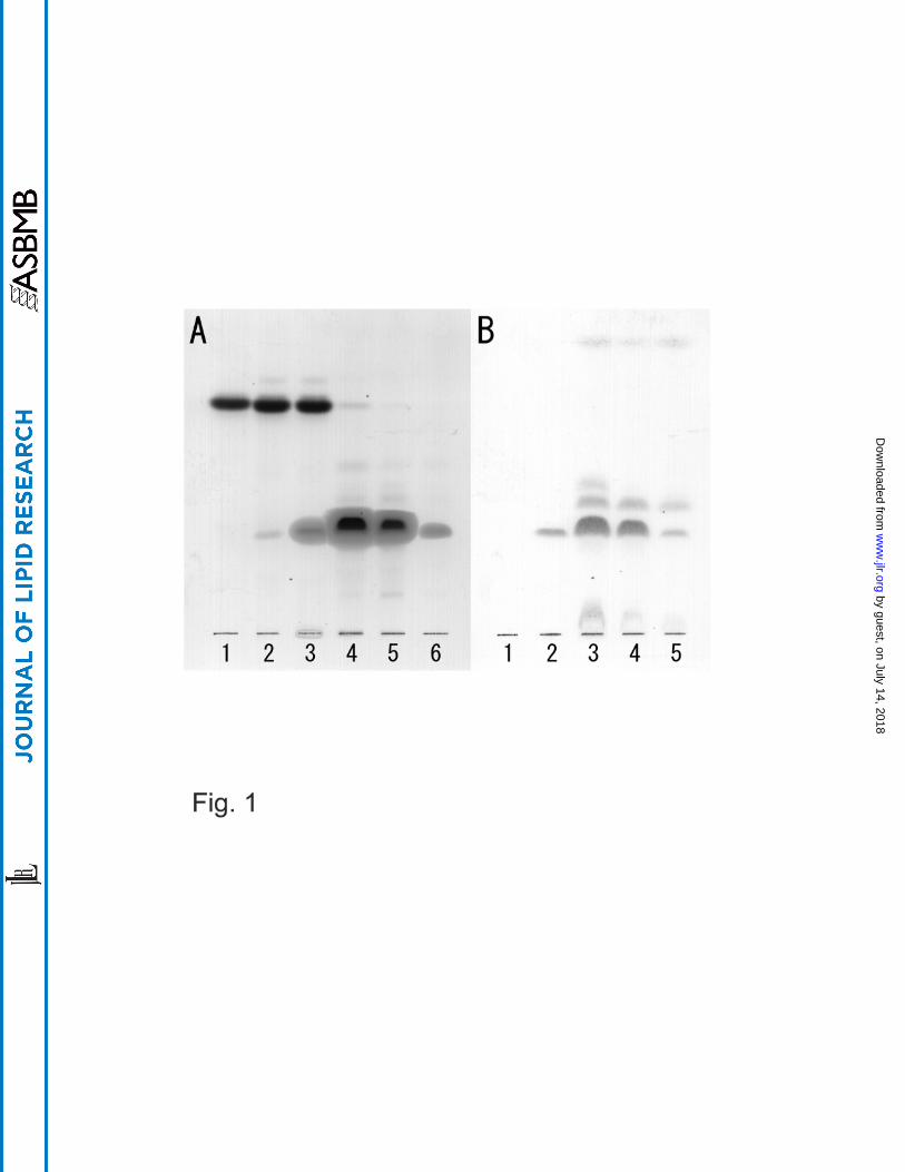
NaOH/CH3OH using a microwave oven. Figure 1 shows the time course of the amide
bond hydrolysis analyzed by TLC. Little lyso-CMS was observed within 1 min 30 sec
reaction time (Figs. 1A, lanes 2 and 3 and 1B, lanes 1 and 2). At 2 to 3 min reaction times,
however, substantial conversion was detected by both orcinol-H2SO4 reagent and ninhydrin
reagent (Figs. 1A, lanes 4-6 and 1B, lanes 3-5). The ninhydrin-positive spot migrating
ahead of lyso-CMS (barely visualized by orcinol-H2SO4 reagent) seems to be the sphingoid
directly formed under this condition (Fig. 1B, lanes 3-5). Prolonging the reaction time for
2 more min led to a lower yield of lyso-CMS due to degradation of products. Using 0.05
M NaOH/CH3OH, mostly intact CMS was observed after an exposure time of 2 min (data
not shown). Therefore, the optimal reaction time for the alkaline hydrolysis was found to
be 2 min with 0.1 M NaOH/CH3OH. It should be emphasized that this hydrolysis was
observed using a Toshiba ER-VS1 oven. Using a Sharp RE-Z3 oven, no lyso-CMS was
produced using an even longer reaction time (data not shown). Although no performance
difference can be inferred based on the technical specification sheets provided with the
Toshiba and Sharp instruments, the Toshiba microwave oven is suitable for this reaction.
This is a key point for selection of an instrument.
[Figure 1]
Moisture contamination in the hydrolysis reagent
The alkaline hydrolysis solution used in this experiment contains hygroscopic reagents.
We examined the effect of moisture contamination by addition of water to the solution.
Complete hydrolysis occurred with 0.25% water in methanol, the same as observed with
distilled methanol alone as a solvent (data not shown). With 0.5% water in methanol,
10
by guest, on July 14, 2018w
ww
.jlr.orgD
ownloaded from
unhydrolyzed CMS was observed and using 1.5% water in methanol, no hydrolyzed
product could be observed (data not shown). We have experienced complete hydrolysis
using the freshly prepared alkaline solution in distilled methanol.
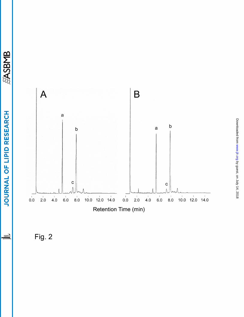
Yield of sphingoids
Lyso-CMS was produced from CMS with freshly prepared 0.1 M NaOH/CH3OH using a
microwave oven for 2 min at maximum power. Afterwards, sphingoids were prepared by
hydrolysis with freshly prepared 1 M HCl/CH3OH in the same tube using the microwave
oven at the maximum power for 45 sec, and were partitioned under alkaline condition.
Sphingoid TMS derivatives produced by this method were compared with those produced
by the conventional method (1 M aqueous methanolic HCl in a conventional oven at 70oC
for 18 h) by calculating areas on their respective GC chromatograms. The internal
standard stearic acid propyl ester was used. The yield of sphingoids was quantitatively
slightly higher (104%) and the error was less (n=3) than in the conventional method (Table
1). This shows that the one-pot reaction system is fully reproducible. Cleavage of
glycosphingolipids with anhydrous methanolic HCl results in the production of
considerable quantities of secondary products from sphingoids, such as O-methyl ethers (17,
22). Although a modified reagent for methanolysis (conventional method), containing
methanol, water and HCl, reduces the yield of these by-products to low levels, by-products
can nevertheless be detected (15). Theoretically, no by-product sphingoids are produced
from lysoglycosphingolipids with anhydrous methanolysis (22). Our results show much
less by-products as artifacts compared with the conventional method (Fig. 2A, B, peak c).
This one-pot reaction system not only reduces the reaction time to 2 min and 45 sec from
18 h, but also produces better yields and fewer by-products. That is, from as little as 20
µg and up to 500 µg of CMS (~3 to ~70 nmole), largely the same gas chromatogram
patterns of the sphingoid, as well as similar by-product/product ratio, were obtained from
the same hydrolysis condition.
[Figure 2], [Table 1]
11
by guest, on July 14, 2018w
ww
.jlr.orgD
ownloaded from

Application to various types of sphingoids and glycosphingolipids
Shorter chain length sphingoids were derived from ceramide heptasaccharide isolated
from the green bottle fly, L. caesar, which contains a moderate-length sugar chain. In
Figure 3, d14:1 and d16:1 sphingoids were detected comparable with the results found
using the conventional method (Fig. 3A, B). Using ceramide nonasaccharide isolated
from the fresh water bivalve, H. schlegelii, which contains a longer sugar chain, d18:1
sphingoid was detected (Fig. 3D). Neither sphingoid chain length nor sugar chain length
in glycosphingolipids appears to affect the yield of sphingoid nor the reactions of alkaline
hydrolysis and methanolysis. In the phosphocholine-containing zwitterionic
glycosphingolipid isolated from the earthworm, P. hilgendorfi, the sphingoid species
contains branched structures. Accordingly, all sphingoid species in this lipid were
detected by the one-pot reaction system, independent of the attached group or substituent
on the sugar molecule (Fig. 3F). Ganglioside isolated from the starfish, A. pectinifera,
contains N-glycolylneuraminic acid as its sugar component and phytosphingoids as
sphingoid species. Every sphingoid species in this ganglioside were detected and no
degradation of phytosphingoid was found using this system (Fig. 3H). Similarly, in the
ubiquitous ganglioside GM1, isolated from bovine brain, typical d18:1 and d20:1
sphingoids were detected (data not shown). In our experience, every type of sphingoid
species in glycosphingolipids could be determined by this one-pot reaction system,
independent of the differences of sugar structures and the types of sphingoid species.
Also it should be noted that the yield of by-products is trivial in most cases.
[Figure 3]
Yield of fatty acid methyl ester
Methanolysis which hydrolyzes the amide bond and esterifies the resulting free fatty acid,
produces fatty acid methyl esters from anhydrous methanolic HCl in boiling water for 3 h,
or from microwave exposure for 1 min (13, 14, 18). In the first step of this one-pot
reaction system, alkaline hydrolysis of glycosphingolipids cleaves the amide bond,
12
by guest, on July 14, 2018w
ww
.jlr.orgD
ownloaded from
resulting in mostly free fatty acids, because no fatty acid methyl esters are detected by GC
at this step (data not shown). Fatty acid methyl esters are produced during the subsequent
acid methanolysis required for cleavage of glycosyl bonds attached to sphingoids. The
resulting fatty acid methyl esters are easily extracted into n-hexane. Their yields by the
new method are compared in Figure 4 and Table 1 with results obtained using traditional
methods, and are quantitatively and qualitatively comparable with results by other methods.
[Figure 4]
Carbohydrate constituent analysis
Under the optimal reaction condition for sphingoid analysis, cleavage of an acetyl group
from a HexNAc residue could occur (20). A GlcNAc-containing glycosphingolipid,
ceramide trisaccharide (CTS, GlcNAcβ1-3Manβ1-4Glcβ1-1Cer) isolated from the green
bottle fly, L. caesar, was hydrolyzed with 0.1 M NaOH/CH3OH using a microwave oven
and analyzed by MALDI-TOF MS (data not shown). The product was represented mainly
by two [M+H]+ ions differing by 28 m/z units and corresponding to the molecular species
containing d14:1 and d16:1 sphingoids. The [M+H]+ ions at m/z 729 and 757 differ by 42
m/z units [-(CH3CO) +H] from the calculated [M+H]+ ion of lyso-CTS at m/z 771 and 799,
respectively. These [M+H]+ ions at m/z 729 and 757 correspond to deacetyl-lyso-CTS
and show the lack of both acetyl and fatty acyl groups from native CTS.
In the one-pot reaction system, without re-N-acetylation of deacetyl-lyso-CTS, only
glucose could be detected still attached to its sphingoid (Fig. 5A). After 30 min
re-N-acetylation (25), the sugar portion was cleaved using microwave exposure for 30 sec
and analyzed by GC as the TMS derivatives. As shown in Figure 5B, the sugar
constituents of CTS, namely Man, Glc and GlcNAc, were detected in molar ratios identical
to those found in traditional methanolysis, as 1:1:1 (Table 2). In this traditional
methanolysis method, complete cleavage of glycosyl bonds was achieved, while the amide
bond on HexNAc was stable and therefore required no re-N-acetylation after methanolysis
(unpublished observation).
13
by guest, on July 14, 2018w
ww
.jlr.orgD
ownloaded from

DISCUSSION
The microwave oven has been used to speed up synthetic reactions in the preparation of
various organic compounds (29-31). In the laboratory, the microwave generator used
early on was a laboratory-grade instrument; for example in 1993 Lagana et al. described the
analysis of sialic acids using a Microwave Digestion System (CEM Co.) (32). More
recently, household microwave ovens have been used for chemical analysis of protein, lipid
and sugar structures in the laboratory (33-38). In our laboratory also, a household
microwave oven has been used for fatty acid compositional analysis of phospholipids and
glycosphingolipids, as well as carbohydrate structural analysis of glycosphingolipids (10,
18).
It is critical in determining the best hydrolysis conditions, to establish the optimal
reaction time. Although the typical household microwave oven has a power control, this
switch permits only on/off control. Therefore, it is necessary to find the optimum reaction
time point at fixed maximum power. We described here that glycosphingolipids were
hydrolyzed by microwave exposure with 0.1 M NaOH/CH3OH for 2 min followed by 1 M
HCl/CH3OH for 45 sec. Taketomi also described the preparation of lysoglycosphingolipid
after exposure for 2 min (likewise found by his group to be optimal) using a Toshiba ER-V
microwave oven (19-21). On the other hand, we should point out that there is a difference
in microwave generating capacity that cannot be estimated from data sheets of the
appliances. For example, the Sharp RE-Z3 oven could not produce lysoglycosphingolipid
from glycosphingolipid under alkaline hydrolysis conditions even after prolonging the
exposure time to 3 min. From another point of view, at the 1 min time point using the
Sharp RE-Z3 oven, the recovery of fatty acid methyl ester was less than when the Toshiba
was used for 30 sec, while some sugar components could be detected only by the latter
microwave-mediated method (10, 18). This was especially true of xylose and its methyl
derivatives, which occur in Protostomia glycosphingolipids, due to the fact that this
reaction is milder than the conventional method, and sugar structures are better retained.
This might be considered a drawback to using household microwave ovens, but it is
15
by guest, on July 14, 2018w
ww
.jlr.orgD
ownloaded from

nevertheless possible to locate a cheap and convenient oven that is acceptable for alkaline
hydrolysis.
We chose the concentration of NaOH in methanol to be 0.1 M. At a lower
concentration, i.e. 0.05 M NaOH in distilled methanol, mostly intact CMS was observed
after 2 min microwave exposure. A concentration of 0.1 M NaOH in distilled methanol is
approximately saturated, because particles of NaOH could be seen in the solution. In
other words, when a clear solution of 0.1 M NaOH in distilled methanol is seen, moisture
contamination of the reagent should be suspected. This one-pot reaction system requires
alkaline hydrolysis followed by acid methanolysis. Although the latter is not affected by
salt or water (15, 37, 39), presence of these in the solution should be kept to a minimum for
subsequent isolation of sphingoids and carbohydrates by partioning under alkaline
conditions. All these considerations dictated our choice of 0.1 M NaOH as the optimum
concentration in this system.
Various methods involving alkaline conditions have been reported for the preparation of
lysoglycosphingolipids, mostly for the preparation (from gangliosides) of deacetyl
lysogangliosides lacking both fatty acyl groups as well as acetyl groups on neuraminic
acids and hexosamines (22, 40-42). A later improved method described the preparation of
lysogangliosides by use of a one-pot reaction (43). Recently, methods have been reported
for preparing various kinds of lysoglycosphingolipids by microwave exposure (19-21).
Using this technique, alkaline hydrolysis of amide bonds could be controlled by careful
timing, resulting in lysoglycosphingolipids and deacetyl lysoglycosphingolipids. In our
experiments mostly deacetyl lysoglycosphingolipids were observed, but from an analytical
point of view, deacylation including deacetylation of carbohydrate moieties does not matter
for the analysis of the sphingoid moiety. For carbohydrate analysis, however,
re-N-acetylation is required for the cleavage of hexosamine-containing glycosyl bonds.
Once the acetyl group has been cleaved from HexNAc, the residual hexosamine glycosyl
bonds are extremely resistant to acid hydrolysis, because of protonation located close to the
glycosyl bond (22). Mannose and glucosamine were practically undetectable at their
corresponding retention times because at this point they exist primarily as the GlcN-Man
16
by guest, on July 14, 2018w
ww
.jlr.orgD
ownloaded from

disaccharide (Fig. 5A). However, for release of sphingoid, acid methanolysis is required
after alkaline hydrolysis. It is therefore necessary that the sugar-containing residual
moiety should be re-N-acetylated and re-methanolyzed. The resistance of hexosaminyl
glycosyl bonds to alkaline hydrolysis is useful for carbohydrate sequential analysis. In
fact, the relative amounts of GlcNAc and Man observed in GC analysis in absence versus
presence of re-N-acetylation implies the GlcNAc to Man link (Fig. 5A), and the method
described here might thus help provide information as to carbohydrate sequences in
glycosphingolipids.
Without a separation of sugars and sphingoids, one-step analysis by GC of our reaction
mixture could determine the products as N-acetyl sphingoid and methylglycoside TMS
derivatives (44). However, the relative intensities of methylglycoside TMS derivatives
compared with N-acetyl sphingoid TMS derivatives (2 to 4-fold higher, data not shown)
complicate the assignment of peaks to various components. Also, less data is available for
mass spectra of of N-acetyl sphingoids as well as for their GC particulars. Therefore, we
chose to separate sugar and sphingoid moieties, and analyze them separately.
The overall conclusion drawn from this study of microwave-mediated reaction in
glycosphingolipid analysis is that the method can be usefully applied for rapid qualitative
analysis. Application of this method to analysis of phosphosphingolipids and inositol
phosphate containing glycosphingolipids is in progress and will be published elsewhere.
ACKNOWLEDGMENTS
This work was supported in part by Grant-in-Aid 14780472 for Young Scientists B (to S.
I.) and 15570097 for Scientific Research C (to M. S.) from the Ministry of Education,
Culture, Sports, Science and Technology of Japan.
17
by guest, on July 14, 2018w
ww
.jlr.orgD
ownloaded from

REFERENCES
1. Hori, T., and M. Sugita. 1993. Sphingolipids in lower animals. Prog. Lipid Res.
32:25-45.
2. Sugita, M., C. Hayata, T. Yoshida, M. Suzuki, A. Suzuki, T. Takeda, T. Hori, and F.
Nakatani. 1994. A novel fucosylated glycosphingolipid from the millipede, Parafontaria
laminata armigera. Biochim. Biophys. Acta. 1215:163-169.
3. Sugita, M., H. Fujii, J. T. Dulaney, F. Inagaki, M. Suzuki, A. Suzuki, and S. Ohta. 1995.
Structural elucidation of two novel amphoteric glycosphingolipids from the earthworm,
Pheretima hilgendorfi. Biochim. Biophys. Acta. 1259: 220-226.
4. Sugita, M., T. Mizunoma, K. Aoki, J. T. Dulaney, F. Inagaki, M. Suzuki, A. Suzuki, S.
Ichikawa, K. Kushida, S. Ohta, and A. Kurimoto. 1996. Structural characterization of a
novel glycoinositolphospholipid from the parasitic nematode, Ascaris suum. Biochim.
Biophys. Acta. 1302: 185-192.
5. Sugita, M., A. Morikawa, J. T. Dulaney, and A. Okada. 1996. Glycosphingolipids with
Galα1-6Gal and Galβ1-6Gal sequences in the leech, Hirudo nipponica. J. Jpn. Oil Chem.
Soc. (J. Oleo Sci.) 45: 731-740.
6. Sugita, M., S. Ohta, A. Morikawa, J. T. Dulaney, S. Ichikawa, K. Kushida, F. Inagaki, M.
Suzuki, and A. Suzuki. 1997. Novel neogala series glycosphingolipids with glucose at the
non-reducing termini in the earthworm, Pheretima sp. J. Jpn. Oil Chem. Soc. (J. Oleo
Sci.) 46: 755-766.
7. Sugita, M., N. Yamake, H. Hamana, K. Sasaki, and J. T. Dulaney. 1999. Structural
characterization of neutral glycosphingolipids, mono-, di- and triglycosylceramides, from
the marine annelid, Pseudopotamilla occelata. J. Jpn.Oil Chem. Soc. (J. Oleo Sci.) 48:
671-679.
8. Sugita, M., S. Miwa, K. Aoki, J. T. Dulaney, S. Ichikawa, F. Inagaki, and M. Suzuki.
2000. Acidic glycosphingolipids in brackish water annelida: Structural analysis of two
novel glycoinositolphospholipids from the lugworm, Tylorrhynchus heterochetus. J. Jpn.
Oil Chem. Soc. (J. Oleo Sci.) 49: 33-43.
18
by guest, on July 14, 2018w
ww
.jlr.orgD
ownloaded from

9. Kurimoto, A., Y. Kawakami, J. T. Dulaney, A. Miyake, and M. Sugita. 2000. Amphoteric
glycosphingolipids in parasitic nematode (I): Chemical structures of glycosphingolipid
series containing cholinephosphoryl(→6)-N-acetylglucosamine as an amphoteric group
from porcine roundworm, Ascaris suum. J. Jpn. Oil Chem. Soc. (J. Oleo Sci.) 49:
127-135.
10. Itonori, S., H. Hamana, N. Hada, T. Takeda, J. T. Dulaney, and M. Sugita. 2001.
Structural characterization of a novel series of fucolipids from the marine annelid,
Pseudopotamilla occelata. J. Oleo Sci. 50: 537-544.
11. Kimura, K., S. Itonori, N. Hada, O. Itasaka, J. T. Dulaney, T. Takeda, and M. Sugita.
2002. Phosphonoglycolipids in marine crustacean: Structural characterization of two
novel phosphonocerebrosides from the crab, Erimacrus isenbeckii. J. Oleo Sci. 51:
83-91.
12. Aoki, K., S. Sugiyama, J. T. Dulaney, S. Itonori, and M. Sugita. 2002. Classification
into a novel mollu-series of neutral glycosphingolipids from the lamp shell, Lingula
unguis. J. Oleo Sci. 51: 463-472.
13. Sweeley, C. C., and B. Walker. 1964. Determination of carbohydrates in glycolipides
and gangliosides by gas chromatography. Anal. Chem. 36: 1461-1466.
14. Vance, D. E., and C. C. Sweeley. 1967. Quantitative determination of the neutral
glycosyl ceramides in human blood. J. Lipid Res. 8: 621-630.
15. Gaver, R. C., and C. C. Sweeley. 1965. Methods for methanolysis of sphingolipids and
direct determination of long-chain bases by gas chromatography. J. Am. Oil Chem. Soc.
42: 294-298.
16. Lie Ken Jie, M. S. F., and C. Yan-Kit. 1988. The use of a microwave oven in the
chemical transformation of long chain fatty acid esters. Lipids 23: 367-369.
17. Khan, M. U., and J. P. Williams. 1993. Microwave-mediated methanolysis of lipids and
activation of thin-layer chromatographic plates. Lipids 28: 953-955.
18. Itonori, S., M. Takahashi, and M. Sugita. 2000. Application of microwave-mediated
reaction to lipid analysis. Proceedings of JOCS/AOCS World Congress, Kyoto, Japan
p.214.
19
by guest, on July 14, 2018w
ww
.jlr.orgD
ownloaded from

19. Taketomi, T., A. Hara, K. Uemura, H. Kurahashi, and E. Sugiyama. 1996. A
microwave-mediated saponification of galactosylceramide and galactosylceramide
I3-sulfate and identification of their lyso-compounds by delayed extraction
matrix-assisted laser desorption ionization time-of-flight mass spectrometry. Biochem.
Biophys. Res. Commun. 224: 462-467.
20. Taketomi, T., A. Hara, K. Uemura, and E. Sugiyama. 1996. Rapid method of
preparation of lysoglycosphingolipids and their confirmation by delayed extraction
matrix-assisted laser desorption ionization time-of-flight mass spectrometry. J. Biochem.
(Tokyo). 120: 573-579.
21. Taketomi, T., A. Hara, K. Uemura, H. Kurahashi, and E. Sugiyama. 1997. Preparation
of various lysogangliosides including lyso-fucosyl GM1 and delayed extraction
matrix-assisted laser desorption ionization time-of-flight mass spectrometric analysis. J.
Biochem. (Tokyo). 121: 264-269.
22. Taketomi, T., and N. Kawamura. 1970. Preparation of lysohematoside
(neuraminyl-galactosyl-glucosylsphingosine) from hematoside of equine erythrocyte and
its chemical and hemolytic properties. J. Biochem. (Tokyo). 68: 475-485.
23. Flowers, H. M. 1966. Substituted cerebrosides: Part II. Synthetic dihydrosulfatides.
Carbohydr. Res. 2: 371-379.
24. Ando, S., and R. K. Yu. 1979. Isolation and characterization of two isomers of brain
tetrasialogangliosides. J. Biol. Chem. 254: 12224-12229.
25. Kozulić, B., B. Ries, and P. Mildner. 1979. N-Acetylation of amino sugar methyl
glycosides for gas-liquid chromatographic analysis. Anal. Biochem. 94: 36-39.
26. Kishimoto, Y., and M. Hoshi. 1972. Isolation, purification, and assay of fatty acids and
steroids from the nervous system. In Methods of Neurochemistry Vol. 3. R. Fried, editor.
Marcel Dekker, New York, NY. 75-154.
27. Svennerholm, L. 1956. The quantitative estimation of cerebrosides in nervous tissue. J.
Neurochem. 1: 42-53
28. Skipski, V. P., R. F. Peterson, and M. Barclay. 1962. Separation of phosphatidyl
ethanloamine, phosphatidyl serine, and other phospholipids by thin-layer chromatography.
20
by guest, on July 14, 2018w
ww
.jlr.orgD
ownloaded from
J. Lipid Res. 3: 467-470.
29. Gedye, R., F. Smith, K. Westaway, H. Ali, L. Baldisera, L. Laberge, and J. Rousell.
1986. The use of microwave ovens for rapid organic synthesis. Tetrahedron Lett. 27:
279-282.
30. Giguere, R. J., T. L. Bray, S. M. Duncan, and G. Majetich. 1986. Application of
commercial microwave ovens to organic synthesis. Tetrahedron Lett. 27: 4945-4948.
31. Caddick, S. 1995. Microwave-assisted organic reactions. Tetrahedron 51: 10403-10432.
32. Laganà, A., A. Marino, G. Fago, and B. P. Martinez. 1993. A hydrolysis method using
microwaves: determination of N-acetyl- and N-glycolylneuraminic acids in biological
systems by fluorometric high-performance liquid chromatography. Anal. Biochem. 215:
266-272.
33. Sun, W.-C., P. M. Guy, J. H. Jahngen, E. F. Rossomando, and E. G. E. Jahngen. 1988.
Microwave-induced hydrolysis of phospho anhydride bonds in nucleotide triphosphates.
J. Org. Chem. 53: 4414-4416.
34. Fountoulakis, M., and H.-W. Lahm. 1998. Hydrolysis and amino acid composition
analysis of proteins. J. Chromatogr. A. 826: 109-134.
35. Dayal, B., G. Salen, and V. Dayal. 1991. The use of microwave oven for the rapid
hydrolysis of bile acid methyl esters. Chem. Phys. Lipids. 59: 97-103.
36. Dayal, B., Keshava Rao, and G. Salen. 1995. Microwave-induced organic reactions of
bile acids: Esterification, deformylation and deacetylation using mild reagents. Steroids
60: 453-457
37. Kunlan, L., X. Lixin, L. Jun, P. Jun, C. Guoying, and X. Zuwei. 2001. Salt-assisted acid
hydrolysis of starch to D-glucose under microwave irradiation. Carbohydr. Res. 331:
9-12.
38. Carrapiso, A. I., and C. Garcia. 2000. Development in lipid analysis: Some new
extraction techniques and in situ transesterification. Lipids. 35: 1167-1177.
39. Schmid, P. C., and H. H. O. Schmid. 1994. Reactions of diazomethane with
glycerolipids in the presence of serum or inorganic salts. Lipids. 29: 883-887.
40. Sonnino, S., G. Kirschner, R. Ghidoni, D. Acquotti, and G. Tettamanti. 1985.
21
by guest, on July 14, 2018w
ww
.jlr.orgD
ownloaded from

Preparation of GM1 ganglioside molecular species, having homogeneous fatty acid and
long chain base moieties. J. Lipid Res. 26: 248-257.
41. Neuenhofer, S., G. Schwarzmann, H. Egge, and K. Sandhoff. 1985. Synthesis of
lysogangliosides. Biochemistry. 24: 525-532.
42. Nores, G. A., N. Hanai, S. B. Levery, H. L. Eaton, M. E. K. Salyan, and S. Hakomori.
1988. Synthesis and characterization of lyso-GM3 (II3Neu5AcLactosylsphingosine),
de-N-acetyl-GM3 (II3NeuNH2LactosylCer), and related compounds. Carbohydr. Res.
179: 393-410.
43. Sonnino, S., D. Acquotti, G. Kirschner, A. Uguaglianza, L. Zecca, F. Rubino, and G.
Tettamanti. 1992. Preparation of lyso-GM1 (II3Neu5AcGgOse4-long chain bases) by a
one-pot reaction. J. Lipid Res. 33: 1221-1226.
44. Carter, H. E., and R. C. Gaver. 1967. Improved reagent for trimethylsilylation of
sphingolipid bases. J. Lipid Res. 8: 391-395.
22
by guest, on July 14, 2018w
ww
.jlr.orgD
ownloaded from
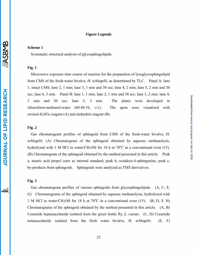
Figure Legends
Scheme 1
Systematic structural analysis of glycosphingolipids.
Fig. 1
Microwave exposure time course of reaction for the preparation of lysoglycosphingolipid
from CMS of the fresh-water bivalve, H. schlegelii, as determined by TLC. Panel A: lane
1, intact CMS; lane 2, 1 min; lane 3, 1 min and 30 sec; lane 4, 2 min; lane 5, 2 min and 30
sec; lane 6, 3 min. Panel B: lane 1, 1 min; lane 2, 1 min and 30 sec; lane 3, 2 min; lane 4,
2 min and 30 sec; lane 5, 3 min. The plates were developed in
chloroform-methanol-water (60:40:10, v/v). The spots were visualized with
orcinol-H2SO4 reagent (A) and ninhydrin reagent (B).
Fig. 2
Gas chromatogram profiles of sphingoid from CMS of the fresh-water bivalve, H.
schlegelii. (A) Chromatogram of the sphingoid obtained by aqueous methanolysis,
hydrolyzed with 1 M HCl in water/CH3OH for 18 h at 70oC in a conventional oven (15).
(B) Chromatogram of the sphingoid obtained by the method presented in this article. Peak
a, stearic acid propyl ester as internal standard; peak b, octadeca-4-sphingenine; peak c,
by-products from sphingoids. Sphingoids were analyzed as TMS derivatives.
Fig. 3
Gas chromatogram profiles of various sphingoids from glycosphingolipids. (A, C, E,
G) Chromatograms of the sphingoid obtained by aqueous methanolysis, hydrolyzed with
1 M HCl in water/CH3OH for 18 h at 70oC in a conventional oven (15). (B, D, F, H)
Chromatograms of the sphingoid obtained by the method presented in this article. (A, B)
Ceramide heptasaccharide isolated from the green bottle fly, L. caesar. (C, D) Ceramide
nonasaccharide isolated from the fresh water bivalve, H. schlegelii. (E, F)
23
by guest, on July 14, 2018w
ww
.jlr.orgD
ownloaded from
Phosphocholine-containing, zwitterionic glycosphingolipid isolated from the earthworm, P.
hilgendorfi. (G, H) Ganglioside isolated from the starfish, A. pectinifera. Peak a,
C14-sphingosine; peak b, C16-sphingosine; peak c, C18-sphingosine; peak d,
C17-sphingosine; peak e, branched C18-sphingosine; peak f, branched C19-sphingosine; peak
g, C19-sphingosine; peak h, iso-C16-phytosphingosine; peak i, C16-phytosphingosine; peak j,
iso-C17-phytosphingosine; peak k, anteiso-C17-phytosphingosine; peak l,
C17-phytosphingosine; peak m, iso-C18-phytosphingosine; peak n,
anteiso-C18-phytosphingosine; peak o, C18-phytosphingosine; peaks *, by-products from
sphingoids. Sphingoids were analyzed as TMS derivatives.
Fig. 4
Gas chromatogram profiles of fatty acid methyl esters from CMS of the fresh-water
bivalve, H. schlegelii. (A) Chromatogram of the sphingoid obtained by anhydrous
methanolysis, hydrolyzed with 1 M HCl/CH3OH for 45 sec by a Toshiba ER-VS1
microwave oven. (B) Chromatogram of the sphingoid obtained by aqueous methanolysis,
hydrolyzed with 1 M HCl in water/CH3OH for 18 h at 70oC in a conventional oven (15).
(C) Chromatogram of the sphingoid obtained by the method presented in this article. Peak
a, C14:0; peak b, C15:0; peak c, C16:0; peak d, C17:0; peak e, C18:0; peak f, stearic acid
propyl ester as internal standard.
Fig. 5
Gas chromatogram profiles of sugar moieties from ceramide trisaccharide isolated from
the green bottle fly, L. caesar. (A) Chromatogram of the methylglycoside TMS
derivatives obtained by following the sequential reactions: alkaline hydrolysis, anhydrous
methanolysis and TMS derivatization. (B) Chromatogram of the methylglycoside TMS
derivatives obtained by the method presented in this article. Peaks a, Man; peaks b, Glc;
peaks c, GlcNAc.
24
by guest, on July 14, 2018w
ww
.jlr.orgD
ownloaded from
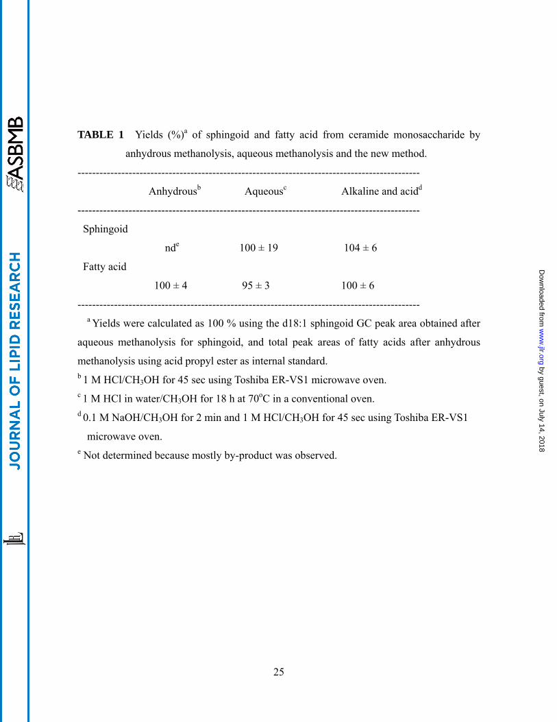
TABLE 1 Yields (%)a of sphingoid and fatty acid from ceramide monosaccharide by
anhydrous methanolysis, aqueous methanolysis and the new method.
----------------------------------------------------------------------------------------------
Anhydrousb Aqueousc Alkaline and acidd
----------------------------------------------------------------------------------------------
Sphingoid
nde 100 ± 19 104 ± 6
Fatty acid
100 ± 4 95 ± 3 100 ± 6
---------------------------------------------------------------------------------------------- a Yields were calculated as 100 % using the d18:1 sphingoid GC peak area obtained after
aqueous methanolysis for sphingoid, and total peak areas of fatty acids after anhydrous
methanolysis using acid propyl ester as internal standard. b 1 M HCl/CH3OH for 45 sec using Toshiba ER-VS1 microwave oven. c 1 M HCl in water/CH3OH for 18 h at 70oC in a conventional oven. d 0.1 M NaOH/CH3OH for 2 min and 1 M HCl/CH3OH for 45 sec using Toshiba ER-VS1
microwave oven. e Not determined because mostly by-product was observed.
25
by guest, on July 14, 2018w
ww
.jlr.orgD
ownloaded from
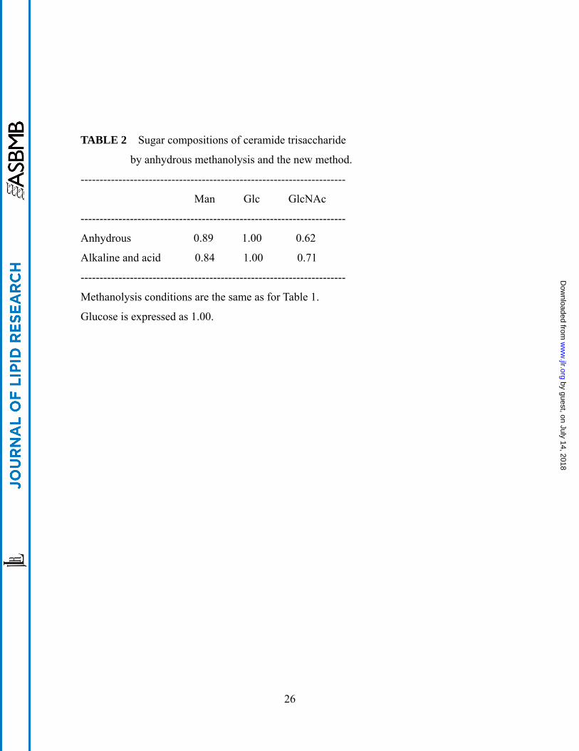
TABLE 2 Sugar compositions of ceramide trisaccharide
by anhydrous methanolysis and the new method.
----------------------------------------------------------------------
Man Glc GlcNAc
----------------------------------------------------------------------
Anhydrous 0.89 1.00 0.62
Alkaline and acid 0.84 1.00 0.71
----------------------------------------------------------------------
Methanolysis conditions are the same as for Table 1.
Glucose is expressed as 1.00.
26
by guest, on July 14, 2018w
ww
.jlr.orgD
ownloaded from
n-Hexane extraction
Fatty acid methyl esters
GC analysisLower phase
Sphingoids
GC analysis asTMS derivatives
Upper phase
re-N-acetylation
1M HCl/CH3OHMicrowave Oven, 30sec
Methylglycosides
GC analysis asTMS derivatives
Glycosphingolipids
0.1M NaOH/CH3OHMicrowave Oven, 2min
Two phase partition
1M HCl/CH3OHMicrowave Oven, 45sec
(lysoglycosphingolipid)
Scheme 1
by guest, on July 14, 2018w
ww
.jlr.orgD
ownloaded from